LEG
Historical document, translated for reference. It reflects medical knowledge of the 1920s–30s and is not medical advice.
Summary
This article examines the leg as a complete anatomical structure and its bones as levers for standing and walking. It discusses the mechanical principles of the leg's skeletal structure, including the femur, tibia, and foot, and how these components function together to support the body and enable movement.
Encyclopedia article (1928–1936)
LEG. The leg as a whole and the bones as levers. The human body in standing and locomotion rests on its caudal extremities and its vertical position sharply differs from that of other mammals. In the process of establishing the bipedal form of humans, the caudal extremities transformed into lower extremities, and according to the peculiarities of their exclusively supportive function, they also gradually changed their external appearance and form. - The skeleton of the lower extremities, like that of the upper extremities, has a quantitative increase in elements and a complication of their connections as they move away from their girdle. Thus, the proximal part of the extremity - the thigh - has as its main axis one long, massive tubular bone, which corresponds to the humerus of the upper extremity. The next, more distal part - the leg - has a support composed of two parallel tubular bones; these are the tibia and fibula, corresponding to the radius and ulna in the hand. Finally, the foot, corresponding to the metacarpus and carpus of the hand, is composed of 12 bones, which terminate distally in five fingers.--Before proceeding to the analysis of the lower extremity as an organ of movement, it is necessary to establish the so-called initial position of the body and extremities. The initial position is taken as the vertically positioned torso on straightened and brought-together legs. The feet are spread apart in such a way that the extensions of lines drawn through the big toe and heel cross at an angle of approximately 60°. The significance of individual bones of the lower extremity and their complexes as levers for standing and walking. The main task of the levers and mechanisms of the lower extremity is the ability at each given functional moment to transform the entire extremity into one mechanical, elastic, strong whole, into one lever suitable for supporting the entire body. At the same time, this extremity must have the ability to quickly fold its levers at the joints, shorten, and in this form be moved to a new initial position, again suitable for support (walking, running). Since, in addition to the supportive function, the main purpose of the extremities is to move the body in space (locomotor activity), the length of the extremities has outstanding significance in this regard. This is quite understandable, since the speed of movement depends both on the individual length of the tubular bones of the lower extremity and on the entire total length. The greatest significance in the length of the legs belongs to the femur and the tibia. The length of the first in an adult male averages about 44 cm, while the length of the leg averages about 38 cm: adding to this the height of the foot (averaging 8 cm), we have the total length of the extremity, averaging 90 cm. Its length somewhat increases when separated from the ground due to straightening in the ankle joint. From this it is evident that if the average length of the entire body is 168 cm, then the length of the lower extremities constitutes 53.5%. The proportional relationship between the length of the legs and the torso has significance not only for artists; it also has production-mechanical significance. Thus, the longer the legs, the higher the center of gravity is located, the less stable the entire human body is as a whole. And, conversely, the body is the more stable, the closer its center of gravity is to the support area (e.g., the sitting position of birds during sleep). These moments have significance, of course, besides walking, also in labor processes. Thus, when, for example, a person creates support on his feet and leans his entire torso forward or backward, he uses the upper part of the torso as auxiliary traction and thus utilizes its dead weight (e.g., pulling a rope, turning a wheel, etc.); greater height then, all other conditions being equal, gives a greater working effect than lesser height. From this point of view, 'macroskelia' (relative long-leggedness) is more advantageous than 'brachyskelia' (relative short-leggedness), not to mention the greater aesthetic harmony of the first type of legs. The bone levers of the lower extremity do not always correspond to one particular bone. They are often composed of several bones. If we consider the pelvis as a solid support on one side
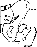
Fig. 2.
for the lower extremities on the other side, it can be graphically represented on the pelvis as
Fig
a solid support on one side /
for the lower extremities on the other side, it can be graphically represented on the pelvis as
divided by a line running from the center of the acetabulum to the anterior ilium; the posterior arm of this same lever is expressed by a line which also runs from the center of the acetabulum, but in the opposite direction—toward the lower end of the articular line of the sacroiliac joint of the corresponding side. These theoretical (graphic) lines express only the direction of the broad bony mass of the pelvis that bears the load, as shown by the arrows, mainly from above (Fig. 1). Depending on which of the three lines the center of gravity of the body is located, the pressure of the torso will be transmitted either to the anterior (arrow 3), smaller lever, or to the posterior (arrow 2), larger lever of the pelvis, or the entire pelvis will be in a state of labile equilibrium, resting on the heads of the femurs. Since the posterior arm of the pelvic lever (and its boreal implementation) is significantly larger than the anterior one, the moment of its force * exceeds that of the smaller anterior arm. Thus, the pelvis, already itself more massive behind than in front, tends to tilt backward, rotating on the heads of the femurs. The load from above of the entire torso many times increases such a tendency. Moreover, as is known, the pelvis can also be held and balanced on one femoral head (e.g., when walking, standing on one leg); then it also plays the role of a broken lever of the 1st kind, but of a different nature, distinct from the case just analyzed. In this position (Fig. 2), it will have an upper larger and lower smaller arm, and then traction on the upper arm will cause the pelvis to tilt in the same direction, while traction on the lower arm will result in the opposite position of it. This moment can be taken into account in explaining the female, more swaying, gait. The moment of rotating force relative to the axis is the product of the acting force by the shortest distance of its line of action from the axis of rotation (arm of the rotating force). * This is characteristic of levers of the 3rd kind (animals), where the points of application of the acting force are between the fulcrum and the point of application of resistance. The foot in relation to the lower leg is set at a right angle (when standing) so that 1/4 of its length falls on the short posterior arm (the heel) and 3/4 on the anterior arm (the tarsus and metatarsus). Thus, the free or supporting (when standing) foot relates to the lower leg as a two-armed lever (1st kind). G. Meyer calculates the load falling on the limb as follows: if we have a human body weighing, for example, 64 kg, then in normal (according to Fick) standing it is evenly loaded on both feet, i.e., 32 kg on each. On each thigh the load is distributed equally: first on both condyles of the thigh and then again concentrated on the bones of the lower leg. Further, on each foot these 32 kg are distributed inversely proportional to the length of its arms: on the posterior arm (the heel) falls 3/4 of this weight (24 kg), and the remaining 1/4 (8 kg) is transmitted (through the talus) to the anterior, longer arm of the foot. When walking, the entire body weight together with the additional reaction force from support alternately falls on each supporting leg. In the free leg (non-supporting), the foot similarly reveals the properties of a lever of the first kind: both its arms freely rise and fall. The absence of the foot, lower leg, or part of the thigh is replaced, as is known, by specially adapted artificial lever-prostheses. Therefore, each centimeter of the animal lever is very valuable, which surgeons always take into account. Character of movement of the lower extremity. Besides the usual processes such as standing, walking, running, jumping, the lower extremity is adapted for a whole series of diverse and more differentiated movements, such as dancing, physical exercises, etc. In the pelvic girdle and in the free lower extremity, the following more or less movable joints can be distinguished: slightly movable joints of the sacrum with the fifth lumbar vertebra and the ilia; the joint of the ilia with the femur—with considerable freedom of movement; the movable joint of the thigh and lower leg in the knee joint; the joint of the lower leg with the foot with a lesser degree of mobility, slightly movable joints of the bony elements of the foot and joints of the phalanges of the toes. More movable joints, such as the hip, knee, ankle, talo-calcaneal, and toe joints can function independently of the others, the movement of which is usually dependent and accompanies displacements of bones in adjacent joints. On the other hand, the more proximal the joint, the more significantly the properties of the 3rd kind lever mentioned above appear in it, where we gain more in speed of movement at the distal end of the lever, but acquire this proportionally at the expense of greater expenditure of muscular energy. Thus, working with the leg as one straight continuous support, we set in motion the mechanism of the hip joint. The effect of action in this joint is passively transferred to the foot and toes. In the hip joint, as in the shoulder joint, all types of movement characteristic of a ball-and-socket joint are possible (see below).—In the knee joint, flexion and extension can be carried out within a range of about 150° and, moreover, in a semi-flexed (up to 90°) position of the joint, circular movement is also possible (see below).—In the ankle joint, three types of movements are possible: 1) flexion-extension—about 60°; 2) adduction-abduction—about 17° and 3) rotation—about 25° (in children the range of these movements is somewhat larger). The joints of the tarsus and metatarsus are very slightly movable, their movements are mostly synergistic and inhibit each other due to the lack of coordination of their axes of movement.—In the metatarsophalangeal joints, flexion-extension up to 90°, adduction-abduction up to 25° are possible. The greatest mobility is possessed by the I, II, V toes. Due to the usual inactivity of the toes of the foot and the wearing of irrational footwear, their degree of mobility is significantly less than that of the fingers of the hand. However, their mobility can be increased by appropriate exercise. In accordance with the supporting function performed, the development of the form and structure of the joints of the lower extremity also proceeded. It is sufficient to point to the size of individual joints of the leg, which exceed the corresponding joints of the arm. Thus, the knee joint is very wide and represents the largest joint in the human body. It is relatively larger than the knee joints of quadruped animals, in which the weight of the body is usually distributed over all four limbs. The joints and bones of the foot have differentiated in the direction of forming elastic and strong arches, on which the entire weight of the body is distributed and supported; ultimately they serve as support when standing and moving. No such adaptations have formed on the upper extremity, which differentiated under the long-term influence of formative labor processes. In the special direction of increasing supporting properties, the evolution and involution of individual bony elements of the lower extremity also proceeds and goes on. The pelvic ring, its mechanical purpose. In holding the pelvis (when standing) in its natural tendency to tilt backward, the main role after muscles is played by the iliofemoral ligament (ligamentum ilio-femorale). Hence, with the first attempt to walk, a mechanical moment arises in the formation of the lumbar lordosis for vicarious compensation of this deviation. However, usually the lig. ilio-femoralis does not become taut, since this role is performed first and foremost with a greater moment of force by the m. ilio-psoas. In a person in an upright position, the centers of rotation of both shoulder joints, promontorium, and femoral heads lie in the same frontal plane. In front, besides this, the pubic bones by their fusion (symphysis) form a second, as it were, additional arch, which is part of the general pelvic structure and at the same time constitutes the anterior transverse connection between both halves of the pelvis. Behind, the ilia are even more strongly connected by the wedge-shaped sacrum between them and their common ligamentous apparatus. The presence of springing (shock-absorbing) properties of the pelvis is of great importance for the elasticity of gait, inevitably associated with jolts from the ground of varying hardness and relief. In this respect, complaints are known from people using lower extremity prostheses about unpleasant jolts in the pelvis. As for the stability of the bones of the pelvic ring against violence, cases are known where when a heavy weight falls from above on the head of a vertically standing person, the heads of the femurs break off, while the pelvis remains intact. On a corpse, the pelvis withstands a load of up to 250 kg without violation of its integrity (Braus). According to Lesgaft, this load can reach 560 and even 2,000 kg. The latter figure belongs to the pelvis of a worker with a well-developed muscular system, the first—to an exhausted woman with flabby musculature. The greatest strength in the pelvic vault is represented by the sacroiliac joint. Due to the strength and almost immobility of the joint, this joint actually stands on the boundary between synarthrosis and diarthrosis. In functional terms, it can be classified as a symphysis.
Often it has an open joint space, which indicates the evolution of this connection from a true joint. This evolution is slowed or stopped due to the special purpose of the pelvic ring, mainly for static purposes (Fik). According to Balandin, mobility in this joint early and significantly increases in pregnant women. In the normal state, however, it is equal to 0; the position of the symphysis itself is quite variable depending on different static and dynamic conditions. G. Meyer introduced the concept of the so-called 'normal conjugate.' This is a line that connects the apex of the symphysis with the apex of the physiological sacral kyphosis. The inclination of the pelvis (to the horizon) when measured by this method is expressed as 50-60°. A pelvic inclination angle of 40° (instead of the normal 50-60°) indicates 'weak holding of the body' in the vertical position; conversely, when this angle increases to 60° and higher, we have the so-called 'strong standing.' If we orient ourselves in the position of the pelvis according to the spina ilii anterior superior, then the protrusion of the latter in front of the symphysis pubis also indicates a simultaneous lifting of the posterior half of the pelvic ring, the posterior arm of the pelvic lever upward; this will be the so-called strong 'military' holding of the trunk (steep inclination of the pelvis). The opposite condition (the spinae ilii ant. sup. receding from the symphysis) indicates a lowering of the posterior arm of the pelvic lever, and such a pelvic position characterizes weak standing. It is self-evident that the magnitude of the pelvic inclination angle varies in one direction or another depending on individual, racial, sexual, and other peculiarities of skeletal structure; but even in one person, this angle often changes depending on the displacement of his center of gravity. The inclination angle of the pelvic inlet varies from 75° to 55°. According to Lesgaft, it averages 65° (deviating to 82' and 56°). The lumbosacral angle (the angle between a line drawn along the anterior surface of L to the same surface of Sr) also due to the displacement of the sacrum gives variations between 120-130° (Lesgaft). When standing, the pelvis tilts forward; in this case, the symphysis pubis lowers, the lumbar lordosis increases, and the sacrum shifts somewhat upward and backward. In this case, Fik determines the inclination of the pelvis (its anatomical conjugate to the horizon) as 67°.-When sitting, there is some displacement of the pelvis upward and forward: the pubic bones assume a more horizontal position, the lumbar lordosis is smoothed out, the sacrum becomes almost in the frontal plane, and the anatomical conjugate angle drops to 7°.-In the lying position, the pelvic inclination line lies below the horizontal line of the ilium. The mechanism of the hip joint. The magnitude of the angle between the mechanical (vertical) axis of rotation of the femoral head and its own body axis averages about 5-7°. The angle between the femoral neck and its body, open inward, averages in both men and women about 125-126°; it often reaches 120-133°, and in exceptional cases 115-140°. In military posture (at the command 'attention!'), the midpoints of the hip, knee, and ankle joints are located on one straight line, but not on the vertical. This straight line forms an angle with the vertical equal to approximately 7° and open upward. In normal standing, the midpoints of the hip joints are located vertically above the midpoints of the knee joints and above the midpoints of the ankle joints. In the so-called calm standing, the midpoints of the hip and knee joints are located one above the other and almost coincide with the frontal plane of the center of gravity of the entire body; however, in relation to the ankle joint, this plane passes in front of its transverse axis. The axis of rotation of the femur. The axis of the femoral neck is somewhat rotated relative to the transverse axis of the knee joint (its 'linea biepicondyloidea'). This rotation, according to Mikulicz, is very variable; it ranges from 25° (deviation of the femoral neck backward from the frontal plane) to 37° (deviation of it forward). To overcome atmospheric pressure on the hip joint, free from its ligaments and muscles, one must add to the weight of the leg itself at least 16 kg. The weight of the limb itself, excluding the muscles that begin on the pelvis, is approximately 7.5 kg; however, such a weight, as is evident, is insufficient to disrupt the continuous contact in the articular surfaces of the hip joint.-Movement in the hip joint is predominantly circular around the midpoint of the spherical femoral head. Flexion and extension here in the sagittal plane generally reaches 121° (flexion) +13° (extension), totaling 134°. When the knee joint is extended, the arc of flexion decreases to 80° (tension of the posterior thigh muscles), while the extension remains the same. The apparent greater extension than indicated for the free leg is in essence a combined movement of the pelvis and trunk in the hip joint when supported on the other, supporting leg. Pure horizontal abduction and adduction is possible up to 74°. Rotation around the long axis of the femur in normal standing reaches 49-43° outward and 36° inward (Fik); when the hip and knee are bent to a right angle, this rotation reaches almost 90° (instead of 49°). One should not confuse circumduction and rotatio femoris, which represent different kinds of leg movements; in the first case, we outline a circle with the foot on the supporting area in three perpendicular planes; in rotation, however, we turn the leg itself around its vertical axis only in one frontal plane. Brakes of movement. In addition to passive brakes such as ligaments, the cartilaginous labrum, the bony edge of the joint socket, etc., the main braking role belongs to the musculature. These brakes with appropriate exercise, as for example in circus artists, can be significantly weakened. Similarly, in young age, the ligamentous type of braking is less expressed; thus, in the degree of mobility of the hip joint, age and individuality are of great importance. When muscles as brakes of movement cease their function, then the ligamentous connective tissue brakes of the hip joint come into play; individually, the ligamentous apparatus here shows greater differences than the muscular one. Thus, the ligamentum teres femoris can support the femoral head in contact with the socket only with the help of the lig. ilio-femorale. Gerdy believes that it is tensed in straight standing. The lig. teres femoris also becomes tense when the raised thigh is turned outward or abducted. It is relaxed in an obliquely bent, somewhat abducted and inwardly rotated thigh. The location of the lig. ilio-femoralis is such that at the extreme degree of thigh flexion, this ligament can limit its adduction and outward rotation; at this time, as is known, the ligamentum ilio-femorale relaxes. Movements of the thigh backward can be limited by the strong Bertini ligament and the ligamentum ischio-femorale. Its movement in the lateral direction is ultimately limited by the tension of the ligamenti pubo-femoralis and the inner part of the capsule, and not by the neck of the femur pressing against the edge of the acetabular fossa. Pronation of the thigh is limited by the lig. ischio-femorale; supination by the lig. ilio-femoraie-lig. pubo-femorale, and when the thigh is bent, also additionally by the lig. teres femoris. More precisely, flexion of the thigh under normal conditions is initially limited not by ligaments, but by the corresponding physiological tension of the muscular antagonists; furthermore, in the last months of pregnancy or obesity, thigh flexion is limited by the protruding abdominal wall wall. With strong spreading or inward rotation of the thighs, the lig. ilio-femorale inevitably becomes tense, and as a result, there is a pulling forward and tilting of the pelvis. Due to the shortness of this ligament (relatively) and the weak expression of the lumbar lordosis, the characteristic position of constantly raised legs in lying infants is observed. If their legs are forcibly straightened (swaddled) into one plane with the trunk, the resulting tilting of the pelvis must be compensated by an increase in the lumbar lordosis. The lig. ilio-femoralis (its pars lateralis) also has a significantly braking effect on lateral movements of the pelvis. Thus, it prevents its deviation to the side of the free leg during walking. Conversely, movement toward the supporting limb is freer. Additionally, the pars lateralis of this ligament also limits outward rotation of the thigh; its pars medialis at the same time partly limits inward rotation of the thigh. Since the lig. ischio-capsular is tense to a greater extent the more the hip is turned forward, this ligament also limits the said movement. Thus, its action is opposite to that of the pars superioris of the lig. ilio-femorale. Together with the ligamentum ilio-femorale, it limits extension in the hip joint. The presence here of 4 more or less differentiated ligaments determines on the joint capsule several places that are relatively weak and thin. These are located: 1) between the lig. ilio-femorale and lig. pubo-capsulare; 2) between the lig. ischio-capsulare and ligamentum ilio-femorale; 3) between the lig. pubo-capsulare and lig. ischio-femorale (here there is even a small sacculated bulge).
Thus, the greatest strength in the whole is presented by the anterior part of the capsule wall, and the weakest part is its lower part (between the pubo-capsular and ischio-capsular ligaments). The mechanism of connection in the knee. The mechanism of the knee joint, in addition to the capsule, ligaments and muscles, is characterized by the presence of the following auxiliary devices: special curved condylar surfaces, paired menisci, cruciate ligaments and the patella. The radii of the femoral condyles are approximately equal only in their posterior part (about 15-17 mm); they gradually increase forward (up to 4 cm), especially on the anterior half of the inner condyle. Thus, the curvature of their articular surface represents a spiral or gradually unfolding curve (Fig. 3). As for the comparative evaluation of the radii of the curvature of the anterior part of both condyles, it is 2-3 mm larger on the inner condyle than on the outer one. The anterior part of the curvature of the condyles belongs specifically to the femoro-patellar articulation. The axes of both condyles do not lie on a straight line, but form an angle of 167° between them (Fick). Such a broken axis itself, as is known, is a movement-inhibiting factor in joint mechanisms. This factor has its application in certain cases; however, it can be turned off as needed, then this broken axis can be turned into a straight one. This transformation is possible due to the presence of articular cartilages or menisci. Since the thickness of the menisci decreases under load on them, for example, when standing, and increases when released from compression due to their elasticity 60S so, in the latter case the broken axis of the joint can be transformed into a straight inclined one, along which flexion and extension can easily occur. Due to the fact that the angle between both axes of the condyles is 167°, the outer part of this broken line deviates from the horizontal by the missing 13° to make it straight. This angle of 13° can be compensated by the elastic menisci. The outer meniscus, being more mobile and thicker, easily adapts to the changing shape of the corresponding lateral condyle during movement, follows it and, in addition to flexion and extension in the knee joint, makes possible additional movements in the form of inward and outward rotations. Thus, the femoral condyles are as if constantly covered in a fine cartilage boot. The greatest displacement of bones during flexion-extension in the knee joint occurs in the menisco-tibial articulation; during this, the menisci can move back up to 1 cm on the tibia. During rotation, however, the movement occurs mainly in the menisco-femoral articulation (lateral). During flexion, the right and left parts of the knee joint are isolated. In this case, the menisco-tibial articulation serves only as a support, and the actual flexion is carried out in the menisco-femoral articulation. Finally, the menisci contribute to the even distribution of body weight on all points of the congruent articular surfaces. The fact that the radii of the condyles increase forward determines the functional significance.
y
joint. Thus, depending on which of these radii is established in the sagittal plane, either tension or relaxation of these ligaments is observed. When their largest radius (during extension) is in the vertical position, we have maximum tension of the lateral ligaments; the knee joint is closed, the menisci are compressed, and vice versa (Fig. 3). The cruciate ligaments connect the knee joint in all flexed positions of its articular surfaces. The fact that both cruciate ligaments are never completely relaxed at the same time, while at the same time freely allowing the tibial platforms to roll along the unfolding femoral condyle curve with continuously increasing radius, is in itself of great interest. It is believed that this femoral condyle curve cannot be exactly compared to any of the mathematically studied geometric curves to date. The attachment on the femur of the anterior cruciate ligament is in a sagittally positioned plane, in which the ligament itself runs obliquely, from back to front, to its origin point at the middle of the anterior-superior edge of the tibia. Since this vertical plane constantly changes its position during femoral excursions, some parts of the ligament tense while others relax at the same time, and vice versa; twisting of the bundle occurs. Thus, the anterior part of lig. cruciati ant. tenses during extension of the knee joint, while the posterior part tenses during its flexion. Similarly, in the posterior cruciate ligament, its anterior and posterior parts alternately tense, but not in coordination with the tension in the anterior ligament, but in regular inverse relationships: during extension of the knee joint, its posterior part tenses, and during flexion, the anterior part tenses. Thus, in whatever state the knee joint may be, certain parts of both cruciate ligaments are always tense. They also act when the lateral ligaments are relaxed (e.g., during flexion); they brake any lateral, anterior, and posterior displacement of the femur and tibia from their proper limits. During rotation in the knee joint, they unwind, diverge, and become more parallel to each other. In this case, their articular surfaces somewhat diverge. When the integrity of the cruciate ligaments is compromised, walking and standing are impossible, as the femur and tibia then lose their normal relationships. The strength of all knee joint ligaments when tested for extension, according to Fessler, on average reaches 240-250 kg. According to Krause, hyperextension of the knee joint of varying degrees often occurs (in 75%), i.e., a condition where the relationship between the femur and tibia forms an angle open forward by 6-9° (beyond the normal 180°). It can be observed especially frequently in young people, military personnel, and almost as a rule in children under 5 years. The range of flexion in the knee joint varies individually depending on the thickness of the musculature and subcutaneous fatty layer of the thigh and tibia (its calf). The tibia flexes at an angle of up to 50°. Thus, the range of free flexion-extension of it is 180°-50° = 130°. Along with knee flexion, as mentioned, the ability of the tibia to rotate appears, and in the following relationships: at 180° flexion, rotation is 0° (standing), at 150°-10°, at 120°-12°, at 90°-21°, at 60°-31°. However, besides this type of rotation (circumductio) of the tibia in the knee joint, one should also distinguish the final rotation outward and inward ('Schlussrotation'). This rotation on average is approximately 5°; individually it fluctuates within small limits. It is connected with movement not along the vertical axis of the tibia, but along its frontal axis in the knee joint. It depends on the different lengths of the curve in the sagittal direction of the articular surfaces of the medial and lateral condyles. As a result, the medial platform (facies articularris tibiae) when sliding along the relatively large internal femoral condyle travels a relatively longer path than its lateral platform. The characteristic sliding of the concave tibial platforms (together with the menisci) along the unequal curvatures of the femoral condyles in the knee joint can be likened to the rolling of two short-connected curved planes along curved rails of unequal length. 60S Thus, due to the fact that flexion-extension and rotation (i.e., movement along two axes) occur in the knee joint, this joint should be classified as of the mixed type - trochoginglymus (pivot-block joint). In forced, e.g., military stance, the body leans forward so much that the line of the shoulders protrudes forward of the hip joints, and the latter are positioned far forward of the ankle joints. The vertical center of gravity then falls almost on the line of the Lisfranc joints of the foot. The knee joint as a result extends ad maximum, and its lateral and anterior cruciate ligaments then strongly tense. For surgeons, the femorotibial angle, so-called physiological abduction angle (valgus), is of great importance. Its magnitude varies individually. It depends primarily on the slight inclination to the horizontal of the articular surface of the proximal epiphysis of the tibia and on the somewhat greater length of the radius of the medial femoral condyle compared to that of the lateral one. Physiological valgus ultimately also depends on the fact that both medial condyles almost touch each other when we stand with adducted thighs, while the heads of the femur are 15-22 cm apart. The greater this latter value, i.e., the wider the pelvis, the smaller the physiological valgus angle, the more pronounced this valgus is (e.g., in women), and vice versa. The magnitude of this angle of inclination of the axis of the femur itself to the articular surface of the tibia in normal cases is about 82°; in children it is up to 90° (Schmidt). This angle in relation to the mechanical axis of the femur reaches 87° (the angle between the anatomical axis of the femur and its mechanical axis is approximately 7°). Some authors consider this angle in women to be even more acute, however this is not a general phenomenon. With more acute angles, a pathological condition of the joint already exists, so-called 'X-legs' or genu valgum. In small, beginning-to-walk children, there is always more or less the opposite of the considered position, namely so-called genu varum ('O-legs'). This position, according to Mikulicz, is a consequence of the usual intrauterine posture of the fetus. The mechanical significance of the patella is: a) facilitating the sliding of the distal tendon of musculus quadriceps femoris along the lower epiphysis of the femur, b) a more convenient and advantageous mechanically transmission of the action of m. quadriceps femoris to the tibia (increasing the moment of force). When acting from the musculus quadriceps femoris, its force is applied to the upper segment of the patella; by raising its lower segment, it thus transmits its force to lig. patellare proprium. In this case, the patella plays the role of a lever. During flexion-extension in the knee joint, the patella moves up and down along the sagittal plane by almost 5-7 cm. In a passively straightened leg, its articular surface corresponds to facies patellaris femoris. When m. quadriceps femoris contracts, the patella shifts upward so that almost a/s of its body is outside the joint, above facies patellaris fem., on the actual femur itself, in the area of bursae suprapatellaris. When the knee flexes, the patella invariably follows the movements of the upper epiphysis of the tibia (traction of ligamentum patellare propr.), shifting downward. During excursions of the femur on the tibia ('lower support'), it actually does not shift, as it is firmly fused with lig. patellare proprium. Therefore, it is always at a certain constant distance from tuberositas tibiae. In the relaxed state of m. quadric. femoris, slight lateral displacements of the patella are possible due to the rather free articular capsule of the knee joint, and when effusion accumulates in the knee joint, its lateral mobility increases. The mechanism of the ankle joint. The sliding of the tibia along the calcaneus can be likened to the sliding movement of a wheel along a rail, with however the difference that here the rail is at the top (tibial bracket), and the moving apparatus - the wheel - is at the bottom (the foot as a whole). One of the bases of the statics of the ankle joint is the fact that the long axis of the calcaneus has an oblique anteroposterior direction, while the bony bracket of the tibia, which encompasses the shaft of the calcaneus, is directed with its long axis at a significant angle to the first and lies approximately in the frontal plane. The crossing of the main axes of the articulating bones' joints is quite common in the animal world and generally serves to strengthen the statics of the organism. In the ankle joint, the vertically standing lever (femur and tibia) has a nearly horizontally positioned, movable base (the foot). With such a relationship, a constant presence of appropriate muscular traction is required.
In the ankle joint, the phenomenon that static and dynamic functions complement each other is very clearly expressed: with the cessation of one of them, the other begins, and the cause of this transition is also the cessation of one of these functions. Flexion (dorsal) of the foot ceases because the block of the calcaneus is wider in front than behind; for this reason, at the end of plantar flexion of the foot, the anterior edges of both malleoli, held by lateral ligaments, press tightly against the anterior part of the block of the calcaneus. Extension of the foot (dorsal), however, ceases when the posterior edges of the malleoli (bony arch) press against the posterior part of the body of the calcaneus. When extending the foot, the calcaneus slides from back to forward so that the posterior part of the block - the narrowest part in cross-section - comes to lie on the widest part of the bony arch. Such freedom of the joint in this position allows for slight lateral movement - rotation of the foot around its anteroposterior axis. Rotation of the foot here is very limited due to the more anterior position of the medial malleolus and the significant protrusion of the anterior part of the medial articular surface of the block of the calcaneus. Similarly, with fixation of the lower leg, rotation of the LEG is possible
510, with the foot fixed, rotation of the shin is possible; then the medial malleolus moves from front to back, and the lateral malleolus from back to front, i.e., rotation of the shin inward around its vertical axis occurs. Thus, in the ankle joint, flexion and extension are possible, and during extension (dorsiflexion), additional rotational displacements are also possible. The axis of this hinge joint lies approximately in the frontal plane, passes from the top of the medial malleolus across the talus, and emerges at the lateral malleolus, through its outer tubercle. The posterior and anterior ligaments of the lateral malleolus descend obliquely downward from the tibia and fibula and tense when the foot is flexed (dorsally). They also tense when the foot is in extreme plantar flexion. The terminal link of the foot, its 'keystone,' is the talus, which is wedged between the calcaneus and navicular bone. The talus is the only bone in the human body that has a significant static-dynamic function and at the same time is not a support for muscles. Its function consists in transmitting the body's weight downward to the foot and at the same time 'rebounding' this weight back. On the other hand, when the arches of the foot are straightened, the talus tends to be pushed upward; however, this is mainly prevented by the so-called apparatus ligamentosus tarsi, which connects the talus with the calcaneus and other bones. The latter is also helped by the pressure of the body's weight from above. The entire weight of the body from the talus, in special positions, can be transmitted either entirely to the anterior part of the foot's arches (when standing on tiptoe) or entirely to the calcaneus (when standing, for example, with the toes raised).- Range of motion of the superior talocrural joint. In normal standing, the longitudinal axis of the foot forms a right angle with the shin; in extreme dorsiflexion of the foot, this angle is about 70°. In extreme plantar flexion, this angle reaches 120-140°. Thus, the range of extension is approximately 55° (130 - 75°); the range of abduction-adduction is 25°; rotation of the foot is 35°.- The strength of the ankle joint ligaments for stretching is 100-320 kg (Fessler).- Mechanism of the foot itself. To the extent allowed by the ligamentous apparatus, movement around its axis is possible in the inferior talocrural joint (talocalcaneal joint). Thus, pronation and supination are possible to the extent of about 13°; abduction and adduction to the extent of about 12.7°; dorsal and plantar flexion not more than 5.8°. These movements in themselves are insignificant but are increased due to the application of the foot's own length - at the tip of the toes, to which they are transmitted. At the heel, due to its shorter length, they are less increased and often barely noticeable. For example, if the foot is maximally supinated, then in the continuation of the movement of the calcaneus around the talus, displacement can also be detected into the cuboid bone. The ligamentous apparatus permits movement in the talotarsal joint up to 21°; it reaches 35° if it acts simultaneously with the calcaneocuboid joint. 'The foot forms an arch in the longitudinal direction, located behind along arcs (circles) of different radii, and in front along a parabola. The constant points of support of the foot (on a cadaver) are the medial tubercle of the heel and the heads of the metatarsal bones, mainly I and V: they are all located on one circle. In a living person, during muscle activity, the points of support are not constant or may be one or another of those mentioned. In the transverse direction, the arch is hyperbolic. - The direction of the foot joints is in most cases perpendicular to the median curves. The arch of the foot, when pressure on it is increased, either does not flatten at all or flattens very insignificantly. Pressure on the foot is distributed between the anterior and posterior points of support as 1:3 to 1:4.5. The ligaments and aponeuroses should be considered as constituent parts of the foot's arch. The muscles play an essential role in supporting the arch not only by not allowing its base to diverge when they contract, but also by attaching to the aponeurosis and ligaments, tensing them and giving them strength that they would not have without muscles' (Kadyan). External irritation of the environment and the corresponding motor reflex keep the arches in a state of proper tone. In the anatomical arches, flattening of the arch occurs by the separation of the individual elements that constitute the arch, due to the presence of stretching, while raising of the arch occurs due to the reverse contraction of the ligamentous apparatus and the active action of the corresponding muscles. Naturally, the longer the arches, the more significant the separation at their freer and more movable ends. This is observed on the foot at the points of greatest support (heel and heads of all metatarsal bones). The mechanical significance of the arches also lies in that they distribute the force falling on the 'lock' of the arches (talus) from above into a series of component parts. On the other hand, the elastic arched structure of the main supporting parts of the skeleton and their mutual mobility help reduce shocks; at the same time, it allows the foot to easily and smoothly adapt to irregularities of the ground. This decomposition of the body's weight along the arches and supporting points of the foot also has great significance in another respect. The above-mentioned structure of the foot significantly softens the jolts against the ground when walking. This is especially felt by persons who have undergone a Pirogov osteoplastic operation with preservation of the calcaneus: such persons experience constant painful bruises of the heel and quickly tire when walking. In addition to the expansion of the foot in the transverse direction under load, there is also a slight simultaneous longitudinal stretching due to the flattening of its longitudinal arches. Thus, when bearing weight on the foot, the latter somewhat lengthens and simultaneously widens due to the fan-like divergence of the toes to the sides; the latter also somewhat move forward. Thus, the arch of the foot represents a spring apparatus that facilitates walking, making it smooth by absorbing the swaying and trembling of the body (Golebiewski). Experiments conducted by Golebiewski showed that the foot reacts very precisely with expansion and lengthening when various weights are loaded onto the subject. The right foot is somewhat longer, its arch is higher. The left is more stretchable. Individual arches of the foot. The bones of the metatarsus and tarsus are connected, as mentioned, by the corresponding ligamentous and muscular apparatus into spring-like arches, which give our gait and stance a considerable degree of elasticity. It is quite correct to compare the half-arches of both feet with a dome or niche. The half-arch of each foot should be considered as consisting in front of five metatarsal bones, five longitudinally placed arches, which are additionally connected together in the form of a transverse arch. Behind, all five arches converge through the tarsus to one supporting calcaneus, while in front each individual arch rests on the head of the corresponding metatarsal bone. Longitudinal arches: the first arch forms the medial edge of the foot. It consists of the first metatarsal, first cuneiform, medial part of the navicular, talus, and calcaneus bones; the second arch includes the second metatarsal, second cuneiform, middle part of the navicular, talus, and calcaneus; the third consists of the third metatarsal, third cuneiform, lateral part of the navicular, and calcaneus; the fourth is the fourth metatarsal, medial part of the cuboid, medial and anterior part of the calcaneus; the fifth forms the lateral edge of the foot and consists of the fifth metatarsal, lateral part of the cuboid, and calcaneus. The longest and highest arch of the foot is the second: in a normal foot, its length is almost 17-22 cm between the points of support, its height above the ground is 5.5-7 cm. The fifth arch has the smallest extent. The highest point of the foot's arch is located between the navicular and talus bones. This place is called the 'instep' of the foot. From here, the bony mosaic supports of the arches radiate in all directions of the foot. The greatest load falls on the second arch, as it is in the middle of the foot, stands higher and is longer than the others. Since the fifth metatarsal forms a smaller angle with the ground than the medial (first) one, and since the medial edge of the base of the first metatarsal and first cuneiform is almost 1 cm lower than the medial edge of the second and third metatarsals, second and third cuneiforms, there is also the so-called transverse arch of the foot on the skeletal foot, which does not directly bear the body's weight. This arch is well expressed only in the anterior part of the foot; as for the area of the talus and calcaneus, there is actually no such arch. The transverse arch is strengthened by the transverse ligaments of the sole and especially by the radially flattened and crossing ends of the tendons of the peroneus longus and posterior tibialis muscles. Mechanical significance of the foot's arches. With the help of radiography, the distance of the foot bones from the ground can be precisely determined. When standing on both feet, the distance of the medial tubercle of the heel reaches 7-10 mm (the lateral tubercle stands somewhat higher) (Virchow H.). The distance of the sesamoid bones of the big toe and the head of the fifth metatarsal reaches 6 mm, of the fourth metatarsal almost 7 mm, of the third 8.5 mm, and of the second almost 9 mm. In a free unloaded foot, these figures are of course larger. These measurements have long provided the basis for the theoretical doctrine of the support of the foot on three main points.
However, one cannot overestimate the significance of these data, since the greatest support on three points still does not indicate an equal division of the load primarily along the two extreme arches. After all, the thickness of the soft padding under the heads of the middle metatarsal bones is greater than under the heads of the lateral ones; from this it can be concluded that the pressure on the middle heads is also greater than on the lateral ones. This occurs, for example, on the inferior surface of the heel, where the fatty layer of the sole is very thick and strong (due to the great pressure on the heel). Nevertheless, it must be admitted as proven that when the foot is loaded (walking), the greater part of the body's weight falls on its lateral edge (Fick). The toes (excluding the big toe) do not play a special supporting role in this, except for increasing contact with the ground and adapting to its irregularities. However, the big toe in special cases, for example in dancers, can also have important supporting value. The bony arches of the foot are fixed by a special ligamentous apparatus, namely: lig. plantare longum, aponeurosis plantaris, and the tendons of muscles supporting the arches. Transversely directed plantar ligaments of the foot support its transverse arch. The inferior Chopart's ligament (ligamentum calcaneo-naviculare) also has special significance in maintaining the longitudinal arches. The dorsal ligaments of the foot are short; they connect the individual bones with each other and are relaxed when the plantar ligaments are tense. Then, in maintaining the arches of the foot, the greatest significance is had both by the muscle tone of the foot itself and by the tension of the long tendons attached to the bones of the foot. G. Virchow, who measured the shape of the foot loaded and free, found that the second arch straightens more than the others under load (19 mm), while the fifth arch less than the others (8 mm). The transverse expansion of the foot varies from 1 to 5 mm in this case. One might have expected that the arch of the foot under load would always lengthen and expand to a corresponding degree (flattening of the arch), as Golievsky discovered. However, it has been observed that in many people the foot, under load, on the contrary, becomes even slightly shorter and narrower. This is achieved by the action of active components affecting the shape of the foot's arch. Muscle tone counteracts the load and 'crushing' of the foot as a strong protective reaction: this also creates shortening of all dimensions of the foot. Contraction of the short muscles of the sole also enhances the fixation of the longitudinal arches. The plantar muscles are stronger and more numerous than the dorsal ones; moreover, the inner arch has the strongest musculature here (mainly due to the tendon of m. flexor hallucis longi). Weakening of this last muscle contributes to the outward deviation of the heel (pronatio calcanei). The latter may be one of the first causes of flat foot. Therefore, we first feel fatigue and even pain in the heel after prolonged standing. The lightest and most economical in terms of muscle energy support on the feet is created under the condition when the foot is turned slightly outward and its long axis forms an angle of approximately 35° with the median and sagittal planes (N. Meyer). The vertical line of the center of gravity, passing 5 mm behind the hip axis and 3 cm in front of the ankle joints (also in front of the transverse axis of the knee joint), falls in the center of the trapezoidal area of support on the ground, bounded by both feet. The stability of the body increases with an increase, up to a certain limit, of this area of support (for example, standing with legs widely apart).-The foot, as mentioned, has in its structure a resemblance to a half-cup, the inner bony arch of which is raised to a certain level above the ground, while the outer one rests entirely on it. The flattening of the inner arch is already a consequence of abnormal relationships of the bones, ligaments, and muscles of the foot, and such a foot is considered as a flat foot - pes planus). However, sometimes the inner arch of the foot is not clearly expressed in appearance due to the strong development of the short muscles or subcutaneous aponeurosis and fat in this place. This phenomenon was noted, for example, in Negroes, who were therefore erroneously attributed racial flat foot (Volkov). Main differences between the leg and hand. The main differences in the form, position, and mechanism of the supporting and grasping foot of monkeys and only the supporting foot of modern man are very significant. The functional and morphological difference lies not only in the fundamental congenital features of the structure of both types of feet, but also to a large extent in the externally disfiguring influence on the foot of modern man of irrational footwear, the wearing of which has long almost completely excluded active movement of the toes. In contrast to this, it is appropriate to say here that in many human races the first two toes serve for grasping objects. Thus, Australians grasp a spear with their first toes, Japanese use the toes in sewing, drawing, writing, etc. The Chinese use the toes in controlling oars, Bengalis participate in weaving, etc.-The heavier the animal's body, the straighter are its supporting limbs (rhinoceros, hippopotamus, elephant); conversely, with a relatively light torso, there are usually non-straight supporting limbs. This is also due to the circumstance that while the weight of the body in large animals increases in cubic degree, the cross-section of all muscles increases only in square, i.e., the strength of the latter becomes relatively less sufficient for supporting the body.
Related articles
Mentioned in
Cite this page
“LEG.” Soviet Medical Encyclopedia. English translation of Bolshaya Meditsinskaya Entsiklopediya, 1st ed. (Moscow, 1928–1936), ed. N. A. Semashko. https://sovietmedicalencyclopedia.pages.dev/article/leg/