Dislocations
Historical document, translated for reference. It reflects medical knowledge of the 1920s–30s and is not medical advice.
Summary
This article from the first edition of the Great Medical Encyclopedia (1928–1936) defines dislocations as the complete separation of articular bone ends with damage to the joint capsule and ligaments. It outlines the classification of dislocations into traumatic, habitual, congenital, paralytic, and pathological types, while noting the epidemiological factors such as age and gender.
Encyclopedia article (1928–1936)
DISLOCATIONS. Contents: I. Traumatic dislocations. Mechanism of origin. - Diagnostic signs. - Course. - Prognosis. - Treatment............ 17. Shoulder dislocations................ 22. Elbow dislocations................ 25. Wrist dislocations............ 28. Finger dislocations............... 28. Hip dislocations................ 29. Knee dislocations........... 33. Foot dislocations................ 37. Vertebral dislocations............... 38. Clavicle dislocations.............. 40. Sternum and rib dislocations.......... 41. Lower jaw dislocations.......... 41. II. Habitual dislocations . . 43. III. Congenital dislocations . . 43. IV. Paralytic dislocations 46. V. Pathological dislocations 47. A dislocation (Latin: luxatio, German: Verrenkung, French: luxation, entorse, English: dislocation of a joint) is the complete separation of the articular ends of two articulating bones with a rupture of the capsule and ligaments—luxatio completa; by luxatio incompleta, or subluxatio, is understood a partial displacement of the articular surfaces. If, upon exceeding the boundaries of normal movement in a joint, the capsule and ligamentous apparatus are torn but still hold the articular surfaces in their normal relationship, such a failed or incomplete dislocation is called distorsio (see). The concept of dislocation can also include the displacement of soft tissues, such as, for example, dislocation of a nerve or dislocation of a tendon. But usually, when speaking of dislocations, one implies separation in the bones of a joint. According to their etiological origin, dislocations are distinguished as: I—traumatic, II—habitual, III—congenital, IV—paralytic, and V—pathological. I. Traumatic dislocations require both predisposing and precipitating conditions for their occurrence. Predisposing factors include sex and age. In men, who are more engaged in physical labor, dislocations are observed 4–5 times more often than in women, with the exception of dislocations of the lower jaw. Age plays a significantly greater role: dislocations belong to middle age—from 20 to 60 years. The causes that produce dislocations at this age result in epiphyseolysis in those under 20 years of age, and epiphyseal fractures in old people. The weakness of certain parts of the capsule, not reinforced by ligaments and muscles, also contributes to the ease and frequency of dislocation formation. Dislocations of "round" joints, which allow for large volumes of diverse movements, are the most frequent. In general, dislocations are observed 9–10 times less often than fractures. Percentage ratio of dislocations (Krönlein) according to Malgaigne: On the head and torso: lower jaw 2.8%, spine 2%. On the upper limb: clavicle 4.8%, shoulder 50%, elbow 15%, wrist, fingers 8%. On the lower limb: hip joint 5%.
[joint ... 2.0] knee ... 1.0} 5.0% 12% patella ... 0.71 | foot ... 1.3} These figures speak to the sufficient stability of the articular apparatus of the spine and lower extremities. Conversely, in the process of the phylogenesis of the biped, after the anterior extremity became the upper one and its load-bearing work shifted to motor work, it is revealed that along with the adaptation of the bone-articular mechanism for new purposes, the strength of the upper extremity for loading it along the long axis has also sharply decreased: the majority of the 92% of dislocations of the upper extremity occur, mainly, during a fall onto the hand. In the mechanism of the origin of a traumatic dislocation, depending on the anatomical features of the joint, three forces producing the dislocation play a role, successively influencing the formation of the dislocation: 1) external violence, most often indirect (as, for example, dislocation of the shoulder during a fall onto the hand). Due to the movement of the joint, which exceeds physiological boundaries, there results 2) the rapid action of an unequal-armed lever with a fulcrum—hypomochlion—on the bony prominences surrounding the joint, or with a punctum fixum on its powerful ligaments, with rupture of the capsule and the exit of the peripheral end into the slit of the torn capsule. Only in the jaw joints does the rather loose capsule stretch strongly, and due to its stretching, a dislocation can occur without rupture of the capsule. Direct, or immediate violence, such as a blow or push, can produce a dislocation only when the force acts in a direction in which it is possible to knock the peripheral end out of the articulation. Here, as with violence acting by traction, a dislocation can result only due to the rupture of ligaments and sometimes muscles. In general, direct violence more often produces a fracture than a dislocation; 3) the third force producing a dislocation, which helps the action of the lever to move the articular surfaces out of their mutual contact and fix them in a new position, is muscle contraction, which places the limb in a characteristic position, creating a so-called typical dislocation. That muscle contraction plays a prominent role in the formation of a dislocation is evident from the fact that in its absence, for example, on a corpse, it is impossible to create a typical dislocation with such ease as it occurs in the living. The tension of strictly defined muscle groups, following the rupture of one or another weak spot of the capsule, contributes to the formation of a typical dislocation. Conversely, extensive ruptures of the capsule can free the articular ends for their positioning in any relation to each other, and upon fixation of the ends by the pull of muscles that vary for each case, they will create a so-called atypical dislocation. Strong muscle contraction can also independently create a dislocation, as, for example, during epileptic seizures. From the point of view of the significance of muscle contraction in the formation of dislocations, the avulsion of tuberosities also becomes understandable. For example, the avulsion of the greater tubercle of the humerus by the posterior muscles of the scapula, noted by Turner, often accompanies a shoulder dislocation and is a factor complicating it. Among other complications of a dislocation, the following are observed: contusions, pinching, or even rupture of muscles, tendons, vessels, and nerves passing in the vicinity of the joint. All these complications are determined by the signs characteristic of each of them—pain, neuritis, paresis, and hyperesthesia upon compression of nerves; paralysis and anesthesia upon their rupture; edema and profuse hemorrhage upon rupture of vessels. The end of the dislocated bone, just as in a fracture, can violate the integrity of the skin and produce an open dislocation. A fracture as a rare complication of a dislocation occurs in the event that external violence continues to act even after the dislocation; sometimes a fracture of the neck is formed first, then a dislocation of the head. Diagnostic signs of a dislocation. Spontaneous pain in a dislocation can be very insignificant; pain increases little even upon palpation and attempts at movement, because the undamaged parts of the capsule and ligaments are tense and the dislocated lever remains fixed and tense in its new position. This fixation with springy resistance of the limb is very characteristic and sharply distinguishes a dislocation from a fracture of the articular end, in which mobility of the lever, sharp tenderness upon palpation, and profuse hemorrhage are easily obtained. The insignificance of hemorrhage in a pure dislocation is explained by the fact that the peripheral end is pinched in the torn slit of the capsule and, thus, compresses the vessels torn and bleeding within it. Due to the displacement of the articular ends, the shape of the joint changes, and a change in the length and direction of the lever can also be observed, depending on the position of the peripheral end, displaced upward or downward, anteriorly or posteriorly, medially or laterally from its normal position. An X-ray of the dislocation confirms its presence, especially in images in different planes. Course. Healing of damaged tissues after a dislocation, as after aseptic damage in general, is so perfect that usually after reduction of the dislocation, the hemorrhage and mild synovitis of the joint disappear within 2-3 weeks. But if the rupture of the capsule was large, and after reduction there was no sufficient fixation and early movements hindered the healing of the capsule, or if, due to damage to nerve branches, a weakening of muscle activity remained and the patient started heavy work early, the dislocation may recur, forming a habitual dislocation—luxatio habitualis. If the dislocation was overlooked or its reduction was unsuccessful, an old, unreduced dislocation is formed—luxatio inveterata. In the latter cases, constant irritation of the dislocated end of the bone in the new place causes fibro-osseous growths with the formation of even a bony depression and a new capsule around the articular end, while the old capsule becomes atrophied. The formation of a new joint subsequently significantly improves the function of the limb. In other cases, especially with complications of a dislocation by avulsions of bony tuberosities and large ruptures of the capsule and ligaments, the articular ends fuse immovably, completely ceasing function. Complicated dislocations generally leave traces behind them significantly more often in the form of limitation of movement. The prognosis, which is quite favorable in a fresh, pure dislocation, worsens, in terms of function, by the presence of complications. Upon rupture of large vessels, hemorrhage, and upon contusion of nerves, a paretic state persists for several weeks, after which function may be restored. Edema from compression of large vessels and as an indicator of vasomotor disorder disappears in parallel with the restoration of function. And only more severe complications, such as avulsions of tuberosities and fractures, significantly worsen function. The prognosis is especially worsened by the presence of a nerve interruption, which may require bloody suturing of its ends; in the presence of an open dislocation, thorough aseptic treatment is required. Treatment of a traumatic dislocation consists of urgent reduction (reductio s. repositio luxationis), maintaining it by immobilization, and subsequent mechano- and physiotherapy—to return the joint to its normal mobility. The earlier the reduction is performed, the easier it is. An unreduced dislocation leaves a person to a significant extent crippled. It is difficult to answer the question of what duration of an old, unreduced dislocation one can still successfully attempt to reduce. There are dislocations with strong pinching of the head in a narrow slit or with pinching of tendons passing through the joint, which are difficult to reduce even in the first 24 hours. But the majority of dislocations in the upper extremity can be attempted to be reduced even after a month and later, in the lower—after 1 1/2-2 months. On round joints, a better result is obtained in this regard. 1. During reduction, the use of brute force, such as the Schneider-Menel pulley system for traction, has been banished with the advent of anesthesia. Reduction proceeds from anatomical considerations. Therefore, it is necessary to recognize during reduction the importance of the principle of following with the displaced peripheral end in the reverse direction along the path along which the formation of the dislocation proceeded. This is the so-called physiological method. In general, as a principle for successful reduction of a dislocation, it is necessary to relax the muscles, the capsule, the opening in it, and the remaining powerful ligaments. Being in a tense state, they all present an obstacle to the reduction of the dislocation. Relaxation of muscles is achieved by anesthesia, and the capsule and ligaments are relaxed by giving the limb a position in which it can be used for reduction as a lever, the fulcrum of which is the punctum fixum from the capsule, ligaments, and muscles on the side opposite to the rupture. Anesthesia and one assistant for counter-traction make successful reduction of dislocations possible with gentle techniques. In particular, movements composed of traction, abduction, and rotation in one direction or the other prove to be especially effective during reduction. 2. After reduction—to prevent recurrence of dislocations and for faster healing of the rupture of the capsule and ligaments—the joint must be immobilized with some light hardening bandage (a splint-starch one is quite sufficient). The duration of immobilization depends on the case and the joint, and on the presence of pain: for typical dislocations, on average, 10-15 days; for atypical dislocations with extensive ruptures of the capsule—from three to six weeks.
Recovery of joint movements to normal is achieved within the next few weeks by means of massage, baths, passive movements, and heat. Cases of assigning patients to work too early, which are encountered at present, also concern dislocations. A dislocated limb should be considered fit for work and without risk of recurrence only after the complete disappearance of pain during movement (in any case, not earlier than 1-2 months after reduction). In old dislocations—1-2 months old—it is necessary, under chloroform, to attempt to loosen the joint in all directions using rotational maneuvers—vigorously, but not roughly, trying to tear the adhesions, and then apply the usual reduction maneuver. It is better not to attempt bloodless correction of an irreducible old dislocation if it has formed a mobile neoarthrosis. Bloodless correction of ankylosed old dislocations—more to improve position than function—is sometimes performed with the help of osteotomy, sometimes with the interposition of fascia to form a neoarthrosis. Secondary surgical intervention is required for dislocations complicated by damage to large nerve trunks, with the aim of restoring their continuity, if prolonged application of mechano- and physiotherapy techniques does not yield more substantial improvement in function. Profuse hemorrhage during a dislocation, indicating its complication by the rupture of a large vessel or avulsion of a tuberosity, does not argue against the fundamental possibility of gentle reduction; however, rotational maneuvers in this case must be performed with great caution, and the application of more prolonged immobilization is required. An open dislocation after reduction requires careful aseptic treatment, with drainage in case of suppuration. Dislocations of the shoulder occupy first place in frequency. According to Malgaigne, out of 489 cases, 321, i.e., 2/3 or 65% of all dislocations, occur in the shoulder joint; according to Krönlein—52%; according to Tikhov—57%. Such frequency of shoulder dislocations is explained, firstly, by its great work and the vast volume of the most diverse movements and, secondly, by the comparatively flat glenoid cavity and the weakness of the capsule, especially in its anterior-inferior part. It is precisely in this place that its rupture occurs when, during a fall on an outstretched and abducted arm, a two-armed lever is formed with the neck resting against the posterior-superior edge of the cavitatis glenoidalis and on the acromion. Under these conditions, excessive abduction of the long end of the lever from the body, continuing beyond the horizontal plane, presses with enormous force with the short end—the head—against the poorly reinforced anterior-inferior part of the capsule. Having ruptured it, the head continues to move in this direction further forward and medially. Tension of the lig. coraco-humerale and spasmodic contraction of the muscles sometimes fix the head in the axillary fossa in the position of a strongly abducted raised arm, and such an inferior dislocation, lux. infraglenoidalis s. axillaris, is called lux. erecta. However, the weight of the arm usually overcomes this sharply abducted position, the arm falls down, the head, pulled by the scapulothoracic muscles, slides upward along the chest wall and the anterior surface of the m. subscapularis and is established most often under the coracoid process—immediately next to the neurovascular bundle, giving the so-called anterior dislocation. Depending on whether the head stands near the fossa, or it has moved under the coracoid process, or has gone further under the clavicle, they distinguish: lux. humeri anterior-praeglenoidalis, subcoracoidea or subclavicularis. Less often than when falling on an outstretched arm and elbow, shoulder dislocations are obtained during a sharp active movement of the entire upper limb in the direction upward and backward (e.g., when throwing a stone, a discus), and also during direct violence to the shoulder from back to front or, with an abducted shoulder, from top to bottom. Lux. humeri posterior-retroglenoidalis, subacromialis, infraspinata is an extremely rare phenomenon and occurs mostly during direct violence. The head, displaced into the fossa infraspinata, is visible to the eye; the coracoid process protrudes sharply from the front. In view of the fact that the practical significance
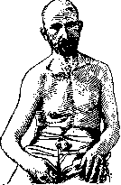
have only anterior shoulder dislocations, the further description refers only to them.—Diagnostic symptoms. The shoulder is abducted, tense, and can remain without support. The region of the shoulder joint has lost its roundness; under the tip of the protruding and, in thin people, even sharply outlined acromion, lying outward from the axis of the shoulder, there is a clearly palpable depression. The axis of the shoulder projects onto the coracoid process or even onto the middle of the clavicle. When attempting movement, the shoulder offers springy resistance, and during rotational movements, the head is palpable medial to the coracoid process (see Figure 1). Among the complications of shoulder dislocations, damage to the vessels and nerves of the axillary fossa is encountered, more often in the form of their compression—with sensory and motor disorders, especially on the part of the axillary nerve, which surrounds the head of the humerus posteriorly and innervates the deltoid muscle. Partial paralyses of this muscle, weakening the function of the shoulder, are not rare and can contribute to the formation of a recurrent dislocation. Also not rare are bone fractures at the lower-anterior edge of the glenoid cavity, as well as avulsions of tuberosities, especially often the greater tubercle (see Figure 32). Avulsion of the latter is diagnosed clinically by tenderness at the corresponding place of the head and the presence of profuse hemorrhage, descending in a strip from the head along the anterior surface of the shoulder to the elbow, sometimes extending to the forearm, even to the trunk. These complications, which cause extensive ruptures of the capsule, can also contribute to the formation of a recurrent dislocation if the patient begins heavy work too early. Recurrent shoulder dislocations are observed in 3-4% of all shoulder dislocations. The prognosis, in general favorable, is worsened by avulsions of tuberosities and ruptures of large vessels or nerves with the formation of either stiffness, or paralysis, or recurrent dislocation.—Treatment. Fresh uncomplicated dislocations must be reduced within the first 24 hours. After a month has passed, reduction is rarely successful. Of the numerous methods, the following are used: 1. A rougher method, practiced since the time of Hippocrates and called the Cooper method, boils down to the fact that the physician sits opposite the patient, lying on a bed or on the floor, and, resting a shoeless heel in the armpit, forcefully performs traction on the arm either along the length of the body or by the abducted arm. 2. The rotational method of Schinzinger. 3. Related to the latter, especially favored at the present time, the method of Ko-
Figure 1. Anterior dislocation of the right shoulder.
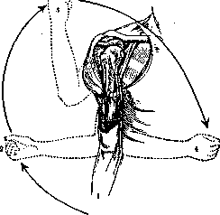
Figure 2. Reduction of a shoulder dislocation according to Kocher;
the numbers indicate the sequence of the four moments of reduction. relax the lig. coraco-humerale; by subsequent rotation of the shoulder inward with its adduction to the chest wall, the head rolls into place. 4. Among the gentlest and, from anatomical considerations, normal maneuvers, one should include the Mothe method, consisting of strong abduction, traction, and direct pressure with the fingers on the head. This method is also suitable for dislocations with avulsions of tuberosities, where rotational methods can increase the avulsions. 5. The Janelidze method—reduction of a dislocated shoulder hanging from a table, with the patient in a lateral position; pressure is applied downward on the forearm bent at the elbow with rotational movements—(see Figure 3). Whatever,
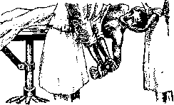
Figure 3. Reduction of a shoulder dislocation according to the Janelidze method.
however, whichever method is used, reduction becomes easy if performed under anesthesia. For old, unreduced dislocations (up to three months) and dislocations with a fracture of the surgical neck of the humerus, Hofmeister suggests attempting reduction after prolonged vertical suspension of the upper limb with traction through a pulley (on the healthy side). In irreducible dislocations, a semblance of a socket with fibrous growths around the head forms on the anterior surface of the neck of the scapula. In cases where such a neoarthrosis later acquires movement, surgical intervention is not needed. It is only necessary to attempt to increase it through mechanotherapy. In the absence of a neoarthrosis, with bony ankylosis, the scapula successfully takes over the movement of the shoulder. Thus, there is no particular need for bloody intervention in old dislocations of the shoulder. When attempting bloody intervention for the purpose of reduction, resection of the head, osteotomy of the neck, or freeing or suturing of a nerve, one must be wary of damaging the neurovascular bundle, to which the displaced head is closely adhered. Habitual dislocations of the shoulder are reduced very easily—often even by the patient themselves. When they recur too frequently due to insignificant causes, they exhaust the patient and force them to seek surgery. In this, the following methods are mainly used: 1) simple suturing of the capsule—capsulorrhaphy; 2) muscle transplantation to strengthen and support the joint from below: the Clairmont-Ehrlich method serves as a model for this method—transplantation of the posterior third of the deltoid muscle, passing it through the axilla via the foramen quadrilaterum and suturing it to the periosteum of the anterior circumference of the surgical neck of the humerus; 3) fascioplasty—according to Kirschner; and 4) fasciosuspension—with the goal of strengthening the capsule and suspending the head to the acromion (see Figure 4). Dislocations of the elbow (luxationes antebrachii s. cubiti) rank second in frequency after dislocations of the shoulder. A feature of the elbow joint is that the trochlea of the humerus articulates with the ulna, and lateral to the trochlea, a spherical elevation (capitulum humeri) serves for articulation with the radius. Thus, in the elbow joint, there is, firstly, simultaneous movement of both bones of the forearm in the form of flexion and extension around the trochlea and, secondly, separate rotational movements of the head of the radius, providing pronation and supination. Connected by strong annular and interosseous ligaments, the bones of the forearm usually dislocate together and most often result in posterior dislocations, somewhat less often in lateral, and most rarely in anterior and isolated dislocations of the ulna or radius. In the mechanism of the origin of posterior dislocation—lux. cubiti posterior (see Figure 5)—in the majority of cases, the influence of excessive extension at the elbow under the
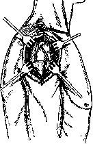
Figure 4. Fasciosuspension of a habitual dislocation of the shoulder by the greater tubercle to the acromion.
action of a single-armed lever is visible. The olecranon thereby becomes a fulcrum for both bones on the posterior fossa of the humerus. During hyperextension of the limb posteriorly at an angle, the trochlea strongly tensions the anterior wall of the capsule, ruptures it, the epiphysis of the humerus protrudes forward through the rupture of the capsule, and the bones of the forearm, connected to each other by the annular ligament,
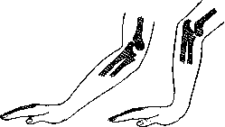
Figure 5. Mechanism of the origin of a posterior dislocation of the elbow: hyperextension followed by flexion.
slide posteriorly and superiorly. During this separation of the articular ends of the elbow, the lateral ligaments running to both sides of the ulna from the epicondyles of the humerus are also torn. The latter, especially the medial one, are often torn and result in profuse hemorrhage. At a young age, epiphyseolysis of the lower end of the humerus can often simulate a dislocation of the elbow. If, in a posterior dislocation, the end of the coronoid process still remains on the trochlea, it is called an incomplete dislocation. In a complete posterior dislocation, the coronoid process is displaced into the posterior fossa. Sometimes, however, it is torn off by the brachialis internus muscle. Symptoms of posterior dislocation: The elbow is extended at an angle of about 140°, and the region of the joint is widened from front to back. The olecranon protrudes sharply posteriorly; above it is a groove with tension of the triceps; when pressing with a finger on the groove, the finger sinks deeply but does not palpate the bone of the humerus. The head of the radius is often even visible to the eye and is easily palpated under the skin during rotational movements of the forearm, posterior to the projection of the humerus (see Figure 6). The smooth articular surface of the trochlea is often well palpated from the front. The relationship of the classic points on the elbow—the tip of the olecranon and the two epicondyles—is disturbed compared to the healthy side: instead of an isosceles triangle (at an angle of 140°), the 3 points lie almost on a single straight line, and, depending on the height of the olecranon's position, its tip may even stand higher than the line connecting the epicondyles. In this case, the distance of the latter to the tip is increased. Springy resistance remains even under anesthesia, since the coronoid process, abutting against the humerus, serves as an obstacle to flexion.
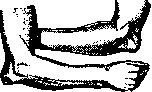
Figure 6. Posterior dislocation of the elbow, dorsal and palmar views (Anger).
into the posterior fossa above the trochlea and forming a fulcrum for a two-armed lever, the upper end of which pulls on the triceps muscle (see Figure 33). Reduction of an elbow dislocation is based on the principle of hyperextension in order to release the coronoid process. Resting the elbow on his knee, the surgeon extends the injured limb, stretches it along its length, and then performs flexion at the elbow. Posterior dislocations of the elbow sometimes allow for successful reduction even after quite long periods (several months). Lateral dislocations usually represent a variation of the posterior one in terms of the basic mechanism. If, for example, during a fall on an outstretched hand, a deviation of the terminal part of the forearm occurs in a lateral or medial direction, then its upper end is dislocated posteriorly and medially or posteriorly and laterally; the former occurs more often due to the normal valgus of the elbow joint (see Figure 34). These dislocations, which are more complex in their clinical manifestations, result in displacement and incorrect relationship of anatomical landmarks, and are accompanied by rupture of ligaments and avulsion of bony prominences. Accurate diagnosis of them in the initial period is hindered by extensive hemorrhage enveloping the site of injury. X-rays are especially useful here. Reduction is based on the basic hyperextension and lateral pressure on the displaced prominences from one side or the other. The rare anterior dislocation of the elbow (luxatio cubiti anterior) usually occurs during a fall on the elbow with its excessive flexion. A distinction is made between an incomplete dislocation, when the olecranon stands, resting against the trochlea, and a complete one, when it stands in front of the trochlea. Often, a fracture of the olecranon also occurs during this. Their reduction, in the presence of a large rupture of the capsule, does not present any particular difficulties with traction and pressure from front to back on the flexed forearm. A divergent dislocation (luxatio cubiti divergens), occurring in the elbow during severe trauma due to the wedge-like impaction of the humerus between the bones of the forearm after rupture of the interosseous and annular ligaments, is encountered very rarely. Reduction is easy due to large ruptures of the capsule. In difficult cases, each bone requires separate reduction: the ulna by extension and traction; the radius by direct pressure with rotation. An isolated or separate posterior dislocation of the ulna arises from a fall on an excessively extended and abducted forearm. It is determined by almost the same clinical signs as a posterior dislocation of both bones. But, due to the upward displacement of the medial bone while the lateral one remains in place, the arm forms a medial shortening and an angle open medially (cubitus varus). Pronation and supination are possible. It is reduced by traction of the supinated forearm with simultaneous straightening of the lateral angle and extension of the elbow. All three of the latter elbow dislocations are very rare. Accurate determination of them is facilitated by fluoroscopy. Of the isolated dislocations, a separate dislocation of the radial head is of much greater importance, which arises mostly from a direct blow from back to front or from the outside. But it can also arise from indirect violence—due to strong pronation of the forearm (twisting of the arm), whereby the annular ligament is ruptured. Most often, a dislocation of the radius forward and outward occurs, when its head lies over the lateral epicondyle of the humerus. Clinically, the forearm is in a position of flexion and pronation and forms an angle with the humerus that is open outwardly—cubitus valgus. The head is palpable in the elbow crease—in front of or above the lateral epicondyle of the humerus—and is recognized by its shape and by rotation during pronation and supination. The posterior anatomical landmarks of the elbow are not displaced (see Figure 35). This dislocation is often preceded by a fracture of the upper third of the ulna. Reduction is usually easily achieved by extension, supination, and pulling on the forearm and by direct pressure on the head. But with the same ease, the head pops out again during movement. Its firm retention in place is hindered by the torn parts of the capsule and the annular ligament. Retention of the head in place is best facilitated by a fixation bandage with pronation and flexion of the forearm at a very acute angle at the elbow. In an irreducible dislocation, the obstacle must be removed operatively—sometimes even by resection of the head. Dislocations of the wrist. Dislocations in the area of the radiocarpal joint are a very rare phenomenon. Injuries that were taken for dislocations here in the pre-X-ray era were classic fractures of the radius. Equally rare are isolated dislocations of the ulna in the dorsal or palmar direction of the wrist. Of the dislocations of the carpal bones, a dislocation of the lunate bone in the palmar direction is observed more often than others. It is almost never possible to put the small bones of the wrist back into place. Therefore, one has to resort to open enucleation, which has little effect on the good function of the wrist. Dislocations of the fingers. Of the dislocations of the fingers, the most frequently observed is the dislocation of the proximal phalanx of the thumb in the dorsal direction of the head of the metacarpal bone, which occurs from forced extension of the finger. In this case, the metacarpal head ruptures the capsule on the palmar side. Symptoms. The thumb, excessively extended at the base, is flexed at the interphalangeal joint, due to which it has a bayonet-like shape. On the back of the metacarpal bone, the protrusion of the base of the phalanx is palpable, and on the palmar surface—the head of the metacarpal
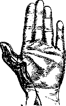
Figure 7. Dislocation of the thumb posteriorly with entrapment of the sesamoid bone and the long flexor of the thumb.
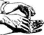
bone (see Figure 7). In cases difficult to reduce, the tendon of the flexor pollicis longus muscle or a torn part of the capsule together with the sesamoid bones, to which the two heads of the flexor pollicis brevis are attached, becomes entrapped between the articular ends. Some subjects can voluntarily create a subluxation of the thumb by strongly tensing the extensor and producing dorsal flexion of the finger at a right angle. Figure 8. Reduction of a dislocation
of the thumb—moment of hyperextension.
of a dorsal dislocation of the thumb is performed by strong hyperextension of it (see Figure 8). This facilitates the release of the entrapped sesamoid bones and the tendon, which are set in place by traction followed by flexion of the proximal phalanx. Anesthesia is necessary. With the difficulties encountered, open reduction is often needed. Dislocations of the thumb in the palmar direction are rare and occur from excessive flexion. Their reduction is achieved with the help of traction, dorsal flexion, and direct pressure on the base of the phalanx protruding in the palmar direction. Dislocations of the phalanges of the other fingers also occur most often from excessive extension and flexion and are divided into dorsal, palmar, and (rarely) lateral, and are recognized by the direction of the angulation of the finger axis and by palpation of the head of the central bone and the base of the dislocated bone. Their reduction
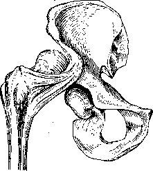
Figure 9. Posterior superior dislocation of the hip—luxatio iliaea (according to Helferich).
is usually easy—with the help of pulling on the end of the finger with pressure on the base of the dislocated phalanx, followed by its flexion—in the case of a dorsal dislocation, or extension—in the case of a palmar one. Dislocations of the hip (luxatio femoris) occur, according to Krönlein, in 2%, according to Russian statistics—somewhat more often: according to Toropov and Kisel—in 7%, according to Tikhov—in 9% of all dislocations. Depending on the position of the head, standing in front of or behind the acetabulum, hip dislocations are divided into anterior and posterior. Depending on the high or low standing of the head, both of these types of dislocations are further divided into superior—luxatio iliaea (see Figure 9) and pubica (see Figure 10) and inferior—ischiadica (see Figure 11) and obturatoria (see Figure 12). In the mechanism of the origin of these dislocations, Bigelow attaches primary importance to a very strong ligament—the iliofemoral ligament of Bertini. Starting from the anterior inferior iliac spine and spreading out like a fan, it attaches to the anterior intertrochanteric line of the femur, dividing here into two legs—medial and lateral. According to Bigelow, it rarely ruptures, and its integrity helps the formation of one or another typical position of the dislocation, while its rupture destroys the typicality of the dislocation. The round ligament—ligamentum teres, on the contrary, almost always ruptures during a dislocation. Hueter divides dislocations by origin: 1) dislocations from flexion
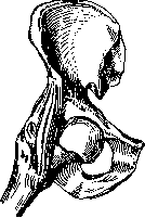
Figure 10. Anterior superior dislocation of the hip—luxatio pubica (according to Helferich).
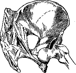
Figure 11. Posterior inferior dislocation of the hip—luxatio ischiadica (according to Helferich).
and adduction (luxatio iliaea and ischiadica) and from flexion and abduction (luxatio obturatoria) and 2) dislocations from extension and abduction (luxatio iliopectinea and suprapubica). Posterior dislocations are the most frequent (up to 80% of all hip dislocations). During sharp flexion, adduction, and inward rotation of the leg, exceeding physiological limits, the neck of the femur rests against the anterior-superior edge of the acetabulum. By forming a fulcrum here, a two-armed lever is created, the short end of which—the head—strongly tenses and ruptures the posterior, poorly reinforced dislocations
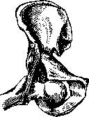
Figure 12. Anterior inferior dislocation of the hip—luxatio obturatoria (according to Helferich).
part of the capsule. If the limb, during forced adduction and internal rotation, is flexed less than a right angle, a luxatio iliaca occurs with a rupture of the capsule above the tendon of the obturator internus muscle. When the thigh is flexed more than a right angle, a luxatio ischiadica occurs, with a rupture of the capsule and the head exiting under the tendon of the obturator internus muscle. This position of the head can change into an iliaca position—in the presence of continued force—in the form of internal rotation of the thigh. In general, the head stands lower the stronger the initial flexion was and the lower the capsule was ruptured posteriorly. Symptoms of posterior dislocation. In luxatio iliaca, the leg is adducted, slightly flexed at the hip and knee joints, sharply rotated internally, and presents springy resistance when attempts are made to extend it at the hip or knee (see Figure 13). The trochanter stands above the Roser-Nelaton line and anterior to its midpoint (the measurement is shown in Fig. 14). Luxatio ischiadica differs little in its clinical picture from luxatio iliaca. There is also a tense, adducted, and internally rotated position of the limb, but it is significantly more flexed at the hip and knee joints and is almost not shortened compared to the same position of the other leg. In thin individuals, the head can be easily palpated in the gluteal region. Sometimes disorders of the sciatic nerve are observed (see Figure 11). Anterior dislocation can be obtained on a cadaver by forced abduction with external rotation and extension. In life, these dislocations occur, for example, when falling from a height onto spread legs with the torso thrown backward. The posterior-inferior edge of the acetabulum serves in this case as a fulcrum for the neck of the femur, and the head easily ruptures the weak anterior-superior part of the capsule and produces a luxatio suprapubica. The head can then move further upward, under the iliopsoas muscle, and produce a luxatio iliopubica. Moving inward to the pecten ossis pubis, it produces a luxatio iliopectinea. In luxatio pubica, the vessels and nerves pass medial to the head; in luxatio pectinea, they pass beneath it and can be compressed by the head. Symptoms of anterior dislocation. In luxatio pubica, the leg is extended or very
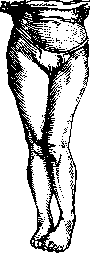
Figure 13. Clinical picture of luxatio iliacae sin.
slightly flexed, abducted, rotated externally, usually not shortened, and, due to the abduction, even appears longer. The tense state of the leg does not allow for active movements; passive movements are slightly possible in the direction of the altered positions. Below Poupart's ligament, the head of the femur is either visible or easily palpable. Medial to it should
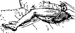
Figure 14. Projection of the Roser-Nelaton line—from the anterior superior iliac spine to the ischial tuberosity.
pulsate the femoral artery, which can be compressed. The femoral and saphenous nerves can also be subjected to compression and cause changes in sensitivity on the thigh and lower leg (see Figure 10). If, however, the torso does not throw backward when falling on extended, spread legs, but on the contrary, the abducted and externally rotated leg maintains a flexed position at the hip, then under force, the head presses against the anterior-inferior part of the capsule. Due to the fixation of the trochanteric region by the strongly taut ligament (iliofemoral ligament), a two-armed lever is formed, the short arm of which—the neck with the head—ruptures the anterior-inferior part of the capsule, settles at the obturator foramen, and produces an inferior anterior dislocation—luxatio infrapubica s. obturatoria (see Figures 12 and 15). Its symptoms are similar to the symptoms of an upper anterior dislocation, but here the position of the leg is even more characteristically expressed—sharply flexed at the hip and knee joints, abducted, and rotated externally. In this position, the head proves to be firmly fixed (see Figure 15). Luxatio supra- and subglenoidalis are very rare forms of hip dislocation. The former is more related to luxatio iliaca than to luxatio iliopectinea; the latter is related to luxatio obturatoria and ischiadica, from which they can form secondarily. Of greater importance is the also rather rare displacement of the femoral head through the fractured floor of the acetabulum, through which the head perforates into the pelvis—luxatio centralis. This fracture-dislocation occurs either from direct force on the trochanter or from a fall on the side. Movements of the thigh, especially abduction, are sharply hindered. The lateral
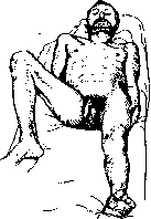
Fig. 15. Clinical picture of luxatio obturatoriae.
distance from the trochanter to the midline of the body is reduced compared to the healthy side. The floor of the acetabulum pushed into the pelvis with the femoral head is well palpable through the rectum. On an X-ray, a sharp protrusion of the acetabular floor into the pelvis is visible, the transverse dimension of which is reduced here. Reduction of a hip dislocation is a rather rewarding task. Even old, 1–2-month-old hip dislocations can be reduced. Cases have been cited where reduction was successful even after a year. Reduction is best always performed under general anesthesia or spinal anesthesia. Of the old methods, traction along the longitudinal axis has been abandoned as irrational due to the sharp tension of the iliofemoral ligament, which hinders reduction. When reducing posterior dislocations, the lever method of Pouteau-Despres, developed by Bigelow and based on flexing the thigh with its external rotation, is very popular among new methods. The patient is placed on the floor. An assistant fixes the pelvis well with both hands, and the surgeon takes hold of the lower leg with both hands. Lifting the leg upward, he flexes the knee and hip joints at a right angle and pulls the thigh upward. The iliofemoral ligament relaxes in this process, and the head settles at the posterior edge of the acetabulum. During subsequent external rotation, the again-tightening iliofemoral ligament will serve as the punctum fixum of the lever, directing the head into the acetabulum. If reduction by this method is not successful, then with continuous traction upward on the flexed thigh, before starting abduction, it is necessary to perform further adduction and internal rotation of the thigh in order to relax the iliofemoral ligament even more with such a maneuver and bring the head as close as possible to the acetabulum, and then, with a quick external rotation and abduction, push it into the acetabulum (see Figure 16). If the dislocation is not reduced even with this technique, Bigelow advises first performing circumduction with the lower end of the thigh to increase the rupture of the capsule. In 1921, Janelidze indicated a method described by Malgaigne and used even earlier by Collin and Colombot—reduction of a hip dislocation in the prone position. The thigh hangs down from the side of the table. Having flexed the knee to a right angle and holding the ankle joint with his hand, the surgeon presses with his knee on the patient's popliteal fossa, thereby producing traction along the axis of the flexed thigh, rotates it internally, and abducts it. Anterior dislocations are also reduced by the lever method, with relaxation of the iliofemoral ligament by flexing the thigh to a right angle; to increase the gap in the capsule, abduction is performed; with subsequent internal rotation of the thigh with adduction, the head falls into place. After reduction of a hip dislocation, a fixation bandage is applied for 2–3 weeks, followed by massage and mechanotherapy. Dislocations in the knee region. Here, three types of dislocations or subluxations are encountered: of the lower leg, the menisci, and the patella. Complete dislocations of the lower leg (anterior, posterior, and lateral) are an extremely rare phenomenon. Flexing only in the sagittal direction, the knee is held from anterior-posterior displacement by powerful intra-articular cruciate ligaments: the anterior cruciate ligament, which runs from the lateral condyle to the anterior intercondylar fossa of the tibia and tightens during strong flexion, and the posterior cruciate ligament, which runs from the medial condyle to the posterior-superior edge of the tibia and prevents hyperextension of the knee. Force acting on the lower leg from behind or in the sense of forced flexion ruptures only the anterior cruciate ligament; the posterior one relaxes in this process; together with the long collateral ligaments, it holds the articular ends from a complete dislocation, resulting only in an anterior subluxation. If, with continued force, the posterior cruciate ligament is also ruptured following the anterior one, a complete anterior dislocation occurs. When force acts on the lower leg from the front or in the sense of hyperextension, the posterior cruciate ligament tightens and ruptures. With this displacement of the lower leg posteriorly, the anterior cruciate ligament relaxes and, remaining
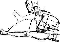
Figure 16. Reduction of posterior hip dislocations. The numbers indicate the sequence of the four moments of reduction.
intact, together with the long lateral ligaments, holds the lower leg from a complete dislocation, forming only a posterior subluxation. A rupture of the anterior ligament by continuing force results in a complete posterior dislocation. Thus, a complete anterior or posterior dislocation of the lower leg requires very great, prolonged force, which must rupture both cruciate ligaments. A lateral dislocation of the lower leg requires not only the rupture of both cruciate ligaments but also both long lateral ligaments, and is observed even less frequently. The clinical picture of a knee dislocation is so typical that it does not present diagnostic difficulties. However, hemarthrosis during the rupture of internal ligaments can significantly hinder the correct diagnosis of a dislocation. Profuse hemorrhage above and below the knee can serve as evidence of the rupture of large vessels, by which a complete dislocation is often complicated. Reduction of a knee dislocation is performed quite easily by traction of the lower leg along its length with counter-pressure on the protruding ends. Fixation with an immobile bandage in the extension position for 4-6 weeks and then massage with cautious passive and active movements. Much more often than a knee dislocation, anterior and posterior subluxations are observed (see Figure 17). They are based on the rupture of only one cruciate ligament, anterior or posterior, and are recognized by the anamnesis and hemarthrosis, whereby the avulsion of the anterior cruciate ligament gives sharp palpation tenderness under the ligamentum patellae—at the site of its attachment to the upper epiphysis of the tibia, and the rupture of the posterior ligament—in the popliteal fossa, at the site of attachment to the posterior surface of the tibia. There is also a symptom of anterior and posterior subluxation of the lower leg, which the French call tiroir du commode ("chest of drawers"): this is a slight displacement of the lower leg along the ends of the femur. With a rupture of the anterior cruciate ligament, with the femur and foot fixed, the slightly bent lower leg is pushed forward somewhat from the femoral condyles with both hands, and with a rupture of the posterior cruciate ligament, the lower leg is pushed backward from the femoral condyles. Patients themselves learn to perform this pushing out or pushing in of the "drawer" of the lower leg: by fixing the foot at the end of the bed or with the other foot, tensing the muscles with the knee slightly bent, they produce the movement of the lower leg from the condyles forward or backward.
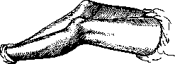
Figure 17. Posterior subluxation of the lower leg after rupture of the ligamentum cruciatum posterius.
Fresh subluxations are treated in the same way as dislocations—by normal setting of the knee ends with a bandage fixing the limb in the extension position for three to four weeks, followed by massage and passive and active exercises. In connection with the development of physical culture, knee subluxations have begun to be encountered more often. Old subluxations, which are frequently recurring and interfere with walking, sometimes have to be operated on. For the restoration of ruptured cruciate ligaments, a number of plastic operations have been proposed, including transplantation of the fascia lata or the tendon of the m. semitendinosus. The graft is passed from the side, through a drilled femoral condyle, along the course of the cruciate ligaments, and is attached through a drilled epiphysis of the tibia or in the region of the tuberositas tibiae or from behind—having passed through a canal in the outer condyle of the tibia. Dislocation of the menisci, predominantly the medial one, was previously considered a very frequent phenomenon. At the present time, it has been clarified that rupture (fracture) of the menisci occurs more often than dislocation. The sickle-shaped menisci, connected by their wide outer side to the capsule, and by their rounded ends to the cruciate ligaments, prevent lateral wobbling of the knee. Their dislocation, like ruptures, occurs most often during rotation of the torso with a fixed foot. A pure dislocation of the meniscus is difficult to differentiate from its rupture or fracture and usually goes under the diagnosis of meniscitis. Dislocation of the patella occurs predominantly in the outward direction (see Figure 18). There are many main factors in the mechanism of lateral dislocation of the patella: genu valgum and as a consequence—lateropositio m. quadricipitis, a reduction of the fossa intercondyloidea, a reduction of the outer condyle of the femur and flattening of the patella, a change in the elasticity of the capsule and laxity of the capsule after debilitating acute infections. The presence of these factors very often creates a habitual dislocation after the first dislocation of the patella. The accessibility of the patella to direct palpation makes easy recognition of its displacement possible. Even a primary dislocation of the patella is too rarely seen and reduced, because often the patient himself, standing on his feet and bending toward the knee, relaxes the quadriceps and, while rubbing the knee, reduces the patella. Even more so, the surgeon does not have to see secondary dislocations, because patients themselves learn to easily reduce them by the indicated method—natural flexion of the hip in the pelvis with the knee extended, with lateral pressure on the patella. A frequently recurring habitual dislocation of the patella
has to be operated on. There are up to 55 different modifications of patellar fixation for habitual dislocation: by means of its enucleation, deepening of the fossa intercondyloidea, bone condyloplasty, excision of pieces of the capsule or its suturing, transplantation of the thigh muscle from the outer side to the inner, etc. Most of these operations leave disfiguring long scars and a lack of knee function; there have even been observed trophic changes on the part of the damaged sensitive branches of the n. cruralis and n. saphenus. Therefore, it is best to perform the following simple operation. According to the idea of G. I. Turner, using a Textor incision, a transplantation of the lateral half of the ligamentum patellae, separated from the capsule, is performed together with the corresponding part of the tuberositas tibiae. The latter is transferred to the medial surface of the tibia and attached under strong tension.
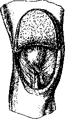
Figure 19. Transplantation of the lateral half of the ligamentum patellae to the medial side of the tibia in habitual dislocation of the patella.
...here under the detached muscles subperiosteally, in a notch of the bone (see Figure 19). Dislocation of the foot (luxationes pedis). Among dislocations of the foot, one distinguishes: dislocation of the ankle joint, isolated dislocation of the talus, and subtalar dislocation. Dislocation of the ankle joint (lux. pedis) is divided into anterior, posterior (the most frequent), and lateral. In pure form, they are all quite rare. Much more often they appear as a result of a fracture of the malleoli and especially often complicate a Dupuytren's fracture of the lower leg. An anterior dislocation of the ankle joint, formed by forced dorsiflexion of the foot, gives a noticeable lengthening of the dorsum of the foot and a reduction of the heel prominence posteriorly. With a pure posterior dislocation, occurring, mainly, during sharp plantar flexion, there is, conversely, a lengthening of the heel and shortening of the dorsum of the foot. Lateral dislocations without fracture of the malleoli are unthinkable. A Dupuytren's fracture causes them especially often. In this case, the foot assumes a position of pes varus or valgus. Reduction of pure dislocations of the lower leg is achieved very easily, especially under anesthesia. An isolated dislocation of the talus (lux. tali) in pure form is encountered rarely. The talus can be displaced in all directions (but mostly outward) and even rotate around its axis. To obtain this dislocation, it is necessary to rupture the ligaments fixing it to the lower leg, to the calcaneus, and to the navicular bone. These ruptures occur either during forced pronation or supination, or during concomitant dorsiflexion or plantar flexion of the foot. The predominant direction of force determines the displacement of the talus in one direction or the other. Often the neck of the talus breaks during this. Reduction of pure dislocations of the talus is possible with traction of the foot with the knee bent and with direct pressure on the protruding bone. In difficult cases, one resorts to open reduction of the dislocated talus into place or even to its removal. A subtalar dislocation (lux. sub talo) is obtained inward or outward during strong supination or pronation of the forefoot fixed on the ground. In this case, the ligaments of the talocalcaneal and talonavicular joints are ruptured; a sharper separation occurs in the latter joint (see Figures 20 and 36). With sharp pronation of the fixed forefoot by a rotational movement of the lower leg inward, a displacement of the navicular bone with the forefoot outward is obtained. Under the same conditions, sharp supination of the foot with rotation of the lower leg outward gives a displacement of the navicular bone and the foot inward. It is easy to recognize a lux. sub talo by this characteristic position of the foot. In the ankle joint, in this case, it is possible to pro...
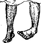
Figure 20. Subtalar dislocation of the foot—clinical picture.
perform flexion and extension. Extremely rare forms of lux. sub talo backward or forward require fractures for their origin. Reduction of pure subtalar dislocations under anesthesia is easily achieved due to the extensive ruptures of the articular connections. Fixation with an immobilizing bandage for 4-5 weeks and approximately the same amount of time for mechano- and physiotherapy provides almost normal foot function after 2-3 months. Among rare foot dislocations, it is necessary to note: dislocation onto the dorsum of one navicular, one cuboid, one, two, or three cuneiform bones, very rare dislocations in the Chopart joint, and, finally, somewhat more frequent dislocations of the metatarsi in the Lisfranc joint: most often of all bones onto the dorsum and outward (total dislocation), less often of one or several bones (partial). These dislocations occur from the action of great forces, for example, when falling on the toes from a height in the presence of sharp plantar flexion or direct pressure on the front part of the foot together with plantar flexion. Fractures and distortions occur more often than dislocations in the Lisfranc joint. Without an X-ray, it is difficult to establish an accurate diagnosis of these injuries. Their reduction is achieved by traction on the end of the foot with counter-pressure on the protruding bones; a plaster cast fixes the foot for 4-5 weeks; the patient can walk in it after a week. Very often these dislocations are combined with fractures and avulsions of bones at the sites of attachment of powerful ligaments and tendons, and with injuries to nerves and soft tissues. In such cases, the restoration of function occurs very slowly, sometimes pain persists for years, forcing one to resort to surgery: excision of small bone fragments, resection of neuromas, or to neurotomy. But before operating, it is necessary to test the effect of an insole made exactly from a plaster cast of the diseased foot. Dislocations of the metatarsophalangeal and interphalangeal joints of the foot occur according to the type of hand ones. Dislocations of the vertebrae are a rare phenomenon; fractures are observed here much more often, and dislocations as a consequence of them. In pure form, these dislocations are encountered almost exclusively in the region of the cervical vertebrae. The upper part is considered the dislocated one. Flexion and rotational dislocations are distinguished. Excessive extension meets an obstacle in the approximation of the vertebral arches, which prevents dislocation. Excessive flexion, however, by bending the head toward the chest, can displace the articular processes of the upper vertebra from the lower one (incomplete dislocation), and upon subsequent straightening of the spine, they can slide further, settling in front of the articular processes of the lower vertebra (see figure 21), having "caught" behind them (complete dislocation). Here the head end acts as the force of a single-armed lever with a fulcrum on both anterior edges of the articular parts of the underlying vertebra. Therefore, a flexion dislocation is always bilateral. In a rotational dislocation, due to excessive lateral flexion with stretching of the ligaments, the articular process of the upper vertebra rests against the arch of the lower one as a fulcrum, and with further violence of the single-armed lever thus obtained, the articular processes on the other side separate, repeating the form of a flexion dislocation, incomplete or complete with locking. On the side of the lever's fulcrum, i.e., where its rotation occurred, the upper articular process shifts posteriorly from the lower one due to stretching of the capsule. Thus, in essence, even with a rotational dislocation, displacement occurs on both sides, but in opposite directions. In view of the fact that locking occurs here on one side, a rotational dislocation is otherwise called unilateral. Both flexion and rotational dislocations are observed only in the cervical part—during sharp flexion in the horizontally located articular processes: flexion—with complete rupture of the joints on both sides, rotational—only with less rupture on the side of the rotation point. Symptoms of flexion dislocation: the head is tilted forward, and the chin almost touches the sternum. Due to significant mobility and sharp pain, patients often support their head with their hands while walking. On the fan of diverging spinous processes, the protrusion of the spinous process of the underlying vertebra is visible to the eye, and an indentation of the overlying one is felt above it. The anterior protrusion of the body of the overlying vertebra makes swallowing difficult; the dislocated vertebra is felt through the throat or determined by laryngoscopy. In a rotational unilateral dislocation, the position is different, depending on the degree of dislocation. With an incomplete dislocation, the neck is extended, and the head is tilted and turned to the healthy side; with a complete one—it is tilted toward the chest and toward the side of the dislocation and rotated to the healthy side, and the spinous processes of the upper vertebrae are deviated toward the side of the dislocation. No less important symptoms of both dislocations are nervous phenomena—in the form of changes in sensitivity of a radicular nature, pareses, and paralyses in the upper extremities as a consequence of compression or damage to the roots. Paralysis of all four extremities will indicate damage to the substance of the spinal cord itself. The severity of this kind of change is decisive for the prognosis. A fatal outcome is not rare. Reduction of vertebrae is not an easy task even under anesthesia. Incomplete unilateral dislocations are reduced more easily than others—by traction along the length of the body and rotation in the opposite direction; complete dislocations, i.e., those where "locking" exists, need to be freed from it.
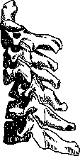
Figure 21. Flexion dislocation of the cervical vertebrae with locking.
by flexing the head toward the side of the dislocation, with rotation to the healthy side, under constant traction. Having thus freed it from the locking and continuing traction, one now acts with the reverse maneuver: abduction to the healthy side and rotation toward the side of the dislocation. It is recommended to first convert a flexion dislocation into a rotational, unilateral one, and then perform its reduction, after which a plaster collar is applied for 4-6 weeks. Most often in the cervical part, these kinds of dislocations occur between the IV-V, V-VI vertebrae, i.e., in the middle part of the cervical vertebrae, where the overall mobility anteriorly is greatest. In the region of the two upper vertebrae, where there are no cartilaginous discs and the joints are wide, they are reinforced by such a powerful ligamentous apparatus that dislocation here is a rare and exceptional phenomenon. Dislocation of the I vertebra from the head, or, more accurately, dislocation of the head from the atlas, is possible either during sharp flexion or during rotation, but almost always with a fatal outcome. The same can be said about dislocation of the atlas from the epistropheus, if it is not accompanied by a fracture of the odontoid process. The latter is firmly connected to the anterior arch of the atlas by powerful transverse ligaments and strong alar and cruciate ligaments with the anterior edge of the foramen occipitale magnum. During sharp tilting of the head with traction (for example, during hanging), these ligaments can rupture; when the atlas moves forward, the odontoid process crushes the spinal cord, and instantaneous death occurs. If the odontoid process is broken off, it moves forward with the anterior arch of the atlas, and the spinal cord is not subjected to compression. In the thoracic part, the spine, being connected to the ribs, is immobile and yields displacements only after fractures of the articular parts. The same can be said about the more mobile lumbar part, reinforced by powerful long and short ligaments. Here the planes of the joints are located in the frontal direction and thus even more prevent dislocation from flexion, the volume of which is greater here than other types of movement. With hyperflexion, fractures of the bodies or articular parts are more often possible here. The phenomenon described for the first time by Killian under the guise of spondylolysis and spondylolisthesis, and by Lambl under the name "self-dislocation of the spine," is a chronic sliding of the body of L. V forward and downward from the sacrum as an inevitable consequence of a congenital defect in the development of the arches in the space between the articular processes, most often L. V. The clinical picture of spondylolysis and spondylolisthesis described by Turner and his school in our time turns out to be a not infrequent phenomenon in both women and men. Acute manifestations of this defect in the form of spondylolisthesis are observed under the influence of sharp physical violence, lifting weights, etc. In women, repeated pregnancy can cause the gradual development of the same symptom. Dislocations of the clavicle at both its ends (lux. claviculae sternalis and acromialis) are observed quite often. Dislocations of the sternal end are almost always anterior and occur from the action of force on the anterior surface.
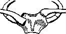
Figure 22.
of the shoulder: by the two-armed lever formed thereby, with a fulcrum on the 1st rib, the anterior end of the clavicle protrudes forward, resulting in an incomplete or complete anterior dislocation (lux. praesternalis; see figure 22)—the most frequent. Superior dislocation—lux. suprasternalis—results from a blow to the outer end of the clavicle, also with the formation of a lever with a fulcrum on the 1st rib. Posterior dislocation—lux. retrosternalis (see figure 23)—usually occurs from a direct blow to the chest at the sternal end of the clavicle. All these dislocations are easily recognized even by the eye by the swelling in the region of the sternal end of the clavicle or by the depression at the site of its attachment in posterior dislocation. In the latter case, the head behind the sternum presses on the trachea and esophagus. The remaining dislocations cause almost no functional disturbances. Acromial dislocations are encountered more often than sternal ones in the form of lux. supra- and infra-acromialis. These dislocations with rupture of only the lig. acromio-clavicularis result in an incomplete dislocation, while a complete one is obtained after rupture of the lig. coraco-clavicularis as well. Supra-acromial dislocations occur from a blow from above onto the acromion—consequently, here one can speak of a dislocation of the scapula. Infra-acromial ones result from direct violence to the outer end of the clavicle, which is displaced downward and produces a step-like notch here. All these dislocations are so typical that they are established even by the eye. All clavicular dislocations are reduced by simple finger pressure, but they just as easily pop out from under the finger immediately. Fixation with bandages and pads also helps little in holding the ends. Sometimes surgical intervention is necessary—sutures on the capsule and ligaments with 3-4 weeks of fixation of the entire limb with a Desault bandage. Posterior sternal dislocations are especially subject to surgical intervention to prevent the occurrence of complications from the trachea. Dislocations of the sternum and ribs. Dislocations of the sternum are very rare; only dislocations of the manubrium and xiphoid process posteriorly from the body of the sternum are known.—Dislocations of the ribs are also such a rare phenomenon that some authors even doubt the possibility of their existence. Dislocation of the lower jaw constitutes about 2.5% of all dislocations and can be unilateral or bilateral (see figure 24); the latter are more frequent. They occur from excessive opening of the mouth during yawning, vomiting, tooth extraction, insertion of a gastric tube, and more rarely from a sharp blow from above downward on the chin. The interarticular cartilage, the meniscus, divides the cavity of the lower jaw joint into two halves—upper and lower. During normal opening of the mouth, the articular heads
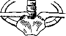
Figure 23. Figure 22 and 23. Anterior and posterior dislocations of the sternal end of the clavicle (according to Bauer). of the lower jaw are displaced forward only in relation to the meniscus. With a stronger opening of the mouth, the jaw head slides anteriorly along the articular fossa already together with the meniscus, until the articular tubercle (tuberculum articulare) stops this movement. A predisposing cause of dislocation is flattening or insufficient development of the articular tubercle, which is often observed in women. For this reason, dislocation is noted in them more often than in men. The mechanism of the origin of dislocation during rapid, wide opening of the mouth consists in the fact that one or both articular heads of the lower jaw roll onto the articular tubercle. The powerful ligament lig. stylo-mandibulare, running from the processus styloideus along the length of the vertical ramus to the angle of the lower jaw posteriorly, as well as the lig. spheno-mandibulare from the inside, are tensed during this. The mandibular angle, held by them like reins and pulled posteriorly and upward, becomes a punctum fixum for the entire jaw as a lever and shifts the articular heads even further anteriorly from the articular tubercle. In this position, the heads with the entrapped menisci, in addition to the tension of the indicated ligaments, are fixed by the tension of the masticatory muscles. Thus, it becomes impossible for the jaw heads to jump back over the articular tubercles. Symptoms of bilateral dislocation: the mouth is springily open, the chin is protruded forward and springs. Occlusion of the teeth is impossible, saliva is secreted, speech is difficult, the cheeks are flattened, anterior to the tragus is a deep fossa. The articular head of the lower jaw is palpable under the zygomatic arch, and beneath it the masticatory muscles bulge like ridges. In unilateral dislocation, these signs are only on one side. The jaw is less fixed. The chin is deviated to the healthy side. The latter circumstance is important, since in a fracture of the articular processes the chin is deviated toward the fracture. Reduction Figure 24. Bilateral dislocation of the lower jaw. is achieved easily and without anesthesia. Its principle consists in guiding the head, stuck anterior to the articular tubercle, backward over the prominence of the tubercle. For this purpose, the patient is seated on a low seat with support under the occiput. The surgeon places his thumbs along both rows of lower teeth, if possible reaching with their ends to the angles, and with the remaining fingers grasps the lower jaw from the outside (see figure 25). Making the angle of the lower jaw the pivot point, the surgeon quickly exerts pressure with his fingers on the angle downward and backward; the chin thereby makes a movement upward and backward. If reduction of both sides does not succeed immediately, it is necessary to try to reduce one side first, then the other. Otherwise
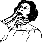
Figure 25. Reduction of a dislocation of the lower jaw.
reduction is easily achieved under anesthesia. A capistrum bandage and liquid food for 2-3 weeks, to avoid recurrence of the dislocation. Cases of reduction of old dislocations of the lower jaw after 8 months have been described. In irreducible dislocations that impede the act of chewing, open reduction or resection of the articular ends is indicated. In the presence of habitual dislocation, injection of Tinctura Iodi or alcohol into the jaw joint or its circumference is recommended, and in any case, it is necessary to forbid wide opening of the mouth. Dislocation of the lower jaw posteriorly is an extremely rare phenomenon. It results from direct violence, e.g., from a blow from the front on the chin with the mouth closed. The articular heads, shifting sharply posteriorly, can perforate the auditory canal. II. Habitual dislocations. Conditions may arise where, after a dislocation, the capsule and ligaments remain sufficiently stretched for a repeat dislocation to easily occur. This is also facilitated by damage to muscles, partially or entirely paralyzed due to their ruptures or damage to the nerve branches supplying them, as, for example, exists in the shoulder in the form of damage to the branches of the n. axillaris. Various factors leading to general weakening of the organism, such as severe infection, hunger, etc., can also be predisposing conditions. More often than others, habitual dislocations are observed in the shoulder, thumb, patella, and lower jaw (as already mentioned above). III. Congenital dislocations (lux. congenitae) have their formation already in the embryonic period. One must distinguish from them dislocations that occur during the act of birth, bearing the name lux. sub partu. Congenital dislocations are predominantly encountered in the hip joint (lux. coxae cong.), with unilateral being observed about twice as often as bilateral. The causes lie in a defect of formation and delayed development of the acetabulum and the head of the femur. A relatively wider pelvis and its earlier formation in a girl are a predisposing factor to
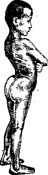
Figure 26. Sharply pronounced lordosis in bilateral dislocation of the hip.
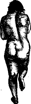
the formation of congenital dislocation of the hip, which is encountered in them 7-8 times more often than in boys. Signs of congenital dislocation of the hip are discovered after the child begins to stand on its legs. Clinical picture of congenital dislocation of the hip: shortening of the length of the limb with protrusion of the trochanter upward in relation to the Roser-Nelaton line; waddling gait, especially sharply pronounced in bilateral dislocation, as a consequence of the positioning of the femoral head posterior to the joint, and as a compensatory phenomenon to this—sharply pronounced lordosis of the lumbar part of the spine with tilting of the pelvis forward and throwing of the upper part of the trunk backward (see figure 26). Active movements of the joint are normal, often its mobility is even greater than normal, with the exception of abduction, which is limited. Often there is mobility of the head along the length of the femur. Upon loading the affected leg, the Trendelenburg sign is observed: the pelvis tilts to the healthy side, and the trunk to the affected one; at the same time, the gluteal fold on the healthy leg stands lower (see figure 27). The Trendelenburg sign is a consequence of the mobility
of the femur along the axis, in the absence of bony fixation of the head and due to the lack of action of the gluteal muscles, the attachment points of which are brought closer together. Under these conditions, upon loading the dislocation, the pelvis drops to the other side until, on the side of the dislocation, it rests against the loaded dislocated femur. Figure 28. "Hourglass" of the coxae capsule by the 7-8-year period of congenital dislocation of the hip. On the X-ray image.
The acetabulum is reduced, flattened, and the head stands above it; sometimes it is deformed and turned anteriorly (anteversio) due to the curvature of the neck, often underdeveloped and small compared to the healthy side (see Figure 37). Under the influence of weight-bearing, the articular capsule, following the upward movement of the head, gradually stretches; bending over the upper edge of the acetabulum, the capsule subsequently adheres here, forming a kind of hourglass (see Figure 28). Therefore, bloodless reduction of the head and its placement into the acetabulum are usually limited to a certain height of the head's position, reaching an irreducible state by 7-8 years of age. Before this time, in the early period, and the earlier the better, it is necessary to make attempts at bloodless reduction according to the principle developed, mainly, by Lorenz. Under anesthesia, with the patient in the supine position, an assistant holds the pelvis with both hands. In this case, it is first necessary to eliminate the obstacle to reduction caused by the shortened muscles. To this end, flexion at the hip and knee is performed with traction along the axis of the thigh. Grasping the bent knee with his right hand, the surgeon
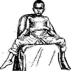
Figure 29. Position of the limbs and fixation of a reduced congenital bilateral hip dislocation.
now tries to advance the head through the narrowing of the articular capsule. By traction, abduction, and external rotation of the thigh, together with pressure from the left hand on the greater trochanter, the head is guided along the posterior edge of the socket and forced into it through the narrowing of the capsule. To hold it in place in this "frog" position, a plaster cast must be applied (see Figure 29) encompassing the lower leg and the pelvic girdle, with pressure applied just above the greater trochanter. The duration of stay in this cast is from 2 to 3 months, followed by gradual adduction of the limb, either naturally (while walking on crutches) or by casts. In cases where the indicated time for bloodless reduction has been missed—at a later age, most often between 15 and 25 years, when the shortening of the limb is significant and when pain during weight-bearing is very troublesome—it is often necessary to resort to palliative operations, which include: 1. Lorenz-Bayer oblique osteotomy, with the placement of the distal fragment at the level of the old acetabulum. With
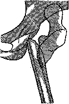
a sharply abducted limb, from the head, neck, and the site of the fracture healing at an angle below the lesser trochanter, a "fork" is obtained with a wide area of contact with the pelvis at both its ends (see Figure 30). 2. A shelf made from the knocked-down outer half of the thickness of the ilium—according to the idea of König. This operation is especially indicated where, in the presence of pain and the Trendelenburg sign, there is also sharp mobility of the thigh along the axis. In the presence of a Luxationspfanne and in those cases where the Trendelenburg sign, thus, depends on the insufficient action of the gluteal muscles (as with coxa vara), due to the convergence of their attachment points, it is advantageous to perform a downward transplantation of the trochanter with a sharply abducted thigh (Veau-Lamy operation). Figure 30. "Fork" of the oblique osteotomy of the thigh below the lesser trochanter in neglected
congenital hip dislocation.
under proper indications, reduce pain and the Trendelenburg sign and improve gait. Among congenital articular developmental defects that can lead to dislocation, one should mention defects of the knee. Luxatio sub partu is more often observed in the shoulder joint and at the head of the radius—often together with damage to the brachial plexus during childbirth. The developmental defect thus created progresses with growth until some trauma forces the patient to pay attention to the paralytic condition, which can therefore sometimes be falsely evaluated Figure 31. Paralytic dislocation of the hip joint with the presence of coxa valga.
as traumatic. In other joints, congenital and birth dislocations are observed extremely rarely. IV. Paralytic dislocations arise on the basis of a flail joint when all its muscles are affected and the capsule is severely stretched, especially during weight-bearing. But even with partially remaining muscles, the work of the latter can contribute to dislocation, which in paralysis is observed mainly in ball-and-socket joints—the shoulder and hip (see Figure 31). Treatment methods include arthrodesis or fascio-suspension for the shoulder joint (see Shoulder dislocation) and the application of König's idea for the hip joint. V. Pathological dislocations are the result of either prolonged stretching of the capsule by exudate or the destructive work of infection on the articular ends of the bones, most often of a tuberculous nature. The displacement depends on the load on the diseased joint and the pull of the muscles. Thus, for example, during an inflammatory process in the head of the humerus, the strong deltoid and scapular muscles pull the shoulder upward, and the trunk muscles (latissimus dorsi and pectoralis)—inward. Thus, a displacement of the shoulder upward and medially occurs. In coxitis, which destroys the acetabulum and the head, the latter, with or without weight-bearing, due to the contracture of the iliopsoas muscle and the gluteal muscles, is displaced upward (see Figure 38). In the knee joint, the muscle contracture of the flexors, which are everywhere stronger than the extensors, gains the upper hand over the quadriceps and, by first bending the knee, then pulls the destroyed end of the tibia backward like reins, forming a posterior subluxation of the knee (see Figure 39). The clinical picture of pathological dislocation, often noticeable to the eye, is supplemented by measuring the limb, the height of the classical points, and an X-ray, which indicates the degree of destruction of the bone ends and their complete dislocation or subluxation. Their treatment by redressment or constant traction with various types of apparatus is usually concomitant with the treatment of the joint diseases themselves.
Related articles
Mentioned in
- Acrobatics
- Aniridia
- Aphakia
- Arachnodactyly
- Arthroplasty
- Arthrotomy
- Avulsion of the Eyeball
- Axillary Artery
- Biological Physics
- Bitter-Salt Lakes
- Bobrov
- Caucasian Mineral Waters
- Coccyx
- Cooper
- Decapitation (or decapitation, is an obstetrical operation)
- Dejerine-Klumpke Paralysis
- Distorsion
- Duchenne-Erb Paralysis
- Dupuytren
- Embryotomy
Cite this page
“Dislocations.” Soviet Medical Encyclopedia. English translation of Bolshaya Meditsinskaya Entsiklopediya, 1st ed. (Moscow, 1928–1936), ed. N. A. Semashko. https://sovietmedicalencyclopedia.pages.dev/article/dislocations/