Axillary Artery
Historical document, translated for reference. It reflects medical knowledge of the 1920s–30s and is not medical advice.
Summary
The axillary artery is a segment of the arterial trunk that carries blood to the upper extremity, extending from the lateral border of the first rib to the lower border of the teres major muscle, where it continues as the brachial artery. It is divided into three parts with multiple branches supplying the thoracic wall and shoulder region.
Encyclopedia article (1928–1936)
AXILLARY ARTERY, subclavian or axillary artery, a defined segment of the arterial trunk carrying blood for the upper extremity. The initial segment of this section is the direct continuation of the subclavian artery (a. subclavia), and the terminal segment directly continues into the brachial artery (a. brachialis). The lateral border of the first rib is conventionally considered the upper boundary of the a. axillaris, and the lower boundary is the lower border of the teres major muscle (m. teres major). The artery itself is also conventionally divided into three parts: the first extends from the lateral border of the first rib to the upper border of the pectoralis minor muscle, the second lies under this muscle, and the third extends from the lower border of m. pectoralis minor to the lower border of m. teres major. From the first part arise: 1) the superior thoracic artery (a. thoracica suprema) and 2) the thoracic and acromial artery (a. thoracico-acromialis); from the second part, the long or lateral thoracic artery (a. thoracica longa s. lateralis); from the third part: 1) the subscapular artery (a. subsca-pularis), 2) the circumflex humeral artery, anterior (a. circumflexa humeri anterior) and 3) the circumflex humeral artery, posterior (a. circ. hum. posterior). The a. a. is accompanied by the v. axillaris, lymphatic glands, and trunks of the brachial plexus. V. axill. lies on the inner side of the artery. Between v. axill. and the artery are placed the inner root of the median nerve, the ulnar nerve, and, for a short distance, the medial cutaneous nerve. Laterally
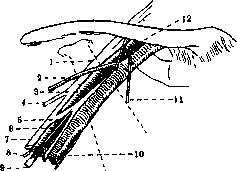
The relationship of nerves and veins to the a. axillaris (two inclined dotted lines indicate the position of m. pectoralis minor): 1-lateral trunk of the brachial plexus; 2-v. cephalica; 3-medial trunk of the brachial plexus; 4-n. musculo-cutaneus; 5-n. cutaneus antibrachii medialis; 6-n. ulnaris; 7-n. medianus; 8-n. axillaris s. circumflexus; 9-n. musculo-spiralis (n. radialis); 10-n. cutaneus brachii medialis; 11-n. thoracalis anterior internus; 12-n. thoracalis anterior externus.
> from the artery is placed the lateral root of the median nerve and the musculocutaneous nerve. Posteriorly-the axillary nerve and the musculospiral nerve (radial). The initial segment of A. a. lies rather deeply, under the pectoral muscles, whereas the terminal segment is covered only by skin and fascia, D. Deshin. AXILLARY NERVE, n. circumflexus, subclavian nerve, encircling the head of the humerus. It emerges from the posterior trunk of the brachial plexus, where its fibers can be traced in the direction of the spine, up to the V and VI cervical nerves. The initial segment of A. n. is placed on the subscapular muscle (m. subscapularis) and lies behind a. axillaris; at the lower border of this muscle, the nerve turns posteriorly and together with a. circumflexa posterior, penetrates into the quadrangular space, the upper border of which is formed by the lower border of the subscapular muscle (m. subscapularis), the lower border by the teres major, the inner border by the long head of the triceps brachii muscle (m. tricipitis), and the outer border by the surgical neck of the humerus. After passing through this space to the posterior surface of the arm, the nerve divides into two branches - superior and inferior. The superior branch surrounds the surgical neck of the humerus and reaches almost to the anterior edge of the deltoid muscle; this branch supplies motor fibers to the deltoid muscle, except for its most posterior part. Several small cutaneous sensory branches, separating from the superior branch, penetrate between the muscle fibers, curve downward, and supply the skin over the lower part of the muscle. The inferior branch, after supplying motor branches to the posterior part of the deltoid muscle and the teres minor (m. teres minor), then curves around the posterior part of the deltoid muscle, slightly below its middle, and ramifies in the skin covering the lower two-thirds of this muscle and the adjacent part of the triceps brachii muscle (m. triceps). Before dividing into its two main branches, n. circumflexus gives off one or two branches to the shoulder joint, piercing the lower part of its articular capsule. The skin covering the upper, and partly the middle, third of the deltoid muscle is supplied by the posterior supraclavicular branches of the cervical plexus (rami supraclaviculares posteriores s. supraacromiales), which arise from the III and IV cervical nerves. In dislocations of the shoulder
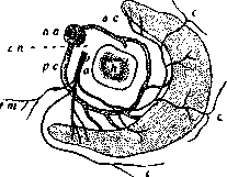
Diagram of vessels and nerve surrounding the surgical neck of the humerus: h-transverse section of the humerus immediately below the tubercles; aa-arteria axillaris; pc-a. circumflexa humeri posterior; ac-a. circumflexa humeri anterior; en-nervus circumflexus; a-articular branch; tm-branch to m. teres minor; c-cutaneous branches.
joint, n. circumflexus is often stretched by the dislocated head and even torn. Since this nerve curves around the humerus at the level of the surgical neck, it is understandable that in fractures of the neck it may also be damaged. In paralysis of n. circumflexus, the deltoid muscle atrophies, and the roundness of the shoulder disappears. Since n. circumflexus passes under the deltoid muscle from behind forward, for penetration into the shoulder joint, incisions (longitudinal) are recommended to be made only on the anterior surface of the muscle. With such an incision, the function of the muscle is preserved more or less completely; atrophy can occur only in the most anterior bundles, since even with an anterior incision, the terminal nerve branches are still damaged. Lesion of A. n. is characterized by atrophy and paralysis of the deltoid muscle and m. teres minor, as well as anesthesia of the lateral surface of the shoulder. In etiology: injuries, infections, intoxications (lead, diabetes), sometimes combined toxic-traumatic effects (some professional cases, paralysis during anesthesia).
Related articles
Mentioned in
Cite this page
“Axillary Artery.” Soviet Medical Encyclopedia. English translation of Bolshaya Meditsinskaya Entsiklopediya, 1st ed. (Moscow, 1928–1936), ed. N. A. Semashko. https://sovietmedicalencyclopedia.pages.dev/article/axillary-artery/