Shoulder Joint
Historical document, translated for reference. It reflects medical knowledge of the 1920s–30s and is not medical advice.
Summary
The shoulder joint is formed by the glenoid cavity of the scapula and the head of the humerus, being one of the most mobile joints in the human body. It allows movement around three axes with a range of approximately 150 degrees, with its surface curvature classified into three types according to Weinstein's research.
Encyclopedia article (1928–1936)
SHOULDER JOINT (articulatio humeri) is formed by the articular (concave) surface of the scapula (cavitas glenoidalis scapulae) and the head of the humerus. This joint belongs to the most mobile ones. Limitation of movements in it to a large extent impedes the use of mobility in other joints of the upper extremity. In the joint, movement occurs around 3 axes: 1) transverse axis-pendulum-like movements, 2) sagittal-abduction and adduction and 3) vertical-rotation. All these movements have a range of about 150°. The shape of the articular surfaces does not lend itself to precise geometric definition: thus, some authors (Langer, Luschka, Meyer H., Gegenbaur) consider them elliptical, others (Hyrtl, Sobotta, Broesike)-spheroidal, still others (Henke, Merkel, Testut, Rauber, Zernov, Fick R.)-incorrectly hemispherical, while Helwig regards it as part of an elliptical ring. Weinstein points to three types of curvature of the articular surface of the humerus on a vertical section, where: "1) the curve of the surface may coincide with one proper circle; 2) the curve of the surface corresponds to two arcs of different radii: the arc corresponding to the larger part of the curved surface has a larger radius (upper part); the arc corresponding to the smaller part of the curve has a smaller radius (lower part); 3) the curve of the surface corresponds to two arcs of different radii: the arc corresponding to the larger part of the curved surface has a smaller radius; the arc corresponding to the smaller part of the curve has a larger radius. The first type occurs in 2%, the second-96.5% and the third-1.5%" (fig. 1). / The articular surface of the humerus is covered with hyaline cartilage 1.5 to 2.5 mm thick, most pronounced in the central part,
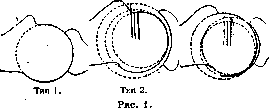
Type 3.
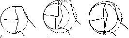
and gradually thinning toward the periphery. The head with the body of the humerus (see Shoulder) forms an angle of 130° to 145° (Poirier, Rauber). Weinstein determines this angle for men as 132-141°, for women-as 127-138°. The articular cavity of the S. j. (see Scapula), corresponding to the articular head, is directed forward, outward and somewhat upward. The vertical diameter is the longest, and the greatest width corresponds to the boundary between the middle and lower thirds. Covered with cartilage forming a rim along the edge (labrum glenoidale), it is in general a shallow depression with curvature more pronounced in the upper-lower section than in the front-back, with greatest depth (up to 10 mm) in the outer-back part. On the inner-front part of the depression there is a notch, which together with the rim forms a depression leading into the mucous bursa (homolog of incisurae acetabuli). The cartilage covering the cavity thins toward the center, where a small bony elevation is noted (tubercule glenoidienne, Poirier); here there is an area of fibrocartilage. Even in the presence of a cartilaginous rim, which significantly increases the congruence of the articular surfaces, their mismatch remains nevertheless large: the length of the articular surface of the head is about 3 times greater than that of the cavity. Corresponding to the above-mentioned types of articular surface of the humeral head, the curvature of the articular surface of the scapula (cavitas glenoidalis) in the first type corresponds to an arc of a proper circle of the same radius; in the second and third types-to an arc with a radius equal to the radius of the circle of the lower pole of the head (fig. 2). In the so-called "ordinary position" of the upper extremity (when the lower part of the head is opposite the upper two-thirds of the articular cavity) complete contact of the articular surfaces is noted; in the abducted position of the extremity these relationships change significantly (in the first type-complete contact is preserved, in the second a slit forms between the articular surfaces and in the third an incomplete "central contact" is obtained)
Type 1.
Type 2. Figure 2 Type 3. Along the outer circumference of the cartilaginous ring begins the articular capsule, covering the articular surfaces and attaching to the humerus at the anatomical neck, medially and dorsally it extends to the surgical neck, i.e., below the epiphyseal line; above sulcus intertubercularis it is thrown like a bridge. Outside, the capsule is closely adjacent to and merges with it at the site of its attachment mm. subscapularis, supraspinatus, infraspinatus and teres minor-in the intervals between the tendons of these muscles it is thickest. The capsule consists of three layers: an outer layer of longitudinal fibers, an adjacent layer of circular fibers and the inner synovial membrane, the latter not being covered with endothelium. The capsule is strengthened by a number of ligaments (fig. 3, 4, 5): 1) lig. coraco-humerale, s. suspensorium capitis humeri- begins at the inner edge and from the base of the coracoid process and, weaving into the capsule, directs itself to the greater
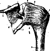
Figure 3. Ligaments of the right shoulder joint 1-collum anatomicum, 2-lig coraco-humerale; 3-processus coracoideus; 4-acromion, 5-spinascapulae, 6-lig transversum scapulae m., 7-scapula; 8-capsula articularis; 9-shoulder.
tubercle, in the area of which it attaches; 2) lig. coraco-brachiale-in the form of a bundle of fibers begins from the articular rim, dips into the joint cavity and attaches in the area of the anatomical neck near the lesser tubercle. Between the named ligaments a groove is formed, in which lies the tendon of the long head of the biceps brachii muscle; between the tubercles, where the tendon enters sulcus intertubercularis, the ligaments are connected by transverse fibers, converting the groove into a bone-fibrous canal (Schlemm, Weinstein); 3) lig. glenoideo-brachiale internum - begins from the front-upper part of the articular rim, goes under the tendon of the subscapularis muscle and attaches to the lower part of the lesser tubercle; between this ligament and the previous one an oval-shaped opening is formed (foramen ovale Weitbrechti), through which passes the tendon
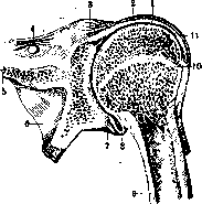
Figure 4. Frontal section of the shoulder joint from behind: 1-caput longum m. bicipitis; 2 and 3-capsula articularis; 3-processus coracoideus; 4 - lig. transversum scapulae sup. 6 - scapula; 7-labrum glenoidale; 9-humerus; 10-boundary of epiphysis; 11-vagina mucosa intertubercularis. 5-spina scapulae;
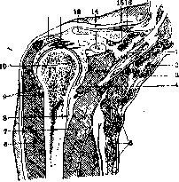
m. subscapularis; 4) lig. glenoideo-brachiale inferius, s. latum-begins from the lower part of the circumference of the articular cavity and attaches to the inner part of the humeral neck; lies in the thickness of the capsule wall, the lower part of which (in the interval between m. subscapularis and teres U 12 Fig 5 Frontal section of the right shoulder joint: 1-plexus brachialis; 2-a. subclavia; 3-v. subclavia; 4-m. subscapularis; 5-m. serratus ant.; 6-m. deltoideus; 7-caput longum m. tricipitis; 8-tendon of m. latissimus dorsi; 9-branches of a. circumflexae humeri post.; 10-head of humerus; 11-acromion; 12-m.supraspinatus; 13-clavicle; 14-processus coracoideus; 15-lig. transversa scapulae; 16-n. suprascapularis. minor) strengthens it with its fibers. The weakest spot of the capsule is under the coracoid process and at the tendon of m. subscapularis. Usually the articular capsule has three protrusions communicating with the joint cavity: 1) vagina mucosa intertubercularis, 2) bursa subscapularis and 3) bursa subcoracoidea. The last two are by some authors combined into one under the name bursa subscapularis. Weinstein notes that the subcoracoid bursa often occurs as a multicameral mucous bursa. Weinstein's experiments show that when filled with fluid under high pressure, the capsule ruptures (with maximum capacity of 63 cm3) usually in the area of the mucous bursae and the more easily the more developed these latter; the fluid found its way into the area of fossae subscapularis. In the immediate vicinity to the joint and participating in its strengthening are: m. pector. minor, caput longum m. bicipitis, mm. subscapularis, supraspinatus, infraspinatus and deltoideus (see Scapula).
Kupriyanov. Pathology. Congenital anomalies of the shoulder joint constitute part of the changes observed in congenital deformities of the entire humerus (see Shoulder), on the other hand, there are also secondary changes associated with developmental anomalies of the shoulder girdle; finally, developmental abnormalities may occur only in the articular end of the humerus or the glenoid fossa of the scapula. Slight impairment of joint function, increased range of passive movements in the direction of adduction, outward rotation of the axis of the shoulder joint are observed with complete or partial defects of the clavicle, which is usually bilateral and is combined with the congenital absence of certain skeletal muscles (Heinecke). In Sprengel's disease (congenital high position of the scapula), there is inward rotation of the shoulder and limitation of movement of raising the hand above the horizontal plane due to mechanical obstacles from the scapula (see Scapula). Congenital absence of the pectoral muscles also causes corresponding impairment of joint functions. Congenital deformations of the shoulder joint itself are encountered in so-called humerus varus, in which the enlarged, mushroom-shaped head is rotated outward and has a greater angle of inclination in relation to the diaphyseal axis. The greater tubercle occupies a higher position than normal. The appearance of the shoulder joint resembles its configuration in anterior-inward dislocation of the shoulder. The lower epiphysis of the humerus in some cases appears thickened but retains normal contours. Humerus varus was first described by Riedinger in arthritis deformans, but as a congenital anomaly it occurs in cretins (Bircher); in another case, besides intellectual underdevelopment in an 11-year-old patient, there were undoubtedly endocrine disorders1-marked hypertrichosis. Some authors, as with coxa vara, distinguish congenital, traumatic, and dystrophic forms of humerus varus. Among congenital dislocations of the shoulder, a significant portion are secondary paralytic deformations. True "congenital dislocations" appear to be a rare anomaly and depend mainly on changes in the head of the humerus. Congenital anomalies from the side of the scapula, such as absence of its neck, rudimentary development of the glenoid fossa, have been described in individual cases. Congenital dislocations are divided into luxatio subacromialis, subcoracoidea, and infraspinata (Smith). Among 39 cases, Ranci found posterior dislocation in 17; bilateral involvement was noted in 5 cases. In congenital dislocations, marked muscular atrophy and significant impairment of joint function are often observed, however this is not a constant symptom. Combination of congenital dislocation of the shoulder with other skeletal developmental anomalies is frequently noted. Based on the study of 428 X-ray photographs, Wolff concludes that the cause of congenital dislocation is delayed development of the head, which depends on intrauterine pressure transmitted to the upper articular end of the humerus. Treatment for congenital dislocations can be bloodless-reduction of the dislocation followed by fixation of the limb for a month in the position of abduction and outward rotation (Whitman), or bloody-excision of part of the capsule and partial resection of the head (Phelps) followed by reduction of the dislocation. Secondary deformations of the shoulder joint are observed on the basis of trauma to it during difficult labor, leading either to epiphyseolysis of the newborn or to obstetric paralysis. In the latter case, the fixed faulty position of the limb leads to the formation of posterior subluxation of the head of the shoulder and subsequently requires surgical treatment, the simplest and most common method being osteotomy of the humerus. Traumatic injuries to the soft tissues of the shoulder joint can be closed and open and are caused by traumatic forces of various nature (bruises, chemical and thermal burns, incised, punctured, gunshot, and lacerated wounds). The localization, extent of injuries, and their outcome in terms of formation of extensive scars are of great importance for joint function (see Shoulder). Among subcutaneous injuries to the muscles of the shoulder joint, besides the deltoid, the rupture of the trapezius muscle is significant, which is most often accompanied by simultaneous injury to nerves and leads to prolonged impairment of joint function and persistent pain. Subcutaneous injuries to blood vessels are observed in dislocation of the shoulder both at the moment of its occurrence due to overstretching of the vessels, but mainly during its reduction. Thus, Korte in 53 cases found injury to blood vessels directly in the dislocation in only 7. According to Guibe's statistics, out of 79 cases, injury to a vessel occurred during reduction of the dislocation in 57 cases, most often in cases where it was performed for old dislocation of the shoulder. Korte and Paget observed cases where rupture of the axillary artery occurred during forced mobilization of the joint for contracture after acute inflammation of it. The condition of the vascular wall in terms of its resistance is of great importance; thus, a case of formation of an aneurysm of both axillary arteries on the basis of pressure from a crutch on the axillary region has been described (Bardeleben). In dislocations, fractures of the neck and head of the humerus, nerve injuries are also observed, with the axillary nerve being affected more often than the main trunk of the brachial plexus. The latter is rarely damaged as a whole, damage to its lower part, so-called Klumpke's paralysis, is somewhat more frequently observed. The most common type of plexus injury is damage to the V and VI roots, so-called Erb's type, with spread of flaccid paralysis to the mm. deltoideus, biceps, brachialis, brachioradialis. The arm hangs along the trunk, the hand is in the position of pronation. In the course of treatment, proper positioning of the limb in the position of abduction up to the horizontal plane and outward rotation of the shoulder, with the elbow joint bent, the hand extended and supinated, is of great importance. This position prevents wrinkling of the joint capsule and in case of restoration of nerve conductivity guarantees quite satisfactory joint function. Proper positioning is achieved by applying a plaster bed with fixation of the affected limb according to Biesalski. Injury to the axillary nerve leads to paralysis of the deltoid muscle and partially the teres minor muscle. Open injuries to blood vessels and nerves of the shoulder joint areav, occurring in incised, lacerated, and mainly in gunshot wounds, give a certain symptom complex depending on the nature and localization of the injury and require appropriate treatment (see Aneurysm, Nerves, Shoulder). Closed injuries to the shoulder joint are caused by the action of direct or indirect force (counterblow when falling on an outstretched hand), but most often occur with hyperextension with simultaneous strong outward rotation of the limb. Injury to the joint can manifest as violation of the integrity of the synovial membrane, joint capsule, and ligamentous apparatus, as well as as avulsion of small areas of articular cartilage or bone tissue. More severe trauma leads to intraarticular fractures and epiphyseolyses, which are encountered mainly in childhood. Clinical symptoms are severe pain during movements and presence of effusion in the joint, which is not always easily determined due to the large muscle layer covering the joint. Fluctuation is most clearly determined on the anterior surface of the joint. Typical for joint injury is the forced position of the limb-its adduction, inward rotation, and forward rotation by approximately 30° (Lange). This position is masked by corresponding rotation of the scapula with approximation of its lower angle to the spine. The primary impairment of joint mobility caused by trauma, even in relatively mild cases, often leads to further persistent limitation of function depending on the organization of blood that has leaked into the joint, wrinkling of the joint capsule and ligamentous apparatus. Such an outcome (stiffness), which is extremely common, depends on the fact that in movements not only the humeroscapular joint participates, but also the joints of the scapula and clavicle-acromioclavicular and sternoclavicular joints, injury to which even with a normal shoulder joint also leads to impairment of limb function. Moreover, full mobility of the joint depends on the absence of pathological changes in the bursae belonging to the shoulder joint. All of the above makes it necessary to pay special attention to functional treatment in traumatic injuries to the shoulder joint. In the first days after injury, elevated position, application of light pressure bandage, and aspiration of serosanguineous effusion in its significant accumulation are indicated. At the end of the first week (in case of mild injury-even from the first days), physio-mechanical treatment should be applied-hyperemia, massage, and active movements. Complete healing occurs in mild and moderately severe cases, however, pains, limitation of joint function, presence of muscular atrophy, and signs of subsequent chronic inflammation often remain, requiring prolonged treatment.
According to Dittmer, out of 28 cases of injury to the S. j., 23 required treatment for 13 months. As a possible complication of traumatic injury to the shoulder joint, purulent inflammation of it and joint tuberculosis should be mentioned. Open injuries to the S. j. occur with incised, punctured wounds, crushed, machine injuries and gunshot wounds. The diagnosis is based on the localization of the wound, the direction of the wound track, the discharge of synovial fluid, the presence of hematoma, limitation of movement and the characteristic position of the limb. Some cases may initially remain undiagnosed, and only subsequent complications—inflammation of the joint, limitation of its function—provide the basis for late diagnosis of joint injury. Gunshot wounds to the S. j. according to old statistics constitute only 1.3-3.4%, but according to data from the last imperialist war, they occupy one of the first places, constituting, according to Geiges, 21% of all gunshot wounds. Of 439 injuries to large joints, Krymov had injuries to the S. j. in 71 cases, with the left side predominating over the right, and bullet wounds were encountered significantly more often than wounds from other types of projectiles. Isolated injury to the joint capsule without bone injuries was noted in 28 cases; indirect injury to the joint is possible in cases where a fracture of the humerus or scapula reaches the joint cavity. Treatment of open injuries to the S. j. depends on the nature and clinical course of the injury. It is extremely important to distinguish between simple and complicated, infected and aseptic, fresh and old injuries. From a clinical point of view, injuries are aseptic where infection is limited to the wound track and has not passed from the synovial membrane beyond the joint capsule, which is usually observed within the first two days after injury (Friedrich, Policard). In these cases, especially with incised, chopped and even torn-contused wounds, primary treatment with excision of all damaged tissues, washing of the joint with antiseptic fluid and subsequent plastic surgery of the joint capsule and skin in case of their defect is possible. This method of treatment in many cases results in healing by first intention with a good functional result. In cases with extensive bone injuries, primary resection of the joint is indicated. Although all gunshot wounds should be considered infected from a bacteriological point of view, clinically many of them have a smooth course without subsequent secondary infection and do not require surgical intervention. Cases with mono-infection range from 50% to 70% and have a better course than with mixed infection. One of the important conditions for treatment is the rest position of the joint, achieved by applying splint bandages in the position of abduction and outward rotation of the limb; bandages are not removed during dressings and allow for early movement in the elbow, wrist and fingers (Bürgi). In many cases of bullet injury to the S. j., such conservative treatment can be carried out even in the presence of small bone fragments, which do not always require their primary removal. In extensive injuries with severe primary infection, surgical treatment is indicated—wide opening of the joint with ensuring good drainage for purulent discharge; in some cases—secondary resection of the S. j. In cases of severe panarthritis and septic condition threatening the patient's life, amputation of the upper limb should be performed. The prognosis for open injuries to the S. j. compared to injuries of other large joints is quite favorable quo ad vitam; good functional results according to Krymov were present in 29.5%. Of 12 cases of Hotz complicated by purulent inflammatory process, in 2 cases a complete ankylosis remained, in 9 cases—limited mobility with a range of motion from 20° to 75° and in 1 case—almost complete restoration of function of the shoulder joint. Intra-articular fractures of bones of the S. j. are most often localized in the area of the articular end of the humerus, fractures of the articular parts of the scapula are observed much less frequently. (Fractures of fossa glenoidalis, fractures of the neck of the scapula, fractures of the acromion, see Scapula.) Fractures of the articular end of the humerus occur mainly in old age, in children injuries are observed along the epiphyseal line. Poirier in 61 cases of fracture of the humerus had fracture of its upper end in 41 cases. According to the localization of the fracture line, Kocher distinguishes: 1) fract. supratuberculares (intra-articular), which include a) fracture of the head and b) fracture of the anatomical neck of the humerus and 2) fract. infratubercularis, which in turn are divided into a) fract. pertubercularis and epiphyseal separations, b) fracture of the surgical neck of the humerus, c) V-shaped fractures and d) fractures of the major or minor tubercle. Pure fracturae supratuberculares (intra-articular fractures) are very rare and can occur mainly in compression fractures. Of these, the most typical is the fracture of the anatomical neck, which, according to Albert, is observed 20 times less frequently than the fracture of the surgical neck of the humerus. The head of the humerus is displaced inward or outward or may be located above it; in some cases, complete rotation of it relative to the horizontal axis is observed, so that the plane of the fracture is facing the glenoid cavity of the scapula. Diagnosis is not always easy. There are symptoms of intra-articular injury present. Crepitus may not be determined with significant rotation of the head or its impaction. Diagnostic signs are: flattening of the humeral region, shortening of the limb and a larger than normal volume of passive movements (especially to the side). In the treatment of intra-articular fracture of the humerus, in addition to reposition and holding the fragments in the correct position, special attention should be paid to the possible early application of hyperemia, massage and active movements. In fractures without displacement, in some cases one can be limited to a light fixing bandage with a pad in the axillary fossa or a splint bandage in the position of abduction and outward rotation. With large displacements, there is usually significant deformation of the articular end of the humerus and limited mobility of the joint. Injury to a. circumflexae ant. can lead to necrosis of bone tissue with subsequent formation of free joint bodies. In the presence of a free head, surgical treatment is indicated (Kocher, Bardenheuer), and the head should not be removed, but if possible, one should strive to restore normal anatomical relationships. According to Romer, blood reposition in intra-articular fracture with or without dislocation gives better results than resection of the head, which should be performed only in cases where setting the fragments is impossible. Too long a period elapsed between the injury and surgical intervention worsens the prognosis in terms of obtaining strong consolidation. Fracturae pertuberculares mostly belong to adduction fractures and occur under the action of a traumatic force directed from the outside and below. The most common form of them are fractures along the epiphyseal line, occurring in newborns, and also often in the first years of a child's life. In older age, together with epiphyseal separation, detachment of a part of the diaphysis of the humerus is observed. Bruns in 49 cases of epiphyseal separation in 21 observed its localization in the upper articular end of the humerus. The distal end is usually displaced inward, upward and forward, the head is rotated outward. To differentiate an epiphyseal fracture from a fracture of the surgical neck, one can mainly rely on X-ray data. In a newborn, clinical symptoms of epiphyseal separation are: the position of the arm extended along the trunk with its inward rotation, the appearance of late hemorrhages, often hematomas in the area of the sterno-cleido-mastoideus and rarely deformation of the S. j. in cases with large displacement of fragments. When an epiphyseal fracture occurs in children, a significant lag in the growth of the limb is often observed later, which is more pronounced the younger the age of the child at the time of injury. In the treatment of epiphyseal separation in newborns, it is necessary to fix the limb for 3 weeks in the position of abduction at an angle of 80-90° with outward rotation. In older age, traction can be applied with the shoulder abducted and the forearm in vertical position. In old cases with large displacement, reposition can be achieved only by blood method with resection of a section of the bone diaphysis. Fractures of the surgical neck are the most common fracture of the upper articular end of the humerus. The fracture line usually passes between the greater tubercle and the site of attachment of m. pectoralis majoris, at the place of transition of the diaphysis into spongy substance. Mostly there are transverse fractures, oblique fractures are observed less frequently. Formation of fragments and avulsion of bone sections is often encountered. Relatively often, fractures of the surgical neck are combined with dislocation of the shoulder, and in some cases there are accompanying injuries of muscles, vessels and nerves. Mostly the fracture is caused by the action of a direct force, e.g. a blow, a fall on the shoulder area, less often—by indirect force, when falling on the elbow, or by the action of muscle traction.
According to the nature of the diaphyseal fragment displacement, abduction and adduction fractures are distinguished. The latter are the most frequent; in them, the distal fragment is displaced inward and upward, while the proximal fragment is displaced and rotated outward. Often there is impaction of the lower fragment into the upper one, in which case the typical symptoms of a fracture—abnormal mobility and crepitus—are absent; the diagnosis is made based on impairment of limb function, pain, hemorrhage, and X-ray data. In the absence of impaction, all the characteristic symptoms of a fracture are present. The differential diagnosis with subcoracoid dislocation, which gives a similar position of the limb, is made based on the presence of limb shortening and palpation of the head in its normal place. Treatment of impacted fractures without significant displacement of fragments can be carried out by applying a light immobilizing bandage with possible early use of physio-mechano-therapy. In significant displacement, traction in the horizontal position is applied, or, according to Bardenheuer, in the vertical position with maximum abduction of the limbs. According to Baumann, in fractures of the surgical neck, no special reduction of fragments is required; Beller, on the other hand, holds the opposite view, considering it a rule to perform reduction of fragments before applying extension splints. Properly conducted treatment of fractures of the surgical neck lasts about 6 weeks. In severe cases with simultaneous combination of fracture with shoulder dislocation, bloodless reduction of fragments is indicated. The formation of pseudarthrosis is observed extremely rarely; more often there is improper union and formation of large bony protrusions, which can serve as a mechanical obstacle to joint movements. Complete and incomplete fractures of the greater tubercle usually accompany other traumatic injuries to the joint, most often observed in shoulder dislocations. In a complete fracture, together with the tuberculum majus, a small area of the humeral diaphysis is also torn off (Gurlt). The traction of the rotator muscles of the shoulder causes the detached greater tubercle to be displaced outward by 2-3 cm. An isolated avulsion can be observed in a fall on the shoulder joint or due to the action of muscular traction during the act of throwing or throwing. From clinical symptoms, there is an absence of active outward rotation, the area of the shoulder joint appears wider than normal, and there is definite tenderness on pressure over the tuberculum majus. Treatment consists of fixing the shoulder in the position of abduction and outward rotation. If in this position there is still a diastasis between the greater tubercle and the humerus, then surgical fixation of the fragment is indicated (Kocher, Hofmeister). In simultaneous shoulder dislocation, especially when the detached greater tubercle is impacted between the head and the glenoid cavity, surgical intervention is inevitable. Fractures of the lesser tubercle are encountered extremely rarely. The function of the limb is impaired in the sense of loss of inward rotation, as well as due to the simultaneous rupture of the tendon of the biceps muscle. Shoulder dislocations constitute more than half of all dislocations encountered in percentage terms (see Dislocations). In men, dislocations are observed predominantly in the age range from 40 to 60 years and occur 4-5 times more often than in women. In children, shoulder dislocations are observed in exceptionally rare cases. Dislocations are divided into 1) anterior—luxatio praeglenoidalis and 2) posterior—luxatio retroglenoidalis. In terms of frequency, the ratio between them, according to Finckh, is determined as 97.5% to 2.5% (see Dislocations). Treatment of dislocations is divided into three moments: reduction, which should be performed as early as possible, keeping the limb in the correct position, and applying measures aimed at restoring joint function (physio-mechano-therapy). Obstacles to reduction of the dislocation can be a whole series of factors, which determine the ease or difficulty of reduction. Among them, mention should be made of: 1) resistance from the stretched ligamentous apparatus, 2) tension of the muscles (which, according to Hofmeister, has greater clinical significance than Busch attributes to it based on anatomical research), 3) the presence of damage to soft tissues with possible interposition of them, and 4) the width of the capsule rupture, through which the head exits the joint. Methods of reduction are divided into impulsive, lever, and rotational. Of the numerous methods proposed, the most widespread is the method of Kocher and Mote (see Dislocations). In recent years in the USSR, the method proposed by Danelidze in 1921 has gained great popularity, which has the following practical advantages: 1) simplicity and ease of technical manipulation, 2) painlessness, 3) possibility of performing reduction with one assistant or even without one, 4) no need for the reducer to apply great physical force (see Dislocations). In infraclavicular dislocation, Kocher modifies the first act of reduction, performing adduction with bringing the elbow backward, in axillary dislocation he performs abduction with outward rotation of the shoulder. After reduction, the limb is placed in a resting position, and massage and careful active movements can be performed from the first days. (Hofmeister, Beller). In young individuals with an uncomplicated dislocation, complete restoration of function occurs in 4-6 weeks. In elderly people, limitation of joint mobility often remains and deformative arthritis often develops. Küttner (Küttner) in 54 cases of uncomplicated dislocations had marked or moderately marked impairment of function of the shoulder joint in 61%. Lexer in 8 complicated dislocations observed restoration of function in only one case. Anterior dislocations, excluding infraclavicular dislocation, are rarely primarily irreducible and in these cases require bloodless reduction. In many cases of dislocations complicated by fractures, surgical intervention is also indicated, which should be performed after a preliminary attempt to reduce the head by direct pressure on it (Hofmeister). Removal of the head during surgery should only be done in case of failure of reduction and repositioning of fragments. One should never wait for consolidation of the fracture in order to later perform reduction of the dislocation, as in such cases the latter is not successful. If surgical treatment is contraindicated, then according to Cooper's proposal, the diaphyseal end of the humerus can be introduced into the glenoid cavity and by applying early movements, promote the formation of a pseudarthrosis. Anterior dislocations, see Dislocations. Posterior shoulder dislocations are rare and occur under the action of a direct force directed from front to back, or indirect, for example, in a fall on an outstretched forward arm with simultaneous inward rotation of it. Kronlein in 207 cases observed posterior dislocation in only one case, Finckh in 201 cases had 5 posterior shoulder dislocations. Posterior shoulder dislocations can only occur with a rupture of the m. subscapularis at its site of attachment to the posterior part of the capsule, which is reinforced here by the rotator muscles of the shoulder outward. This is possible only under the action of a significant traumatic force. A more common form of posterior dislocations is subacromial dislocation, in which the head is placed on the collum scapulae under the acromion. In this form, avulsion of the tuberculum minus is often observed (Malgaigne). The shoulder appears wider than normal, the axis of the limb is deviated backward and outward, and the head is palpated in an unusual place. Reduction often succeeds with traction of the limb along its axis. Kocher proposes to perform reduction in three moments: 1) strong inward rotation with the arm bent, 2) traction in this direction, and 3) outward rotation and extension. Hofmeister performs reduction by applying traction. In infraspinatus dislocation, the head is displaced even further backward and is placed in the fossa infraspinata, and the tuberculum minus is pressed tightly by the coracohumeral ligament to the edge of the glenoid cavity. For reduction, Kocher performs 1) inward rotation of the bent arm, 2) abduction, 3) traction, and 4) outward rotation with adduction of the limb. Reduction usually succeeds, but habitual shoulder dislocation often develops. According to Russian statistics, old dislocations constitute about 10-12% of all shoulder dislocations (Tikhov, Toropov), and they are mainly anterior dislocations. Factors contributing to their formation are, on the one hand, the unculturedness of patients who resort to treatment with folk healers, late timing of the reduction performed, and erroneous diagnosis, and on the other hand, anatomical-pathological factors, such as aversion and displacement of the greater tubercle, impingement of the tendon of the m. biceps and the coracohumeral ligament (Turner), and complications of the dislocation with a fracture of the anatomical or surgical neck of the humerus.
The period when a shoulder dislocation becomes chronic is established at approximately 4-6 weeks, however, bloodless reduction of the dislocation was achieved by Finke up to 14 weeks, by Kocher up to 4 months after the dislocation, Simon describes cases of reduction after 1 year 8 months, while in Vreden's clinic, reduction by the Danelidze method was even possible after 2-21/2 years.--Patho-anatomical changes in an unreduced dislocation consist of the development of scar tissue at the site of the former hemorrhage, which can form a new joint capsule, changes in the cartilage covering of the head, bony growths in the area of the edge of the fossa glenoidalis and proc. coracoidei, obliteration of the old joint socket and formation of a new one in the area of collum scapulae, changes in blood vessels with frequent adhesions to the newly formed socket, and changes in muscles in the sense of their retraction. On the basis of experimental data, Dollinger attaches great importance to the contraction and subsequent scar changes of m. subscapularis, which in his opinion serves as the main obstacle to the reduction of the dislocation.-Functional disorders in chronic dislocation are expressed to varying degrees, in some cases when a neoarthrosis forms, function can be quite satisfactory, and such cases do not require other therapeutic measures besides mechanotherapy procedures that help increase the range of motion. In most cases, there is significant limitation of joint mobility, sometimes accompanied by vascular-nerve phenomena caused by pressure from the displaced head on the axillary region.--Indications for bloodless reduction should be given with great caution, since it requires the use of great physical force, which often leads to fractures. Among the operative methods, first place is occupied by bloody reposi-tion of the head, which, according to Schoch, gives about 64.5% good results. However, even by the operative method, reduction is not always successful or in many cases gives a poor functional result (Kpar). For access to the joint, both posterior (Kocher) and anterior (Langenbeck) incisions are used. Vreden proposed a modification of the anterior incision with the addition of a horizontal one along the line of attachment of the deltoid muscle to the acromion and the acromial end of the clavicle. To facilitate reduction, Dollinger considers it necessary to sever the attachment of m. subscapularis. In the presence of significant deformation of the articular end of the humerus, resection of the head is indicated, which gives a satisfactory functional result (according to Schoch's data in 51.2%). In habitual shoulder dislocation, redislocation of the head occurs both under the influence of external, sometimes very insignificant force, and due to involuntary or voluntary contraction of muscles. Cases have been described where the dislocation repeated 100-200 times (Schlange). Among all shoulder dislocations, habitual ones constitute according to Lexer's and Beck's (Vosk) statistics about 3-4%, and occur predominantly in the age of 20-30 years (Seidel). The occurrence of habitual dislocations is especially often observed in epileptics.--Patho-anatomical changes that play a role as predisposing factors can be varied. Great importance is attached to the expansion of the joint capsule, especially of its lower-inner part, which is observed in almost 90% of all cases of habitual dislocation (Franke). Often there is also a detachment of limbi cartilaginei at the anterior edge of the joint socket, sometimes the joint cavity has direct communication with a newly formed joint capsule in the area of colli scapulae. Detachments of muscles, especially external rotators of the shoulder, and detachment of the greater tubercle are further factors favoring the occurrence of habitual dislocation. Often on the posterior surface of the head of the humerus there is a groove, the formation of which is explained by polishing and pressure from adjacent bony prominences or by a congenital anomaly of development of the head, which creates a certain predisposition to the occurrence of habitual dislocation (Lenormant, IOzerov). Function with frequently recurring dislocation is impaired, and the fear of its recurrence causes such patients to significantly limit movements of the extremity, especially those associated with raising and abduction of the shoulder, so that on average the working capacity of the extremity is reduced by 25-50%. The main task in treatment is to keep the head in its normal place while preserving maximum function of the extremity. The proposed conservative measures consist of wearing special splints and bandages (Hol'fa, Gaugele, Well), administration of preparations causing shrinkage of the joint capsule (iodine, iodoform, blood), and prolonged fixation of the extremity in the position opposite to that in which the dislocation occurs (Volkmann). However, all these methods do not give a long-term effect and can lead to joint stiffness. Many operative treatment methods have been proposed and they can be divided into 1) methods narrowing the joint capsule-extraarticular capsulorrhaphy or with opening of the joint (in some methods with addition of partial excision of the joint capsule), 2) methods strengthening the joint capsule by means of muscles, fasciae and tendons, 3) methods strengthening the joint capsule with suspension of the joint by means of fasciae, muscles and tendons, 4) methods creating bony obstacles for dislocation of the head. Among the old operative treatment methods, mention should be made of resection of the head and arthrodesis of the shoulder. Recurrences after operative treatment are not uncommon, according to Biru in 53.8%, according to Pair in 33%, and with the Clairmont-Ehrlich method with formation of a muscle flap from the deltoid muscle-in 25-33%. The observation period for judging the durability of the achieved results is determined from 2 (Schultze) to 3-4 years (Milller). The so-called clicking shoulder joint is observed with voluntary dislocation or subluxation of the shoulder, which is caused and reduced by voluntary movements of the musculature. According to Kappis's research in this condition, there are extracapsular causes, such as displacement of the deltoid or biceps muscle from normal or newly formed bony prominences and changes in the bursae, which can give a similar picture. In most cases, no therapeutic measures are required, only in cases of severe functional disorders is operative treatment indicated- elimination of one or another cause underlying this condition. Diseases of the bursae of the shoulder joint can be acute and chronic, occur primarily or secondarily, due to the spread of the inflammatory process from surrounding tissues or by the hematogenous route (in pneumonia, acute suppurative osteomyelitis, etc.). Tuberculous lesions are often observed; thus, Kreuter in 26 cases had only in 7 an inflammatory process of non-tuberculous nature. Tuberculous bursitis proceeds with the clinical picture of hydrops tuberc. or granulating forms with outcome in suppuration. More often it accompanies tuberculous lesions of the shoulder joint (Konig). Traumatic injuries most often affect bursa acromialis as having the most superficial location. Clinical diagnosis in non-infected cases is easy; there is a circumscribed swelling and fluctuation corresponding to the location of the bursae. With spread of the process to surrounding tissues, recognition is significantly complicated.--Treatment consists of puncture followed by introduction of tincture of iodine or iodoform emulsion in tbc. In case of suppurative bursitis, excision of the bursa is indicated. In inflammatory processes of deeply located bursae (bursa subscapularis, subdeltoidea, subacromialis), excision is not so simple, and in many cases it is possible to limit oneself to scraping them out followed by injection of T-rae Jodi. Lesions of the bursae (b. subacromialis, b. subdeltoidea) at the attachment sites of tendons of m. supraspinati, long head of biceps and periarticular connective tissue give a picture of a disease which, according to Duplay, is called periarthritis humero-scapularis. The clinical picture consists of limitation of movements in the joint in the sense of abduction and external rotation and pain on palpation in strictly localized points corresponding to the location of the bursae, most often b. subdeltoideae. On the radiograph in the area of the bursa or at the attachment sites of tendons, deposition of salts is often determined, which consist of carbonate and phosphate of lime and are not a consequence of gouty diathesis. According to Wrede's research, in cases of salt deposition, bursitis calcarea is not always present, often the paraarticular tissue is primarily affected, and the salt deposition should be considered, in his opinion, a secondary reactive phenomenon. In differential radiodiagnosis, one should keep in mind fracture of the greater tubercle of the humerus, calcification of tuberculous foci in soft tissues and free joint bodies. The cause of periarthritis humero-scapularis cannot always be1 established with accuracy.
A single or repeated trauma, infections, mainly acute and chronic joint rheumatism, metabolic disorders, and deforming arthritis may play a significant role. Treatment can be conservative—heat, massage, iodo-ionotherapy and diathermy; in some cases, especially those with an acute onset, X-ray therapy gives remarkably rapid and lasting recovery. In severe cases, surgical intervention is indicated—removal of the synovial bursa and foci of calcareous deposits (Lexer, Stieda, Vrede): In cases of significant limitation of joint function, mobilization of the joint is proposed with preliminary injection of a physiological solution with novocaine into the cavity of the affected synovial bursa. The shoulder joint is affected by the same inflammatory processes as other large joints, with a corresponding clinical and pathoanatomical picture. The sufficiently powerful muscular layer often obscures the presence of effusion in the joint; fluctuation is best determined in the sulcus bicipitalis behind the m. deltoideus or when palpating the joint from the axillary fossa while simultaneously pressing on the area of the m. deltoideus. The muscular atrophy accompanying joint inflammation leads to greater mobility of the head, and its destruction—to pathological dislocation or subluxation. According to the nature of the disease, serous, sero-fibrinous, and purulent forms of inflammation are distinguished. The purulent inflammatory process may be limited to only the synovial membrane—synovitis purulenta—or spread to the entire joint capsule and para-articular tissue, giving a picture of severe panarthritis. Inflammations of the shoulder joint can arise from purely local causes, such as aseptic, serous synovitis from contusion or distortion, or purulent arthritis from open injury to the shoulder joint. Serous and sero-fibrinous inflammations of the shoulder joint are observed in suppurations localized near the shoulder joint. The process can then spread to the joint per continuitatem from surrounding tissues; finally, lesions of the shoulder joint occur in general diseases, acute and chronic joint rheumatism, gouty diathesis, or arise by the metastatic route in purulent septic and acute infectious diseases (in puerperal sepsis, typhoid, scarlet fever, smallpox, etc.). The shoulder joint is affected more often than others in pneumonia, which is especially noted in childhood. This complication is observed mainly during the first 9 days of the disease. In the presence of purulent forms of inflammation, the prognosis should be considered poor, since pneumonia with such a complication usually indicates the development of a septicopyemic process. Treatment of acute serous forms consists in proper immobilization of the limb, the application of physiotherapy, and in cases of large effusions—joint puncture. In purulent forms, especially in cases where there is panarthritis, wide opening of the joint with drainage is indicated, in the most severe cases—resection of the joint. Keeping in mind that lesions of the shoulder joint often lead to its stiffness, it is necessary to begin active and passive movements as early as possible during treatment. In chronic forms, some authors recommend treatment with injections of Sanarthrit (Knorp e lextract). Gonococcal inflammation of the shoulder joint occurs according to Benecke's data in 5%, Nolen's—in 11.4%, Hol'tsov's—in 7% of all gonococcal arthritis. Women aged 15-49 years are more often affected, which is explained by their greater tendency to generalization of gonococcal infection (Veliaminov). According to König and Benecke, sero-fibrinous synovitis and purulent arthritis occur. Phlegmonous forms in the area of the shoulder joint are rarely observed. In many cases, conservative treatment can be limited to; the application of congestive hyperemia according to Virchow and vaccine therapy according to Wright (Hol'tsov) is recommended. In severe cases, König and Wilms performed arthrotomy with subsequent washing of the joint with a carbolic solution. As the outcome of the disease, greater or lesser limitation of joint function often remains, sometimes subluxations are observed on the basis of destruction of the ligamentous apparatus. Luetic arthritis of the shoulder joint occurs more often in women than in men, usually after the age of 40, constituting one of the late manifestations of syphilis. Serous forms may be observed along with lesions in other joints in the secondary period of the disease. Among the clinical forms, epiphyseal gummas, hydrops fibrinosus, synovitis hyperplastica with periarticular gummatous infiltration, and more rarely osteoarthritis hyperplastica, giving a clinical picture similar to that of tuberculosis of the joint, are encountered.
Differential diagnosis between them is established mainly on the basis of anamnestic, serological, and X-ray data. Examination of the punctate for RW gives, according to Reschke, a more refined result than examination of the blood. Tuberculous inflammation of the shoulder joint (omarthritis tuberculosa) compared to other joints occurs not often (according to Billroth in 15%, according to Tikhov's data in 3%) and affects persons mainly in the age group from 14 to 30 years. A preceding trauma was noted by Tikhov in 23%. The disease is observed in the form of primary synovial and primary bone forms, the ratio between which, according to König, is determined as 25% to 75%, according to Tikhov—16% to 84%. In the synovial form, hydrops of the shoulder joint and the formation of tubercles are rarely encountered, more often there are scattered nodules on the synovial membrane and a tendency to develop fibrinous forms and the formation of rice bodies. Often the synovial bursae are involved, mainly the bursa subdeltoidea. In bone forms, the formation of round or wedge-shaped granulation and purulent foci with frequent subsequent sequestration is observed. Most often in the shoulder joint, a special form of tuberculosis, so-called caries sicca, is encountered, which according to König constitutes 46% of all bone forms. According to Veliaminov's data, in children fungous osteoarthritis are more often observed, after 15 years—almost exclusively caries sicca. In the latter, there is a granulation process leading to significant atrophy followed by resorption of bone substance, as a result of which ankylosis with a defect of the head is formed. In some cases, there is a transition of caries sicca into the purulent form with the formation of fistulas. Caries sicca gives characteristic clinical symptoms: early atrophy of the deltoid muscle, change in the position of the head, which is displaced inward and downward, simulating a shoulder subluxation, pain, mainly on pressure over the tuberculum majus, significant limitation of movements, sometimes the appearance of crepitation during rotational movements. In differential diagnosis, syphilitic, gouty, rheumatic lesions of the shoulder joint and arthritis deformans must be excluded. In the treatment of tuberculous omarthritis, conservative therapy is in the forefront: general strengthening treatment, rest in position, congestive hyperemia according to Virchow, hemotherapy. (Treatment with tuberculin—see Tuberculin). According to Rollye's data, 66% recoveries are obtained with conservative treatment, of which in % restoration of joint function is noted. However, with conservative treatment, relapses are often observed; thus, König found fresh foci in cases with a 10-year history of the disease. Garre, König, and Kocher recommend early resection of the joint in omarthritis tubercul, which according to their data gives better results than conservative treatment. Thus, König had 74% of stable recoveries in 69 followed cases. Garre reports cases with restoration of almost full working capacity, which were under his observation for 12-17 years. The prognosis quo ad vitam in tuberculous omarthritis is favorable, but it worsens due to the frequently observed complication of pulmonary processes. From the point of view of the function of the limb, the prognosis is significantly worse, as in the vast majority of cases, stiffness or ankylosis of the joint remains.
The shoulder joint is often affected by deforming arthritis. Changes in the joint consist of thickening of the joint capsule, connective tissue proliferations with their subsequent ossification, while simultaneously the destruction and new formation of cartilage and bone tissue occur, leading to significant deformation of the head and edge of the fossa glenoidalis and the formation of free joint bodies. Very often there are changes in the tendons of the m. supraspinatus and biceps, which can lead to their rupture. The so-called malum senile differs from arthritis deformans by the absence of cartilaginous and bony proliferations. The disease can be characterized as arthritis ulcerosa chronica sicca, depending on impaired nutrition due to age-related changes in blood vessels (Ziegler). The course of deforming arthritis is chronic, giving fluctuations in the direction of decrease and increase of clinical symptoms, which consist of pains, mostly increasing at night and after prolonged rest of the limb, limitation of joint mobility, muscular atrophy, crepitation on movement, sometimes the presence of serous effusion, especially in the presence of joint bodies. Treatment—conservative: hyperemia, diathermy, iodo-ionotherapy, balneotherapy, peloid therapy.
In the shoulder joint, in rare cases, osteochondritis dissecans of König may be observed, with localization of wedge-shaped necrosis in the head of the humerus. Three cases of the disease in children (aged from 2 to 7 years) have been described, with clinical and X-ray pictures typical of osteochondropathy of the type of Calvé-Legg-Perthes disease. In one case that underwent surgical intervention, it was possible to histologically exclude tuberculosis and other inflammatory infectious processes. Neuropathic diseases of the upper extremity occur most frequently in syringomyelia, with the shoulder joint being affected in almost half of all cases (44% according to Schlesinger). According to the data of Rotter, Sonnenberg and others, out of 50 cases of syringomyelitic arthropathies, 30 to 40 cases are tabetic. The picture of the disease resembles arthritis deformans, expressed much more sharply, with a more rapid and severe course. The deformation affects not only the joint but also the diaphysis of the humerus. In the shoulder joint, an atrophic form is often observed with complete disappearance of the articular ends, and it often gives rise to spontaneous and habitual dislocations. In many cases in syringomyelia, the disease is complicated by purulent inflammation of the joint on the basis of exogenous infection (developing with injuries to the extremity, which go unnoticed due to analgesia). Treatment is useless, reducing only to wearing orthopedic appliances in cases of habitual shoulder dislocations. Traumatic injuries and diseases of the shoulder joint, depending on special anat.-physiol. conditions (see above), often result in the development of stiffness or ankylosis of the joint. This latter may be of connective tissue or bone character. When ankylosis forms in early childhood, retardation in development is noted not only of the affected extremity but of the entire corresponding half of the chest wall. Of particular importance in terms of impairment of function of the extremity is the formation of ankylosis or stiffness in the position of adduction and internal rotation, while fixation of the extremity in the abducted position and external rotation largely preserves the working capacity of the extremity, with the absence of movements in the art. humero-scapularis being compensated for by movements due to the scapula. In joint stiffness developing on the basis of changes in the capsule, muscles, or para-articular tissue, physio-mechanotherapy is indicated, in some cases with the application of traction. If mobilization of the joint under anesthesia is undertaken, it should be performed with great caution, since on the one hand, the resulting hemorrhage into the joint may lead to even greater stiffness, on the other hand there is danger of fracture and fat embolism. In complete ankyloses, especially in a faulty position, surgical intervention is indicated - osteotomy of the humerus with subsequent rotation of the lower fragment outward or resection of the head. Arthroplasty in the shoulder joint is used comparatively rarely, since here there are a number of unfavorable conditions: after removal of the head, its new formation is produced from a short neck, the bone marrow cavity is easily opened, the short muscles are severed at their place of attachment (mm. infra-, supra-spinatus, m. subscapularis), relaxation of m. pectoralis and m. deltoidei is easily obtained; in the latter, significant atrophy often develops after the operation, leading to impairment of function of the extremity; there is a very limited range of active movements with good passive mobility in the joint (Bereznegovsky). Klapp and earlier König and Tuffier in some cases apply temporary resection of the head with its subsequent reimplantation on its articular end shortened by 2 cm. Lexer uses this method in fracture of the neck with simultaneous dislocation of the head. Another outcome of injuries and diseases of the shoulder joint and surrounding muscles is the formation of a flail joint, which arises on the basis of a bone defect of the articular end of the humerus (in wounds, epiphyseolysis, after resection of the joint), relaxation of the articular capsule, in arthropathies and mainly in paralysis of the shoulder musculature, more rarely of traumatic character, most often developing on the basis of a past childhood spinal paralysis (see Heine-Medina disease). The function of the extremity is sharply impaired, the arm hangs along the trunk, passive movements are possible in significantly greater than normal ranges, there is a subluxation of the shoulder, the roundness of the shoulder is smoothed, a clear space is formed between the projecting acromion and the head. Treatment. Restoration of function by conservative methods is possible only in mild cases, depending mainly on changes in the articular capsule. In bone defects, bone-plastic operations with a transplant taken from the tibia or fibula, more rarely from the humerus itself, are indicated. In a flail joint of paralytic origin, wearing orthopedic appliances usually brings little benefit (Biesalski). The proposed surgical treatment amounts to intervention on the nerves - transplantation according to Stoffel (in paralysis of the n. axillaris) or to muscle plastic surgery, first applied by Hoffa, who replaced the paralyzed deltoid muscle with a part of the trapezius. The best functional result in paralysis of m. deltoidei is given by myoplasty with transplantation of m. pectoralis majoris according to Hildebrandt and the trapezius muscle, in many cases leading to complete restoration of function of the extremity. Hesse's clinic with the indicated therapy emphasizes the importance of preserving the anterior chest vessels and nerves supplying m. pectoralis major, fixation of the trapezius muscle by bone-periosteal method, giving the extremity a horizontally abducted position, application of early movements and treatment by massage and electrization, started no later than 2-3 weeks after the operation. Despite the excellent results obtained in some cases with muscle plastic surgery, the most common surgical method in a flail joint of paralytic origin remains arthrodesis of the shoulder joint, proposed by Albert, which with preserved scapular muscles gives no worse, and sometimes better, functional results (Vulpius). The simplest method is decartilaginization of the articular surfaces with subsequent fixation, preferably by a bone pin, of the head to the glenoid cavity. The extremity is given a position of abduction to the horizontal plane, slight forward rotation, and a median position between pronation and supination. Formation of a more solid ankylosis is achieved by adding fixation of the joint by means of a bone-periosteal flap taken from the humerus, coracoid process (Biesalski), acromion (Gocht) or from the scapula with formation of a posterior strut (Rokitsky) (see also Arthrodesis). The range of motion after shoulder arthrodesis according to Vulpius can reach the following limits: raising the arm forward - 75°, to the side - from 60° to 90°, backward - 30°. In childhood, the shoulder joint should not be arthrodesed, since the resulting high position of the scapula may lead to the formation of scoliosis. The clinical picture, course, and outcome of malignant and benign neoplasms, osteodystrophic and cystic processes in the shoulder joint present nothing peculiar (see Bone, Fibrous Osteitis, Shoulder).
Operations and operative approaches to the shoulder joint. Puncture of the shoulder joint is used for diagnostic or therapeutic purposes for removal of serous exudate, poured-out blood, or for introduction of medicinal substances. It can be performed along the anterior or posterior edge of m. deltoidei (according to Huter) or through the thickness of the latter, with the point of puncture lying in front, between the acromion and the most prominent part of the deltoid muscle. With these methods, the needle passes through the cavity of the mucous bursae located here, and the puncture may lead to infection being carried into them. The best is the posterior method of Müller (Muller) with the site of puncture in the fossa formed between the posterior edge of m. deltoidei and the horizontally running m. infraspinatus. The needle is directed toward the proc. coracoideus.
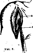
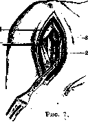
Figure 6. Arthrotomy according to Langenbeck. 1-tendon of m. bicipitis; 2-m. deltoideus.
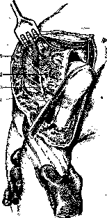
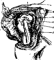
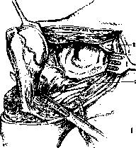
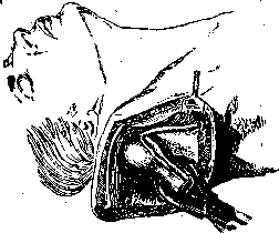
Figure 7. Arthrotomy according to Langenbeck: 1-tendon of m. bicipitis; 2-sulcus bicipitalis; 3-tuberculum minus. For removal of pathological contents of the shoulder joint, mainly in its purulent inflammation, opening of the joint (arthrotomy) with subsequent drainage is applied. The proposed incisions can be divided into 2 groups - anterior and posterior. Anterior ones are made either through m. deltoideus or along its anterior edge (Langenbeck). Posterior incisions, according to König, are made along the posterior edge of the deltoid muscle, they give a narrower access to the joint, but ensure better drainage. In many cases, a combination of two incisions is indicated. For wide opening of the joint
Figure 8. Arthrotomy according to Langenbeck: 1-tuberculum majus; 2-tendon of m. bicipitis.
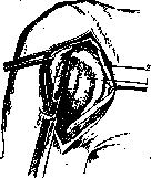
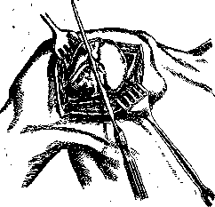
Figure 9. Sawing off the head.
Usually arthrotomy is insufficient, and if the process has spread to cartilage and bone tissue, then resection of the joint is indicated. In addition, resection can be applied in ankyloses, in irreducible dislocations, in some cases of fractures of the neck complicated by dislocation (see above) and in neoplasms. Approaches to the shoulder joint are divided into anterior, posterior, and transverse. One of the basic conditions recognized by all authors is the possible preservation of the integrity of the fibers of the n. axillaris in order to avoid subsequent complete or partial paralysis of the deltoid muscle. The most typical methods are the methods of Langenbeck, Kocher, and Neudorfer.
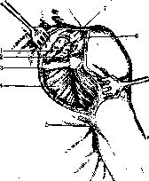
Figure 10> Arthrotomy with resection of the joint according to Kocher: 1-m. trapezius; 2-m. supraspinatus; 3-spina scapulae; 4-m. infraspinatus; 5-posterior surface of the axillary fossa; 6 - acromion; 7 - clavicle.
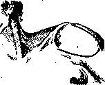
Figure 11. Incision with formation of a skin-deltoid flap.
methods are the methods of Langenbeck, Kocher, and Neudorfer. According to Langenbeck's method, the incision begins slightly away from the acromion and goes vertically downward through the fibers of the m. deltoidei (fig. 6-9). Upon opening the tendon sheath of the biceps, with lateral abduction of it and rotation of the head outward, the joint capsule is opened and the tendon of the m. subscapularis is severed from the tuberculum minus. With rotation of the head inward, the tendons of the mm. infra-, supraspinati and teretis majoris are detached from the tuberculum majus. The head is dislocated into the wound and the dorsal part of the joint capsule is severed; a fairly wide access is obtained, and it is easy to perform trimming or partial removal of the head. To preserve joint mobility, it is proposed to perform interposition of soft tissues, such as a muscle flap from the m. deltoideus or m. pectoralis major according to Pyr. Bardenheuer fixes the upper joint end of the arm to the joint socket in the abducted position of the limb.
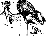
Figure 12. Extracapsular resection of the joint: 1-m. deltoideus; 2-extracapsularly freed joint; 3-saw cut.
According to Kocher's posterior method, the incision begins from the apex of the acromion and goes along the spina scapulae to its middle, then descends vertically downward almost to the angle of the scapula (fig. 10). After detaching the m. trapezii and part of the m. deltoidei, the neck of the scapula is freed with a periosteal elevator and the acromion is sawed off at its base, which is pulled outward. The attachments of the tendons of the mm. infra- and supraspinati and teretis majori are detached subperiosteally, or the tuberc. majus along with the attachment of the mentioned muscles is chiseled off. Upon opening the joint capsule, a wide access to the posterior-superior parts of the joint is obtained.
In Neudorfer's transverse method, the incision is made from the spina scapulae to the clavicle and then along the sulcus deltoideo-pectoralis. The clavicle and acromion are temporarily sawed through, and the entire flap is turned outward. This method provides wide access to the upper parts of the shoulder joint. For severe cases of tuberculosis of the shoulder joint, Bardenheuer proposed a method of extra-articular resection of it (fig. 11 and 12). The incision is made according to the outline of the m. deltoideus, and the skin-muscle flap is turned upward. The humerus is sawed through, the muscle tendons are severed outside the joint, as close as possible to their attachment; the neck of the scapula is freed extra-articularly, and after detaching the tendons of the mm. biceps and triceps, it is sawed through. The entire joint with the capsule is removed as a foreign body. The humerus is fixed to the scapula with a nail in the abducted position of the limb.
Exarticulation of the shoulder joint (fig. 13-16) has very limited application in clinical practice, since in neoplasms it turns out to be insufficiently radical, and in traumatic injuries, in most cases, it is possible to perform a high amputation of the arm, which is much more advantageous from the point of view of prosthetics (see Arm).
Figure 13. Exarticulation of the shoulder joint - incision of the capsule, II moment: 1-m. supraspinatus; 2-m. infraspinatus; 3-m. teres minor; 4-n. axillaris.
Figure 14. Exarticulation-III moment: 1-tendon of m. bicipitis; 2-a. circumflexa humeri ant.; 3-tuberculum minus; 4-tuberculum majus; 5-m. subscapularis; 6-lig. coraco-humerale.
Figure 15. Exarticulation-IV moment: 1-a. circumflexa humeri ant.; 2-tendon of m. subscapularis.
The most common methods are: 1) the method of Chalot-Lysenkov, in which the vessels are ligated at the beginning of the operation from an incision starting from the pr. coracoideus and carried downward to the edge of the m. pectoralis majoris, and 2) the circular method of Esmarch, which is divided into two moments: a) high circular amputation of the arm with exsanguination of the limb by Esmarch's tourniquet followed by ligation of the vessels, and b) extirpation of the entire upper end of the humerus from an additional vertical incision.
Figure 16. Exarticulation in combination with bone resection.
To improve the stump, Pyr proposed making a bony protrusion from the spina scapulae, which is covered with a flap of soft tissue and can serve as a lever for fitting a prosthesis.
Related articles
Mentioned in
Cite this page
“Shoulder Joint.” Soviet Medical Encyclopedia. English translation of Bolshaya Meditsinskaya Entsiklopediya, 1st ed. (Moscow, 1928–1936), ed. N. A. Semashko. https://sovietmedicalencyclopedia.pages.dev/article/shoulder-joint/