PLETHORA
Historical document, translated for reference. It reflects medical knowledge of the 1920s–30s and is not medical advice.
Summary
PLETHORA (plethora, from Greek plethore-filling), plethora, a rarely used term that formerly denoted various conditions, such as an increase in the total mass of blood, polycythemia, as well as an increase in the amount of water in the plasma. The latter condition was designated as plethora serosa.
Encyclopedia article (1928–1936)
PLETHORA (plethora, from Greek plethore-filling), plethora, a rarely used term that formerly denoted various conditions, such as an increase in the total mass of blood, polycythemia, as well as an increase in the amount of water in the plasma. The latter condition was designated as plethora serosa. In connection with the clarification of the concept and methodology for determining the amount of circulating blood and the ratio in it of the mass of blood corpuscles and the mass of plasma, as well as in connection with a more precise differentiation of individual types of polycythemia, the former concept of plethora becomes obsolete (see also Hydremia, Blood-total mass, Polycythemia). BRACHIAL PLEXUS (plexus brachialis) is formed by the connection of the last four cervical roots (from C5 to C8) and the first thoracic, and gives rise to the nerves of the upper limb and shoulder girdle. This connection occurs as follows: C5 merges into one common trunk with C6, forming the primary upper trunk (fasciculus primarius sup.); C7 connects with C1 in the primary lower trunk
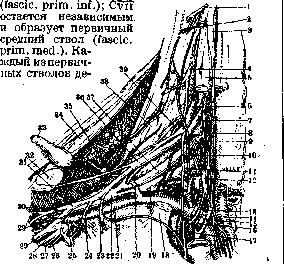
Figure 1. Brachial plexus: 1-C5; 2-C6; 3-ganglion sympathicus cervicale sup.; 4-C7; 5-m. rhomboidus capitis; 6-C5; 7-C6; 8-a. vertebralis; 9-C7; 10-n. phrenicus; 11-C8; 12-gangl. sympathicus cervicale inf.; 13-ansa Vieussenii; 14-a. carotis int.; 15-n. vagus; 16-a. subclavia; 17-apex of lung; 18-m. scalenus ant.; 19-fasciculus primarius inf.; 20-a. transversa scapulae; 21-a. axillaris; 22-fasciculus secundarius post.; 23-nerve for m. subclavius; 24-nerve for m. pectoralis major; 25-fasciculus secundarius inf.; 26-n. cutaneus antibrachii med.; 27-n. cutaneus brachii med.; 28-n. ulnaris; 29-n. medianus; 29'- its outer root; 30-n. musculo-cutaneus; 31-fasciculus secundarius ext.; 32-clavicle; 33-m. subclavius; 34-m. trapezius; 35-n. subscapularis; 36 and 37-fasciculus primarius sup. et inf.; 38-m. scalenus post.; 39-nerve for m. scalenus.
divides into two branches-anterior and posterior; The posterior branches connect into one common trunk-the secondary posterior (fascic. post., s. radio-axillaris). The anterior branch of the upper primary trunk connects with the anterior branch of the middle primary trunk, forming the secondary outer, or upper (fascic. later., s. superior), trunk, the anterior branch of the lower primary trunk remains independent-the secondary lower, or inner (fascic. medians, s. inf.) trunk (fig.-see Nerves). Fascic. post. continues into n. radialis (fig. 1). The inner and outer trunks again divide into 2 branches; both middle branches resulting from this division connect at a right angle to form n. medianus; the outer branch of fascic. lat. gives n. musculo-cutaneus; the inner branch of fascic. med. gives rise to n. ulnaris and two cutaneous nerves of the arm-n. cutaneus brachii medialis et n. cutaneus antibrachii medialis. The three upper roots of the plexus have a vertical descending direction, the fourth-horizontal, and the fifth-vertical ascending. The length of the brachial plexus is 15-20 cm. The corresponding roots of the plexus exit from the intervertebral foramina between mm. inter-transversarii anter. et poster., penetrate between m. scalenus medius, located posteriorly, and m. scalenus anter., located anteriorly. Then P. s. passes obliquely through the lower part of the supraclavicular fossa, heading toward the axillary fossa, in the upper parts of which it divides into terminal or peripheral nerves. The clavicle divides P. s. into two parts-pars supraclavicularis et pars infraclavicularis.. Pars supraclavicularis of P. s. is located posterior to the lower part of m. sterno-cleido-mastoidei, crosses with m. omohyoideus. Pars infraclavicularis is located between m. subscapularis et m. serratus anter., under mm. pectoralis minor et major. A. subclavia lies initially below the roots of the plexus, then curves around its anterior surface, under the name a. axillaris penetrates between the two roots of n. medianus and goes to its posterior surface.-P. s. receives blood from the arteries surrounding it. C5 receives an anastomosis from C7, C1 - from C2 (C3)'; these anastomoses are almost constant, in case of absence of one, the other is more developed. In addition there are anastomoses with n. sympathicus, carrying fibers from cervical sympathetic nodes and from gangl. stellatum. On its way P. s. gives branches to the neck, shoulder girdle and to the upper limb. Nerves for the neck arise from the roots of the plexus soon after their exit from foram. inter-vertebralia. These are muscular nerves, ending in the lower segments of mm. scaleni anter., med., poster, et longi colli. For the muscles of the shoulder girdle P. s. sends: 1) nn. thoracales poster., originating from the upper primary trunk and dividing into n. dorsalis scapulae for mm. rhomboidei and n. thoracalis longus for m. serratus anter. 2) Nn. thoracales anter. usually in the amount of two-primus, (externus) from the upper secondary trunk and secundus (internus) from the lower secondary trunk; both innervate mm. pectorales major et minor. 3) N. subclavius (primary upper trunk) for m. subclavius. 4) N. supra-scapularis (primary upper trunk), giving branches to mm. supra- and infraspinatus, as well as to the articular capsule of the shoulder. 5) N. subscapularis, dividing into three branches: inferior (primary upper trunk)-to m. latissimus dorsi; medius (posterior trunk)-to mm. subscapularis et teres major; superior (also origin)-to m. subscapularis. 6) N. axillaris (fig. 2), emerging from the secondary posterior trunk; it surrounds the humerus and ends in m. deltoideus; gives branches for the shoulder joint; its branch-n. cutaneus brachii lat.-innervates the skin over the posterior surface of m. deltoideus and the upper half of the arm. The terminal nerves of the brachial plexus intended for the upper limb are in most cases mixed nerves (nn. medianus, musculocutaneus; radialis, ulnaris) and only two (nn. cutaneus brachii medialis, cutaneus antibrachii medialis)-exclusively sensory. (Ontogenesis and phylogenesis of the brachial plexus - see Lumbosacral plexus.) Physiology of the brachial plexus, P. s. controls all movements of the shoulder girdle and upper limb. In this case, nerves originating from the upper roots of the plexus or from its primary upper trunk innervate the muscles of the shoulder girdle and to some extent the arm, while the lower-roots of the plexus are related to the muscles of the forearm, hand and fingers. The function of the nerves of P. s. and the muscles innervated by them-see Muscles of man and Nerves of man.
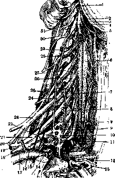
Figure 2. Brachial plexus: 1-ramus spinalis n. accessorii; 2-n. vagus; 3-n. hypoglossus; 4-gangl. sympathicus cervicale sup.; 5-meninges; 6-spinal cord; 7-C7; 8-section of transverse processes; 9-nn. vertebralis; 10-gangl. stellatum; 11-C1; 12-esophagus; 13-trachea; 14-rami communicantes; 15-thorax; 16-truncus primarius inf.; 17-a. axillaris; 18-tr. secundarius inf.; 19-tr. secundarius post.; 20-tr. secundarius sup.; 21-tr. primarius med.; 22-a. cervicalis; 23-tr. primarius sup.; 24-ramus communicans ad gangl. stellatum; 25-C6; 26-C5; 27-gangl. spinale; 28-C7; 29-truncus sympathicus cervicalis; 30-C8; 31-C1.
Pathology of the brachial plexus. Being a large anatomical formation extending over a considerable distance from the spine to the axilla, the brachial plexus can be affected by various processes, and depending on the level of the lesion, the roots, primary trunks, or secondary trunks of the brachial plexus may be damaged; in the axilla, the process usually involves the entire brachial plexus. Etiology. Diseases of the brachial plexus (plexitis, or neuritis of the brachial plexus) are most often of traumatic origin: direct injury to the brachial plexus by some object, pressure on the brachial plexus in fractures of the clavicle or humerus, caused not only by bone fragments but also by accompanying hemorrhage; pressure in dislocations of the head of the humerus, in tumors in the axilla (tumors, aneurysm of the axillary artery), pressure from crutches; strong abduction of the arm can also cause paralysis of the brachial plexus (paralysis of the arm during general anesthesia when the arm is strongly abducted and placed behind the patient's head). Injury to the brachial plexus is also observed during childbirth when attempts are made to rotate the fetus or apply forceps; paralysis may also be due to skeletal anomalies - false ribs. More rarely, the cause of paralysis of the brachial plexus is infection, intoxication, and auto-intoxication (tuberculosis, malaria, gonorrhea, diphtheria, syphilis, rheumatic polyarthritis, various typhus, meningitis, etc., diabetes, poisoning from sausage, arsenic, alcohol, carbon monoxide oxide, chlorosis, cachexia, anemias). Among professions, it should be noted porters carrying heavy loads on their back, on their shoulders, and workers with lead. In general, paralysis of the brachial plexus is not a particularly common disease, it was more often observed during wartime. More common in men than in women; in manual laborers more often than in mental workers. Pathological changes in diseases of the brachial plexus-see Wallerian degeneration and Neuritis. Symptomatology. As a result of the localization of the process in various parts of the brachial plexus, the most diverse clinical syndromes are observed. In diseases of the entire brachial plexus, paralysis of the muscles of the entire upper limb and shoulder girdle is observed; the muscles are atrophied, with complete or partial reaction of degeneration: the periosteal and tendon reflexes are absent; sensory disturbances are also present throughout the upper limb and in the area of the shoulder girdle, with the exception of a small area on the inner surface of the arm innervated by the Dn; trophic and vasomotor symptoms and Horner's syndrome are also observed. In neuralgia of the brachial plexus, there is an indefinite, not strictly localized spread of pain throughout the upper limb, from the supraclavicular fossa to the fingers; pains occur in attacks or are constant, sometimes exacerbating; they have a tearing, burning, drilling character; they intensify with movement; painful points can often be found in the supraclavicular and infraclavicular fossae (Erba's points), and sometimes along the course of peripheral nerves; paresthesias and hypesthesias; complete paralysis is not observed in this case, diffuse pareses are present. It should be noted that in reality, diseases of the entire brachial plexus are very rare, some branches (motor or sensory) always remain unaffected, which changes the clinical picture. In lesions of the roots or primary trunks of the brachial plexus, root syndromes are observed-root distribution of sensory and motor disturbances. Known are: 1) the upper type, or Erb-Duchenne plexus syndrome, observed in lesions of the C5-C6 roots, the supraclavicular part of the brachial plexus, or the upper primary trunk; it is characterized by paralysis of the muscles of the shoulder girdle and arm (see Erb-Duchenne paralysis); 2) the posterior type, or Remak's syndrome, described in lesions of the C7 root or the posterior primary trunk, it manifests as paralysis or paresis of the muscles innervated by the radial nerve, with the exception of the supinator muscle (see Remak's paralysis); 3) the lower type, or Dejerine-Klumpke syndrome, observed in lesions of the C8 and T1 roots or the lower primary trunk; it is characterized by paralysis of the flexors of the wrist and fingers, sensory disturbances, and Horner's syndrome. The syndrome observed in lesions of the secondary trunks gives the same picture as diseases of peripheral nerves. Lesion of the upper or lateral secondary trunk is characterized by paralysis of the biceps, brachialis, pronator teres, palmaris longus muscles, and weakening of the function of the finger and wrist flexors. In lesions of the posterior secondary trunk, to the symptoms of radial nerve disease are added symptoms of axillary nerve lesion (see Neuritis). In lesions of the lower or medial secondary trunk, complete paralysis of the ulnar nerve is observed (see Ulnaris nervus) and partial manifestations of median nerve lesion. From the collateral branches of the brachial plexus, isolated lesions can be observed: 1) of the thoracic nerves, which manifests as paralysis of the serratus anterior muscle-the scapula recedes from the chest wall and moves medially, elevation of the arm is limited to a right angle; paralysis is sometimes accompanied by pain, sensory disturbances do not occur; 2) of the axillary nerve, accompanied by paralysis of the deltoid muscle-inability to raise the arm upward, sensory disturbance of the skin over this muscle. The nerve is compressed by crutches and when carrying heavy loads on the shoulder; this nerve disease is also observed in diabetes. Other branches of the brachial plexus are rarely affected in isolation, and their disease manifests as paralysis of the corresponding muscles (disease of the terminal branches of the brachial plexus-see the respective nerves). Differential diagnosis of diseases of the brachial plexus and various peripheral nerves can be established on the basis that the paralyses and anesthesias do not correspond to a single peripheral nerve. It is rather difficult to distinguish diseases of the brachial plexus from lesions of the spinal cord roots, especially its complete disease, which gives the same picture. The main help is provided by etiological factors and the further course. Paralyses of the plexuses depending on severe wounds give a poor prognosis for complete recovery. Treatment and prevention of diseases of the brachial plexus are the same as in diseases of individual nerves (see Neuritis, Neuralgia, Polyneuritis, and individual nerves): rest, heat, diathermy, galvanization; if there are causes that can be eliminated surgically (tumors, false ribs, bone fragments), surgical treatment should be resorted to. In injuries to the brachial plexus, if the ends of the nerves are not too far apart, sutures are applied (see Nerve suture, Neurorrhaphy).
Related articles
Mentioned in
Cite this page
“PLETHORA.” Soviet Medical Encyclopedia. English translation of Bolshaya Meditsinskaya Entsiklopediya, 1st ed. (Moscow, 1928–1936), ed. N. A. Semashko. https://sovietmedicalencyclopedia.pages.dev/article/plethora/