PLETHYSMOGRAPHY
Historical document, translated for reference. It reflects medical knowledge of the 1920s–30s and is not medical advice.
Summary
Plethysmography is a method for measuring the volume of different body parts, particularly useful for studying blood redistribution during work and fatigue. The technique uses specialized instruments to record volume changes, revealing how blood flow varies with physical and mental activity.
Encyclopedia article (1928–1936)
PLETHYSMOGRAPHY (from Greek plethyo- I fill and grafeo - I write), a method for measuring the volume of various parts of the body. The volume of all parts of the body in animals and humans increases and decreases within small limits depending on the expansion and constriction of blood vessels. When certain organs are more active, blood flows into their dilated vessels while simultaneously flowing away from organs at rest. Thus, during digestion, blood flows to the stomach and intestines, flowing away from the muscles and brain. The study of such redistribution of blood in the body through measurement of organ volumes is performed with plethysmographs. The simplest plethysmograph for measuring the volume of the arm consists of a metal or glass cylindrical vessel lying horizontally with one large lateral opening for inserting the arm inside the vessel. Through another opening, water heated to skin temperature is poured into the vessel; to this opening is attached a vertical tube, which in turn is connected by a rubber tube to Marey's recording drum. The arm fits tightly into a special rubber sleeve attached to the edges of the lateral opening, which prevents water from flowing out of the vessel. Since water is a liquid that is practically incompressible, when the arm's vessels expand and its volume increases, water is displaced from the vessel by exactly the increased volume of the arm, and the water level in the upper tube rises and presses on the air, causing the pen of Marey's drum to rise. Further recording is done in the usual physiological way on a kymograph. The curve thus recorded - a plethysmogram - shows that the volume of the arm increases, although very slightly, with each cardiac systole and decreases during diastole. In addition, respiratory movements are clearly reflected on the plethysmogram. Plethysmographs vary greatly in construction, depending on the purpose for which they are built. There are plethysmographs only for fingers of the hands, for the foot, for individual organs, for example the kidney, etc. Plethysmography was used to study the distribution of blood during physical and mental work, as well as to determine the onset of fatigue. E. Weber studied the distribution of blood during fatigue particularly extensively. He studied simultaneous changes in blood filling in the arms, legs, abdominal cavity and head during
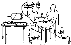
Arrangement of instruments for studying the expansion of vessels in the arm during work and fatigue of the hand muscles. A foot ergograph is attached to the leg, driven by the calf muscle, which is stimulated by electrical shocks. The hand is placed in a glass vessel P with water, from the top of which a rubber tube leads to the recording apparatus. When the leg muscles become fatigued, the arm muscles also become fatigued, and the blood vessels in them constrict; during this constriction, the volume of the hand in the water decreases, which is recorded on a rotating cylinder.
work of the leg or arm, etc. (see figure). To accurately study the influx and efflux of blood to different parts of the body simultaneously, Weber developed a method of recording several plethysmograms at once. Special modified plethysmographs, depending on the organ being studied, were adapted to the arms, leg, abdominal area, and ear. Using this method, Weber found that during work, blood flows not only to the working muscles but to all muscles, and during fatigue, conversely, flows away from all of them. Therefore, if one makes completely fresh, unworked muscles work, they will cause an influx of blood to the tired muscles and thereby increase their capacity for work. This struggle against fatigue has its limits and is possible only when small groups of muscles are working. If the whole body or many muscles are working, then rational complete rest is necessary. That one can increase the capacity of tired muscles not only by rest but also by working other muscles was shown by E. Weber himself on human muscles. If a small group of muscles contracts voluntarily, this affects the capacity of other muscles. This was first shown by I. M. Sechenov. He found that if the left hand works, the capacity of the right hand (or vice versa) increases. Sechenov explained this by the influence of the sensory nerves of the working hand on the nervous system of the other hand. Weber attributes this phenomenon to the activity of the vasoconstrictor nerve centers. Having confirmed the known fact that blood flows to a working organ, he discovered that the influx of blood, for example to the arm, can occur not only during actual physical work but even with just the thought, the vivid representation of physical work with the arm. But not all people show a 'positive', i.e., rising plethysmogram with only psychomotor representation - overly nervous people do not show this phenomenon. A positive plethysmogram, although very weakly expressed, was also obtained during mental work. A hypnotized person, to whom it was suggested that he was performing heavy physical work, showed a positive plethysmogram. Conversely, during fatigue, after prolonged and heavy work, a 'negative' plethysmogram is obtained, showing that in the long-working organ blood circulation worsens, vessels constrict and blood flows away. Obviously, during long and tiring work, vasodilators become less excitable than usual, as if they tire. Therefore Weber says that in this case there is fatigue of the central nervous system. For a negative plethysmogram to appear, 15-20 minutes of fast running is sufficient. In mental fatigue, the negative plethysmogram is much less pronounced than in physical work. In trained people, the negative plethysmogram appears much later compared to untrained people. The works of E. Weber and the method of P. were subjected to very sharp criticism by Bruns, who pointed out the great difficulty of obtaining accurate data with a plethysmograph. With every movement of the arm in the vessel, the water immediately comes into motion, and a positive plethysmogram appears. He also showed by recording the movements of the arm itself that it is extremely difficult to keep the arm in the plethysmograph completely still during psychomotor representation of muscle movement: involuntary movements occur that distort the experiment and give incorrect results and conclusions. To eliminate the influence of these arm movements, the entire plethysmograph is suspended on chains.
Related articles
Mentioned in
Cite this page
“PLETHYSMOGRAPHY.” Soviet Medical Encyclopedia. English translation of Bolshaya Meditsinskaya Entsiklopediya, 1st ed. (Moscow, 1928–1936), ed. N. A. Semashko. https://sovietmedicalencyclopedia.pages.dev/article/plethysmography/