Abdominal Cavity
Historical document, translated for reference. It reflects medical knowledge of the 1920s–30s and is not medical advice.
Summary
This article from the 1st edition of the Great Medical Encyclopedia describes the anatomy, boundaries, regional divisions, and contents of the abdominal cavity, detailing retroperitoneal and peritoneal structures as understood in the early 1930s.
Encyclopedia article (1928–1936)
ABDOMINAL CAVITY, cavum abdominis, is bounded on the sides by the abdominal walls (see), its upper boundary is formed by the diaphragm, and its lower by the linea innominata; however, the peritoneal sac also extends into the cavity of the lesser pelvis, forming the so-called cavum pelvis peritoneale. The shape of the abdominal cavity varies depending on age and sex and is determined by the dimensions and shape of the thorax (apertura thoracis inferior) and pelvis. In children, it has an approximately ovoid shape; in individuals with a narrow thorax and wide pelvis (women), it is pear-shaped, with the wide end pointing downward; in subjects with a wide thorax and relatively narrow pelvis (men), it has the same shape, but with the wide end pointing upward. Each shape corresponds to a specific arrangement and form of the organs enclosed within the abdominal cavity (see Figure 1). The abdominal cavity is divided by conventional planes into nine regions. The most accepted division is as follows: two horizontal planes, of which one passes through the lowest points of the 10th ribs and the other through the highest points of the iliac crests, divide it into three sections: upper—epigastrium, middle—mesogastrium, lower—hypogastrium; two vertical planes passing through the middle of the inguinal ligaments divide each of these sections into three: the upper into regio hypochondriaca dextra et sinistra and regio epigastrica propria, the middle into regio lumbalis dextra et sinistra and regio umbilicalis, the lower into regio inguinalis dextra et sinistra and regio pubica (see Figure 2). The abdominal cavity contains the greater part of the digestive apparatus and part of the urinary system. By the posterior layer of the peritoneum (see), it is divided into two sections: the retroperitoneal space (cavum s. spatium retroperitoneale) and the peritoneal cavity (cavum peritonaei). Accordingly, the organs located in the abdominal cavity are divided into two groups: retroperitoneal organs (organa extra saccum peritonaei) and intraperitoneal (organa intra saccum peritonaei). It should be noted that this division is somewhat conventional, since in reality no organ is located entirely inside the peritoneal sac, but rather, depending on whether the peritoneal fold covers certain organs incompletely, the latter are isolated into a group of extraperitoneal organs. In the full sense, extraperitoneal organs are the kidneys, adrenal glands, ureters, part of the rectum, abdominal aorta and inferior vena cava with their branches, lumbar sympathetic trunk, lumbar plexus, lumbar lymph nodes, and cisterna chyli. Practically, into this group
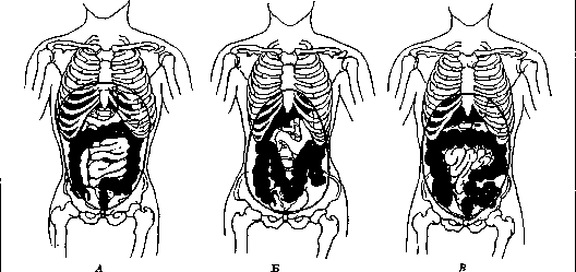
Figure 1 forms of the abdominal cavity: pear-shaped in the man; pear-shaped in the woman; in the child.

Venous system.
Gland.
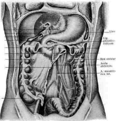
Figure showing abdominal organs and vessels.
Visceral organs and retroperitoneal space structures.
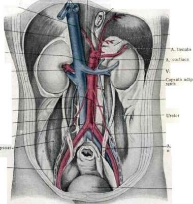
Left adrenal gland, kidney capsule, vessels, and pelvic organs.
Mesentery of the small intestine, sigmoid colon, uterus, and sagittal cross-section of the abdominal cavity after Corning, showing peritoneal reflections schematically.
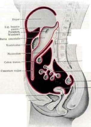
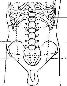
Figure 2. Division of the abdominal cavity into regions: 1—regio epigastrica; 2—regio umbilicalis; 3—regio pubica; 4—regio hypochondriaca; 5—regio lumbalis; 6—regio inguinalis. The posterior wall of the retroperitoneal space is the posterior abdominal wall, and the anterior wall is the dorsal parietal peritoneum. Above, this space reaches the place where the peritoneum passes to the liver, stomach, and spleen, and below—to the promontory. It is filled with cellular tissue in which the listed organs are located, partly surrounded by their own connective tissue layers. Cavum peritonaei is a voluminous cavity that is completely closed in the male, while in the female, through the openings of the Fallopian tubes, it communicates with the cavity of the tubes, uterus, and vagina. There is essentially no cavity as such; it appears only in pathological cases of fluid accumulation within it, whereas in a normal physiological state the internal organs protruding into it (being under a certain pressure—"intra-abdominal pressure"), closely adhering to each other, leave only narrow slits between them. The cavity is lined from the inside by the peritoneum (see). The parietal layer lines the entire inner surface of the anterior and lateral walls and passes to the posterior, continuing then into the visceral layer. The latter along its path forms five "pouches": hepatic, gastric (and splenic), colic, for the small intestines, and for the genital organs. The course of the visceral layer is as follows (see Figure 3 and Table II, Figure 4): from the anterior wall to the diaphragm and then to the upper surface of the liver; rounding its sharp edge, it lines the lower surface to the porta hepatis, from here it heads to the lesser curvature of the stomach, forming the anterior layer of the hepatogastric ligament or lesser omentum, covers the anterior wall of the stomach, hangs down from the greater curvature in the form of an apron and, bending over, heads to the posterior wall of the abdominal cavity, toward the spine. From here, descending again, it encloses the colon, then also the small intestine, and thus forms the transverse mesocolon and mesentery consisting of two layers, in the thickness of which (between the layers) vessels and nerves are enclosed; after this it descends along the spine to the lesser pelvis, encloses part of the rectum, and passes in males onto the urinary bladder, covering its posterior and upper walls, and in females passes successively onto the uterus, vaginal vault, and urinary bladder, and then heads toward the anterior wall of the abdominal cavity.
The translation of the remaining anatomical index labels and figure captions follows the structured list above.
Related articles
Mentioned in
Cite this page
“Abdominal Cavity.” Soviet Medical Encyclopedia. English translation of Bolshaya Meditsinskaya Entsiklopediya, 1st ed. (Moscow, 1928–1936), ed. N. A. Semashko. https://sovietmedicalencyclopedia.pages.dev/article/abdominal-cavity/