Aeroperitoneum
Historical document, translated for reference. It reflects medical knowledge of the 1920s–30s and is not medical advice.
Summary
This article describes the historical medical procedure of aeroperitoneum, which involved injecting oxygen or air into the abdominal cavity to improve visualization during X-ray examinations. It outlines the clinical indications, diagnostic utility, potential complications, and contraindications for the method as understood in the early 1930s.
Encyclopedia article (1928–1936)
AEROPERITONEUM (from Greek aer—air and Latin peritoneum—peritoneum), the artificial introduction of oxygen or air into the abdominal cavity for the purpose of creating a contrast image during X-ray examination. Aeroperitoneum was first applied for the purposes of laparoscopy in 1902 by Kelling. Beginning in 1914, the technique of aeroperitoneum was systematically developed by Rautenberg, and aeroperitoneum was introduced into clinical diagnostic practice. The experience accumulated since then, which has been reflected in extensive literature, gives the right to recognize the enormous significance of this method for the diagnosis of complicated cases. Diseases of the diaphragm, liver, spleen, and kidneys, in the presence of anatomical changes, are clearly revealed thanks to aeroperitoneum. Structural changes in the pancreas and retroperitoneal glands can be recorded on photographic film when using aeroperitoneum. All kinds of adhesions of the gastrointestinal tract with surrounding organs can be detected or clearly seen after the insufflation of oxygen into the abdominal cavity. The presence of adhesions of the anterior abdominal wall with organs of the abdominal cavity can be indisputably proven by aeroperitoneum. It is sufficient for this to place the patient on their back and give the X-ray beam a latero-lateral direction. All kinds of anatomical changes in the upper part of the abdominal cavity can be established with the patient in a vertical position (see separate table). The position of the patient with the pelvis raised upward makes it possible to judge the state of the genital system in women (used in obstetrics and gynecology). The downside of the method is that, in the case of extensive development of any tumor in the abdominal cavity and in the presence of extensive adhesions, oxygen, accumulating in one place, does not make it possible to make a general overview of the topography of the abdominal organs, and in such cases, aeroperitoneum provides almost no additional information beyond that which can be obtained by ordinary methods of clinical examination. The use of air as a contrast medium must still be recognized as far from safe for the patient, since its delayed absorption from the abdominal cavity (3–4 weeks) has been repeatedly observed. From Case's questionnaire material, collected by surveying 223 surgeons who used aeroperitoneum, it follows that the following complications may occur: 1) injury to the intestine, which, admittedly, did not lead to serious consequences; 2) perforation of mesenteric vessels, established by laparotomy; 3) perforation of viscera, namely: a) puncture of the liver, b) of the urinary bladder, c) puncture of a hydronephrotic kidney; 4) peritonitis (a fatal case described by Barringer from New York); 5) gas embolism, possible when using air (Joseph's case); 6) subcutaneous emphysema; 7) rupture of peritoneal adhesions and the spread of infection throughout the peritoneum; 8) decline in cardiac activity. In total, from Case's questionnaire material, four fatal cases are seen (puncture of the liver, peritonitis, and two cases without autopsy verification). Talalaev observed a fatal case of rupture of the splenic artery with simultaneous severe arteriosclerosis of the latter. As for contraindications for the use of aeroperitoneum, it is clear that cases of severe heart defects, myocardiopathies with decompensation, cases of kidney disease with symptoms of intoxication, as well as cases where one can assume an encapsulated focus of pus in the abdominal cavity, are incompatible with aeroperitoneum. Decker considers intestinal meteorism a contraindication for aeroperitoneum. In Uspensky's cases, aeroperitoneum was sometimes successfully used even with meteorism, provided that a cautious technique of oxygen introduction was observed. Contraindications must be strictly individualized, and the environment in which the aeroperitoneum is performed must be precisely taken into account; therefore, in outpatient cases, where it is impossible to verify absorption from the abdominal cavity through repeated examinations, aeroperitoneum is absolutely excluded.
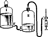
For the introduction of oxygen, the patient is given a position approaching the Trendelenburg position, and the puncture of the abdominal wall is made along the linea alba below the navel or along the outer edge of the rectus abdominis muscle. Various forms of needles with blunt and sharp ends have been proposed, or the puncture is made first with the needle of a syringe filled with physiological saline, and slowly, by raising the vessel (see figure) filled with mercuric chloride or physiological saline, the oxygen is displaced from the vessel into the abdominal cavity. Some surgeons check the correctness of the puncture performed with an abdominal manometer, a U-shaped tube with physiological saline, or are guided by a peculiar sensation upon puncturing the peritoneum. The amount of oxygen must be strictly individualized, from 0.5 to 3 liters, depending on the patient's reaction and the goals pursued by the aeroperitoneum.
Related articles
Mentioned in
Cite this page
“Aeroperitoneum.” Soviet Medical Encyclopedia. English translation of Bolshaya Meditsinskaya Entsiklopediya, 1st ed. (Moscow, 1928–1936), ed. N. A. Semashko. https://sovietmedicalencyclopedia.pages.dev/article/aeroperitoneum/