Gallbladder
Historical document, translated for reference. It reflects medical knowledge of the 1920s–30s and is not medical advice.
Summary
A detailed anatomical description of the gallbladder, covering its location, structure, dimensions, and vascular supply, as presented in the 1930s Soviet medical literature.
Encyclopedia article (1928–1936)
GALLBLADDER, bile ducts. Contents: I. Anatomical-topographical data... 202 II. X-ray examination... 219 III. Pathological anatomy... 225 IV. Pathological physiology and clinical picture... 226 V. Surgery of the gallbladder and bile ducts... 229 I. Anatomical-topographical data. The gallbladder (vesica fellea) is a hollow organ serving as a reservoir for bile, lying on the inferior surface of the liver in the fossa vesicae felleae, between the right and quadrate lobes of the liver. With its superior-anterior surface, the gallbladder touches the liver tissue directly and is loosely connected to it by connective tissue and a network of blood vessels. Its inferior-posterior and lateral surfaces are covered by the visceral peritoneum, which transitions onto it directly from the inferior surface of the liver. Thus, the connective tissue, vessels, and, mainly, the peritoneum fix the gallbladder in the fossa vesicae felleae and prevent its mobility. The fundus and neck of the gallbladder are more pliable. In some cases, the entire gallbladder is covered by the peritoneum so completely that a mesentery of the gallbladder of varying degrees of development is formed (Figures 1 and 2): according to Suslov—in 4%, according to Brewer and Kehr—in 5%, according to Testut—in 10%. In the region of the gallbladder, peritoneal folds are also quite often observed: lig. cystico-colicum and lig. cystico-duodenale. The first
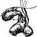
Figure 1. Lig. cystico-duodenale (a).
represents a simple duplication of the peritoneum and connects the gallbladder with the transverse colon, transitioning subsequently into the leaflets of the lig. hepato-duodenale; the second connects the ductus cysticus, and in cases of strong development, also a part of the gallbladder with the duodenum. The gallbladder most often has a pear-shaped or cylindrical form; in pathological cases, it can be shriveled, contracted, empty, or, conversely, distended and assumes an egg-shaped form, sometimes reaching significant dimensions (up to the size of a child's head). The length of a normal gallbladder fluctuates within the following limits: according to Langenbeck from 10 to 14 cm, according to Testut 9–11 cm, according to Volyntsev 8–14 cm, according to Suslov 5–8 cm. Its greatest width according to Kehr is 2.5–3 cm, according to Testut 3.5–4 cm. Capacity according to Luschka and Kehr is 30–40 cm3, according to Testut 50–60 cm3, according to Fedorov 40–70 cm3. The gallbladder, however, is easily distended. When filled with water (in cadavers), up to 200 cm3 of fluid can be freely introduced into it without damage. Upon further injection, rupture of the wall occurs, always in one specific place (Testut), namely, on the right wall, at the place of transition of the body of the gallbladder into the neck. In the gallbladder, one distinguishes the fundus, body, and neck, which form a slight arc, with the concavity facing downward, to the left, and posteriorly. In the vertical position of a person, the fundus of the gallbladder is located below all other parts, then the body, and the neck is the highest (Figure 3).
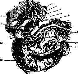
Figure 2. Gallbladder and bile ducts with the excretory ducts of the pancreas: 1—liver; 2—fundus of the gallbladder; 3—body of the gallbladder; 4—ductus cysticus; 5—ductus hepaticus; 6—arteria hepatica; 7—vena portae; 8 and 13—ductus choledochus; 9—ductus pancreaticus; 10 and 11—arteria et vena mesenterica superior; 12—duodenum. (According to Spalteholz.)
The fundus of the gallbladder is usually considered a special section; being located at the anterior edge of the liver, it usually protrudes beyond the edge and has a complete peritoneal covering. The liver in this place has a slight notch, sometimes, especially often in the elderly, its tissue is as if eroded here and replaced by scar tissue. Most often, the fundus protrudes slightly beyond the sharp hepatic edge (according to Siraud—32 cases out of 40, according to Charpy—23 out of 27, according to Suslov—70 out of 100). Much less frequently, cases are encountered where the fundus does not reach the hepatic edge, and finally, it can be more or less deeply and completely covered by the liver parenchyma. In the case where the fundus protrudes from under the lower edge of the liver, it lies directly against the anterior abdominal wall and is projected onto it in the angle formed by the right costal arch and the lateral edge
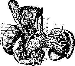
Figure 3. Relationship of the bile ducts with surrounding organs: 1 and 2—glandula suprarenalis; 3—arteria cystica; 4—ductus cysticus; 5—ductus hepaticus; 6—ductus choledochus; 7—arteria gastrica dextra; 8—arteria gastro-duodenalis; 9—vena portae; 10—vena cava inferior; 11—arteria hepatica; 12—aorta abdominalis; 13—arteria coeliaca; 14—arteria gastrica sinistra; 15—arteria lienalis; 16—ligamentum gastro-lienale; 17—arteria mesenterica superior; 18—vena mesenterica superior. (According to Kehr.)
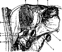
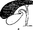
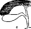
of the rectus abdominis muscle, approximately at the height of the transition of the VIII costal cartilage into the costal arch (Corning). According to other authors, the fundus of the gallbladder corresponds to either the end of the X or the IX and VIII ribs. The body of the gallbladder is usually covered by the peritoneum on 3/4 of its surface. Along its long axis, it describes a more or less slight curvature and, with its free inferior-posterior surface, usually lies against the colon transversum and flexura coli dextra (Figure 4). Located medially are the pars superior duodeni and the pylorus. These relationships, however, can change depending on the position of the gallbladder itself. With a more medial position, it will touch the horizontal part of the duodenum and even the pylorus. With a lateral position, the gallbladder approaches the vertical part of the duodenum, and the distal section of its body, and partly the fundus, touch the flexura coli dextra, sometimes even the right kidney. With a strong displacement downward, the body of the gallbladder can lie on loops of the small intestine. The place of transition of the gallbladder into the neck is usually indicated by a well-defined bend. One can establish two extreme types of this transition: a) when the body transitions into the neck at a sharp angle and b) when this transition occurs gradually (Figure 5). A valvular fold corresponds to this bend. The structure of the entire valvular apparatus of the neck of the gallbladder and the ductus cysticus is not the same in both types (Figure 6). Usually, the neck is compared in shape
Figure 4. Relation of the gallbladder to surrounding organs: 1—gallbladder; 2—liver; 3—probe inserted into foramen Winslowii, under the ligamentum hepato-duodenale; 4—lobus Spigelii hepatis; 5—cardiac section of the resected stomach; 6—bursa omentalis; 7—mesocolon transversum (resected); 8—flexura coli transversi; 9—duodenum (horizontal part). (According to Spalteholz.)
to a bird's beak or to the Latin letter S, formed by the angle open to the left between the neck and the body of the gallbladder on one side and the reverse bend of the ductus cysticus on the other. Usually, the neck, upon the formation of its angle, deviates forward and upward and lies against the left side of the gallbladder, but there are cases of right-sided adherence (according to Suslov, five times less frequent than left-sided); even more rarely, the neck lies against the inferior and posterior surface of the gallbladder (Suslov). In cases of the second type (i.e., with a gradual transition of the body of the gallbladder into the neck), all bends are weakly expressed. The neck of the gallbladder, being in the highest part of the fossae vesicae felleae, touches the right branch of the vena portae above (Figure 2), and below—the place of transition of the horizontal part of the duodenum into the descending part. The lumen of the neck is 7–8 mm, the length on average equals 15–18 mm (Fedorov).
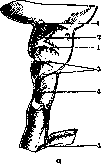
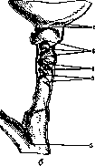
Figure 5. Two types of transition of the gallbladder neck into the ductus cysticus. The wall of the gallbladder is composed of three layers: the mucous membrane, the muscular layer, and the outer fibrous layer, which in turn consists of a connective tissue layer adjacent to the muscle, followed outwardly by the subserous layer (tunica subserosa), and finally (the peritoneal covering) the tunica serosa. In the place where the gallbladder lies directly against the liver tissue, the serous layer is absent. Arteries, veins, lymphatic vessels, and nerves are embedded in the wall of the gallbladder. The muscular layer in the body of the gallbladder is developed extremely weakly, more strongly in the region of the neck. The normal mucosa, thanks to folds running in various directions, has a reticulated appearance. It is velvety, as it is studded with small, uniform villi, and is usually colored dark brown. In the neck of the gallbladder (and according to some authors, also in the body and in the fundus of the gallbladder), there are mucous glands. In addition, in the wall of the gallbladder, there are so-called Luschka ducts, which represent an outpouching of the mucous membrane penetrating between the weak, separated muscle fibers. The gallbladder receives its nutrition from the arteria cystica, which usually branches off from the arteria hepatica dextra; the arteria cystica constitutes the upper side of the so-called Calot's triangle, the ductus hepaticus—the base of the triangle, the ductus cysticus—the lower side (Figures 7 and 8). (Regarding variants of the origin of the arteria cystica, see below—bile ducts and Figure 24.)
Figure 6. Types of arrangement of the valves of the ductus cysticus (a and b): 1—neck valve; 2—intermediate valve; 3—semilunar valve; 4—spiral valve; 5—terminal valve.


Upon approaching the neck of the gallbladder, the arteria cystica divides in a fork-like manner into two branches: one of them runs along the superior surface of the gallbladder, which is attached to the liver, the other, usually larger, along the inferior surface, which is covered by the peritoneum. Much less frequently, both branches are found under the peritoneum. The terminal branches anastomose widely with each other. According to Cavalier, the arteria cystica forms three networks in the gallbladder: rete submucosum, subserosum, and a network in the fibrous-muscular layer. The system of branches of the arteria cystica has anastomoses with the branches of the arteria hepatica. Sometimes cases are encountered where the arteria cystica divides into its two branches immediately after its origin, and both branches then approach the gallbladder independently. Finally, cases of a paired cystic artery are encountered. The veins of the gallbladder do not run as such isolated trunks as the arteries, but form
Figure 8. Figure 7. Common arrangement of the cystic artery. Figure 8. Fairly common arrangement of the cystic artery: 1-liver parenchyma of the cystic fossa; 2-visceral peritoneum; 3 and 4-right and left cystic artery; 5-gallbladder. (According to Kehr.) ... a wide network of anastomoses, the various branches of which partly enter the liver parenchyma, and partly drain into the right branch of the portal vein. - The lymphatic vessels of the gallbladder form two networks: one is located in the mucosa, the other in the subserosa (Clairmont, Poirier, and Charpy). According to Franke, the lymphatic vessels of the gallbladder anastomose with the lymphatic vessels of the liver (Fig. 9). The terminal trunks of the network extend to the left and right of the gallbladder and head toward the glands of the neck. Some of the branches bypass these glands. Subsequently, some lymph flows reach the glands near the head of the pancreas, with whose lymphatic network the lymphatic vessels of the gallbladder have a close connection. - The gallbladder receives innervation from the vagus nerves and sympathetic nerves, apparently in a plexus that gives nerves to the gallbladder; the phrenic nerve also participates. - Of great practical interest are the anomalies of the ga-
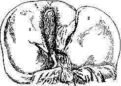
Figure 9. Lymphatic vessels of the gallbladder: 1 and 2-right and left lobes of the liver; 3-gallbladder; 4-hepatoduodenal ligament; 5-duodenum; 6-stomach. (According to Kehr.)
...llbladder. The gallbladder may be completely absent. Sometimes the gallbladder is present but completely embedded in the liver tissue, so that it is not visible from the surface. More often, only its fundus is buried in the liver parenchyma. Furthermore, cases are encountered where the gallbladder is of significant length, protrudes strongly beyond the edge of the liver, and makes various bends or folds over onto the upper surface of the liver, or, turning back, lies parallel to its body. The gallbladder may be double. Accessory ducts from the liver (hepatocystic ducts) may open into it. These ducts may end blindly at its wall. The position of the gallbladder on the lower surface of the left lobe of the liver, in the left longitudinal sulcus of the liver, has been described. Finally, the gallbladder may be directed toward the lumbar region. Biliary tracts (external) represent a system of ducts that drain bile from the place of its formation—the liver—into the intestine. Their beginning consists of two trunks from the bile ducts (hepatic ducts) of both liver lobes, which merge at an obtuse angle (Fig. 10) in the porta hepatis, and the common hepatic duct (ductus hepaticus communis) created from them. The latter subsequently heads downward and to the right until it meets the duct of the gallbladder (ductus cysticus). The continuation of both the common hepatic and cystic ducts is the common bile duct (ductus choledochus), which maintains the direction
of the hepatic ducts and runs within the thickness and along the free edge of the hepatoduodenal ligament up to the place of attachment of the ligament to the duodenum. Below, the duct descends,
descending,
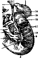
crossing behind the horizontal part of the duodenum, enters into one or another relationship with the head of the pancreas and, having finally approached the inner wall of the descending part of the duodenum, obliquely pierces it and opens into the lumen of the intestine at the apex of the papilla of Vater, separately or jointly with the pancreatic duct (Fig. 11). The connection of these ducts, as well as other anatomical relationships of the gallbladder with the pancreas, is explained by the embryological development of both the liver and the pancreas from the same "glandular ring" of the duodenum, from which both Brunner's glands and both these glandular organs are formed (see Pancreas - development). - Comparative anatomical data regarding lower stages of vertebrate development indicate that as soon as a clear division of the liver into two lobes appears, there are Figure 10. Duodenum and pancreas: 1-common hepatic duct; 2-celiac artery (III branch); 3-common bile duct; 4-uncinate process of pancreas; 5-opening of pancreatic duct; 6-head of pancreas; 7-papilla of Vater; 8-inferior horizontal part; 9-longitudinal fold of duodenum; 10-descending part; 11-circular folds of Kerkring; 12-muscularis; 13-superior horizontal part; 14-gallbladder; 15-edge of liver; 16-cystic duct; 17-right and left hepatic ducts. (According to Braus.)
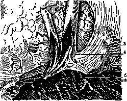
Figure 11. Opening of the common bile duct and pancreatic duct into the duodenum: 1-head of the pancreas; 2-descending part of the duodenum; 3-common bile duct; 4-pancreatic duct; 5-papilla of Santorini; 6-circular folds of the duodenum. (According to Kehr.)
also two initial hepatic ducts (cyclostomes). With very rare exceptions, there is always a gallbladder, and the cystic duct, connecting with one or two or several hepatic ducts, forms the common bile duct,
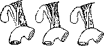
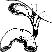
Figure 13.
which goes to the beginning of the midgut or into one of the pyloric appendages. With a well-developed pancreas (amphibians, reptiles), it opens with one or two ducts, sometimes separately, sometimes together with the common bile duct. In birds, the gallbladder is rarely absent. Besides the hepatic duct and cystic duct, which Fig. 12. Relation of the biliary tracts to the portal vein. always open into the intestine separately, there is a hepatocystic duct leading from the right lobe of the liver into the gallbladder; the pancreas has 2 ducts opening near the liver ducts (Kholodkovsky).
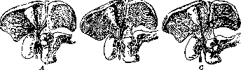
The liver of mammals most often has 2 lobes, less often 3 or 4 (many monkeys, bats, edentates, monotremes), even less often many small lobes (Phascolarctos). The gallbladder may be absent (whales, horses, elephants, tapirs, rhinoceroses, deer, camels, mice, hamsters). Rarely it is double (Orycteropus) or divided inside by a septum (lion). The common bile duct often opens into the intestine, connecting with the pancreatic duct (platypus, most marsupials, many carnivores, ungulates, cetaceans). Sometimes, upon opening into the intestine, it forms a more or less bladder-like dilation, equipped with a valve inside. In some bulls, hepatocystic ducts open into the gallbladder, or these additional ducts open into the cystic duct (sheep, dog) or into the common bile duct (some seals); sometimes two hepatic ducts lead into the gallbladder, which then discharges all bile into the intestine through the cystic duct. Such ducts as an anomaly are also found in humans. The human biliary tracts are located within the thickness of the hepatoduodenal ligament, which is a peritoneal duplicature connecting the porta hepatis with the superior part of the duodenum. At the porta hepatis, the layers of the ligament diverge to cover the lower surface of the gallbladder. Approaching the duodenum, they diverge in the same way, with the anterior layer passing directly onto the anterior surface of the intestine, while the posterior layer, covering part of the head of the pancreas, continues to the left in the form of a serous covering of the posterior wall of the omental bursa and, rising upward, turns back, lies on the inferior vena cava, and forms the posterior wall of the Winslow's foramen. The maximum length of the hepatoduodenal ligament is 6 cm, the minimum is 1 cm (Suslov); most often its extent is 3.5 - 5.5 cm. In addition to the biliary tracts, the portal vein (posteriorly; Fig. 12), hepatic artery (medially), lymphatic vessel, glands, and nerves are located within the thickness of the hepatoduodenal ligament. The bile ducts lie closest to the free edge of the ligament, but in very rare cases, when the peritoneal covering of the cystic duct forms a duplicature connecting this duct with the duodenum, the so-called cystico-duodenal ligament, all structures of the hepatoduodenal ligament are significantly removed from the free edge of the ligament. By their external appearance, the bile ducts have the form of hollow tubes, which in the living are distinguished from the hepatic artery by their thickness and lack of pulsation, and from the darker portal vein by their brownish color (Fedorov). The trunk of the hepatic duct has a lumen of about 4 mm and a rather varied length (most often 2-6 cm). But there are cases when the hepatic duct is completely absent and the cystic duct opens into the place of confluence of both initial hepatic ducts (in this case, longer than usual). Even rarer are cases when one of the initial branches (usually the right) opens directly into the cystic duct (Fig. 13). On its way, the hepatic duct crosses the right branch of the hepatic artery (see below). There are no folds or valves in this duct. Cystic duct. Before transitioning into the cystic duct, the neck of the gallbladder makes a more or less sharp bend, usually downward and inward, and is somewhat twisted along its axis (Fig. 5). The lumen of the cystic duct is always somewhat narrower than the lumen of the hepatic duct and averages 3 mm. Its length varies from 3 to 7 cm. Heading from right to left and somewhat upward, the duct connects with the hepatic duct. Three methods of this connection can be encountered (Fig. 14). - The wall of the cystic duct, just like the hepatic duct, consists of three layers and is covered on all sides by peritoneum. The muscular layer is weak, contains especially few
fibers.
Figure 14. Variations in the connection of the ductus cysticus with the ductus hepaticus: A-the relationship previously considered typical; connection of the ductus cysticus with the ductus hepaticus at a more or less acute angle; B-the ductus cysticus runs partly parallel to the ductus hepaticus; C-the ductus cysticus runs in a spiral around the posterior side of the ductus hepaticus and empties into it from the left or also from the front. (According to Huge.) The mucous layer has a large number of well-developed glands and bears a system of folds-valves (Figure 6). The following valves are distinguished (Frik, Karlmark): 1. The neck valve (Collumklappe), lying at the border between the neck of the gallbladder and the body; there are usually two of them; they are especially well-developed when the neck of the ductus cysticus is strongly curved, they hang down like sails and occupy 3/4 of the lumen, and sometimes almost completely close it. 2. Between these two valves, a third, obliquely running valve is encountered-the intermediate valve (Intermedialklappe). In the second type, this valve is encountered more often (57%) than in the first (30%). 3. After a small space, free of folds, valves begin again, irregularly arranged around the entire circumference of the lumen-these are the semilunar valves (Arcusklappen). Their number varies-from two to six. 4. In one-third of cases, the last or one of the last valves forms the so-called spiral valve
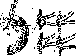
Figure 15. Ductus choledochus and the duodenum from behind: 1-pars supra-duodenalis; 2-retro-duodenalis; 3-pancreatica; 4-intra-parietalis. (According to Kehr.)
Figure 16. Types of connection of the ductus choledochus and ductus Wirsungianus at their entry into the duodenum: 1-stone in the papilla Vateri; 2-stone in the ductus choledochus, compressing the ductus Wirsungianus; 3-stone in the ductus choledochus without compression of the ductus Wirsungianus. (According to Kobson.)
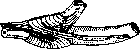
Figure 17 (valvula Heisteri, Spiralklappe). Sometimes there are two of them. 5. Finally, after some free space, occasionally interrupted only by weak folds, at the site of the entry of the ductus cysticus there is the last valve-the terminal valve (Terminalklappe), which represents the edge of the duct wall rather than a true fold. Histologically, the above-described valves are formed not only from folds of the mucous membrane but also contain weak muscle fibers (Berg, Hendrickson). Ductus choledochus. The common bile duct is significantly wider in its lumen than the hepatic duct and twice as wide as the cystic duct. Its length is quite variable, on average 7-8 cm, and sometimes 2-4 cm; in 25% of cases, the duct is longer than 8 cm and reaches 12 cm. It is customary to divide the ductus choledochus into several sections (Fig. 15). In the pancreatic part, the duct has close relations with the pancreas. In its last section, the ductus choledochus, merging with the ductus Wirsungianus, opens into the intestine. The fusion of these ducts is not always the same (Fig. 16). Two extreme types can be seen: either both ducts empty into a common ampulla (diverticulum Vateri) (1) or they empty independently into the intestine with or without the formation of a papilla (2 and 3). Schirmer finds these two types equally often. Letulle considers the 2nd type to be the most frequent. The size of the ampulla is from 4x6 mm to 5x12 mm. These two types are not equivalent from the point of view of ascending infection or obstruction of the ducts by a stone. In the region of the papilla Vateri, there is a special muscular sphincter, first described by Oddi (Figure 17). According to Hendrickson, this sphincter contains longitudinal musculature, and this sphincter is common to both ducts. The function of this sphincter, according to Oddi, consists in regulating and alternating the flow of bile into the intestines. The walls of the ductus choledochus are constructed in the same way as those of the cystic and hepatic ducts, but it contains many more muscle fibers. The mucous membrane does not bear any folds until the very end and only in the last section has a whole series of fold-pockets facing toward the intestine (Vanovsky). The introduction of a probe from the intestine into the ductus choledochus is almost impossible due to the folds on the mucous membrane, while the introduction of a probe into the ductus Wirsungianus in the same direction can be done relatively easily, as its wall is completely smooth. The place where the ductus choledochus pierces the intestine is located (according to Sappey) at a distance of 14-15 cm from the pylorus. The blood supply to the bile ducts is carried out from the system of the arteria hepatica, with the branches of which they enter into known relationships. Most often (55%, Rio Branco), the so-called "normal type" of branching (main trunk) is encountered; the arteria hepatica divides into two branches: the arteria hepatica propria and the arteria gastro-duodenalis (Fig. 20). The former, having given off a branch to the lesser curvature of the stomach
Figure 18. Relationship of the arteria hepatica, arteria gastrica dextra, or arteria pylorica, runs in front of the vena portae to the porta hepatis, where it divides into a left and-longer-right branch, which, having crossed the ductus hepaticus, gives off the arteria cystica. Significantly less often (in 20% of cases) one can observe the so-called
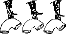
42%
26%
18%
14%
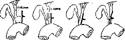
Figure 19. Relationship of the arteria hepatica to the ductus choledochus. (According to Kehr.) scattered type: the right and left branches of the arteria hepatica, the arteria gastrica dextra, and the arteria gastro-duodenalis depart in one bundle-the arteria hepatica propria thus does not exist at all. The trunk of the hepatic artery, passing in front of the vena portae, in the majority of cases (52%, according to Kehr) runs almost along the middle of the anterior wall of the vein (Figure 18). The artery is usually removed from the bile ducts by 15-25 mm, less often it passes closer, 5-15 millimeters, but it can also approach the ducts closely and even cover them (Figure 19). The right branch of the arteria hepatica, as indicated above, crosses with the ductus hepaticus; in the majority of cases (60%) the artery lies under the duct, but in 26% of cases it passes along its anterior surface and sometimes (10%), being located first on the anterior surface of the duct, it bends spirally and goes under it (Fig. 21). Thus, in 36% on the anterior surface
Rio Branco and others, according to Kehr-in 60%); Fig. 24 presents various variants of the origin of this artery. Sometimes there are two of these arteries. Rarely, the artery can start from the arteria mesenterica superior. Especially great importance is attached to those cases where
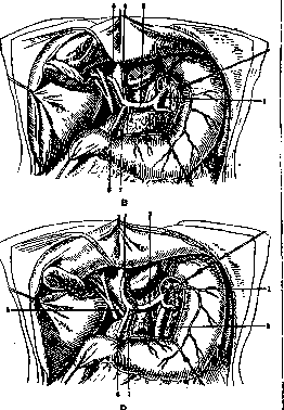
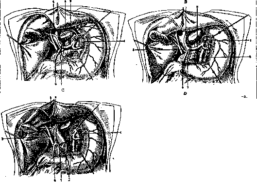
Figure 20. Types of branching of the arteria hepatica: 1-coeliaca; 2-arteria hepatica; 3-arteria hepatica propria; 4 and 5-ramus sinister et dexter arteriae hepaticae; 6-arteria gastro-duodenalis; 7-arteria pylorica; 8-arteria mesenterica superior-A-normal (main trunk) type of division of the arteria hepatica; B-scattered type; C-origin of the left branch of the arteria hepatica from the arteria gastrica sinistra; D-origin of the right branch of the arteria hepatica from the arteria mesenterica superior; E-origin of the arteria hepatica from the arteria mesenterica superior. (According to Kehr.)
of the hepatic duct one can encounter a large arterial trunk (Kehr). The relationships of the right terminal branch to the neck of the gallbladder and the ductus cysticus are presented in Fig. 22. The right terminal branch of the arteria hepatica, after crossing with the ductus hepaticus, gives off a significant trunk (arteria cystica), which, having approached the neck of the gallbladder, divides into anterior and posterior branches running along the corresponding sides of the gallbladder. From the arteria cystica, a fairly large small trunk usually departs along the wall of the cystic duct and the ductus choledochus (Fig. 23). The above-described origin of the arteria cystica is encountered in 88% of all cases (Suslov), the artery crosses the bile duct from the front (23%, Rio Branco) and can be injured during its dissection. The arteria gastro-duodenalis, besides several branches to the upper horizontal part of the duodenum, gives off 2 large trunks running in the groove between the head of the pancreas and the duodenum-one along the anterior surface, the other along the posterior, and anastomosing with two branches-posterior and anterior-of the arteria pancreatico-duodenalis inferior. The posterior of these main trunks (Fig. 25) always crosses the ductus choledochus, entering into the closest connection with it and giving off ascending branches to its wall.- The veins of this region are constructed according to the same type of two main trunks (Tonkov) and empty independently as a trunk into the vena portae. The lymphatic system of all bile ducts and the gallbladder consists of two networks in the mucous and in the outer membrane. At the neck of the gallbladder, there are small glands, from which small trunks stretch along the ducts; some of them, crossing the ductus cysticus, go to the head of the pancreas. The larger

Figure 21. Relationship of the right
branches of the arteria hepatica to the ductus hepaticus (According to Kehr). The trunk of the lymphatic vessels of the bile ducts collects into two glands located to the right of the ductus choledochus. A finger inserted into the foramen Winslowi feels these glands first (Clairmont). Along the course of the ductus cysticus there are several glands, from which vessels cross the arteria hepatica and terminate in the glands along the course of this artery near the pancreas to the left of the artery. From these and the above-described glands lying to the right of the ductus choledochus, efferent pathways extend to the posterior wall of the head of the pancreas (Franke; Figs. 26 and 27). The most constant are the glands at the neck of the gallbladder at the entry of the ductus cysticus, to the right of the ductus choledochus and to the left of the arteria hepatica. The connection of the lymphatic pathways of the bile ducts and the pancreas is of great importance in the pathology of these organs. The innervation of the bile ducts involves the nervi splanchnici and the nervi vagi; there are indications that the nervus phrenicus also participates (via the solar plexus; studies by Shevkunenko and Vorobyov) in the innervation of the ducts. Irritation of the peripheral ends of the nervus splanchnicus causes contraction (Doyen) of the lumen of all bile ducts; thus, the nervi splanchnici are motor nerves for the musculature of the excretory apparatus. Upon irritation of the central ends of the nervus splanchnicus
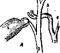
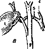
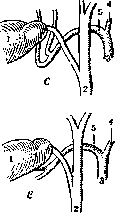
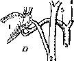
Figure 22. Relation of the ramus dexter arteriae hepaticae to the ductus cysticus and the gallbladder: A and B—common arrangement; C and D—fairly common; E—rare; 1—gallbladder; 2—ductus choledochus; 3—arteria hepatica; 4—ramus sinister arteriae hepaticae; 5—ramus dexter arteriae hepaticae. (According to Kehr.)
and the nervi vagi, complex relationships are obtained, to a certain extent indicating the normal course of irritation of the central centers and bile secretion. Irritation of the central ends of the nervus splanchnicus causes relaxation of the ducts; irritation of the central ends of the nervus vagus excites contraction of the bile ducts and simultaneously relaxes the sphincter of Oddi (Babkin). The center of this sphincter, according to Oddi, lies in the lumbar part of the SPINAL CORD.
V. Vanovsky. Structure of the bile ducts. The beginning of the bile ducts are thin (1.5–2 µ) bile capillaries (canaliculi biliares), located inside the hepatic lobule between the liver cells and analogous to the secretory capillaries of other glands (see Liver).
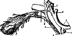
Figure 23. Relation of the arteria cystica to the ductus cysticus and ductus hepaticus according to Calot: 1—arteria cystica; 2—ramus dexter arteriae hepaticae; 3—ramus sinister arteriae hepaticae; 4—arteria hepatica; 5—arteria gastro-duodenalis; 6—vena portae; 7—ductus hepaticus; 8—ductus cysticus; 9—ductus choledochus. (According to Kehr.)
At the periphery of the lobule, the capillaries flow into the bile ducts (ductus biliares), which encompass the surface of the lobule, anastomosing and forming networks (Fig. 28); the bile ducts at this place have the appearance of thin tubules (19–24 µ in diameter, injected—up to 50 µ), the wall of which consists of a membrana propria and a single layer of low epithelium (intermediate ducts of French authors). These ducts, in the number of 2–4, unite into larger tubules (perilobular), which head into the connective tissue layers between the lobules (por
tal spaces
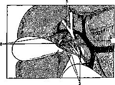
Figure 24. Various variants of the origin of the arteria cystica: 1—arteria hepatica propria; 2—normal origin of the arteria cystica; 3—variants of origin. (According to Rio Branco). tal spaces, Kiernan's spaces) and here they flow into large ducts (interlobular, canaux de distribution Renaut), whereby tubules from 3–4 lobules bordering at this place flow into one such duct. From here, the bile ducts head toward the porta hepatis, forming by their fusion the bile duct (ductus hepaticus). In general, the distribution of the bile ducts inside the liver corresponds to the distribution of the branches of the vena portae and arteria hepatica, which accompany them. Bile ducts of small caliber, up to 90–100 µ in diameter, consist of a membrana propria and low or cuboidal epithelium; starting from here, they receive a sheath of connective tissue with elastic fibers and cells, and the epithelium becomes high, cylindrical. At the same time, their walls receive glands (glands of the bile ducts), first in the form of simple diverticula, then in the form of branched tubulo-acinar glands, which bulge the connective tissue sheath from the outside; their excretory duct flows into the lumen of the duct in an oblique direction. The largest intrahepatic ducts are constructed in the same way as the ductus hepaticus. Injections of the bile ducts (technique—see Liver) show that small-caliber ducts anastomose repeatedly with each other, and large ones give a large number of outgrowths, branching, forming networks and sometimes ending blindly (vasa aberrantia); part of them serves to connect the ducts of the right and left lobes. Extrahepatic bile ducts, ductus hepaticus, ductus cysticus, ductus choledochus, are constructed identically (Figs. 29 and 30). Their wall consists of a mucous membrane (tunica mucosa) and an outer fibrous one (tunica fibrosa), containing Figure 25. Retroduodenal part of the ductus choledochus: 1, 2, 3—three parts of the duodenum; 4—ductus choledochus; 5—vena portae; 6—arteria hepatica communis; 7—arteria gastro-duodenalis; 8—arteria pancreatico-duodenalis superior. (According to Kehr.)
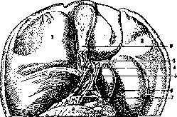
Figure 26. Lymphatic vessels in the ligamentum hepato-duodenale: 1—right lobe of the liver; 2—left lobe of the liver; 3—ductus cysticus; 4—ductus hepaticus; 5—ductus choledochus; 6—arteria duodeno-jejunalis; 7—pancreas; 8—duodenum; 9—incision of the peritoneum of the gallbladder. (According to Kehr.)
containing bundles of smooth muscles. The mucous membrane is covered with cylindrical epithelium, which in the region of the ductus choledochus bears a border on its surface, as in the intestine; in the ductus hepaticus and ductus cysticus it gradually disappears. Among the cylindrical cells, goblet cells are always encountered. The fibrous sheath consists of circular bundles of connective tissue and contains bundles of smooth muscle fibers running longitudinally, to which circular ones can be added from the outside. In humans, in the ductus hepaticus, muscles are almost absent; they are more strongly developed in the ductus choledochus, especially near the entry into the duodenum, where a strong circular layer appears. In the walls of all ducts there are in
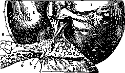
Figure 27. Lymphatic vessels in the ligamentum hepato-duodenale and in the pancreas: 1 and 2—right and left lobes of the liver; 3—vena portae; 4—pancreas; 5—arteria lienalis; 6—vena lienalis; 7—arteria mesenterica superior; 8—arteria coeliaca; 9—fatty tissue around the arteria coeliaca; 10—duodenum. (According to Kehr.)
a large number of branched, highly convoluted glands of various sizes—from 0.5 mm to 2 mm. In these glands, there are cells similar to the epithelium of the duct and others, lighter, in the upper part of which there is granularity. Physiology of the bile ducts is poorly developed. Upon irritation of the nervus splanchnicus, contraction of the smooth muscles of the gallbladder and ducts is observed, as a result of which bile is secreted; upon irritation of the central end of the same nerve, their relaxation occurs. Conversely, irritation of the central end of the nervus vagus after its transection causes contraction of the walls of the gallbladder. Microphysiology is also poorly developed. Contrary to common assertions about the secretion of mucus by the glands of the bile ducts and the mucous membrane of the gallbladder, histologists cannot establish the mucous character of the cells in the indicated glands, and the very presence of glands in a normal gallbladder is often disputed. Probably, the secretion of the gallbladder epithelium occurs in small portions while preserving the membrane on the surface facing the lumen, as has been proven for the surface epithelium of the stomach, which is why some authors classify the gallbladder and bile ducts as excretory epithelial surfaces (see Glands).
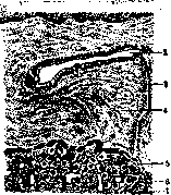
Figure 28. Interlobular bile ducts in the human liver: 1—hepatic lobules; 2—interlobular bile duct; 3—perilobular duct; 4—connective tissue between lobules; 5—intermediate canal (Hering's canal); 6—bile capillaries. (According to Prenant.)
The finding of cholesterol in the epithelium of the gallbladder was interpreted as proof of its secretory activity (Robin), although it is quite possible to explain its presence by the absorption of the components of bile. On the part of the epithelium of the ductus hepaticus and ductus choledochus, as in the gallbladder, absorption of fat can take place. V. Karpov. P. Radiological examination. Radiological examination in cholelithiasis and other diseases of the extrahepatic ducts comes down mainly to
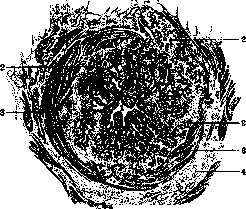
Figure 29. Cross-section of the ductus choledochus: 1—lumen of the duct; 2—glands of the bile duct; 3—muscle bundles; 4—loose connective tissue with blood vessels and fat cells. (According to Kölliker.)
its part in the detection of stones in the gallbladder and in the ducts and in the clarification of anatomical and functional changes of the gallbladder and ducts. At the present time, all methods available to radiology can be divided into the following five groups: 1) obtaining radiographs with an image of stones in the gallbladder and ducts; 2) obtaining an image of the gallbladder without the use of contrast agents; 3) the use of air insufflation into the abdominal cavity to create contrast around the gallbladder; 4) the introduction of contrast agents with bile into the cavity of the gallbladder and into the bile ducts; 5) the determination of changes in the bile ducts and the gallbladder in particular on the basis of indirect signs. I. Finding gallstones. In positive cases, shadows are visible on the radiograph, partly round, partly with straight edges, located under the edge of the liver; they are either of uniform intensity or ring-shaped. The size of gallstones is most often from a pea to a hazelnut, the number of stones is from one to several dozen. Some authors believe that in 80% of all cases of gallstones in the gallbladder, the stones can be seen on a radiograph. The highest percentage of positive results is noted by American authors. A series of images is taken (about 40) in all possible directions; however, stones are visible only on a few of them. Pribram, Altschul, and others consider finding stones by this method a rarity. Technical requirements boil down to the greatest possible shortening of the exposure time when taking pictures and to the use of the Bucky-Potter diaphragm. The criterion for assessing the quality of the image is the obtaining of clear outlines of the kidney. The chemical composition of gallstones is of great importance for obtaining positive results. Stones with a high cholesterol content absorb X-rays twice as weakly as water, and it is extremely rare to find them. Bilirubin stones absorb rays in the same way as water; finding them is somewhat easier than cholesterol ones. Mixed stones, consisting of bilirubin, cholesterol, and lime, are clearly visible on the image, because the lime contained in them absorbs X-rays six times more strongly than water. Practically, by this method, it is possible to determine the presence of stones only with a sufficient content of lime. Romang considers it possible to judge the nature of the process that caused their formation by the character of the stones depicted on the image; according to Aschoff, mixed stones are the result of an inflammatory process, and cholesterol stones are the result of a metabolic disorder. II. Obtaining an image of the gallbladder. As early as 1896-99, attempts were made to obtain a shadow of the gallbladder on a radiograph. It was established that the gallbladder can be seen on an image under the following conditions: 1) with thickening of the gallbladder wall due to chronic inflammatory processes, 2) with an increase in its volume, in its dropsy, or 3) with thick bile with a high content of salts or even sand. This method is of no significant importance for diagnosis
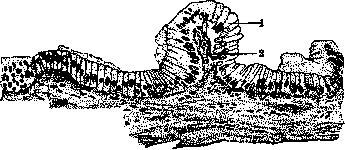
Figure 30. Mucous membrane of the human gallbladder: 1-epithelium; 2-connective tissue forming folds; 3-bundles of muscle fibers. (According to Prenant.)
...cannot have, as positive results are obtained extremely rarely.
III. Air insufflation to create contrast. Inflation of the colon with air can in some cases create sufficient contrast around the gallbladder for the shadow of the gallbladder to be visible on the radiograph.
In 1913, a method of insufflating gas into the abdominal cavity by puncturing the abdominal wall was proposed—pneumoperitoneum (see Aeroperitoneum).
The gallbladder is sometimes clearly visible if it is altered by a tumor or enlarged by dropsy. Sometimes it is possible to establish the presence of adhesions. The disadvantages of this method lie in the complexity and danger of its application and in the fact that only advanced cases with major anatomical changes in the gallbladder are amenable to observation.
IV. Introduction of contrast agents into the gallbladder. Many attempts have long been made to obtain an X-ray image of the gallbladder by filling its lumen with a contrast agent.
Only in 1924 was this task solved, thanks to the fact that for this purpose American physicians (Graham, Cole, Copher) chose a substance that is: 1) excreted from the body in bile, 2) strongly absorbs X-rays, and 3) is harmless to the body in the necessary doses.
They used tetrabromophenolphthalein for this, replacing chlorine with bromine in the compound that Rosenthal used to determine the functional capacity of the liver, as bromine, being a substance of higher atomic weight, absorbs X-rays more strongly.
Tetrabromophenolphthalein, introduced into the blood intravenously, is excreted with bile through the liver and enters the gallbladder.
The mucous membrane of the gallbladder concentrates its contents by absorbing, mainly water, and after 7-8 hours, the bromine content in the gallbladder reaches such a degree that it produces a distinct shadow on the radiograph.
Later, Graham replaced bromine with iodine, which produces fewer toxic effects.
According to Hoesch, tetraiodophenolphthalein introduced into the blood, which has a bright blue color in solution, changes rapidly upon passing through the liver; and in the blood serum, bile, and urine, this substance is already found in the form of a colorless iodine compound.
In the bile, iodine is detected 20 minutes after introduction into the blood; its quantity increases intensively over 40 minutes, remains unchanged for several hours, and then slowly falls.
From the urine, iodine normally disappears simultaneously with the iodine of the blood serum at the beginning of the 3rd day; in case of impaired liver function, it can persist for 14 days.
At the present time, tetrabrom- or tetraiodophenolphthalein-sodium, or, otherwise, "Bromtetragnost" or "Jodtetragnost" from the Merck company, is used in medical practice.
Substances proposed later by other authors have not received wide application, either due to low contrast or harmfulness to the body.
Such are the following substances: 1) a 20-percent solution of sodium or strontium bromide (Sabatini and Milani); 2) "Dijodato-phan-biloptin," proposed by Pribram; 3) Chohimbrm (Mort; England); 4) Foriod (Wangermetz; France).
The application of Jodtetragnost is performed in various ways. 1. Intravenously. For introduction into a vein, the iodine compound is recommended—bromine is more toxic.
Among the complications described are: nausea, vomiting, headaches, diarrhea; phenomena of collapse may also occur. The entry of the substance under the skin causes necrosis of the subcutaneous tissue.
With the use of excessive doses, severe degenerative processes in the liver are possible. If the established rules are followed, all unpleasant complications are completely absent.
The technique of introduction is as follows: the dry substance is dissolved at a rate of 0.05 per 1 kg of the patient's weight in 20-30 cm3 of distilled water. After filtration, the solution is sterilized for 20 minutes in a water bath, as the solution cannot be boiled.
The patient's intestine is thoroughly cleansed, and complete fasting for 8-12 hours is prescribed.
Half an hour before the introduction, most authors (Kalk, Schoendube, and others) inject 2 cm3 of Merck's hypophysin intramuscularly to induce emptying of the gallbladder and allow greater access for the influx of bile containing the contrast agent.
The solution of Jodtetragnost, prepared ex tempore, is introduced with an ordinary 20-30 gram syringe into the cubital vein.
The slower the introduction is performed, the greater the guarantee of avoiding phenomena of collapse. It is introduced over 10-15 minutes, then, without removing the needle, it is flushed with a small amount of physiological saline.
To prevent phenomena of irritation of the vagus nerve by Jodtetragnost, 1 mg of atropine is injected under the skin after the injection.
After 8-13 hours, the first X-ray image is taken. Technique: 50-60 kV, 80 mA, exposure 0.5-1 sec., double-coated film and—preferably—a Bucky-Potter diaphragm.
Within this time, the shadow of the gallbladder is best seen. After 18-20 hours, the shadow of the gallbladder gradually disappears.
2. Introduction through the mouth. Tetraiodo- and tetrabromophenolphthalein-sodium, being absorbed in the stomach and duodenum, irritate the mucous membrane of the stomach and duodenum to a very high degree.
They are therefore given in gelatin capsules hardened with formaldehyde, or in keratinized pills, or in special tablets that dissolve only in the alkaline environment of the intestine.
Kirklin proposed giving the contrast agent as a drink in a solution with the addition of grape juice; in the presence of fruit acids, the sodium salt of tetraiodophenolphthalein apparently converts into tetraiododioxyphthalophenone, i.e., tetraiodophenolphthalein, which is insoluble in gastric juice, and in the alkaline environment of the intestine, it converts back into the sodium salt and is absorbed.
The Merck company released a special powder, "Oral-tetragnost," which is dissolved in 200 cm3 of water before use and given as a drink.
The method of introduction per os is very simple, but the image of the gallbladder is significantly weaker than with the intravenous method (the bromine compound is usually used per os); the result depends on the conditions of absorption in the intestine and on the timely dissolution of the capsules.
Diarrhea occurs as a complication.
Conditions necessary for the gallbladder to fill. 1. The liver cells must be capable of excreting the contrast agent with the bile.
2. The cystic and hepatic ducts must be patent, and the cavity of the gallbladder must contain a sufficient amount of bile.
3. The mucous membrane of the gallbladder must concentrate the bile, and consequently the contrast agent in it, to a sufficient degree so that a shadow of the gallbladder can be obtained.
4. The gallbladder must possess the ability to empty.
An unchanged gallbladder produces an intense shadow with regular contours on the radiograph and shifts well when the position of the body is changed.
The shape is very diverse: oval, pear-shaped, lanceolate (see separate table, figs. 1-4); in hypersthenics—wide, in asthenics—elongated.
The height of its position depends on the position of the liver. Atonic (i.e., wide, with flaccid walls), congested, and ptotic (lowered) gallbladders are distinguished.
If the gallbladder does not produce a shadow, there may be the following reasons for this.
1. Impairment of the ability of liver cells to excrete the contrast agent introduced into the blood.
In various forms of jaundice, in cirrhosis, in degenerative processes of the liver, neoplasms, echinococcosis, and other processes in the liver that involve a large part of the hepatic parenchyma, cholecystography yields a negative result even with a completely healthy gallbladder.
Cholecystography is contraindicated in cases with undoubted clinical data pointing to damage of the liver parenchyma.
In the absence of clear symptoms of liver damage, in those cases where cholecystography has yielded a negative result, one can verify sufficient excretion of iodine by the liver by determining it in bile obtained via a duodenal tube.
The presence of iodine in the urine later than 3 days after cholecystography can also serve as a sign of iodine retention by the liver.
2. Obstruction of the bile ducts by stones. A stone located in the cystic duct mechanically blocks the access of the contrast agent into the cavity of the gallbladder, and then the shadow of the gallbladder is not obtained on the radiograph.
If the stone does not completely block the duct, the shadow of the gallbladder may be obtained, but it is very weak due to the small amount of iodine that has entered the cavity.
The presence of stones in the gallbladder itself is not in itself an obstacle to filling it with a contrast agent (if the number of stones is not so great as to prevent sufficient iodine from entering the cavity of the gallbladder).
In such cases, the shadow of the gallbladder is sometimes obtained significantly weaker than normal, or against the background of the shadow, round areas of translucency are visible—filling defects caused by stones, which do not themselves produce shadows.
In rare cases, it is possible to see the shadows of stones after the gallbladder has emptied of the contrast agent, and the cholesterol stones have become imbibed with iodine.
3. Impairment of the absorptive and contractile function of the gallbladder prevents the obtaining of the gallbladder shadow.
The cause of such impairments of the functional capacity of the gallbladder is various inflammatory processes of its wall—both acute and chronic.
Depending on the degree of impairment of the absorptive capacity of the gallbladder, its shadow is either completely absent or is significantly weakened.
In acute cholecystitis, the shadow of the gallbladder is absent at the height of the disease; as the process subsides, a distinct shadow can be obtained. If, as a result of an inflammatory process due to cholecystitis or cholelithiasis, atrophy of the gallbladder mucosa develops, a persistent negative result of cholecystography is obtained. 4. Cicatricial shrinkage of the gallbladder wall with impairment of its contractile capacity or with obliteration of its lumen or duct prevents the filling of the gallbladder with contrast medium. The shadow of the gallbladder in these cases is either absent or appears blurred and deformed. Cicatricial processes in neighboring organs can involve the gallbladder in adhesions, deform it, and reduce its lumen. In these cases, a deformation of the shape of the gallbladder is sometimes obtained without impairment of its absorptive capacity (see separate table, fig. 5). 5. Compression of the gallbladder or ducts by tumors originating both from the gallbladder and from neighboring organs also often purely mechanically interferes with the filling of the gallbladder with contrast medium. 6. Apparently, reflex-nervous factors can also paralyze the contractile capacity of the gallbladder or cause spasm of the sphincters; in some cases with increased neuromuscular excitability, the shadow of the gallbladder may be absent without the presence of pathological processes in the gallbladder. In these cases, a repeated check of the cholecystography results is necessary. Only a persistent negative result of cholecystography obtained during repeated examinations gives the right (in the presence of other clinical symptoms as well) to consider a persistent anatomical and functional lesion of the gallbladder as certain. The contractile function of the gallbladder can be investigated with the help of cholecystography almost as in experimental conditions. After obtaining the shadow of the gallbladder and introducing certain chemical substances that cause the emptying of the gallbladder, serial radiographs are taken and all phases of emptying into the bile ducts and into the duodenum are monitored. Often, it is possible to obtain an image of the ducts: ductus cysticus, ductus choledochus, and even ductus hepaticus (see separate table, figs. 3-4). Of the substances acting to empty the gallbladder, hypophysin, resp. pituitrin (Kalk and Schöndube), a mixture of cream and egg yolks (Boyden), or (best of all) egg yolk (Bronner) are used. The time of gallbladder emptying in response to the use of stimulants varies depending on the individual characteristics of each patient, and mainly on the degree of impairment of the corresponding function of the gallbladder. An important auxiliary method is the combined X-ray examination of the gallbladder and the gastrointestinal tract filled with contrast medium. In this way, the influence of pathological processes of the gallbladder on neighboring organs and vice versa is clarified. The indirect X-ray method boils down to fluoroscopy.

Figure 31. Types of pressure of the bulbus duodeni on the anterior wall in the 2nd position. (According to Berg.)
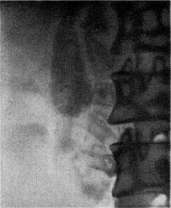
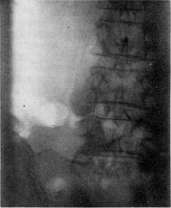
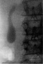
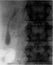
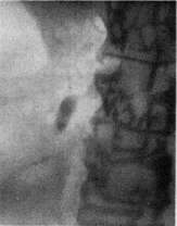
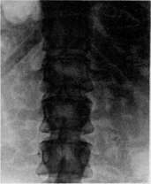
Figure 1. Cholecystography, normal filling of the gallbladder. Figures 2, 3, and 4. Cholecystography.
Gradual emptying of the gallbladder. Figure 5. Cholecystography of a shrunken gallbladder. Figure 6. Gallbladder stones (posterior view). To the entry: Gallbladder.
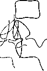
Figure 32. Schematic representation of the most frequent changes in the bulbus duodeni in diseases of the gallbladder.
Gallbladder: ----- indentation by an enlarged gallbladder without displacement to the right; .......the same with displacement to the right due to adhesions of the stomach, duodenum, and intestines. A number of changes in the X-ray picture of these organs have been established, which are considered characteristic in the presence of biliary tract disease; the most important of them are the following: 1) indentation on the anterior wall of the duodenal bulb during enlargement of the gallbladder (Fig. 31); 2) pulling of the duodenal bulb upward and to the right in pericholecystitis, however, unlike adhesions due to duodenal ulcer, there is no persistent deformation of the bulb in pericholecystitis (Fig. 32); various disturbances of passage through the duodenum (dilation of the bulb - "megabulbus" - and stasis in it or its rapid emptying) are often encountered in biliary tract disease (Berg). All the above-described methods can be of significance for the diagnosis of gallbladder and duct disease only in the closest connection with other clinical methods of examination, N. Potte. Sh. Pathological anatomy. Malformations of the biliary tract are most often expressed in hypoplasia, atresia, or even complete absence of any part of the extrahepatic biliary system, and sometimes of this entire system as a whole. Underdevelopment of the gallbladder is of the least significance, since even its complete absence does not cause any symptoms during life. If there is an isolated atresia or agenesis of the cystic duct with an existing gallbladder, the latter becomes cystically distended, and the mucus contained in it often turns out to be colored with bile due to the secretion of bile pigment by the epithelium of the gallbladder mucosa or the glands of the neck. Of much greater significance is atresia or absence of the common bile duct or the hepatic duct. If it is a matter of atresia, it is usually observed not along the entire length of these ducts, but in certain "favored" places, which include: 1) the lower segment of the common bile duct at the place of its entry into the duodenal wall and 2) its uppermost part at the place of the entry of the cystic duct. In all such cases, children are either born with jaundice or develop it in the first days after birth. Their life expectancy, due to cholemia and the developing biliary cirrhosis of the liver, in most cases does not exceed 5-6 months, and all this time the intestinal contents in them may remain slightly colored with bile due to the secretion of bile pigment by the intestinal glands. Also worthy of mention is idiopathic dilation of the common bile duct immediately before its entry into the duodenum - the so-called cyst of the common bile duct, sometimes reaching colossal sizes (the size of an adult's head). The causes of this dilation are not entirely clear, as probing always establishes the absence of any stenosis whatsoever. It is assumed that a congenital weakness of the walls plays a role here in connection with the excessively oblique direction of the duodenal part of the common bile duct in relation to the intestinal wall, as a result of which conditions are created for valve-like closure of the duct orifice. - Inflammation of the biliary tract - see Cholangitis, Cholecystitis. The majority of benign primary tumors described in the gallbladder and ducts belong to the category of papillomas and adenomas. Among malignant ones, sarcomas are rarely encountered, but predominantly carcinomas, which in most cases have a glandular or papillary character and are located most often at the fundus or in the neck of the gallbladder, as well as in the region of the papilla of Vater. In general, for Moscow, carcinomas of the gallbladder and ducts account for 2.8% of all carcinomas (according to data from hospital prosectoriums for 1923-1927). - Narrowing and obstruction of the large bile ducts can be caused by stones, tumors, parasites, and less often by scars (e.g., after the healing of decubital ulcers). Obstruction of the common bile duct causes bile stasis in the entire biliary system with all the consequences of such stasis. If the hepatic duct becomes obstructed, there occurs, on the one hand, dilation of its superior part with the entire system of intrahepatic bile ducts and the development of obstructive jaundice, and on the other, collapse of the gallbladder. In this case, if the obstruction continues for a long time, then over time the bile in the dilated biliary tracts may become almost colorless; normal bile is replaced by the so-called "white bile." Blockage of the cystic duct leads to the so-called false dropsy of the gallbladder. Since there is no inflow of bile into the gallbladder in this case, the bile contents that were there are gradually absorbed and replaced by a clear liquid, secreted by the gallbladder mucosa and accumulating in it sometimes - especially in the case of simultaneous catarrh of the gallbladder - in a very large quantity. The same phenomenon can be observed in some cases even without mechanical closure of the cystic duct. Thus, in severe acute infectious diseases or sharp exhaustion - especially in children - false dropsy of the gallbladder often develops on the basis of atony of the gallbladder musculature. Disorders of the corresponding nervous apparatus in the form of spasm of the gallbladder neck or, conversely, paresis of the gallbladder muscles can lead to the same thing. Attacks of such dyskinetic disorders of nervous origin can sometimes create a symptom complex almost identical to the picture of cholelithiasis (see). M. Skvortsov. Pathological physiology and clinic. Inflammatory processes (see Cholecystitis) and the formation of gallstones (see Cholelithiasis) play a dominant role in the pathology of the gallbladder. Other diseases are encountered significantly less frequently, resemble hepatic colic or cholecystitis in their course, and often remain clinically unrecognized, being diagnosed only on the operating or autopsy table. These include: a stagnant gallbladder of non-calculous origin, tumors, chronic infectious (tuberculosis, syphilis) and parasitic diseases. - In the absence of gallstones, stasis in the gallbladder arises due to mechanical or functional causes. Mechanical stasis most often develops due to the closure of the bile ducts by a tumor (carcinoma of the head of the pancreas, papilla of Vater, biliary tract; mucous polyps, adenomas, adenofibromas of the common bile duct, etc.). Often, mechanical stasis is caused by plastic perivisceral processes, mainly pericholecystitis, complicating cholecystitis, gastric and duodenal ulcer, gastric carcinoma, chronic appendicitis, etc. Changes in the omentum and other organs adjacent to the gallbladder can also serve as an obstacle to the outflow of bile. The cause of stasis is also congenital anomalies of the gallbladder and ducts: diverticula of the gallbladder, kinks of the cystic duct, oblique position of the common bile duct in the duodenal wall, elongation or, conversely, complete absence of the mesentery of the gallbladder. Congenital atresia of the common bile duct in children leads to huge dilation of the entire biliary system. Gosset collected 23 cases of congenital atresia of the duct. Functional stasis in the gallbladder is explained by spasm of the sphincter of Oddi, which entails relaxation and atony of the bile duct. Some authors believe that atony of the gallbladder is caused by its loss of the ability to contract, although the question of the contractility of the gallbladder itself is controversial. Others believe that hypokineses of the gallbladder depend on a decrease in the tone of the vagus nerve. With atony of the gallbladder, the gallbladder reflex to the introduction of magnesium sulfate, peptone, etc., into the duodenum is delayed, and sometimes completely absent. The introduction of pilocarpine accelerates the appearance of "B" bile. Systematic drainage of the gallbladder by means of a duodenal tube (repeated triggering of the reflex according to Meltzer-Lyon) in the opinion of some clinicians contributes to the improvement of gallbladder function. In contrast to hypokineses, hyperkineses of the gallbladder are observed in vagotonia, which sometimes simulate the picture of cholelithiasis. Hyperkineses give spastic pains in the right hypochondrium. With the Meltzer-Lyon test, "B" bile does not enter the duodenum; pilocarpine does not cause its inflow; after the injection of atropine, the spasm of the gallbladder relaxes, the pains cease, and "B" bile enters the duodenum. Dyskinesias of the gallbladder and biliary tract have been attracting great attention recently. Their study is carried out with the help of fluoroscopy and duodenal probing. Tumors of the gallbladder, especially carcinoma, are of great practical interest. According to Kaufmann's statistics, the gallbladder accounts for 5% of all cases of carcinoma of internal organs. Carcinoma is often combined with gallstones (according to Futterer and Haberfeld, in 70%; according to Courvoisier, in 91%). According to data from Mayo, Moynihan, and Riedel, carcinoma occurred in 5-7% of all cases of gallstones. Most often, carcinoma is localized in the fundus or neck of the gallbladder, at the site of scars after previous inflammations, or in areas subjected to trauma by stones. The tumor can be barely noticeable or, conversely, fill the entire gallbladder and block the cystic duct. It often has the appearance of a cauliflower; its surface is often ulcerated. Metastases to the liver develop rapidly; distant metastases appear late. In some cases, the only symptom of the disease is cachexia; in others, pain and jaundice are observed. Sometimes a tumor is palpable. Operative cure is possible only in the very early period.
Among other tumors, adenomas are of practical interest, which, according to Aschoff, often degenerate into cancer. Papillomas of the gallbladder, which present a picture of cholecystitis, are cured by surgery. Sometimes they also present a picture of cancerous degeneration. Fibromas and myxomas are extremely rare. Among the rare diseases of the gallbladder is its torsion, which occurs at the age of 60-80 years. Clinical symptoms: sharp pains in the epigastric region and hypochondrium, sometimes vomiting; temperature reaches 38°, pulse 90-100 beats per minute. The difference from intestinal obstruction is that gases can pass, and the characteristic sharpening of facial features is absent. With timely surgical intervention, the disease always ends in recovery; in its absence, exitus letalis occurs on the 5th-6th day. Of the 30 cases described in the literature, a clinical diagnosis was made only once. Tuberculosis of the gallbladder arises primarily or secondarily due to lesions of adjacent organs. Only 10 cases of primary gallbladder involvement have been described in the literature. Pathoanatomically, tuberculosis is expressed by the appearance of small, limited foci of necrosis on the mucous membrane, and in chronic cases, by the development of irregularly shaped ulcers and fibrous thickening of the wall. Tuberculosis of the ducts is almost unknown. The gallbladder appears enlarged, its walls are thickened and uneven. Pus often accumulates in the cavity. Clinically, pain is observed, jaundice is absent; a painful tumor is palpable; general weight loss and weakness are observed. Sometimes spontaneous fistulas or cold abscesses form. Until now, the diagnosis was made only after surgery. Treatment consists of excision of the gallbladder. Syphilis and actinomycosis are extremely rare. Syphilitic changes are predominantly secondary in nature, arising as a result of the process spreading from the liver parenchyma. Actinomycosis rapidly spreads to adjacent organs and the abdominal wall. The picture of cholecystitis is sometimes simulated by parasites penetrating into the gallbladder from the intestine. In gallbladder bile obtained during surgery or by means of a duodenal probe, Lamblia intestinalis, amoebae, and ascarids were sometimes found. Coccidia (Eimeria Stiedae), which are frequent parasites of the rabbit liver, develop in the epithelium of the bile ducts. It is possible that in rare cases this coccidium also parasitizes the bile ducts of the human liver. Dysenteric amoebae can also metastatically enter the bile ducts (see Amoebae). Among parasitic worms, the inhabitants of the bile ducts are mainly flukes (Trematodes), namely: Fasciola hepatica, the common liver fluke—a normal parasite of cattle and small ruminants; F. gigantica; Opisthorchis felineus, the cat liver fluke—a parasite of dogs and cats, which also occurs in humans; Opisthorchis viverrini; Opisthorchis noverca; Pseudamphistomum truncatum; and Dicrocoelium lanceolatum, the lancet fluke. In all cases of liver flukes found in humans, it is a matter of accidental parasitism of these parasites. Among large worms, ascarids can abnormally localize in the bile ducts and in the common bile duct (see Ascarids—ascariasis, ascarids in children). In one case, up to 90 ascarids were found in the gallbladder. Treatment of parasitic diseases of the gallbladder is very difficult, because the gallbladder serves as a reservoir from which parasites re-enter the intestine. For treatment in cases of Lamblia intestinalis, salvarsan and osarsol are used; for amoebiasis, emetine. Treatment of gallbladder ascarids is complicated by the fact that this disease is almost always accompanied by a severe secondary infection of the bile ducts; the prognosis is extremely unfavorable. N. Stotsik.
V. Surgery of the gallbladder and bile ducts. Gallbladder. The most frequently performed operation on the gallbladder is its excision (cholecystectomy). Indications for excision: stones in the gallbladder, calculous and non-calculous cholecystitis, dropsy and empyema of the gallbladder, and neoplasms. Indications for stones are relative: surgery is proposed only in cases of failure of internal therapy (diet, regimen, mineral waters, pharmacotherapy, duodenal probe), and in cases of frequently recurring colic that interferes with work. In chronic recurrent calculous cholecystitis, the indications for ectomy are more persistent the stronger and more severe the manifestations of inflammation are. In general, in 'calculous' diseases, removal of the gallbladder should be preferred to other operations—the creation of a fistula and the so-called ideal cystotomy; in the vast majority of cases, ectomy removes the site of stone formation (see Gallstone disease) and allows for examination of the state of the ducts. Non-calculous cholecystitis is sometimes resolved by the creation of a fistula, but often
requires an ectomy. In phlegmonous cholecystitis, the gallbladder should also be removed, provided that the patient's condition and the circumstances of the operation allow it; otherwise, one has to create a fistula of the gallbladder. Dropsy and empyema indicate only ectomy. The same should be said about neoplasms; however, patients with malignant tumors of the gallbladder usually reach surgeons late, and ectomy proves to be unfeasible. Excision—if there are no special contraindications—is performed under anesthesia. Many incisions of the abdominal wall have been proposed. It is not so easy to satisfy two requirements at once: to obtain wide and convenient access to the bile ducts and at the same
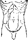
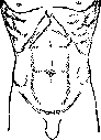
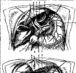
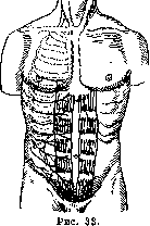
time not to disturb the innervation of the abdominal muscles. From Figure 33 it can be seen that the innervation is not disturbed by: a) an incision along the midline and b) an incision along the midline almost to the navel, and from there obliquely upward and to the side in the direction of the nerve fibers (Rio-Branco); through the first incision, the gallbladder can be removed in thin women with a compliant abdominal wall; however, this incision gives so little space that it can be limited only in the case where it was made in anticipation of another operation, e.g., on the stomach. The Rio-Branco incision is too cumbersome for most cases. The transverse Gosset incision (Fig. 34) hardly cuts any nerves and provides good access to the gallbladder, especially with a ptotic liver. The Kocher incision (Fig. 35) parallel to the right costal margin provides good access, but it cuts nerve branches and in any case should not go beyond the right anterior axillary line. Kehr performed a wave-like (or bayonet-shaped) incision (Fig. 36) with a transverse cut across not the entire right rectus muscle. The incision is convenient for ectomy and often for choledochotomy. However, in difficult cases, when it is necessary to mobilize the duodenum, the Kehr incision is insufficient: the transverse part of the incision has to be continued to the side, and then the lower vertical
part becomes superfluous. If the transverse part of the Kehr incision is continued parallel to the costal margin and the lower vertical part is not made, an incision is obtained which is used by Fedorov in more difficult cases (Fig. 37). Of the other incisions, one can also mention the Sprengel incision, shown in Figure 38, and the Kausch incision (Fig. 39). The operation is performed with a bolster placed at the level of the lower angles of the scapulae. After opening the peritoneum, in cases of chronic cholecystitis with pericholecystitis, the gallbladder is first of all separated from adhesions (with the greater omentum, stomach, duodenum, transverse colon, etc.), after which the bile ducts are examined. In the absence of changes (stones, strictures) in the main duct, the gallbladder is removed after being isolated with napkins. Removal can be started either from the fundus, which is more convenient with large adhesions, or from the cystic duct. In the first case, the cystic artery is cut and ligated at the end of the gallbladder isolation; in the second, before or after cutting the cystic duct (Fig. 40). Separation of the gallbladder from the liver is sometimes extremely easy; in other cases, the layer of loose connective tissue between the gallbladder and the liver turns out to be scarred due to previous pericholecystitis. Besides other circumstances affecting the isolation of the gallbladder, much depends on how much the liver is brought out of the wound. With a high and rigid rib cage and the impossibility of mobilizing the liver, the operation is technically less convenient than in thin subjects with a small anterior
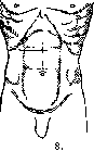
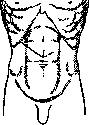
Figure 34. Figure 35.
Figure 36.
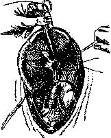
Figure 37. Figure 38. Figure 39. Figure 40.
anteroposterior diameter of the chest and a mobile liver, easily brought into the wound by its sharp edge. In the absence of adhesions, the gallbladder is inspected and palpated. It may be altered in color (whitish, with engorged vessels, etc.). The walls of the gallbladder may be thickened. Stones may be palpable in the gallbladder. In other cases, no clear changes are found. If, after a thorough examination of the stomach, duodenum, head of the pancreas, right kidney, and appendix, no deviations from the norm are found in them, then the gallbladder, after being isolated with napkins, is punctured; the contents are aspirated, which may turn out to be normal or altered bile. Upon emptying the gallbladder, even if incomplete, it is sometimes possible to palpate small calculi that were not detectable when the gallbladder was full. In cases of uncertain examination results, the gallbladder should be opened. After removing all the bile, one sometimes (quite rarely) finds a small stone in the neck of the gallbladder. If there is no stone, attention is paid to the mucosa: an inflammatory state indicates acalculous cholecystitis. In pronounced cases, the mucous membrane is raspberry-colored; against this background, a delicate yellowish network, consisting of cholesterol deposits, is sometimes visible. In other cases, the mucosa is found to be smooth, devoid of folds—an indication of previously occurring overdistension of the gallbladder. If the surgeon decides to remove the gallbladder in cases of acalculous cholecystitis, the ectomy does not differ in any essential way from an ectomy for stones. Also, there are no essential differences in an ectomy for hydrops or empyema of the gallbladder. Excision in the case of a tumor must be preceded by a detailed examination to accurately determine the feasibility and extent of the operation. After removal of the gallbladder, the abdominal wound can be packed with gauze or sutured tight. A blind suture has the advantages of rapid healing and protection to a certain extent against postoperative hernia. An argument against the blind suture is the unreliability of closing the stump of the cystic duct, through which bile can flow into the abdominal cavity; bile leakage is also possible through existing accessory ducts, which sometimes go from the liver and empty directly into the gallbladder: when the gallbladder is removed, they are inevitably injured. There are, however, a number of cases in which suturing tight is fully indicated, namely, when removing a non-functioning gallbladder, when it is 1) completely filled with stones, 2) shriveled and contains no bile, 3) filled with mucus or pus (hydrops, empyema). With a functioning gallbladder, a blind suture is possible only if the surgeon is confident in the reliability of closing the cystic duct stump; in other words, it comes down to technique. More or less significant accessory hepatic ducts can be noticed in the gallbladder bed. To ensure against the possible entry of bile into the abdominal cavity, Rozanov inserts a rubber drain to the stump of the ductus cystici for a short time. Suturing tight is contraindicated when it is necessary to drain the biliary tract, as well as in cases of widespread inflammatory changes and in cases of bleeding that yields only to tamponade. If the surgeon decides to suture tight, the stump of the cystic duct is ligated (preferably with catgut) and then peritonized—usually by sliding the peritoneum of the posterior wall. Rotter, one of the first surgeons to begin suturing tight after an ectomy, proceeded as follows: after ligating the cystic duct, he would bend it and apply a common ligature to the end and beginning of the duct. He then placed the entire cystic duct behind the peritoneum. The weak point of Rotter's method is the closed cavity along the entire length of the cystic duct. Martynov recommends the following method. The stump of the cystic duct
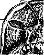
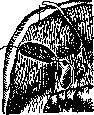
Figure 41.
Figure 42.
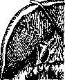
Figure 43.
is ligated with catgut at a distance of less than 1 cm from the line of excision. The rosette formed peripheral to the ligature is sutured with catgut, which is then tied; one end of the suture is left long. The stump of the duct is pulled toward the gallbladder bed by this end (Fig. 41). 4–5 sutures are placed on the gallbladder bed, i.e., on the liver parenchyma, in such a way that the edges of the bed cover the duct stump and the remaining catgut thread, which is then tied to one of the sutures on the liver (Figures 42 and 43). This is followed by a layered suture of the abdominal wall. If tamponade is necessary, the cystic duct is ligated or drained, and strips of gauze are introduced laterally—to the beginning of the lateral canal (Zernov), to the stump of the cystic duct, to the Winslow foramen, and to the transverse colon; part of the surgical wound is sutured. The tampons are left in for a relatively long time. They are removed, if possible, together with the removal of the sutures. The tampon closest to the liver is removed first. The others are removed in the second week; in this case, it is almost always necessary to use ether anesthesia. The edges of the wound are stretched with retractors, and one or two strips of gauze are re-inserted into the wound. Removal of the gallbladder is often an easy operation. With significant adhesions, it requires great caution, as it is sometimes easy to injure the hepatic artery or its right branch (liver necrosis!), or the portal vein. Injury to the stomach, duodenum, and transverse colon is less critical. The creation of a gallbladder fistula (cholecystostomy) is performed in the poor condition of the patient, when the removal of the gallbladder requires much time (adhesions), and also sometimes in severe phlegmons, although in the latter case, ectomy is preferable. Cystostomy is performed either in two stages (suturing the gallbladder—opening it) or—as is now done almost exclusively—in one stage. Cystotomia idealis, s. cholecystendysis is an operation consisting of an incision of the gallbladder, removal of a stone, and suturing of the incision. The abdominal cavity is closed tightly. For the purpose of removing a stone, this operation is currently almost never performed, except perhaps in cases where a gallbladder stone, which had not given clear symptoms, is accidentally discovered during laparotomy for another reason. The incision of the gallbladder may also end in suturing in cases of exploratory opening of it (see above). When suturing the gallbladder, one should not pierce the mucous membrane so that the sutures do not provide a cause for the formation of stones.
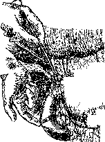
Anastomoses between the gallbladder and the gastrointestinal tract are created in cases of narrowing or obstruction of the common bile duct. The following are used: anastomosis with the stomach (cholecystogastrostomy), with the duodenum (cystoduodenostomy), with the jejunum (cystojejunostomy), and finally, there have been cases of anastomosis with the transverse colon (cystocolostomy). In cases of obstruction of the common bile duct, the creation of an anastomosis is indicated primarily for: chronic sclerosing pancreatitis, cicatricial strictures after stones, and neoplasms. In cases of impacted stones, an anastomosis is not indicated: the stones must be removed through an incision of the common bile duct. An anastomosis with the jejunum was first performed by Monastyrsky (Leningrad) in 1887 for cancer of the head of the pancreas, and an anastomosis with the stomach by Terrier (1896). An anastomosis with the stomach is in most cases simpler in technique than anastomoses with the intestines. Usually, the prepyloric part of the stomach is taken for the anastomosis. The distended gallbladder is freed from bile. If it is necessary to pull the stomach toward the gallbladder, it is useful to separate the body of the gallbladder from the liver to avoid tension on the sutures. The opening is made 1–2 cm in diameter. The suture is in two layers. It is more convenient to suture the mucosa with catgut and the serosa with silk. The abdominal wound (much smaller in size than for an ectomy) is usually closed tightly. Stools become colored in the very first days; itching disappears, and then, little by little, jaundice as well. Cystogastrostomy has no visible harmful effect on gastric digestion. In the future, one may fear cholecystitis and ascending cholangitis, although in this respect, an anastomosis with the stomach should be considered more favorable (due to the scarcity of the stomach's microflora) compared to intestinal anastomoses.
Cystogastrostomy as a palliative operation finds application in unresectable cancers of the ampulla of Vater and the head of the pancreas. It is also used in chronic indurative pancreatitis, which is easy to mistake for cancer during surgery, and also in high cicatricial strictures of the common bile duct; cystojejunostomy and cystoduodenostomy are used somewhat less frequently. With an anastomosis with the jejunum, one can expect cholangitis sooner than with an anastomosis with the duodenum. As for the choice of method, in cases of cancerous tumors, one should perform the operation that is more convenient in the given case (usually cystogastrostomy). Considerations about the possibility of cholangitis should not have decisive significance, since the life of patients with a cancerous tumor of the pancreas or the ampulla of Vater is generally short. As for benign strictures, in these cases, one should prefer the operation of anastomosis of the common bile duct with the duodenum, which is discussed below. Operations on the cystic duct are only a supplement to ectomy and consist of an incision for the removal of stones or the removal of the cystic duct itself. The main duct (hepatic and common bile ducts). The most common operation is opening the duct (hepaticotomy, choledochotomy). It is performed for the removal of stones and drainage of the ducts in cases of cholangitis. A distinction is made between: incision or opening of the hepatic duct, the common bile duct above the duodenum (supraduodenal choledochotomy), the common bile duct behind the duodenum (retroduodenal choledochotomy), and the common bile duct through the duodenum (transduodenal choledochotomy). Hepaticotomy is rarely used. Usually, stones located in the hepatic duct can be brought down into the common bile duct or reached directly with forceps or a blunt spoon through an incision of the common bile duct. However, sometimes the stones turn out to be immobile, and it is necessary to dissect (longitudinally) the hepatic duct. In this case, caution must be exercised 1) regarding the cystic artery, which often (in 27% of cases according to Rio-Branco) passes across, in front of the hepatic duct, 2) regarding the right branch of the hepatic artery and the hepatic artery itself, which also sometimes lie in front of the duct. Injury to the cystic artery is of no particular significance, although it forces one to resort to ligation of the artery; injury to a branch, and even more so to the hepatic artery itself, threatens liver necrosis and a lethal outcome. After the removal of stones, one must try to suture the wound of the hepatic duct and place a drain through the incision of the common bile duct.
Supraduodenal choledochotomy is the most common of the operations for opening the main duct. It is used for stones in this part of the bile duct, as well as for stones in the hepatic duct and the lower part of the common bile duct. Preliminarily, as with hepaticotomy and choledochotomy of the lower section, it is technically more convenient to remove the gallbladder first, which is usually severely altered; there is no need to remove a shrunken gallbladder that does not contain bile or stones. The common bile duct is often significantly dilated in cases of bile stasis. In other cases, it has adhesions that displace it from its usual location. Due to these changes, it is sometimes not so easy to find the duct and verify that the common bile duct has indeed been discovered, especially if stones are not clearly palpable. In such cases, it is permissible, after walling off the abdominal cavity with napkins, to perform a trial puncture with a thin needle. Upon obtaining bile, a longitudinal incision is made above the duodenum or in the place where the stone is palpable, along the lateral side of the duct. After the removal of the stone, the duct is examined, if it is wide, with a finger or, if possible, with the thickest probe. The examination is performed both in the direction of the hepatic duct and its branches, and downward—in the direction of the common bile duct, while trying to pass into the duodenum. If a thick probe does not pass, then thinner ones are tried. The ducts are felt with a finger over the probe. Stones of the main duct discovered during probing (above and below the incision) are removed if possible from the same incision by pushing them from the outside with a finger or with an instrument introduced into the duct (Fig. 44).
canal with an instrument - a blunt spoon, forceps. When a thick probe passes freely up and down into the duodenum (Fig. 44) and the finger does not palpate any stones, it remains to drain the ducts, which are usually infected. To do this, a soft drainage tube (caliber 16-18 catheter size) is inserted upward into the hepatic duct, and the excess part of the incision of the common bile duct at the inserted tube is sutured with catgut. The suture closest to the tube is not cut, but is used to tie the drain. Martynov never uses Kehr's T-drains and has not seen any bad consequences from this. In case the probe at the bottom of the common bile duct encounters an obstacle - a stone, which cannot be moved upward and cannot be removed with a spoon or dressing forceps, it is necessary to mobilize the duodenum, and then perform a retroduodenal choledochotomy or - if the stone is determined to be at the very papilla of Vater - a transduodenal one. Mobilization of the duodenum is performed as follows: the parietal peritoneum is incised with a scalpel parallel to and to the side of the vertical part of the intestine, 2-4 cm away from it (Fig. 45). Both edges of the peritoneum are grasped with Pean or Luer clamps, and the duodenum is carefully detached with a finger. Then the posterior wall of the intestine, the retroduodenal section of the common bile duct, and the section of the pancreas closest to it become visible. A small incision is made on the palpable stone (Fig. 46) of the duct (avoid injuring the duct of Wirsung!), the stone is extracted, and the papilla of Vater is probed. The duct is drained through an incision above the duodenum, and the retroduodenal incision is sutured. The wound is tamponed, with a separate strip of gauze being brought to the duodenum. In transduodenal choledochotomy, it is also advantageous to mobilize the duodenum. If a gauze tampon is placed under it, the intestine
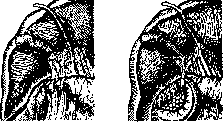
Figure 45.
Figure 46.
approaches the abdominal wound, which significantly facilitates the operation. The incision in the duodenum is made small - vertical or transverse at the level of the papilla of Vater. The stone is extracted through an incision of the duct orifice (Fig. 47), after which the edges of the duct incision are sutured to the intestinal mucosa with two to four stitches (choledochoduodenostomy interim). The intestine is sutured. A drain is inserted into the common bile duct through an incision above the duodenum. Transduodenal choledochotomy is especially suitable for large stones stuck at the orifice of the duct (Fedorov), because making a large incision through the duct behind the duodenum is associated with the risk of injuring the duct of Wirsung. An anastomosis between the common bile duct and the duodenum is performed in case of narrowing or obstruction in the retroduodenal part of the duct. The narrowing can be either due to a scar of the duct or due to an inflammatory process in the head of the pancreas. In such cases, i.e., in non-cancerous narrowings, choledochoduodenostomy should generally be preferred to an anastomosis of the gallbladder with the stomach; with an anastomosis
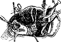
Figure 47. of the common bile duct, there is much less risk of subsequently developing ascending cholangitis, especially if the operation is supplemented by the removal of the gallbladder as a possible accumulator of infection. Of course, such a general position is not applicable to individual cases: thus, with a functioning gallbladder and in a severe condition of the patient, one has to use the gallbladder for the anastomosis, which takes significantly less time. There are end-to-side and side-to-side anastomoses of the duct with the duodenum. In the first, the common bile duct is transected, and the upper segment is sutured into a small incision in the intestine. In the second (Fig. 48), an incision of the duct is made over the intestine, which is connected to the intestine with two rows of sutures. The first method is more complex and dangerous, the second is simpler and therefore is currently predominantly used. In the absence of complications, the operation yields excellent results. In addition to the indications given, one should also mention the anastomosis between a cyst (congenital) of the common bile duct and the duodenum, which also yields very good results. The same cannot be said about the anastomosis of the hepatic duct with the duodenum, which is always performed end-to-side during resections of the common bile duct due to neoplasms and its narrowings. Resection of one or another part of the main (hepatic-biliary) duct is performed for neoplasms. The continuity of the biliary tract can be restored in various ways: by connecting the hepatic duct with the common bile duct end-to-end, by an anastomosis of the hepatic duct with the gastrointestinal tract, by replacing the common bile duct with the gallbladder (anastomosis with the stomach or duodenum), by flap pla
sty of the duct from the walls
The upper end of the rubber tube must be
located in the hepatic duct, the lower one in the duodenum. If the tube is inserted before the plasty or the formation of the anastomosis, it is more convenient to apply sutures on it as on a frame; after the operation, the tube ensures the free discharge of bile into the intestine and thus protects the operative adhesions of the duct incisions from rupture. If the end of the tube descends into the duodenum by 6-8 cm, then gradually, thanks to the peristalsis of the intestine, it descends lower and after some time (from several days to months) exits through the anus. A. Martynov.
Related articles
Mentioned in
Cite this page
“Gallbladder.” Soviet Medical Encyclopedia. English translation of Bolshaya Meditsinskaya Entsiklopediya, 1st ed. (Moscow, 1928–1936), ed. N. A. Semashko. https://sovietmedicalencyclopedia.pages.dev/article/gallbladder/