Liver
Historical document, translated for reference. It reflects medical knowledge of the 1920s–30s and is not medical advice.
Summary
The liver is a vital organ found in all vertebrates, with complex anatomical structures and functions. This article details its comparative anatomy, embryological development, physical properties, and fixation in the human body.
Encyclopedia article (1928–1936)
VI. Functional diagnosis of liver .... 575 VII. Surgical diseases of the liver.....583 VIII. Syphilis of the liver................ 591 I. Anatomy of the liver. Comparative anatomy. The liver, hepar, is an organ peculiar to all vertebrates. Connected in its origin with the anterior wall of the upper part of the intestinal tube (see below), during its development it grows into the mesentery of the intestine (pushing apart its layers) and thereby remains in connection with both the elements of the biliary-intestinal section and the wall of the abdomen.-The structure of the liver in all vertebrates, including humans, has very similar features: there is always an excretory duct, and in most there is also a reservoir for bile-the gallbladder (cystis i'ellea). A separate group is Acrania, in which the liver is an unpaired organ, projecting forward on the right side of the body as a blind sac (I. E. W. Ihle), connected with the midgut, and remains in the stage of a tubular non-branching organ. In Craniota, the liver is always a compact voluminous organ of brown or brown-red, sometimes green color, located caudally from the heart, in the vicinity of the stomach. As a rule, this gland is always complex and divided by a more or less deep groove into 2 lobes-right and left (in bony fish they are barely distinguishable). Very often each half is divided into new lobes, which differ in length and shape. In animals with an elongated body, the liver is elongated, the lobes are unequal and follow one after another. In birds, the right and left lobes are sharply separated from each other and each has a separate duct; they open (one above the other) into the upper part of the ascending limb of the duodenum, with the right, caudal one being connected to the gallbladder. In pigeons, the left duct opens into the descending, the right into the ascending limb of the duodenum. In addition to the right and left lobes, some mammals also have a middle lobe (lobus centralis). In carnivorous animals, the liver is larger than in herbivores. Embryology of the liver. The liver develops from the abdominal wall of the endoderm of the intestine, from the so-called hepatic groove, as a forward-directed blind outgrowth lying in the abdominal mesentery. From the posterior part of the groove, the gallbladder develops. In lower vertebrates, numerous hollow branches then develop from the liver rudiment. In higher vertebrates, compact 5-27
LIVER
628 initially cellular strands appear. Later, a lumen appears in these strands, and the rudiment transforms into a tree-like branched complex tubular gland. Subsequently, numerous anastomoses develop in the epithelial cellular mass between the branches, and the liver acquires the characteristics of a reticular gland.
Physical properties. The human liver is a massive organ of dense consistency, but at the same time possesses great plasticity, which is manifested in the change of its shape (see below) under the influence of compression by adjacent organs. Its tissue is very fragile and tears easily. The abundance of blood gives the organ an intensely red color, especially sharply expressed in early childhood. With age, this color more and more acquires a brownish or brownish tint.-The relative weight of the liver to the weight of an adult is determined, according to Meckel, as 1 : 33, according to Charpy, as 1 : 38 with variations up to 1 : 15. In the early period of development, it is significantly higher and decreases with age. The weight of the liver of a living person reaches 2,300-2,500 g. The size of the liver is subject to individual variations. On average, the liver has 28 cm in transverse (frontal) diameter with variations from 20 to 40 cm; 8-9 cm in vertical (variations from 5 to 12) and 10 cm in anteroposterior (variations 15-27 cm). The outer surface of the organ under normal conditions is even, smooth, and shiny. Prolonged pressure from adjacent organs can cause persistent changes in the outer configuration, leaving traces in the form of depressed grooves and indentations. Impressions from ribs can, deeply indenting, constrict the liver and give it a characteristic appearance ["corset liver" during the wearing of waist-constricting corsets, rachitic liver with severe rachitic deformity of the chest cavity (see separate table, fig. 1), etc.]. The shape of the liver is generally subject to very large variations depending on its size, position in relation to surrounding organs, and the condition and shape of these organs. Therefore, a liver removed from a corpse during autopsy does not reflect the configuration it had in life, if the corpse was not before dissection fixed with sufficient strength by formalin or other hardening reagents. In general, the liver is usually compared in shape to a wedge, the wide base of which is directed backward and fixed to the diaphragm, while the sharp edge is directed forward and downward. Thus, one can speak of two free surfaces of the organ: the upper convex, adjacent to the diaphragm, and the lower concave, directed downward and backward and in contact with the organs of the biliary-intestinal tract. Fixation. The larger part of the liver surface lies freely in the abdominal cavity, the smaller part is fixed to the upper posterior wall of the latter. If not considering the intraperitoneal pressure that holds the liver in place, its fixation is ensured by three connections: 1) hepatic veins, 2) adhesion with the vena cava itself, 3) the ligamentous apparatus, which is formed by the transition of the visceral peritoneum to the parietal. The hepatic veins firmly fix the liver in the area of the foramen quadrilaterum of the diaphragm (at the site of their entry into the vena cava). Due to the firm adhesion of the vena cava with the edges of the opening, displacement of the organ is possible only together with the diaphragm. The strength of the second connection is subject to individual variations, mainly depending on the shape of the aperturae thoracis inf.: displacement is possible within wider limits in the narrow-chested type. The parietal ligamentous apparatus of the liver consists of 2 main ligaments: the coronary (lig. coronarium, s. hepato-phrenicum), directed frontally backward, and the suspending ligament of the liver (lig-suspensorium, s. falciforme), which is a continuation of lig. teres hepatis and located sagittally forward and upward. The degree of fixation by these ligaments depends on the completeness of the covering of the organ by visceral peritoneum. The more it is, the closer the anterior and posterior sheets of it come together and the more mobile the liver becomes. In places of complete convergence, membrane-like ligaments resembling mesenteries are thus formed: lig. suspensorium hepatis and lig. triangulare in the right and left angles of the coronary ligament (fig. 1). The left part of the coronary ligament ends with a thickened ligamentous strand, noted by some authors as lig. fibrosum hepatis, or appendix fibrosa. The continuation of the left part of the coronary ligament backward and downward (in the sagittal direction) is lig. venosum Arantii, which surrounds the obliterated anastomosis in adults between the umbilical and vena cava (ductus venosus Arantii) (fig. 5). At a right angle to lig. venosum approaches and continues into it the duplication of the peritoneum (lig. hepato-gastro-duodenale), or the lesser omentum. In the thickness of this ligament, along its free right edge, pass the common bile duct, the hepatic artery, and the main trunk of the portal vein. Of the latter ligaments, only the coronary and venous ligaments have fixing significance. They firmly hold the posterior part of the liver in the least mobile part of the diaphragm. The width of the area of this support is greatest, according to the teachings of Prof. Shevkunenko's school,-in persons with a wide thoracic aperture.-Due to the strengthening of the liver mainly in the posterior upper part, in the least mobile part of the diaphragm, and moreover in such a way that the main mass of the organ remains free, the liver is characterized and mainly accessible to two types of movement: tilting upward, to the dome of the diaphragm, and downward displacement (tilting) of the free anterior edge downward. During these movements, the posterior part remains immobile, and the organ moves relative to it, like on a hinge (Zernov). The most essential condition for the fixation of the liver is, however, not so much the ligamentous apparatus as the abdominal pressure in conjunction with the pressure from the diaphragm. Weakening of this pressure leads to displacement of the liver downward, and conversely, paralysis of the diaphragm promotes the elevation of the liver upward. In the human liver, 3 surfaces are distinguished: anterior-superior, posterior, and inferior (facies-superior, posterior et inferior). The upper surface has a convex shape, exactly repeating the curvature of the dome of the diaphragm. The lower surface is concave (in a child more or less flat) and is directed partly downward, partly backward. The posterior-the smallest surface of the liver-is turned toward the spine. The upper and lower surfaces meet at the anterior edge of the liver (margo anterior, acutus), which on the left
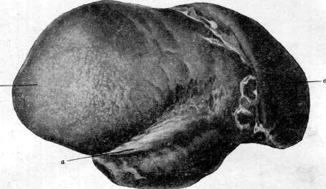
Figure 1.

:.<
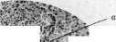

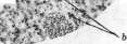
Figure 3.
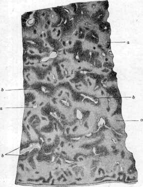
Figure 2. Figure 1. Deep groove along the upper-anterior edge of the liver. The groove formed in areas of indentation by ribs with severe curvature of the chest cavity in kyphoscoliosis. Sclerosis of the capsule in the area of the groove (a); b- right lobe; c-left lobe. Figure 2. Mottled liver (enlarged with a magnifying glass): a- sharp atrophy of the central parts of the lobule; b- areas of preserved parenchyma. Figure 3. Liver with congenital syphilis: a- miliary gummas; b- giant cell metamorphosis of the parenchyma.
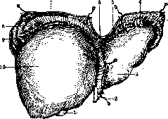
Figure 1. Ligamentous support of the liver: I-gallbladder; 2-lig. teres; 3-left lobe; 4-lig. triangulare sin.; 5 and 7-lig. coronarium hepatis; 6-lig. suspensorium hepatis; 8-diaphragm; 9-lig. triangulare dextr.; 10-right lobe. (After Testut-Latarjet.)
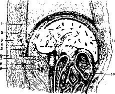
Figure 3. Sagittal section of the liver at the porta: 1-lung; 2-lig. coronarium; 3-pleural sinus; 4-lig. hepato-duodenale with its vessels and bile duct; 5-diaphragm; 6-v. cava inf.; 7-foramen of Winslow; 8-kidney and adrenal gland; 9-duodenum; 10-colon transversum; 11-liver. (After Testut-Latarjet.)
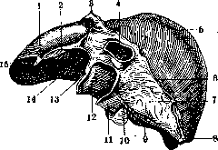
Figure 5. View of the liver from the diaphragmatic side: 1-lobe sin.; 2-lig. triangulare sin.; 3-lig. falciforme; 4-v. cava inf.; 5-lobe dexter; 6-lig. coronarium; 7-retroperitoneal field under lig. coronarium; 8-lig. triangulare dextr.; 9-lig. hepato-renale; 10-gallbladder; 11-v. cava inf.; 12-Spiegel's lobe; 13-lig. venosum (Arantii); 14-impressio oesophagea; 15-appendix fibrosa. (After Braus.)
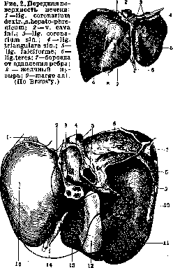
Figure 4. Inferior surface of the liver: 1-appendix fibrosa hepatis; 2-tuber omentale; 3-papillary process of Spiegel's lobe; 4-Spiegel's lobe (lobus caudatus); 5-v. cava inf.; 6-rhomboid retroperitoneal field under lig. coronarium; 7-lig. triangulare dextr.; 8-impressio renalis; 9-bridge of serous covering between right and Spiegel's lobes; 10-impressio duodenalis; 11-impressio colica; 12-gallbladder; 13-lobus quadratus; 14-lig. teres; 15-impressio gastrica. (After Braus.)
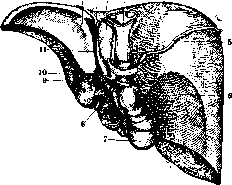
Figure 6. Posterior surface of the liver: 1-lig. triangulare sin.; 2-impressio oesophagea; 3-Spiegel's lobe; 4-v. cava inf.; 5-lig. coronarium; 6-impressio renalis; 7-gallbladder; 8-biliary passages; 9-v. portae; 10-a. hepatica; 11-lig. venosum (Arantii). (After Testut-Latarjet.)
In the left lobe, the anterior edge is thinner and sharper, while in the right lobe it is thicker and more blunt. In the absence of pathological changes, the liver is soft, especially in children, and usually cannot be palpated through the abdominal wall. Along its course, the anterior edge of the liver is interrupted by 2 notches: the left one (incisura umbilicalis), into which the free thickened lower edge of lig. teres enters, and the right one, which is more blunt and corresponds to the gallbladder (incisura vesicae felleae). On the superior surface, 2 lobes are distinguished-right and left (lobus dexter et sin.). The boundary between them is lig. suspensorium hepatis (fig. 2). The space between the liver and diaphragm, i.e., the anterior subphrenic 'space,' is divided by this ligament into two parts. In adults, the right lobe is significantly smaller than the left, and lig. suspensorium always deviates to the right. Both lobes of the liver are covered on top by visceral peritoneum, with a break only under the root of lig. suspensorii. If the latter is cut off, a defect of the serous covering remains on the liver in the form of a narrow sagittal groove, which, widening fan-like, transitions into a rhomboid area of the posterior surface devoid of peritoneum, located under the root of the coronary ligament. The inferior surface is divided by grooves into 4 lobes: right, left, and the median section with two lobes lying one behind the other-the square lobe (lobus quadratus) in front and Spiegel's lobe (lobus Spiegelii, s. caudatus) behind. The deep transverse groove between them, filled with areolar tissue with vessels and bile ducts, is called the porta of the liver (porta, s. sulcus transversus hepatis) (fig. 4). These middle lobes are bounded on the left by lig. teres and lig. venosum Arantii, while on the right, Spiegel's lobe has the inferior vena cava, and the square lobe has a groove covered by the gallbladder and its duct. The inferior surface of the liver is covered with peritoneum, the covering of which is interrupted by the root of the ligament, composed of two duplicatures of peritoneum converging at right angles: the frontal one, reinforced along the groove of the liver's porta-lig. hepato-gastro-duodenale, and the sagittal-lig. venosum Arantii. In these ligaments are located: 1) in the transverse, or in the 'porta'-vessels, bile passages, lymph, glands, and nerves. From the liver tissue forward, mostly two converging liver ducts (ductus hepatici) emerge at an angle. Somewhat to the left of them, 2-3 branches of the hepatic artery enter the liver. Behind the liver ducts and artery is the wide trunk of the portal vein. Between these structures, lymph nodes and associated lymph vessels of the liver are sometimes (not constantly) found. 2) In the longitudinal ligament (lig. venosum Arantii) is contained the obliterated ductus venosus Arantii in adults. The ligaments converging at an angle in the porta of the liver cover behind them Spiegel's lobe, hanging down with a broad papilla into the bursa omentalis, and cause the division of the serous covering of the liver into 2 unequal fields: the smaller one with the just-named lobe and the larger one, encompassing all the remaining lower-posterior surface of the liver, namely the covering of the right lobe, gallbladder, square lobe, and left lobe. Both fields communicate through a narrow bridge of serous covering between the porta of the liver and the inferior vena cava. This intermediate area forms the upper wall of for. Winslowi (see Peritoneum), leading into the bursa omentalis (fig. 3). A finger introduced into it encounters the free end of Spiegel's lobe-processus caudatus, covered in front and behind by peritoneum. The left part of this sometimes develops to the degree of a strongly protruding papilla, distinguished as a special formation-processus papillaris. After separation of the gallbladder from the liver, the serous covering of the liver is interrupted by a defect of the corresponding area. Depending on the type of covering of the gallbladder by peritoneum, this defect can be wide and extend to the anterior edge of the liver (with a less mobile and insufficiently complete gallbladder) or be reduced to a narrow strip (with a well-expressed mesentery of the gallbladder). The anterior end of such a defect sometimes does not reach the edge of the liver, and the gallbladder can be hidden under its body. The area of the liver under the gallbladder bears the name fossa vesicae felleae. The posterior surface of the liver (fig. 6) has a shape close to a right triangle, the boundaries of which are not as clearly defined as in the other parts of the liver. One leg is short, located vertically and borders the outer edge of the right lobe of the liver, the other, longer one, lies horizontally and forms the posterior edge of the liver. The hypotenuse passes behind its porta and (conditionally) crosses obliquely the inferior surface of the liver from the lower point of the right free edge of the liver to the tip of its left triangular ligament (lig. triangulare sinistrum). In projection on the posterior abdominal wall, this line, along an oblique, ascending line from right to left, crossing the upper pole of the right kidney, goes from its outer edge upward through the body of the diaphragm to the cardiac part of the stomach, bordering along the way with the upper edge of the head of the pancreas. This line is the most constant of all other boundaries of the liver and in its main parts is preserved with all movements and congenital deviations in the position of the liver, being that axis of the hinge around which the movements of the liver occur. The largest part of its posterior section is devoid of peritoneal covering. The defect of the latter on a liver removed from the abdominal cavity appears in the form of a rhombus. In the course of the posterior section of the liver, 2 formations are noted: the inferior vena cava, more or less embraced by the liver tissue, with 3-4 hepatic veins flowing into it at the very dome of the diaphragm, and the upper part of Spiegel's lobe, covered here by serous membrane. The vena cava borders it but lies extraperitoneally, adjacent to the diaphragm either directly or partially separated from it by the tissue of the liver extending onto the posterior surface of the vein. Only a small section of this vein below the liver (fig. 11) is covered with peritoneum, which bends over from the aforementioned bridge between the serous fields of the inferior surface of the liver and is a continuation of the peritoneum lining the right kidney. This area forms the posterior wall of for. Winslowi. Topography and syntopy. The liver occupies the upper part of the abdominal cavity, with the largest part of the organ in an adult located in the right hypochondrium. In the first half of intrauterine life, the liver occupies more than half of the abdominal cavity, pushing away other abdominal organs from the diaphragm. In the second half, even more sharply after birth, under the influence of the increase in volume
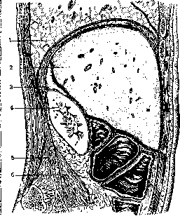
Figure 7. Sagittal section at the level of the right kidney: 1-lung; 2-liver; 3-adrenal gland; 4-right kidney; 5-m. quadratus lumborum; 6-m. psoas. (After Testut-Latarjet.)
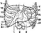
Figure 9. Projection of the liver on the anterior wall: 1-liver; 2-stomach; 3-12-duodenum. (After Testut-Latarjet.)
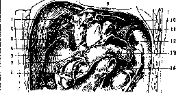
Figure 11. Relations of the liver to surrounding organs: 1-pyloric part of the stomach; 2-lig. hepato-duodenale; 3-kidney; 4-lig. triangulare dextr.; 5-v. cava inf. (below covered by a bridge of peritoneum); 6-right adrenal gland; 7-defect of peritoneum-area of attachment of the liver to the diaphragm; 8-hepatic veins; 9-parietal layer of peritoneum behind Spiegel's lobe; 10-lig. triangulare sin.; 11-esophagus (cut); 12-greater omentum; 13-pancreas; 14-duodenum. (After Braus.)
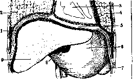
Figure 8. Frontal section of the hepatic region (diagram): 1-diaphragm (muscular part); 2-right lung; 3-heart; 4-left lung; 5-tendinous part of the diaphragm; 6-stomach; 7-spleen; 8-gallbladder; 9-liver. (After Testut-Latarjet.)
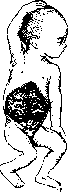
Fig. 10. Liver in a fetus of 3 1/2 months. (After Testut-Latarjet.)
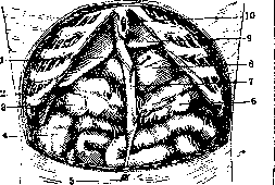
Figure 12. Relationship of the anterior edge of the liver to the costal arch in an adult: 1-right lobe of the liver; 2-gallbladder; 3-hepatic angle of the large intestine; 4-ascending large intestine; 5-navel; 6-transverse colon; 7-lig. teres; 8-stomach; 9-left lobe of the liver; 10-xiphoid process. (After Testut-Latarjet.)
of the thoracic cage and a decrease in the amount of blood (emptying of the umbilical vein), the size of the liver decreases, its left half lags behind in growth, and its relationship to the diaphragm becomes asymmetrical. The liver departs from the left hypochondrium, but its left lobe remains for a long time under the left dome, wedging its sharp end between the diaphragm and spleen. The upper border of the liver coincides with the borders of the diaphragm and during respiration or changes in intra-abdominal pressure or from the side of the thoracic organs, it follows its movements and displacements. In the average resting position in an adult, the projection of the upper edge onto the anterior wall reaches its highest point on the right along the nipple line at the level of the upper edge of the V costal cartilage (figs. 8 and 9). From here, the border curves steeply to the right, intersecting the VI and subsequent ribs up to the X, meeting the VII intercostal space on the axillary line. To the left of the highest point, this line slopes gently downward toward the VI costal cartilage, meeting and crossing along the way on the median line the lower part of the sternum above the base of the xiphoid process and along lin. parasternalis - the cartilage of the VI rib. Further to the left, the edge of the left lobe is subject to individual variations. It may descend to the VII cartilage and extend beyond the nipple line or end near the cartilage of the VI rib. The lower border of the liver in projection forms a broken line. In most cases in an adult, under normal conditions, the lower edge in the right hypochondrium does not extend beyond the edge of the ribs, on the median line of the body it occupies half the distance between the xiphoid process and the navel, and rising to the left, it intersects the costal arch in the area of the VII-VIII costal cartilage (fig. 12). Individual variations in the position of the anterior edge of the liver lead to 2 extreme types of its rotation around (obliquely) the transverse axis; to the type of liver thrown backward--'dorsopetal position,' and to the type turned forward-'ventro-petal' (Melnikov). Behind, the upper border of the organ is projected at the level of the lower edge of Dxi, the lower-at the middle of Dxi-In adolescents, the relationships are almost the same as in adults. In early childhood, the soft lower edge is often 1-2 fingers below the costal margin (Filatov), while the upper level is located higher, and its height decreases with the child's growth. According to the data of Engel's roentgenoscopic examination from 1 to 3 months after birth, this level reaches Dviji-ix, by 11-13 months it can reach Dx-xi; the excursion of the highest point of the diaphragm of the newborn, according to Vogt, occurs between the IV and VI ribs.-On the anterior surface of the liver, corresponding to its curvature, 2 surfaces can be distinguished: anterior and superior. As can be seen from the sagittal section of a corpse (fig. 7), the anterior surface is in contact with the muscular part of the diaphragm, and along the course of the costal angle of the epigastric region, it lies directly against the abdominal wall. These relationships divide the surface into 3 areas: costal, where it is in contact through the diaphragm with the pleural sinus and lung and in penetrating wounds is damaged simultaneously with these organs; upper median, behind the xiphoid process, where it is connected only with the mediastinal sinus of the pleura and in injury the pleura may participate without the lung, and lower median, in which in injury of the liver the peritoneum is affected. The same conditions explain the possibility of the liver being displaced downward, below the costal margin, in pleural empyema, the inevitability of the surgical approach to the liver abscess through the pleural sinus, and the impossibility of reaching it through the abdominal wall, etc. The superior surface of the liver lies against the- tendinous part of the diaphragm and through it is adjacent to the bottom of the pericardial cavity and the inferior surface of the heart. Correspondingly to this, on the superior surface a limited, somewhat concave area-planum cardiacum-is usually noticeable. In the case of a poorly developed left lobe, it corresponds only to part of the cardiac field of the diaphragm and then, if the spleen is not excessively large, the rest of the field is occupied by the stomach. With a large left lobe of the liver, it completely displaces the stomach from the planum cardiacum of the diaphragm and may come in contact with the spleen, especially when it is enlarged. The inferior surface of the liver over a large extent is adjacent to various parts of the gastro-intestinal tract and kidney, consequently on the liver, especially fixed (in a corpse) by formalin, there remain indentations, imprints repeating the shape of the adjacent organ. Thus, the entire area of the left lobe in an adult represents a concavity corresponding to the curvature of the stomach floor (impressio gastrica). Only a small area of it (between the venous ligament and the lesser curvature of the stomach) protrudes in the form of a tubercle, pressing together with the lesser omentum into the area of bursae omentalis, and for this reason it bears the name tuberculum omentale hepa-tis (fig. 4). Immediately above it at the posterior edge of the liver, a groove-like depression (fig. 6) is clearly outlined from the pressure of the cardial part of the esophagus (impressio oesophagea). The latter is embraced here in front by lig. triangulare sin. hepatis and the initial part of lig. venosi Arantii and is closely adjacent to Spigelian lobe. The quadrate lobe is in contact with the pyloric part of the stomach and the initial part of the duodenum, the vertical segment of which is already adjacent to the medial part of the inferior surface of the right lobe, forming a longitudinal vertical groove (fig. 4) (impressio duodenalis). The remaining part of the right lobe is divided into 2 fields. In front lies the depression from the hepatic angle of the large intestine (impressio colica), behind-from the right kidney (impressio renalis). The gallbladder is adjacent partly to the pyloric part of the stomach and partly to the duodenum, and its apex touches the transverse colon. The degree of this contact varies depending on the position and filling of the stomach, large intestine, and type of duodenal structure. Changes in these relationships are possible depending also on the degree of development of the right lobe of the liver itself.
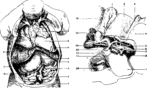
Figure 13. Dissection of the cavity of a newborn child: 1-right lobe of the liver; 2-gallbladder; 3-left lobe of the liver; 4-lig. suspenso-rium hep.; 5-v. umbilicalis; 6-navel with skin cut along the edge; 7-bladder with two umbilical arteries; 8-diaphragm cut along the costal margin; 9-heart; 10-left lung; 11-right lung; 12-thyroid gland. (After Esipov's preparation.)
Figure 15. Porta hepatis: 1-gallbladder; 2-quadrate lobe; 3-lig. teres; 4-left lobe; 5-hepatic duct; 6-branch of the portal vein; 7-left hepatic artery; 8 and 14-lymphatic glands; 9-Spiegelian lobe; 10-portal vein; 11-inferior vena cava; 12-common bile duct; 13-right hepatic artery; 15-accessory portal vein; 16-right lobe of the liver.
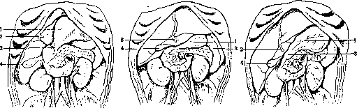
Figure 11. Three types of liver position in a child under 1 year of life: 1-greater curvature of the stomach; 2-round ligament of the liver; 3-pylorus; 4-gallbladder. (After Natiyev.) 4 5^
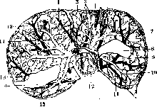
Figure 16. Scattered type: 1-right branch of the middle hepatic vein; 2-quadrate lobe; 3-branches of the vein of the quadrate lobe; 4-ramus umbilicalis; 5-ramus marginalis; 6-v. arcuata ant.; 7-anterior branch of the left hepatic vein; 8-unworked areas of the preparation; 9-v. arcuata sup.; 10-v. arcuata post.; 11-posterior branch of the left hepatic vein; 12-v. lobi Spigelii; 13-branches of the right hepatic vein; 14-deep branch. (After Melnikov.)
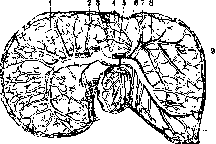
Figure 17. Main type of branching of ducts and portal vein inside the liver: 1-a. obliqua angularis; 2-a.ascendens; 3-recurrent branch; 4, 5 and 6-lower and upper veins of the quadrate lobe; 7-v. arcuata anterior; 8-a. arcuata anterior; 9-v. arcuata superior. (After Melnikov.)
the peritoneum covering the muscular part of the median legs of the diaphragm, and on the sides it closely abuts the inferior vena cava on the right and the esophagus on the left. The entire posterior part of the Liver through the medium of the diaphragm is in contact with the pleural sinus. And on the peculiarities of the Liver, its growth and development in the child. The Liver in the newborn is large-approximately 4.5% of body weight (in adults-2.8%), or, otherwise, 1/th-x/i3 of body weight, while in adults-1!ya-V34 of this weight. The weight of the Liver in the newborn is determined differently: according to Lereboullet, it averages 91.5 g, according to Wallich and Gubendorf-120-130 g, according to Arnold, Aeby and Benecke--130-180 g. It occupies not only the right half of the abdomen, but with its strongly developed left lobe it extends into the left half of the abdomen, reaching the spleen. As for the shape of the liver, peculiarities are noted in the configuration and position of Spiegel's lobe and lob. quadrangularis, which affects the position of the vessels and excretory ducts in the gates of the Liver (Jaschke). With age, the relative weight of the Liver decreases, but the absolute weight increases, so that in a one-year-old child it weighs on average 320 g, in a three-year-old-457 g, in a ten-year-old-800 g, in a sixteen-year-old-1 260 g. Starting from the 5th month of life, the left lobe lags in growth. The lobular structure of the Liver in the newborn is absent and appears only by the end of the 1st year of life, by 2-4 years the characteristic radial arrangement of liver cells already appears, and by 8 years the Liver does not differ in its macro- and microscopic structure from the Liver of an adult. The boundaries of the Liver (in an infant): upper boundary-V intercostal space or upper edge of V rib along the mamillary line; along the axillary line-VII rib. The lower edge of the Liver in the newborn is determined 1-2 cm above the navel (Mettenheimer), in an infant it protrudes 1-2 cm from under the rib edge, and by 2-4 years it ceases to be palpable. These boundaries are subject to individual fluctuations depending on the shape of the chest. In any case, the transverse (vertical) dullness of the Liver by the end of the 1st year is 4-6 cm; about 5 years-6-7 cm; after 8 years of age it is approximately equal to the number of years of the child, but does not exceed 14 cm. In childhood, the relationships of the Liver with other organs are somewhat different than in adults, especially in the embryonic period and in the 1st year of life. The embryonic Liver is almost symmetrical and with its anterior surface descends downward to the pubis, slightly not reaching it (fig.12). In the 1st year of life in 58% (according to Nativiev) the right angle of the Liver does not reach the iliac crest (fig. 13 and 14). The left lobe completely or partially covers the stomach, the thin left edge of it reaches the spleen, the lower surface of the liver is flat, there is only a concavity in the area of contact with the bottom of the stomach. In 28% (especially in the first 2 months of life and in premature infants) the left lobe is very large, sometimes almost equal to the right. Its massive edge separates the spleen from the diaphragm and displaces it inward and downward. The left notch of the Liver is close to the midline. The anterior edge protrudes from under the ribs. Its lower level is almost at the navel. The lower surface is mostly flat. In 14% the liver is positioned obliquely, approaching the vertical, i.e. it lies mainly in the right hypochondrium; the anterior edge is close to the rib edge (iy2-3 cm) along the mamillary line. The right angle along the axillary line reaches the iliac crest. The left lobe is small, mostly lies to the right of the midline, often not reaching the spleen. The left and right notches are almost on the same vertical. Blood supply. The Liver is supplied with blood from a. hepatica (fig. 15), which originates from a. coeliaca, more rarely-from a. mesenterica sup., a. renalis dextra or independently from the aorta. In addition to the Liver, additional arteries may approach in the form of branches from neighboring main vessels: from aa. gastrica dextra, spermatica dextra, renalis, mesenterica superior, pericardiaco-phrenica and suprarenalis dextra. The main trunk of the hepatic artery in the gates of the Liver mostly divides into 2 branches--for the right and left lobes. Each branch, however, can supply not only its own lobe, but also two, three and even all four lobes of the lower surface of the Liver. The right branch of the hepatic artery is usually larger in caliber than the left, initially lies to the left, from the hepatic duct, then curves around it posteriorly and crosses it; along the way it gives off a branch to the gallbladder and the so-called middle hepatic artery, which at the left notch of the Liver divides into branches for the square and left lobes. Before entering the tissue of the Liver, the main trunk of the artery divides into 2, more rarely into 3 or 4 branches. The left hepatic branch ascends to the left of the portal vein and its left branch, medially from the hepatic duct. Before entering the left lobe of the Liver with one, two or more branches, it mostly also supplies Spiegel's lobe.
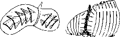
Venous blood from the Liver is drained by the hepatic veins (vv. hepaticae), of which the smallest ones in large numbers open with pinpoint holes into the inferior vena cava, while the larger ones gather into 2 or 3 very short trunks up to 15 mm in diameter. The latter, after leaving the Liver, flow into the vena cava at an acute angle, immediately before it passes through the diaphragm. Into such a left terminal trunk of the hepatic vein, in embryonic life, Aranzio's duct opens. The vascular branching within the liver, according to Mel'nikov, occurs according to the main or scattered types (fig. 16 and 17) and is so regular that it allows for proposing certain planes of incisions during operations to avoid injury to large vessels (fig. 18). The lymphatic system of the Liver can be divided into superficial and deep, differing by drainage on the upper and lower surfaces and very rich in variations. Most of the lymph from the Liver flows into the thoracic duct through cisterna chyli via abdominal pathways, while part is drained through thoracic lymphatic vessels into the supraclavicular area, into angulus venosus (fig. 19 and 21). The nerves of the Liver originate from two sources: from the solar plexus and the vagus nerve. Some authors have established a connection with the thoraco-abdominal nerve, but it is not recognized by all. The nervous system in the form of a complex network reaches the liver, passing through the thickness of lig. hepato-gastro-duodenale. Topographically, two anastomosing plexuses (plexus hepatici) are distinguished, which are located in front of and behind the vessels of the said ligament (Latarjet). The anterior (fig. 22) arises from the left part of the solar plexus, gives a network along the course of the hepatic artery and, approaching the liver, scatters into a thin surrounding network for the bile ducts and vessels, from which branches depart to neighboring organs, in particular to the gallbladder and Liver. The posterior (fig. 20) originates from the right node of the solar plexus and reaches the Liver with branches, located behind the portal vein and forming numerous anastomoses with plexus ant. In addition to these nerve branches, there is an independent branch to the Liver from p. vagus sin., which, receiving a twig from p. vagus dext., approaches the Liver in the thickness of the lesser omentum behind the left and in front of Spiegel's lobe. The nerve at the gates of the Liver divides into branches, of which some anastomose with the anterior sympathetic plexus, others are connected with the accompanying the hepatic artery. Inside the tissue of the Liver, branches from the mixed nerve plexus scatter into small fibrils and, following the vessels, penetrate deep into the liver lobules. (Anatomical structure of the bile ducts and gallbladder--see Gallbladder.)
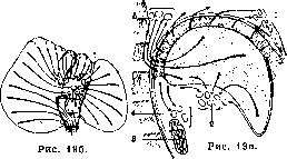
Fig. 18. Scheme of liver incisions according to Mel'nikov.
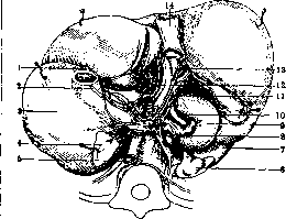
Fig. 19 (a, b, c). Scheme of directions of lymphatic currents: 1- lymphatic glands behind the xiphoid process; 2-same at the gates of the liver; 3-pancreas; 4-lymphatic glands along the upper edge of pancreas; o-lymphatic glands of the thoracic surface of the diaphragm. (According to Testut-Latarjet.)
Fig. 20. Posterior nerve plexus of the liver (the latter is deviated forward): 1- p. vagus sin.; 2-p. vagus dext.; 3-stomach; 4- left adrenal gland; 5-aorta; 6- right adrenal gland; 7-kidney; 8-14-v.cavainf.;9-12-duodenum; 10-plexus coeliacus; 11-plexus hepaticus post.; 12-plexus hepaticus ant.; 13-portal vein. (According to Testut-Latarjet.)
k. Egipov.
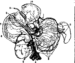
Fig. 21. Lymphatic vessels of the lower surface of the liver, anterior surface of the stomach and right adrenal gland and kidney: 1-glands of the lesser omentum and entrance of the stomach with the vessels of the liver and stomach flowing into them; 2-glands of the splenic hilus with vessels of the bottom of the stomach flowing into them; 3-chain of glands of the greater omentum, lying along the course of the inferior coronary artery of the stomach and receiving lymphatic vessels directed to the greater curvature of the stomach; 4-aorta; 5-glands on the aorta and inferior vena cava, receiving lymphatic vessels of the liver, adrenal gland and kidney; 6-adrenal gland; 7-gallbladder; 8-glands in lig. hepato-duodenale with lymphatic vessels flowing into them, emerging from the network of the gallbladder and left longitudinal groove of the liver; 9-round ligament of the liver; 10-central glands, through which lymph from the stomach, spleen and most vessels of the lower surface of the liver passes.
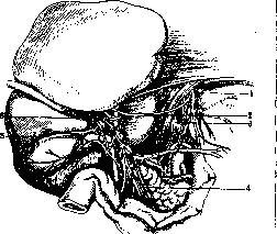
Figure 22. Anterior nerve plexus of the liver: 1- left vagus nerve; 2- hepatic branch of the vagus nerve; 3- right vagus nerve; 4- duodeno-pyloric nerves; 5- cystic nerves; 6- anterior hepatic plexus (According to Latarjet). II. Histology of the liver. General architecture of the liver. The liver is a rather homogeneous formation in its microscopic structure. It differs sharply from typical glands in that it consists only of small lobules, closely pressed against each other and not collecting into higher-order lobules, as well as in that the terminal sections do not have lumens. Often, individual lobules, by fusing together at their bases, form a complex lobule. On the outside, the liver is covered with a connective tissue capsule (capsula Glissoni), which is further covered on top with a thin layer of peritoneum. In the area of the liver hilum, the connective tissue of Glisson's capsule penetrates into the liver, accompanying the blood vessels and bile ducts, and in the form of increasingly thinner septa reaches the individual lobules. In the pig, bear, and camel, these septa are well developed, in humans they are expressed very weakly and therefore the individual liver lobules merge with each other [see separate table (pp. 223-224), fig. 4 and 5]. In humans, this connective tissue is preserved only in the triangles formed at the point of contact of three lobules. In these areas pass: a branch of the hepatic artery, a bile duct, and a branch of the portal vein, the so-called interlobular vein. In the center of each lobule passes the so-called central vein, carrying blood out of the lobule. The blood flow through the liver proceeds as follows. Blood enters the liver through the portal vein and hepatic artery (along with them go the branches of the bile duct) [see separate table (pp. 223-224), fig. 4 and 5]. The branch of the portal vein - interlobular vein - goes along the edge of the lobule. From the interlobular vein, capillaries branch off into the lobule, which, by forming anastomoses into a network, penetrate the lobule to its center, where these capillaries flow into the central vein. The central veins flow into collecting veins (or otherwise sublobular veins), and the latter, connecting, give rise to the hepatic veins that carry blood out of the liver. The lobules (lobuli, s. insulae) have a prismatic shape and a diameter of 1-2 mm; each lobule consists of the so-called liver cords, which converge radially toward the center of the lobule. All cords are connected to each other in a network, located in the loops of the capillary network. Thus these two networks are as if inserted into each other. On a reconstruction of the liver {see separate table, figure 1} the liver cords are marked in yellow, and the dense capillary network (blue). On a cross-section through a cord, it is seen that the cord consists of two rows of epithelial cells, between which passes as an intercellular channel the bile capillary. The liver cords correspond to the terminal sections of glands of the usual tubular type, but, unlike the latter, first, they are connected to each other by anastomoses, and second, their central lumen is extremely narrowed and is located only between two rows of cells. Thus, if in a normal gland each glandular cell has its basal end turned toward the basal membrane and its apical end toward the lumen, then in the liver cord both surfaces are adjacent to blood vessels, and the 'lumen' - the bile capillary - lies between the lateral surfaces of two adjacent cells. Due to this structure, liver cells are in very close contact with the blood, all the more so since the basal membrane is absent and the cells are directly adjacent to the capillary wall. Liver cells have predominantly polyhedral shape and usually one nucleus, but two- and multi-nucleated cells are not uncommon. The amount of chromatin in the nucleus varies depending on the physiological activity of the cell. The nucleolus is always well expressed. Mitoses in liver cells are extremely rare. The cells are clothed in a well-expressed membrane, which is a thickening of the peripheral layer of protoplasm. Chondriosomes in liver cells are abundant, somewhat concentrated around the nucleus, and have either a rod-like or granular character. In the liver lobule, three zones can be distinguished: the zone around the central vein - the zone of rest, where the chondriosomes look like long, thin threads with a few granular forms scattered between them; the active zone (on the periphery of the lobule) has swollen and thickened chondriosomes; here there are many rounded forms; finally in the middle zone the chondriosomes have an intermediate character. Near the nucleus, the Golgi network is found, as well as the cell center. In addition, in the protoplasm of the liver cell, with appropriate reagents, protein clumps, fat droplets, and glycogen clumps are revealed, the amount of which varies depending on the functional state of the organ. Glycogen is deposited in clumps in the cytoplasm, first in cells lying closer to the central zone, and then in more peripherally located cells. Conversely, the dissolution of glycogen probably begins in the peripheral cells of the lobule. After giving the animal food rich in carbohydrates (sugars), liver cells already after 4-5 hours show signs of activity, increasing in volume, and after 10-12 hours the cytoplasm is filled with clumps of glycogen. Then comes the period of reverse transformation of glycogen into glucose and its release into the blood. After 24-48 hours, all glycogen disappears from the protoplasm and the cell passes into a state of rest. - The type of nutrition strongly changes the structure of liver cells. When feeding with fat, liver cells become larger and fill with fat droplets, and the number of chondriosomes decreases. When feeding with carbohydrates, in addition to glycogen clumps, small iron-containing particles and fat droplets appear. With a protein diet, the cells become very large and fill with protein inclusions in the form of irregularly shaped granules, often large. Bile ducts. In lower vertebrates (reptiles), each liver cord has a tubular shape and contains inside a well-expressed lumen, which is formed by 8-9 rows of cells. In the human embryo, the structure of the liver cord is analogous, but soon after birth the tubular character of the cords is finally lost and in the adult liver, as shown above, each cord consists of two rows of cells, between which remains a narrow slit - the bile capillary [see separate table (pp. 567-568), fig. 1]. These bile capillaries run along the cord, having a zigzag course, and give off short lateral branches that go between individual liver cells and end blindly. The old idea that these lateral branches go inside the cells has not been confirmed. Since the liver cords anastomose, the bile capillaries passing inside them also form anastomoses. The bile capillaries do not have their own membrane, and their walls are formed by a layer of more 1 condensed protoplasm of the limiting
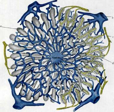
FIG. 1.
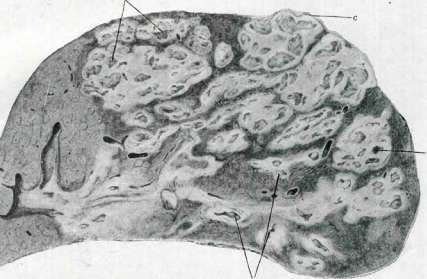
Fig. 2.
Fig. 1. Scheme of a liver lobule: a-liver trabeculae; b-branch of v. portae; c-biliary duct; d-biliary capillary (according to Pañ'egu). Fig. 2. Suppurative thrombophlebitis (a) of the liver with abscesses (b), partially protruding under the capsule (c). The liver lobule is built from hepatic cells, between which run blood capillaries. Due to this density of their protoplasm, it is sometimes possible to isolate these capillaries. At the periphery of the lobule, the biliary capillaries flow into the small branches of interlobular ducts. These transitional sections are called Hering's canals (Hering). Here the biliary capillary directly continues into the biliary duct, but the limiting larger hepatic cells are immediately interrupted and replaced by small cells of the duct epithelium. At the same time, collagenous connective tissue appears around the biliary duct, which is absent around the hepatic cells. The epithelium of the biliary ducts is low cuboidal. Small biliary ducts, anastomosing, form a network around v. inter-lobularis; then, increasing in diameter, they collect towards the gates of the liver. As the caliber of the duct increases, its epithelium becomes progressively higher, changing from cuboidal to cylindrical (see Gallbladder). The blood capillaries inside the lobules (often called hepatic sinusoids), densely anastomosing with each other, form, as shown above, a network between v. interlobularis and v. centralis, thus forming the so-called "wonderful network". Since all these venous sinusoids originate from the vv. interlobulares and go to v. centralis, their general arrangement is radial. The finest branches of a. hepaticae pour into the aforementioned sinusoids at the periphery of the lobule. The blood sinusoids of the lobule are lined with cells of two types. Some of them have a small nucleus, which is so dense that no structure can be discerned in it. Their protoplasm is stretched into a thin layer along the sinusoid. Cell boundaries cannot be detected in the "walls of the hepatic sinusoids". The cells of the second type are rich in protoplasm, which often stretches into long processes directed in all directions if these cells are viewed from the surface; when viewed from the side, however, they are spindle-shaped. Due to their processes, these cells received the name stellate cells (Kupffer's). Because of these processes, Kupffer's cells somewhat protrude into the lumen of the venous sinusoid and are bathed in blood on all sides. The nuclei of Kupffer's cells are large, light, with a small nucleolus. The protoplasm of these cells often contains grains of green pigment, as well as erythrocytes in various stages of digestion and granules of hemosiderin. Kupffer's cells, having a pronounced ability to phagocytosis and to accumulate vital colloidal dyes and particles of fine suspensions in a granular form, belong to the ret.-end. system, constituting the reticulo-endothelial apparatus of the liver. These Kupffer's cells can detach from their connections and, as free ameboid cells, enter the bloodstream, forming macro-phages or free histiocytes. The often observed microscopic differences in the cells of the sinusoid walls are an expression of different func. states. When lithium carmine or trypan blue is introduced into an animal, Kupffer's cells accumulate large amounts of these dyes in the form of grains, while indifferent cells do not show the ability to accumulate these dyes. However, when ink is injected, particles of the latter are deposited in cells of both types, indicating their relationship to each other. In addition, they are connected by numerous transitional forms. The wall of the hepatic sinusoid is a continuous membrane, maintaining its syncytial structure from the embryonic period. Kupffer's cells are apparently the most active elements of the ret.-end. system, as they are the first in the entire body to capture foreign substances introduced into the body, and only after them do these substances begin to accumulate in other elements of the ret.-end. system. Connective tissue of the L. It was indicated above that connective tissue, accompanying the branches of v. portae, enters the spaces between the lobules. This periportal (since it surrounds the branches of v. portae) dense connective tissue directly continues into a dense network of reticular (argyrophilic) fibers surrounding the intralobular capillary-sinusoids. From these argyrophilic fibers, the larger ones run along the sinusoid, while the thinner ones form a dense network between them (see Fibrous lattices). This network of reticular fibers supports the tissue of the lobule. The reticular fibers and the cells of the sinusoid wall (Kupffer's and indifferent) form a continuous wall of the sinusoid without any gaps or openings. Lymphatic spaces and vessels. According to some definitions, from one-third to one-half of all the body's lymph is formed in the L. However, lymphatic vessels have been found only in the interlobular connective tissue along the vessels. No lymphatic capillaries have been found inside the lobules. Some authors admit the existence of only lymphatic spaces between the sinusoid wall and the hepatic cells, although not detectable by the methods used so far. Thus, in essence, we cannot determine the exact place of lymph formation in the L. and its method of exit into the periportal lymphatic vessels. The latter, merging, form a network of deep vessels, vasa profunda. The second network, vasa superficialia, is located directly under the serous membrane. A feature of the lymph draining from the L. is its abundant protein content. Cultivation of liver tissue in vitro to date has only been possible with the explantation of embryonic L. (chick on the 8-16th day of incubation). According to observations by Lynch (Lynch), growth is membranous, by an epithelial membrane formed by polygonal cells. The mitochondria of hepatic cells in tissue culture conditions are large and granular. In liver cultures, mitoses are observed in the cells (Levi). In addition to hepatic cells, connective tissue elements and epithelium of the biliary ducts also grow. Nordmann (Nordmann) showed that hepatic cells, even under tissue culture conditions, retain the ability to accumulate glycogen, thus remaining specifically differentiated. Of particular interest are the observations of Dolzhansky, who showed that bile formation occurs in hepatic cells cultivated in vitro. In this case, bile formation occurs only in those cultures where mesenchymal elements grow simultaneously with epithelial cells. In pure cultures of L. epithelium, bile formation does not occur. Benevolenskaya observed hematopoiesis in the L. in vitro, by cultivating in tissue culture the liver of a human embryo aged 1-3 months, and the growth of mesenchyme and its hematopoietic transformations turned out to be more intense than the growth of hepatic epithelium. Hematopoiesis in vitro proceeds according to the same scheme as in the body. Methods of histological investigation of the L. Methods of injection of biliary ducts, a) General investigation. For the purpose of a general survey of the structure of the L., any commonly used microscopic techniques are suitable. For fixation, mercuric chloride and mercuric chloride mixtures (saturated solution of mercuric chloride + 5% Ac. acet. glac; saturated solution of mercuric chloride-3 parts + formalin-1 part; Zenker's fluid with formalin) are especially recommended, as well as formalin. After these fixations, any staining is permissible, and even ordinary staining with hematoxylin and eosin gives satisfactory results. After mercuric chloride fixations, staining according to Mallory should especially be recommended, mainly its modification "Azanfar-bung". This staining very well reveals, in addition to glandular epithelium and connective tissue, also on thin sections shows the reticular fibers, b) Fat. Fixation with formalin, sections on a freezing microtome, staining with Sudan III according to Chiaccio. c) Glycogen. Fixation with absolute alcohol or Carnoy's fluid, embedding in celloidin, staining with carmine according to Best, as well as the iodine test for glycogen. d) Chondrium. Fixation and staining by ordinary methods, e) Kupffer's cells are detected after subcutaneous or intravenous injection of lithium carmine, trypan blue, colargol, ink, etc. It is better to use lithium carmine, after which the L. is fixed with mercuric chloride or formalin, embedded in celloidin or celloidin-paraffin, and on sections the nuclei are restained with hematoxylin*. The dosage of lithium carmine depends on the weight and size of the experimental animal (e.g. for a rabbit 10-15 cm3, for a rat-2.5-3 cm3; 5-6 days daily one injection). f) Blood vessels are detected by injection through vena portae (with the vena cava inf. ligated), as well as through vv. hepaticae or through a. hepatica. g) Reticular fibers are best revealed by silver impregnation according to Belinovsky. h) Biliary capillaries can be revealed by injection, impregnation and staining. Injection of biliary capillaries is performed with colored liquid masses (a concentrated aqueous solution of Berlin blue is recommended) through d. hepaticus or d. choledochus. In the latter case, the dye first fills the gallbladder, and from there through ductus hepaticus enters the L. In herbivores (e.g. rabbit), injection is easier.
The injection is performed carefully until the dye appears on the surface of the Liver. The physiological injection according to Chrzhonshchevsky is more successful. A saturated aqueous solution of indigo carmine is injected into the v. jugularis ext. (to a dog 50 cm3, to a cat 30 cm3, to a rabbit 20 cm3) three times within 11/2 hours, or a concentrated solution of sodium indigosulfate in doses of 15-20 cm3 every 10 minutes (for a dog). Then the animal is killed, and small pieces of the Liver are fixed in absolute alcohol, or the entire Liver is injected with absolute alcohol through the aorta. On the sections, the bile capillaries are found to be filled with indigo carmine, which has been secreted into them by the liver cells from the blood and lymphatic vessels. - Impregnation of the bile capillaries is performed as follows: pieces of fresh liver are placed in a mixture: 3% solution of K2Cr2O7 - 4 parts, 1% solution of OsO4 - 1 part, for three days, changing the liquid every day. From here, for 24-48 hours, into a 3/4% solution of AgNO3. Then the pieces are washed with distilled water, quickly hardened in alcohols and cut. The bile capillaries turn black. As for the staining of the bile capillaries, it is complex, inconsistent, and difficult to achieve.
B. Aleshin. III. Normal Physiology of the Liver. Although the functions of the L. are far from completely studied, what is already known indicates the active participation of the L. in most of the most important chemical processes in the organism. Neither carbohydrate metabolism nor protein, fat, and fat-like substances and some salt metabolism can do without a certain indispensable participation of the L. Methodology. Experimental physiology has applied three methods to the study of the physiological activity of the L.: 1) the perfusion method, 2) Eck's method and the method of excision, and 3) the angiotomy method. The perfusion method in relation to the L. has the peculiarity that the perfusion fluid is not introduced into the artery, as is usually done, but into the portal vein, because under natural conditions substances absorbed from the intestine enter the L. by this path.--The method of excision of the L. in dogs also differs from the usual method of organ excision. For a dog to be able to live for several hours after the operation, holding itself as a dog usually holds itself in the first hours after the operation g, three operations are necessary, which follow one another at intervals of two to three weeks. The first operation begins the same as the Eck operation, i.e., an anastomosis is made between the portal and vena cava, but it ends differently: not the portal vein is tied off, but the vena cava between the anastomosis and the place where the renal vein trunk flows into it. As a result of this operation, blood is diverted from the inferior vena cava into the portal vein. As a result of this operation, collateral circulation is created; when the latter has already fully established itself and compensated for the disturbance in blood flow caused, the second operation is performed, which consists in tying off the portal vein at the point where it enters the L. As a result of this second operation, blood from both large veins—the vena cava and the portal vein—is diverted into the collaterals. Then follows the third operative session, in which excision of the L. is performed. The perfusion and excision methods are auxiliary methods. The first can show what the L. is capable of when all anatomical connections between it and the organism are completely severed. The second method gives an idea of what changes occur in the organism with complete removal of the L. Neither of these methods can give a complete picture of what functions the L. performs under normal anatomical-physiological conditions. Eck's operation, in which blood from the portal vein is diverted into the vena cava, can give some indications regarding the functions of the L., but these indications cannot have exhaustive value, because first, in Eck's operation the hepatic artery is left, through which some substances still penetrate into the L., and second, the omentum, which adheres to the L., also to a large extent compensates for the defect in portal circulation in the L. The third method—angiotomy—is free from all these objections. In relation to the L., the angiotomy method is applied as follows: two cannulas are applied—one to the portal vein near the bifurcation, and the other—to the hepatic vein. From a comparison of the composition of the blood in these veins, the functional activity of the L. is accurately accounted for in each of the cases studied. If the perfusion and excision methods can still give in some cases an idea of the statics of the processes of the L., then the dynamics of these processes, their course in time, can only be shown by the angiotomy method. Carbohydrate metabolism. Of all organs, the L. appears the richest in glycogen content. The ability to form and accumulate glycogen is already present in the newborn child. The main source of the latter in the L. is sugar absorbed from food by the intestine. The first few grams of sugar absorbed from the intestine pass through the L. freely, almost without being detained by it. As a result, the sugar concentration in the blood rises. Hyperglycemic blood excites the center of the vagus nerve, which gives an impulse to the pancreas, which begins to secrete insulin in response to irritation. The appearance of the latter in the blood leads to the deposition of sugar in the L. in the form of glycogen. In later periods of digestion, the L. retains on average 3/4 of the sugar passing through it, which apparently completely turns into glycogen. It is clear that the glycogen content in the L. is strongly dependent on the carbohydrate content in food. After a plentiful carbohydrate meal, the glycogen content in the L. can reach 16-18% of the weight of this organ, usually its content does not exceed 4%. In a frog, a percentage ten times greater can sometimes be found. The glycogen content in the liver of a growing organism is especially influenced by the protein content in food (Schiff's experiments on puppies). When introducing food rich in protein (and with the condition of limiting water intake), the liver becomes poor in glycogen; with prolonged introduction of protein food, fatty infiltration of the L. begins. Approximately the same data were found by Rosenbaum in infants with acute water loss. In addition to food, work also influences the glycogen content in the L. During intense work, the glycogen content in the liver can drop to its minimum. Fasting together with intense muscular work can almost completely deprive the L. of glycogen. To completely deprive a dog's L. of glycogen, it is made to fast, and then floridzin is administered. The formation of glycogen in the L. occurs not only at the expense of carbohydrates. It is undoubtedly that protein can also serve as a source of glycogen. If a dog deprived of glycogen by preliminary fasting and floridzin is fed boiled meat, a large amount of glycogen (up to 7%) can be found in the L. of this dog. Fat can also give rise to glycogen of the L. The main source for the formation of glycogen of the L. are carbohydrates. Among them, glucose and fructose occupy the first place. In second place are galactose and mannose. It is known that various monosaccharides easily pass into one another in the organism. From more complex sugars, the L. can form glycogen only if this sugar was previously broken down to monosaccharides. An example can be maltose, which, when introduced under the skin, is broken down under the influence of maltase with the formation of glucose; only a small part of maltose, which escaped the action of the enzyme, is excreted outside with urine. Such disaccharides as cane or milk sugar, which are broken down in the organism only in the intestinal tract, are excreted intact with urine if they were introduced into the organism parenterally. Judging by experiments on a surviving liver, there is a whole series of substances that lead to the deposition of glycogen in the L.--Protein metabolism. Foreign protein. All kinds of foreign substances—passing through the circulatory system of the L., are detained in the latter for some time. Apparently in connection with this peculiarity stands the delay in the L. of foreign proteins that have entered the circulatory system in one way or another. They can come from the intestine, especially in cases of ulcer formation in it, or by the parenteral route. The attitude of the L. to amino acids. The influence of the liver on protein metabolism is not limited to the delay of foreign proteins. The L. affects all protein derivatives passing through its bloodstream, regardless of whether they come from the intestine during the digestion and absorption of proteins or by the parenteral route.--Formation of urea—see Urea.--Bile formation constitutes the specific function of the L. The amount of bile produced by the L. is quite significant. In a dog, the L. produces per day depending on food from 3 to 36 g of bile per 1 kg of weight. In man, it is difficult to determine the amount of bile excreted with such accuracy as in animals. It is assumed that the amount of bile excreted in man per day fluctuates within the limits of from 1/2 to 1 liter. Since the L. as an organ producing bile acts as a digestive gland, the corresponding function of it must change depending on food. During fasting, the solid part of bile appears diminished in its absolute magnitude. During digestion this magnitude increases. With carbohydrate food, the maximum of excretion falls on the 2nd and 3rd hours of digestion; with protein—on the 3-4th hours and with fatty food—on the 5th, 6th and 7th hours. Not only the character of the bile excretion curve depends on the kind of food, but also the total amount of bile flowing out. The most bile flows out on fat, less on proteins and even less on carbohydrates. Fat metabolism. According to the latest research by S.V. Nedzvedsky on angiotomized dogs, the L. retains less fat than other organs. Nevertheless, judging by the experiments of L. Stotsik, again made on angiotomized dogs with blood transfusion, it must be assumed that in the L. there is a fat depot, from which fat is released under the influence of irritation caused by the transfused blood. Finally, the L. is indicated as the place where fats are converted into carbohydrates. That the transition in the organism of fats into carbohydrates really takes place, this can now be considered firmly established.--Lipoid metabolism. If one analyzes the blood flowing to the L. through the portal vein and flowing away from it through the hepatic vein for lecithin (for phosphorus), it is easy to be convinced that the blood flowing through the L. is enriched with lecithins.
The hepatic vein is richer in lipoid phosphorus than all other veins. From this it is permissible to conclude that lecithins are produced by the liver. All that has been said about the complex metabolic processes taking place in the liver naturally presupposes the presence in the liver of a large number of enzymes. Indeed, catalase, oxidases, aldolases, carboxylases, various hydrolytic enzymes (proteases, amylase, lipases, nucleases), deamidases, and finally enzymes that form and destroy uric acid have been found in the liver. Detoxifying action of the liver. The detoxifying action of the liver is already evident in the embryonic stage of development (Charrin); from Petru's experiments with alkaloids, it can be concluded that the liver of a child has a greater ability to bind poisons than the liver of an adult. Substances toxic to the body are detoxified in several ways. 1. Through oxidation or reduction, or hydrolysis, deamination, decarboxylation, dealkylation (in most cases with the help of enzymes). 2. Through synthesis with sulfuric, glucuronic, aminoacetic, carbamic acids or through methylation or sulfhydrylation (and in these processes enzymes are largely involved). 3. The substance is simply deposited as a depot. Among the reactions of destruction in the liver of toxic substances, the most common is the oxidation reaction. Aliphatic alcohols and aldehydes are completely oxidized in the liver; benzene is oxidized to muconic acid, with the intermediate products of the reaction being benzene catechol and orthoquinone. In all these oxidative processes, glutathione, which is present in the liver in large quantities, plays a significant role. It has been found that the glutathione content in the liver varies between 0.22% and 0.35%. The oxidative activity of the liver is also manifested in the fact that the hepatic vein contains 7-8% more oxidized sulfur than the portal vein. In diseases of the liver, its oxidative activity decreases, and therefore, for example, in cirrhoses, the peripheral blood appears enriched with unoxidized sulfur (thiemia). The detoxification of plant alkaloids in the liver also rests partly on the process of oxidation. First, they are deposited in the liver, and then oxidized. This has been proven by experiments on surviving livers of various warm-blooded animals when passing through it such poisons as curare, nicotine, aconitine, muscarine, digitalin, atropine, strychnine, ricin, picrotoxin, phenol, alcohol, and adrenaline. That poisons are destroyed when passing through the liver can be judged by the fact that most alkaloids lose half of their toxicity in the process. Some authors obtained different effects from the same poison depending on whether it was administered to the animal in a peripheral vein or directly into the portal vein. For example, for sulfuric acid atropine, the lethal dose when injected into a peripheral vein is 0.041, and when injected into the portal vein it is 0.192, i.e. almost 5 times more. Direct chemical analysis showed that oxidation is only one of the types of degradation of pharmacological agents, that some of them, as mentioned above, undergo reduction, hydrolysis, dehydrogenation, deamination, decarboxylation, etc. The liver is richer in all kinds of enzymes than any other organ, and therefore it is not surprising that it is the central organ for the destruction of various pharmacological agents. Many poisons are detoxified by coupling them with sulfuric or glucuronic acid, carbamic or acetic acid. The detoxifying action of the liver with respect to microbes is not manifested in all cases. If rabbits or guinea pigs are injected with anthrax bacilli or golden staphylococcus into a peripheral vein, these animals always die from bacteremia, but when these microbes are introduced into the portal vein, the animals remain alive in the vast majority of cases. The reverse relationship is obtained with respect to staphylococcus. It is further assumed that the liver is the central place for the formation of various immune bodies, such as alexins, agglutinins, precipitins, opsonins, etc. In this respect, the reticulo-endothelial tissue plays a major role. With respect to inorganic substances, the liver also shows a delaying action. While sodium and potassium salts pass through the liver completely unimpeded, salts of heavy metals are to a greater or lesser degree retained in it. Thus, ferric lactate is three times less toxic when injected into the portal vein than when injected into the general bloodstream. Copper albuminate loses half of its toxicity when passing through the liver. Mercury, lead, and arsenic salts are held by the liver very firmly, while magnesium, silver, and zinc salts are quite quickly excreted from the liver with bile. Arsenic salts are held by the liver firmly but not for long. When salvarsan is injected, arsenic is deposited in the liver, but after 4 days, 12 times more arsenic is found in the spleen than in the liver. If a metal is introduced into the blood in the form of a colloidal solution, a large part of it is deposited in the reticulo-endothelial tissues of the liver. For experimental pathology, the relationship of the liver to toluylenediamine and hydrazines is of great importance. As far as toluylenediamine is concerned, two effects must be distinguished: hemolytic and icterogenic. The peculiarity of toluylenediamine is that it has a very strong toxic effect on liver cells, so that when injected into the portal vein, it acts more strongly than when injected into a peripheral vessel, as is the case with phosphorus, with hydrazines. It is believed that in all these cases, the injected substance forms toxic chemical compounds with the ingredients of liver cells. The detoxifying activity of the liver is not associated with the function of the mass of liver cells, but with the reticulo-endothelial group of cells, especially with Kupffer's star-shaped cells. Substances accumulated in the reticulo-endothelial system subsequently have different fates: bilirubin passes into bile; cholesterol and bile acids are also completely excreted from the liver; heavy metals remain in the star-shaped cells for a fairly long time and are then gradually transferred to the circulatory system, from where they are gradually excreted by the intestine or kidneys. Some authors also point to the role that the terminal capillaries supplied with circular muscle fibers may play in the distribution of various substances in the liver. The contraction of these branches helps the liver to remove toxic substances from stagnant blood. By detoxifying the blood, extracting foreign elements from it, and processing excesses of normal metabolic products, the liver fully justifies its reputation as an organ regulating the blood volume. In addition, the liver is rightly considered an excretory organ with respect to certain toxic substances, certain metabolic wastes, and cellular excreta. In this respect, the liver in its function approaches the kidney. It is believed that the liver is an excretory organ for derivatives of hemoglobin, iron, cholesterol, lecithin, mucus, proteins, fats, lipoids, calcium, magnesium, and other mineral constituents. The excretory activity of the liver is especially easy to trace on dyes. Congo red, introduced into the blood, is captured by Kupffer's cells and is completely excreted with bile within two hours. Acid dyes like Wasser blue are excreted with bile only in the amount of 7%, while alkaline dyes of the same series are not excreted at all with bile or urine. Tetraiodophenolphthaleins, used for radiography of the liver, are excreted with bile for more than half, and with urine only in the amount of 4%. In view of the fact that some substances absorbed from the intestine pass through the portal vein to the liver, from there with bile they return again to the intestine, from where they can be absorbed again and pass to the liver, and then return again to the intestine, one might expect that this enterohepatic circle would continue indefinitely. In reality, however, this usually does not happen, because these substances are chemically changed and become less soluble when passing through the liver. As an example, bilirubin can be mentioned, which becomes more difficult to absorb from the intestine due to its transition into urobilin. The same happens with cholesterol when it passes into coprostanol, with many organic substances when coupled with sulfuric acid, with easily absorbable divalent iron compounds when they pass into trivalent compounds, etc. With respect to the child's liver, its connection with blood formation should be noted. In the embryonic period of life, the liver is the main center of hematopoiesis; but already from the 5th month of intrauterine life, this function of the liver ceases; nevertheless, in a young child, depending on various pathological causes, foci of blood formation again develop in the liver. Along with the muscles, the liver is an extensive reservoir for water. The role of the liver in the distribution of blood in a small child should also be mentioned: recent works by Mautner (Mautner) and others indicate the existence in the venous valve apparatus of a mechanism subordinate to the sympathetic nervous system and regulating the blood filling of the liver.
E. London. IV. Pathophysiology of the liver.- The pathophysiology of the liver developed through clinical observations as well as through experiments on animals. One of the basic experiments in this context is the removal of the liver (see above - normal physiology of the liver). The significance of experiments on animals must be very strongly limited because the results obtained from these experiments cannot be transferred to humans without a critical reworking of these results in accordance with clinical facts, whereas clinical observations often, even in far-advanced pathological conditions of the liver, do not reveal special deviations in its function, which must be explained by the enhanced compensatory function and enhanced regeneration of the liver, by virtue of which a relatively small part of the liver can take on the function of the entire liver. This explains why modern pathophysiology of the liver is predominantly the pathophysiology of very severe disorders of the liver. It is necessary to mention the method of isolated liver, developed especially by the school of Embden, but it is understandable that the results of this method must be used to clarify questions of pathophysiology of the liver in humans with even greater caution, since here the liver is taken completely isolated from the organism, from the influence of its nervous system and metabolic processes throughout the body. 1. Pathology of the liver and disorders of carbohydrate metabolism. In any disorder of carbohydrate metabolism, the function of the liver plays an essential role, and basically this function is determined by the so-called glycogenization of the liver, i.e., the process of formation and accumulation of glycogen in it. The significance of glycogenization of the liver for carbohydrate metabolism became especially clearly evident from the study of liverless animals. Mann and Magath showed that removal of the liver leads to the most severe disturbance of carbohydrate metabolism, which consists of a sharp drop in blood sugar. This phenomenon had been noted repeatedly before, but this process could not be studied so thoroughly by previous researchers because dogs after removal of the liver died too quickly. Mann and Magath established that after removal1 of the liver, after 3-8 hours of good condition, usually suddenly severe phenomena occurred: muscle weakness, fading of reflexes, drowsiness. This period, lasting about an hour, was usually accompanied by blood sugar reaching 0.05%, sometimes 0.06%, often 0.04% instead of the normal level of 0.08-0.11%. Subsequently, reflexes suddenly recovered, even increased, after which convulsions set in, and the animal quickly died. By the time of convulsions, blood sugar usually did not exceed 0.03%. Mann and Magath thus noted a clear parallelism between the change in the amount of blood sugar and the change in the general state and showed (which is especially important) that the severe phenomena described above could be completely prevented by intravenous infusion of glucose; glucose introduced even during convulsions saved the animal. By repeated infusions of glucose, they succeeded in keeping the dog alive in a fairly satisfactory condition for up to 34 hours. Thus it was finally proved that in the absence of the liver, blood sugar cannot be maintained at a sufficient level for long, since the glycogen reserve present in other tissues, mainly in muscles^, is quickly exhausted, and from other products (proteins, fats and their derivatives) glycogen, and consequently sugar, can apparently be formed only in the liver. It was clearly established that a sharp drop in blood sugar is the immediate cause of death of a liverless dog. A similar state of carbohydrate metabolism had been experimentally induced earlier than by Mann and Magath by Fischler, but by a different method. Subjecting dogs with Eck's fistula to prolonged starvation and injecting them with florizin, Fischler observed in them severe intoxication leading to death. This intoxication Fischler called glycoprivic intoxication, as he noted in it a sharp drop in blood sugar. In dogs with Eck's fistula, the glycogen content in the liver decreases; under the influence of prolonged starvation and florizin, glycogen completely disappears from the liver, which leads to a drop in blood sugar. Thus a very sharp decrease in liver glycogen leads to such a drop in blood sugar and tissue sugar, at which the animal's life is impossible. In humans, in diseases of the liver, the matter apparently usually does not go to such a sharp decrease in liver glycogen as would lead to a catastrophic drop in blood sugar. Such hypoglycemia in humans became known only recently as a result of hyperinsulinization, but the mechanism of insulin hypoglycemia is different (see Insulin). Disorders of glycogenization play a significant role in the mechanism of hyperglycemias and the associated glycosuria. From the side of the liver, hyperglycemia and the associated glycosuria can be caused by two mechanisms. The first mechanism consists in an increase in the function of the liver cell in the sense of converting glycogen into glucose with its transition into the blood; in this case, liver glycogen decreases or even disappears. The second mechanism consists in insufficient function of the liver in the sense of a violation of the ability of the liver cell to fix glycogen, which also leads to a decrease in liver glycogen. In experiments on laboratory animals, glycosuria can be caused by the introduction into the portal vein of a diluted solution of any acid, alcohol, ether; here the first mechanism is involved, i.e., increased glycogenolysis (Roger). The same mechanism is involved in the glycosuria that occurs under the influence of intoxication with chloroform, carbon monoxide, strychnine. Glycosuria due to increased glycogenolysis also explains adrenalin glycosuria and the glycosuria sometimes observed in Basedow's disease. Finally, the same mechanism underlies the glycosuria that occurs after Claude Bernard's sugar injection and probably also those glycosurias that are often observed in various lesions of the nervous system: cerebral hemorrhages, brain tumors, multiple sclerosis, etc.- To the glycosurias caused by a violation of sugar fixation in the liver, one must include those alimentary glycosurias that are sometimes observed in cirrhoses of the liver, severe jaundices, fatty degeneration and amyloidosis of the liver. However, it should be pointed out that alimentary glycosuria may not occur even in far-advanced lesions of the liver; the explanation for this fact must be sought, as already mentioned, in the very highly developed compensatory and regenerative ability of the liver parenchyma, so that with the preservation of even a small part of the parenchyma, phenomena of insufficiency can be absent for a long time. Therefore, in recent years, attention has been paid less to glycosuria than to the nature of the curve of alimentary hyperglycemia, trying to find in it a reflection of the pathological state of the liver (see functional examination of the liver). The glycogen content of the liver decreases under many conditions. It has already been mentioned above that starvation leads to a decrease in the glycogen reserve of the liver. Existing experiments on dogs show that the degree of decrease in liver glycogen under the influence of starvation varies greatly in different animals.(Pfluger, Jun-kersdorf). Michailesco was able to show that the liver of a dog is completely deprived of glycogen by the time the starving dog has lost 40% of its weight. Muscular work also leads to a decrease in liver glycogen. As for the effect of various diseases on liver glycogen, a decrease in glycogen occurs in all diseases of the liver accompanied by damage to the liver parenchyma, in severe forms of diabetes, in prolonged tbc, eclampsia of pregnant women. Roger notes that after injection of rabbits with cultures of anthrax, streptococci, a decrease in liver glycogen occurred, but only when severe phenomena of general intoxication began. Experimentally, disappearance of liver glycogen can be achieved by cutting the vagus nerve in the neck (Claude Bernard), by ligation of the hepatic artery, d. choledochi, by poisoning with arsenic, phosphorus, curare, strychnine. For the restoration of the glycogen reserve of the liver, nutrition is of the greatest importance, and liver glycogen is best restored under the influence of a mixed diet in which carbohydrates predominate, but there is also a minimum of proteins and fats. Attaching great importance to maintaining the glycogen reserve of the liver at a sufficient level, Fischler calls such a diet a sparing diet for patients with lesions of the liver.--The ability of the liver to convert levulose and galactose into glycogen suffers under pathological conditions to a significantly lesser degree with respect to glucose than with respect to other monosaccharides. As clinical (Strauss, Bauer and others) and experimental studies have shown, a decrease in this ability of the liver with respect to levulose and galactose appears especially easily in various lesions of the liver (see functional diagnosis of the liver). In addition to appropriate nutrition, the deposition of sugar in the liver can be increased with the help of insulin. Under the influence of insulin injection, especially with simultaneous administration of glucose, abundant deposition of glycogen in the liver of a dog deprived of the pancreas was observed (Macleod); Cori showed that even a single dose of insulin <without sugar> promotes glycogen accumulation. On this basis, in recent years insulin has been increasingly used for therapeutic purposes in diseases of the liver. 2. Pathology of the liver and disorders of protein metabolism.
Among the violations of protein metabolism in pathological conditions of the L., the main one is the so-called meat intoxication, changes in urea, amino acid, and uric acid metabolism. Pavlov and his colleagues, when giving meat to a dog with an Eck fistula, observed a clinical picture described by them under the name of meat intoxication. This picture comes down mainly to a sharp decrease in appetite, reduction in the animal's mobility, ataxia of the forelimbs, amaurosis, decreased skin sensitivity, and finally convulsions and a comatose state. In this severe condition, the dog often died. If some authors disputed the very fact of meat intoxication, the overwhelming majority of experimenters confirmed this fact, and Fischler pointed out that meat intoxication is caused as a rule only when a large amount of meat is given to Eck dogs. Fischler also established a parallelism between the severity of the intoxication and the amount of meat eaten by the dog. The pathogenesis of meat intoxication has not yet been finally clarified. Observing in meat intoxication in dogs increased excretion of ammonia by the urine, Pavlov's school connected this with the formation of carbaminic acid ammonium, which was also assigned a causal role in meat intoxication all the more so because intravenous administration of carbaminic acid ammonium to dogs caused a clinical picture resembling meat intoxication. But soon Pavlov and his colleagues abandoned this theory. Fischler, relying on the fact established by Pavlov's laboratory of increased ammonia excretion and on a number of other data, put forward the theory of alkalosis to explain meat intoxication, showing that administration of acid quickly restores the dog's good condition and that simultaneous intake of meat and acid even prevents the onset of meat intoxication in an Eck dog. In the still not entirely concluded discussion as to whether the L. is the only place of urea formation, the experiments of Mann and Magath with removal of the L. have brought much clarity. After removal of the L., a decrease in blood urea always occurs; the degree of decrease depends on the activity of the kidneys; the role of the L. is further emphasized by the fact that the increase in blood urea that occurs after bilateral nephrectomy ceases after removal of the L. On this basis, Mann and Magath came to the conclusion that after removal of the L., urea production ceases. It is therefore natural to think that with lesions of the L., a decrease in urea excretion with urine may occur. And indeed, Frerichs had already noted decreased excretion of urea by the urine in acute yellow atrophy of the liver. Charcot especially insisted that the decreased excretion of urea is explained by a lesion of the L. Recent research has also shown that the nitrogen of urea, which in a healthy person ordinarily constitutes 89-90% of the total nitrogen of urine, in acute yellow atrophy can fall to 52% (Münzer), in cirrhoses to 70% (Gumlich), and in phosphorus poisoning to 44% (Frankel). However, experimental research shows that even with fairly severe destruction of the liver parenchyma, urea in urine can remain at rather high figures (Fischler), and on the other hand, we now know that the amount of urea in urine is primarily determined by the composition of the food. In dogs with an Eck fistula, urea in urine is often not decreased, but it immediately falls if these dogs are made to fast (Fischler). And clinical practice indeed with certainty establishes the fact of a sharp decrease in urea in urine during fasting, on a diet poor in proteins. Many connect changes in urea metabolism in pathological conditions of the L. with disturbances in ammonia metabolism; they try to confirm this by the fact that in pathological conditions of the L., simultaneously with decreased urea excretion, increased ammonia excretion often occurs. On the basis of research by Pavlov and his colleagues, as well as Schroeder and others, it must be thought that in the L. urea is synthesized from ammonia; therefore, increased ammonia excretion in pathological conditions of the L. is connected with disturbed urea synthesis. But it must be taken into account that the formation of urea in the L. most often occurs in the form of two successive processes: first, deamination of amino acids occurs, i.e., the splitting off of nitrogen from them in the form of ammonia, and then the synthesis of urea from this ammonia. The study of liverless animals has shown that in them the amount of amino acids in the blood sharply increases, while the ammonia in the blood changes insignificantly (Mann and Magath); consequently, the process does not reach the synthesis of urea, because the splitting of amino acids is disturbed, i.e., their deamination is disturbed. If this is so, then ammonia under such conditions should not be formed in the L. Indeed, Fischler in dogs during glycerol intoxication found, along with decreased urea excretion, not increased, but decreased ammonia excretion. Therefore, the increased excretion of ammonia by the urine, which in some severe liver patients (in acute yellow atrophy, in the final stages of cirrhoses, in phosphorus poisoning, etc.) is observed simultaneously with decreased urea excretion, should not be connected with disturbance of urea synthesis; the increase in ammonia in these cases, at least partially, must be explained by acidosis. Disturbance of amino acid metabolism in pathological conditions of the L. began to be given importance from the time of the work of Frerichs and his colleagues, who discovered in the urine of patients with acute yellow atrophy of the L. crystals of amino acids—leucine and tyrosine. Recent research has established that increased aminoacidemia and increased aminoaciduria indeed occur (although not always) in acute yellow atrophy of the L., sometimes also occur in other diffuse diseases of the L., such as cirrhoses, syphilis of the L., purulent cholangitis, fatty degeneration, phosphorus poisoning, intoxication with chloroform, mushroom poison (Masuda, Frey, Labbe, Jacoby, Wells), etc.; hyperaminoaciduria in acute yellow atrophy of the L. Frerichs explained by disturbance of urea synthesis, assuming that leucine and tyrosine represent a preliminary stage in the synthesis of urea. This theory met substantial objections. According to this theory, a parallelism should always be observed between the decrease in the amount of urea in the blood and urine and the increase in the amount of amino acids in them, which in reality does not always happen (Neubauer). On the other hand, in dogs with an Eck fistula with severe destruction of the L., caused by ligation of the d. choledochi, increased excretion of amino acids does not occur (Fischler). However, the research of Mann and Magath, which established a sharp increase in amino acids in the blood (from 5 to 12 mg%) after removal of the L., undoubtedly speaks in favor of Frerichs' theory. This research thus showed that in the destruction of amino acids the L. plays a predominant role in the animal organism, therefore disturbance of this function can be assumed in marked pathological conditions of the L. At the same time, it must be pointed out that in connection with the doctrine of autolysis, hyperaminoacidosis acquires a new light. Salkowski had already discovered in the self-digesting L. crystals of leucine and tyrosine, and the works of Hoppe-Seyler brought many facts supporting the view that hyperaminoacidosis in acute yellow atrophy of the L. occurs as a result of autolytic breakdown of the diseased L. Autolysis of the L. is often characterized by the formation of such a large amount of amino acids that they begin to precipitate (Jacoby), and some researchers are inclined to see in autolysis the only source of hyperaminoacidosis in severe diseases of the L. It is certain that one of the most important functions of the L. is the destruction (deamination) of amino acids, and therefore it must be thought that in some far-advanced diffuse lesions of the L. this function may prove to be disturbed, and then hyperaminoacidosis should result. It is possible that hyperaminoacidosis arises as a result of a combination of both factors: autolytic breakdown of the L. and the loss or weakening of the amino acid-destroying function of the L.; perhaps the variability of hyperaminoacidosis proceeds from the fact that both these factors in one case or another may be differently expressed (O. Neubauer). Disease of the L. can also lead to disturbance of uric acid metabolism, because in the L. both the formation and destruction of uric acid occur. In dogs with an Eck fistula, Pavlov and his colleagues observed a temporarily occurring increase in the excretion of uric acid by the urine; this increase conspicuously and constantly appeared in the so-called meat intoxication. Fischler, however, regularly noted in dogs with an Eck fistula increased excretion of uric acid by the urine, while when applying a reverse Eck fistula, which, as Fischler thinks, stimulates the function of the L., the excretion of uric acid by the urine sharply decreased, sometimes even completely ceased. Experiments with a surviving liver show that in it both the formation and destruction of uric acid take place, but the above-mentioned research makes one think that the function of the L. in relation to uric acid is more expressed in destruction than in formation of uric acid. This was fully confirmed by removal of the L., which causes a sharp increase in the amount of uric acid in the blood, and this increase still further increases with simultaneous removal of the kidneys.
On this basis, it must be assumed that in pathological conditions of the L., the process of destruction of uric acid in the organism may be disrupted, which can lead to its accumulation in the blood and urine. Mann and Williamson observed, although not constantly, an increase in blood uric acid in dogs when poisoned with phosphorus, chloroform, i.e., poisons that particularly affect the hepatic parenchyma. 3. Fat metabolism in a diseased L. It is known that under the influence of food rich in fats and carbohydrates, along with filling the body's fat depots, there occurs fatty infiltration of the L., which thus occurs in unaffected liver cells. The fat content in the L. in this infiltration can reach enormous proportions—up to 45% of its weight (compared to 3% in normal). Such a large increase in the amount of fat in the L. already constitutes pathological infiltration of the L., when the liver cell with the nucleus strongly pushed to the periphery resembles a fat droplet (Brugsch). Of greater significance is the fatty infiltration occurring in a pathologically altered L., e.g., in phosphorus poisoning, arsenic poisoning, floridzin poisoning, chloroform poisoning, alcohol poisoning, in pancreatic diabetes, in exhausting chronic diseases, e.g., tuberculosis, and in acute infections. Fatty infiltration of a pathologically altered L. was formerly called fatty degeneration; it was customary to explain it by a local, intrahepatic transition of cellular albumins into fats. This viewpoint proved incorrect. At present, it must be considered established that even in a pathologically altered L., fatty infiltration occurs by the entry of fat from the body's fat depots into the L. This conclusion follows from the fact that in exhausted animals with depleted fat reserves, it is impossible to cause fatty infiltration of the L. even with phosphorus poisoning (Shibata, Rosenfeld, Fibiger). On the other hand, fatty infiltration of the L. in phosphorus poisoning is accompanied by a decrease in fat reserves in other tissues. Fatty infiltration of the L. is inversely proportional to the glycogen content in the L. (see below). Damage to the L. also leads to a disturbance in the metabolism of acetone bodies, which are mainly products of the breakdown of fatty acids, although they can also be formed from amino acids. The formation of acetone bodies in the L., first1 established by Embden on a surviving L., also occurs under physiological conditions. But it is in the L. that the process of destruction of acetone bodies also occurs, leading to the formation of carbonic acid and water. This process of destruction of acetone bodies is disrupted in a diseased L., which is apparently connected with the insufficient glycogen content in it (see below) (Roger). This becomes understandable from the viewpoint of Neubauer, who thinks that acetone bodies, especially acetoacetic acid, can only be destroyed after they have entered into combination with glucose or with some product of its breakdown. One must agree with Fischler, who considers ketonuria a manifestation of impaired liver function. 4. Pathology of the L. and disturbances in the metabolism of bile and its components (bilirubin, bile acids, urobilin, and cholesterol). Damage to the L. often causes jaundice, but not every jaundice indicates suffering of the hepatic parenchyma. It is known that mechanical jaundice is caused not by damage to the hepatic parenchyma, but by an obstacle to the outflow of bile from the bile ducts, and the obstacle can be located either in the L. itself or outside it. On the other hand, it is known that the basis of the pathogenesis of hemolytic jaundice is not damage to the liver cell, but increased function of the reticuloendothelial system, connected with increased breakdown of red blood cells. The third form of jaundice—parenchymatous—is essentially hepatic jaundice, connected with damage to the liver cell. But jaundices most often have a complex pathogenesis. Mechanical jaundice can lead to damage to the liver cell and thus ceases to be purely mechanical. In the pathogenesis of jaundices, sometimes (e.g., in some cirrhoses) all three factors participate: mechanical, hemolytic, and parenchymatous (hepatic) (see Jaundice). It is known that every jaundice is ultimately caused by hyperbilirubinemia. The question of the role of the hepatic parenchyma in bilirubinemia has not yet been finally clarified. The teaching of Aschoff advocates the origin of bilirubin in the reticuloendothelial system and assigns to the liver cell only the function of an excretory organ in relation to bilirubin. This viewpoint is based mainly on the experiments of Mac Nee and Lepenhne. Mac Nee proved that in rabbits, in Kupffer's cells under the influence of poisoning with hydrogen arsenide, bilirubin is formed from the hemoglobin of disintegrated red blood cells. Lepenhne confirmed the role of Kupffer's cells in the formation of bilirubin by the following experiment: by injecting rabbits with collargol beforehand, he could no longer cause jaundice by subsequent poisoning with hydrogen arsenide. Lepenhne explained the result of this experiment by the fact that collargol blocked the reticuloendothelial system and thus functionally inactivated the organ where bilirubin is formed. In the light of these new facts, the famous experiment of Minkowski was reinterpreted, which showed that in a goose after removal of the L., it is impossible to cause jaundice by inhalation of hydrogen arsenide. As is known, Minkowski explained the results of this experiment by the absence of liver cells and drew the conclusion that bilirubin is formed only in the liver cell. The school of Aschoff explains the results of this experiment by the removal not of liver cells, but of the reticuloendothelial elements contained in the L. along with it. The experiments of Mann and Magath strongly supported the teaching of Aschoff. These researchers in liverless dogs observed an increasing yellow coloration of the plasma, sclera, mucous membranes, fatty tissue, caused by a pigment giving the same reactions as bilirubin. Thus, the possibility of extrahepatic formation of bilirubin must be considered indisputable, but this does not yet refute the possibility that bilirubin is also formed in the liver cell (Lubarsch, Fischler, Brugsch, Rosenthal, Licht, Melchior, Windaus). In jaundices, the blood and urine usually contain an increased amount not only of bilirubin but also of bile acids, which cause a slowing of the pulse by affecting the nervous apparatus of the heart, especially the sinus node; the bile acids may have an exciting effect on the center of the vagus nerve (Biedl, Kraus). In mechanical jaundice, the amount of bile acids in the blood usually increases in parallel with the amount of bilirubin; this parallelism is absent in hemolytic jaundice, and it may sometimes also be absent in parenchymatous jaundice ('dissociated jaundice'). Bile acids have a significant influence on digestive processes in the intestine. In mechanical jaundice, when the entry of bile into the intestine, and consequently of bile acids, is stopped, there occurs a sharp decrease in the absorption of fats, a decrease in the digestion of proteins and carbohydrates, and in connection with the disturbance of fat absorption, putrefactive processes in the intestine are intensified. Disease of the L. leads to a disturbance in urobilin metabolism. Urobilin, formed in the intestine, enters the L. through the portal vein, and with its normal function, it is probably at least partially converted back into bilirubin, partially may be destroyed or used as material sent to the bone marrow for the formation of new hemoglobin. In pathological conditions of the L., these processes are disrupted, and urobilin in increased amounts passes into the general circulation and into the urine, i.e., urobilinuria occurs (see). Urobilinuria is a very sensitive indicator of suffering of the L. But it must be remembered that urobilinuria is not observed in mechanical jaundices when bile does not enter the intestine at all, i.e., when the possibility of enterogenous formation of urobilin is excluded. On the other hand, urobilinuria occurs in pleochromia, caused by increased breakdown of red blood cells—then it is not an indicator of a pathological process in the L. Hayem and Tissier assumed that urobilin is formed in the L. itself. In opposition to this viewpoint, it must be considered established that in a normally functioning L., urobilin is not formed, but it can apparently be formed in the L. in some pathological processes. Fischler observed urobilinuria in dogs with glycerin intoxication and after ligation of the bile ducts, i.e., when enterogenous formation of urobilin was excluded, but on the other hand, Mac Master and Elman showed that with complete closure of the d. choledochus, urobilinuria occurs only with infection of the bile ducts. Cholesterol also belongs to the components of bile. The L. is in any case an excretory organ for cholesterol. In mechanical jaundice, hypercholesterolemia occurs, caused by the increased entry of cholesterol into the blood along with bile. Later, however, a decrease in cholesterolemia—hypocholesterolemia—may also occur, which may be partially due to the fact that in connection with the non-entry of bile into the intestine, the absorption of cholesterol from the intestine is disturbed, since this absorption is closely connected with the absorption of fats from there.
But with severe acute and chronic diffuse lesions of the Liver [acute hepatopathy (icterus simplex, s. catarrhalis), acute yellow atrophy of the Liver, severe forms of chronic hepatitis (cirrhosis)], hypcholesterolemia is usually observed (Adler, Miasnikov), which cannot be explained by a violation of cholesterol absorption from the intestine. Therefore, at present, they tend to attribute to the Liver also the function of producing cholesterol and to consider the hypcholesterolemia observed with these severe lesions of the Liver as a manifestation of a violation of this function. 5. Pathology of the Liver and the so-called antitoxic function of the Liver. How antitoxic processes change in a diseased Liver and to what extent any of these processes are specific to the Liver is still insufficiently studied. Pelkan and Whipple, introducing phenol to dogs before and after poisoning them with chloroform and phosphorus, before and after the application of Eck's fistula, came to the conclusion that the process of forming paired compounds with phenol occurs mainly in the Liver. On the other hand, Mann and Magat established that in dogs with complete extirpation of the Liver, this process of forming paired phenolic compounds is not impaired. 6. Disorders of blood circulation in the Liver. Disorders of water metabolism in connection with pathology of the Liver. Disorders of blood circulation in the Liver are caused, on the one hand, by weakness of the right heart, on the other - by portal hypertension. Already after a plentiful meal, after taking a large amount of liquid, the system of the portal vein is filled with blood, which causes the Liver to swell: in this case the Liver delays the influx of too large a quantity of blood into the right atrium. On the other hand, the Liver, especially with weakness of the right heart, thanks to its abundant network of capillaries, becomes a huge reservoir for blood, capable of holding four, five times more blood than in normal conditions. The intralobular capillaries of the Liver then strongly expand, thereby causing compression of the liver cells and bile ducts. Lesion of liver cells is often manifested in these cases by urobilinuria, sometimes by reduced excretion of urea with urine and alimentary glycosuria. A prolonged stagnation of blood in the liver leads, as is known, to cirrhotic changes. Cirrhotic changes in the Liver, causing portal hypertension, lead to the subsequent development of a network of venous anastomoses and bleeding from them. But bleeding (subcutaneous and parenchymatous) are observed with severe insufficiency of the Liver not accompanied by portal hypertension; in these cases the bleeding is apparently due to insufficient formation in the Liver of 'fibrinogen, fibrin-enzyme and toxic changes in the capillaries. In diseases of the Liver, disorders of water metabolism are often observed. The mechanism of this violation has not yet been fully elucidated. The frequency of ascites in portal hypertension and the subsequent edema of the legs, caused by compression of the inferior vena cava by ascites, are well known. Gilbert and his students explained by portal hypertension the alternating sometimes polyuria and oliguria, as well as opsiauria, i.e., delay in the excretion of the liquid taken. But in cirrhosis of the Liver, edema of the legs may precede ascites; there may be large edema with small ascites, and conversely, edema may be not only on the legs but also on the upper extremities and on the face. On the basis of these facts, French clinicians distinguish 'hepatic edema' (Hanot), which, unlike edema in kidney lesions, is characterized by the fact that the urine of these patients does not contain protein, that this edema disappears after a few days of rest; a load of chlorides does not prevent the absorption of these edemas. Thus, it is clear that the pathogenesis of disorders of water metabolism in lesions of the Liver is not exhausted by portal hypertension. In this violation of water metabolism, the condition of the Liver capillaries probably also plays a role, as well as intoxication caused by the pathological function of the Liver. 7. The mutual connection of individual most important pathological processes in the Liver. The violations of one or another processes in the Liver in connection with its pathology were described above mainly analytically, in isolation, e.g., changes in carbohydrate, protein metabolism, etc., without an internal connection between them. In reality, the pathological processes in the Liver are of course not isolated from each other, however, the questions of synergy of individual processes in the Liver are still insufficiently studied. Apparently, the glycogenic and sugar-forming functions of the Liver are among the basic functions, the violation of which is associated with a number of pathological processes both in the Liver itself and outside it, processes affecting not only carbohydrate metabolism. Indeed, insufficient glycogen content in the Liver prevents the destruction of ketone bodies in the liver; this leads to acidosis, more precisely to ketosis, caused by the accumulation of acetone bodies in the blood, and to the associated ketonuria (see above). It is therefore understandable that ketonuria is best eliminated by the introduction of a sufficient amount of carbohydrates, which is confirmed by the practice of combating ketosis in diabetes and starvation. Insufficient glycogen content in the liver is also closely related to its fatty infiltration. In the process of fatty infiltration of the liver, Rosenfeld ascribes primary importance to the decrease in liver glycogen, and indeed he succeeded in showing that with carbohydrate nutrition, fatty infiltration of the Liver can be limited or even stopped. But apparently the fat of the Liver can under certain conditions become a source of glycogenesis. This was confirmed by the fact that Fischer succeeded in causing glycosuria in dogs only after prolonged fasting, which led to the depletion of the body's fat reserves. This is also confirmed by the fact that Wertheimer, under the influence of insulin, observed in a dog poisoned with floridzin, increased deposition of glycogen in the Liver along with a rapid decrease in fatty infiltration. Thus, a decrease in glycogen content in the Liver leads to its fatty infiltration, and the latter in turn can under certain conditions become a source of glycogenesis. The processes of deamination of proteins are probably also connected with the glycogen content in the Liver. We have reason to think so because Abderhalden, feeding dogs on an empty stomach with amino acids and thereby causing them aminoaciduria, observed a decrease in this aminoaciduria when he simultaneously fed the dogs carbohydrates with amino acids. There are studies indicating a close connection between the processes of urea formation and bile formation (Noel Paton, Meissner, Bcuppert), between the glycogen content in the Liver and biligenesis (Arthus, Hoffmann), between the glycogen content in the Liver and the fixation of alkaloids in it (Roger). Clinic also teaches that in diseases of the Liver, a violation of a number of processes is simultaneously observed: decreased fixation of sugar in it, decreased formation of urea, increased excretion of amino acids, increased excretion of uric acid, increased fatty infiltration. Of the violations of bile metabolism, only in hemolytic jaundice are the processes mentioned above, connected with the function of the Liver, very little violated or even not violated at all; in mechanical jaundice, only at the beginning does the metabolism of carbohydrates, proteins and fats not undergo special changes in the Liver; in parenchymatous jaundice, along with the violation of bile metabolism, other most important processes are also violated to a greater or lesser degree. The Liver of a child reacts very sensitively to all pathological factors (infections, disorders of nutrition and digestion, etc.) to which the body is exposed. Here it is important to note first of all the changes in the liver in exsicosis, especially in severe diarrheal diseases of young children. Rosenbaum traced the content of water and fat in the liver; he found that the greater the loss of water by the liver, the more fat in it; fatty liver is a consequence of its depletion of glycogen. Such an antagonism between fat and glycogen of the liver exists even in children who were exclusively on a carbohydrate diet (Hiibschmann, Dubois, Saito). The fat of the liver does not come from food, but from other fat depots.
E. Gelyshtein. V. Pathological anatomy of the liver. Postmortem changes quickly set in after death and manifest themselves on the surface in parts adjacent to the intestine as spots of greenish-gray or greenish-black color (pseudo-melanosis of the liver); sometimes, especially after septic diseases, very abundant development of gas bubbles in the liver tissue is observed—postmortous emphysema of the liver, foamy liver (German Schaumleber).—Developmental defects of the liver are more often expressed in the underdevelopment of individual lobes (usually the left), and less often in the complete absence of parts of the liver. Unusual lobulation of the liver is often observed, depending on the presence on its surface of fairly deep grooves running in various directions, which produce significant changes in the shape of the organ. This also includes the sometimes observed tongue-like elongation of the left lobe of the liver. Grooves resulting from developmental defects of the liver should be distinguished from the often encountered—sometimes deep—groove running in a transverse direction on the anterior surface of the liver, which sometimes causes significant deformation of the organ and resulting from pressure on the liver from the anterior abdominal wall due to tight lacing of the body—so-called laced or corset liver [see separate plates (pp. 527-528), fig. 1].—Anomalies in the position of the liver are expressed in the organ being found in the left hypochondrium in connection with the general congenital displacement of other internal organs as well (see Situs viscerum inversus), or this anomaly is limited to an incorrect position of the liver only. Acquired displacement of the liver upward or, more often, downward is encountered much more frequently: for example, when pathological contents accumulate in the right pleural cavity, with tumors, etc., pressing on the surface of the liver, or from relaxation of the ligaments supporting the liver.—Developmental defects also include the sometimes large number of cysts observed in the liver, reaching large sizes, filled with a transparent light liquid (sometimes colored yellowish). When cysts are strongly developed, most of the liver appears to be penetrated by such cysts—cystic 'degeneration' of the liver. The formation of these cysts is most often associated with anomalies in the development of the bile ducts. Of circulatory disorders, the most frequently encountered is congestive hyperemia of the liver, observed with all kinds of difficulties in the outflow of venous blood from the liver with weakened heart function, especially the right, for example, in heart defects, in emphysema of the lungs, in pericarditis, in compression of the hepatic veins and the upper part of the inferior vena cava (for example, by tumors), etc. In congestive hyperemia, the liver appears enlarged, firm, with a rounded lower edge; its capsule is tense; the color of the organ is dark red with a cyanotic tint, and then acquires a somewhat variegated character because the dark red central parts of the lobules (dilation of the central veins and the capillaries flowing into them) alternate with areas colored grayish or yellowish, corresponding to the peripheral parts of the lobules, which often undergo fatty degeneration (so-called nutmeg liver) [see separate plates, pp. 567-568 (fig. 4) and pp. 527-528 (fig. 2)]. In cases of longer-lasting congestion, significant atrophy of the liver cell cords occurs from pressure on them by the dilated central veins and capillaries, especially in the central parts of the lobules, sometimes up to the death of liver cells, which leads to the collapse of these parts of the liver lobules and reduction in the size of the organ—cyanotic atrophy of the liver. Subsequently, proliferation of connective tissue often occurs here, which causes some hardening of the liver tissue—cyanotic induration—and gives it a granular appearance on cross-section. In place of the disappeared parts of the liver parenchyma, regeneration of its tissue sometimes occurs in the form of pale gray areas or also nutmeg areas of various sizes and shapes with a more prominent lobular pattern, somewhat protruding on the cross-section of the liver. When there is an obstacle to the outflow of blood only in one of the branches of the hepatic vein, such changes are limited only to a certain part of the liver, not spreading to the entire organ. This occurs, for example, in thrombosis of the mentioned veins, in their external compression, or in narrowing of their lumen, for example, in obliterating endophlebitis. With external compression or blockage of the portal vein or especially its branches by thrombi and emboli, which most often have their source in thrombosis in the tributaries of the portal vein (for example, in the mesenteric veins after appendicitis), a wedge-shaped, dark red, limited area with a fine nutmeg pattern develops in the liver, so-called atrophic red infarct of the liver. Anemic infarcts in the liver are extremely rare (blockage of branches of the hepatic artery in very weak patients). Anemia spread throughout the entire organ is most often a manifestation of general anemia of various origins. With larger, sometimes fatal, hemorrhages into the liver, these are mainly encountered with various traumatic effects on the liver, sometimes accompanied by rupture of liver tissue. In particular, such ruptures in the fetus may be associated with the act of birth, with artificial respiration according to Schulze. Widespread, sometimes small pinpoint hemorrhages into the liver are often encountered, especially under the capsule. They are observed with various poisonings, especially with phosphorus, arsenic, etc. Particularly abundant hemorrhages along with necrotic changes in the liver are observed in eclampsia. In cases of hemorrhagic diathesis in leukemia, acute yellow atrophy of the liver, and in some infections, hemorrhages into the liver are also observed. Phenomena of edema of the liver manifest in the form of accumulation of transudate between capillaries and liver cells and are often observed with general edemas and dropsies.—Atrophy of the liver, spreading uniformly throughout the entire organ, sometimes leads to a sharp reduction in the liver, which in these cases is distinguished by the sharpness of its edges and very often a change in color, acquiring a clearly expressed brown tint—brown atrophy of the liver. The liver cells appear significantly reduced in size, and in their protoplasm there is an accumulation of a large number of granules of brown pigment 'wear' (lipofuscin). The causes of liver atrophy are various exhausting, especially chronically occurring, processes; liver atrophy is also observed in old age, during starvation, in some cases of particularly prolonged congestive hyperemia (cyanotic atrophy, see above), in so-called acute yellow atrophy, etc. Atrophic changes limited to only a certain part of the liver depend mainly on pressure on the corresponding parts of the liver from the proliferation of tumors, parasites, etc., within the liver itself or in its vicinity, or from mechanical pressure on the liver from the outside, for example, in 'laced' or 'corset' liver (see above). Various dystrophies and degenerations of liver cells, recently grouped by Rossle under the term 'hepatosis', are very common; among them, most often in various infectious-toxic processes, some poisonings, etc., phenomena of cloudy swelling, granular or parenchymatous degeneration of the liver are observed; this is expressed by some enlargement of the organ, its flabbiness, a dull appearance of the tissue, and blurring of the lobular pattern. Microscopically, there is some swelling of liver cells and the appearance of small droplets of liquid in their protoplasm. These changes are associated with the swelling of protoplasm colloids, a change in their dispersity, and the accumulation of water in the cells. The deposition of amyloid in the liver is very often observed and mainly simultaneously with the deposition of this substance in many other internal organs (spleen, kidneys, etc.) under the same general conditions (see Amyloid degeneration). In this case, the liver appears enlarged, firm, pale, and has a special, so-called fatty appearance on cross-section. Amyloid is deposited in the intermediate tissue of the liver along the course of capillaries, between them and liver cells, compressing the latter, as well as in the walls of larger blood vessels, giving everywhere the usual characteristic reactions.—The very common deposition of fat in liver cells, which is also observed in normal conditions, can in disorders of fat metabolism in liver tissue reach extreme degrees. In some cases, the fatty degeneration of cells spreads quite uniformly throughout the entire liver; it appears enlarged, of doughy consistency, grayish-yellow in color, with an indistinct lobular pattern (so-called 'goose liver'). In other cases, focal distribution of fatty degeneration is observed throughout the organ, and the liver acquires a mottled appearance due to the appearance of limited yellowish areas of various sizes and shapes. Microscopically, in fatty degeneration of the liver, the appearance of small droplets of fat in the protoplasm of liver cells is observed, which subsequently show a greater or lesser tendency to merge into larger droplets. The quantity and size of fat droplets can vary greatly; sometimes they completely fill the protoplasm of the cells, pushing the nucleus to the periphery. The cell at the same time significantly enlarges and rounds. The arrangement of fat-containing cells in the liver lobules can be varied: sometimes fatty degeneration spreads quite uniformly throughout the entire lobule, starting from its peripheral parts, sometimes fatty degeneration mainly affects the cells of the central parts of the lobules.
Fatty infiltration of the liver, which is an infiltrative process, is often observed as a manifestation of general obesity of the body when oxidative processes are reduced, but it also occurs in severely emaciated individuals, for example, in pulmonary tuberculosis; furthermore, fatty infiltration of the liver is observed in stagnant conditions in the liver (predominantly in the cells of the peripheral parts of the lobules, see above), in severe, especially malignant forms of anemia (predominantly fatty infiltration of the cells of the central parts of the lobules), as well as in various infectious-toxic processes (often focal distribution of fatty infiltrated areas in the liver tissue), especially in acute toxic digestive disorders in infants, predominantly of parenteral origin (Czerny), and in some poisonings, for example, alcohol and especially phosphorus. In the latter case, the liver, initially increasing in size, acquires a lemon-yellow color, then in later stages, due to the death of liver cells, it appears mottled yellowish-red; a characteristic feature of diabetes mellitus is predominantly anisotropic fatty infiltration of the Kupffer cells of the liver. If in some cases the process of fatty infiltration of liver cells is easily reversible and transient (so-called functional fatty infiltration, for example, in excessive nutrition), then in other cases, with a deeper disturbance of metabolism in the cells, fatty infiltration of the liver is a severe process leading to the death of cells (phosphorus poisoning, etc.). Particularly severe necrosis of liver tissue in conjunction with fatty infiltration is observed in acute
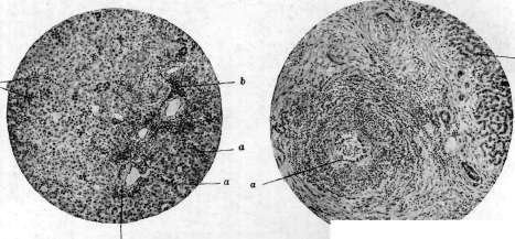
Figure 1.
Figure 2.
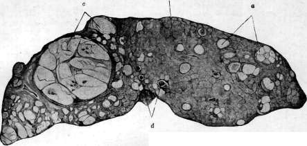
Figure 3.
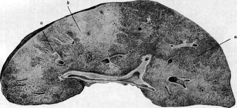
FIG. 4.
Fig. 1. Liver of a newborn. Hematopoiesis along the course of the interlobular tissue (b); a-accumulation of hemocytoblasts between the liver trabeculae. Figure 2. Polyarteritis nodosa. Proliferation of granulation elements around a small artery (a); the structure of the latter is strongly erased; around-infiltrate; b-liver. Figure 3. Cirrhosis and nodular hyperplasia (b) (regeneration) of the liver; numerous adenomas (a); at points c-cancerous nodules of the liver; d-thrombosis of a branch of the portal vein. Figure 4. Nutmeg liver: a-typical nutmeg pattern; b-area of significant fatty degeneration and slight nutmeg appearance. Yellow atrophy of the liver (see Acute yellow atrophy of the liver). In cases that are somewhat prolonged (subacute and rare chronic), along with phenomena of fatty degeneration and death of liver cells, reactive and reparative processes are observed, proliferation of small bile ducts, areas of regenerating liver cells, foci of infiltration with round cells of the connective tissue. Foci of regeneration sometimes appear macroscopically in the form of more palely colored, different in shape and size foci, slightly raised on the section (nodular hyperplasia of the liver). Recently, Beneke considers it possible in some cases to attribute necrotic changes of the liver of the acute yellow atrophy type to angioneurotic spasms of the hepatic artery. In the liver, small scattered necrotic foci of infectious-toxic origin are often observed, occurring mainly in typhoid fever, paratyphoid, scarlet fever, diphtheria, Botkin-Weil disease and some others. In cases of bile retention in the liver, very abundant small focal necroses sometimes arise (also in eclampsia). The liver cells themselves are most readily and easily subject to necrotic changes; Kupffer cells and elements of the supporting connective tissue are more resistant. The deposition of glycogen in the cells of the liver is subject to quantitative fluctuations; especially much glycogen is deposited in the cytoplasm and nuclei of cells in cases of disturbance of the general carbohydrate metabolism in the body (e.g. in diabetes mellitus).-Pathological pigmentations occur in the liver very frequently. Of endogenous pigments, the most frequently observed is the deposition in liver cells of fine-grained brown autochthonous pigment lipofuscin, which gives the liver a brown color in brown atrophy of the liver (see above). Very often the deposition in the liver of breakdown products of Hb in the form of iron-containing as well as non-iron-reacting pigments is observed, which is connected with the ability of the liver to destroy red blood cells. In this process, the widely distributed elements of the reticulo-endothelial system in the liver tissue in the form of Kupffer's stellate cells play an important role, playing an essential role not only in the process of destruction of red blood cells but apparently also in the formation of bile pigments from breakdown products of Hb. In cases of increased breakdown of red blood cells in poisoning with hydrogen arsenide and other hemolytic poisons, in anemias, especially pernicious, in some septic processes, in some forms of cirrhosis of the liver etc., accumulation of a large amount of iron-containing pigment hemosiderin is observed, mainly in Kupffer cells, and sometimes also in liver cells, especially in the peripheral parts of the lobules. In severe cases of hemosiderosis of the liver, it even takes on a rusty-brown color macroscopically. Particularly pronounced deposition of hemosiderin in the reticulo-endothelium of the liver occurs in chronic nutritional disorders in small children; in acute nutritional disorders this deposition is usually insignificant (Saito, Stephan, Dubois). In chronic malaria in the liver, mainly in Kupffer cells, a dark, almost black pigment is deposited, from which the liver can take on an ashen-gray color. In the deposition of bile pigments in large quantities (see Jaundice) the liver acquires a greenish-yellow color. In cases of obstructive jaundice, a sharp dilation of the bile ducts is observed (especially in the presence of factors hindering the outflow of bile from the larger ducts-stones, tumors etc.) and their filling with dense dark bile. In the smallest bile ducts, and sometimes even inside the liver cells, branching bile cylinders or thrombi are sometimes observed. The deposition of bile pigments in the liver cells occurs partly in a granular form, partly in the form of diffuse impregnation. The deposition of exogenous pigments in the liver is observed much less frequently; sometimes in cases of entry from the lungs into the blood of dust particles and their being carried by the blood current to the liver, their deposition in Kupffer cells results. For example, in the therapeutic administration of silver preparations, silver can be deposited in a granular form in Kupffer cells of the liver (argyria). The liver has a highly pronounced ability to regenerate; regenerative and hypertrophic changes are observed in it very frequently in cases of death of larger or smaller parts of the liver parenchyma, which occurs in cirrhoses, in acute yellow atrophy, in stagnation of blood in the liver, in necroses of infectious-toxic origin, in destruction of liver tissue by proliferating tumor nodes, parasites etc. The appearance of regenerated areas of liver tissue can be different: sometimes these are small microscopic foci, sometimes clearly defined foci with a paler gray, sometimes yellowish tint, of diverse shape, scattered throughout the liver, often reaching quite large sizes and appearing on the section in the form of nodules and nodes (nodular and adenomatous form of hyperplasia). Microscopically in such areas large hypertrophied liver cells are found, sometimes containing several nuclei, in other cases the proliferation of small bile ducts in the form of solid cords or narrow tubes is particularly pronounced. However, the question of the participation of bile duct epithelium in the regeneration of liver cells is still not resolved. In some forms of diseases of the blood and hematopoietic organs, the development of foci of hematopoietic tissue is observed in the liver, sometimes reaching such a degree that the entire organ appears significantly enlarged, e.g. in cases of leukemic and aleukemic lymphadenosis (see Leukemia). In some forms of severe anemias and in some infections in cases of development of extramedullary hematopoiesis, cellular foci consisting of erythroblasts, myeloblasts and myelocytes appear in the liver. The appearance in the liver of foci from young cellular forms of blood is connected with their autochthonous development right here in the liver tissue by analogy with the same process in the embryonic period [see separate table (pp. 567-568), figure 1]; at the same time Kupffer cells of the liver probably play a large role as a source of their formation. Recently, cases have been described characterized by intense proliferation in the liver of cells of the reticulo-endothelial apparatus (Kupffer cells), going in parallel with the proliferation of similar elements in other parts of the body (spleen, lymph nodes etc.); such processes as systemic diseases are assigned the name reticulo-endotheliosis (see). Acute inflammations of the liver (acute hepatitis) most often belong to exudative forms, namely to purulent inflammation with the development of abscesses. Most often the development of abscesses in the liver occurs secondarily, as a result of the spread of infectious agents causing purulent inflammation, through the branches of the portal vein (pyelephlebitic abscesses), sometimes with simultaneous thrombophlebitis of the latter. They have their source in purulent processes in the roots of the portal vein and develop mainly following purulent appendicitis, in dysentery, especially amoebic, etc. Much less frequently foci of purulent inflammation of the liver develop as a result of transfer here of infectious agents through other blood vessels: through the branches of the hepatic artery (e.g. in cases of general septic processes, ulcerative endocarditis etc.), hepatic vein, in newborns sometimes from infection of the umbilical wound through the umbilical vessels. Pylephlebitic abscesses of the liver can reach very large sizes and are usually represented in the form of groups of abscesses located along the course of the branches of the portal vein in the liver; often they merge with each other [see separate table (pp. 543-544), figure 2]. Sometimes in the center of such an abscess a vessel thrombosed with a purulent thrombus is noticeable. Another fairly frequent path of development of purulent inflammation of the liver is the system of bile ducts-cholangitic abscesses. In these cases purulent inflammation of the liver has its source in purulent cholangitis, and the infection penetrates into the bile passages from the gallbladder (e.g. in gallstone disease) or from the intestine (in this case the bacillus coli often has etiological significance in the development of abscesses in the liver). An important facilitating factor for the spread of purulent inflammation to the liver through the bile passages is difficulty in the outflow of bile through the bile ducts. Much less frequently purulent inflammation develops in the liver by the lymphogenous path from the periphery (e.g. in the transition of infection from the area of the porta hepatis in cholecystitis and cholangitis) or in secondary suppuration in nodes of tumors and parasites of the liver (e.g. hydatid of the liver).
Non-suppurative inflammations of the liver occur in sepsis and various infectious diseases, manifesting as the development, for the most part, of microscopic inflammatory cellular infiltrates, most often in the interlobular connective tissue, mainly around foci of degeneration and necrosis of tissue of infectious-toxic origin. Chronic inflammations of the liver belong predominantly to productive forms of inflammation, expressed by the proliferation of interstitial connective tissue; as is generally believed, they form the basis of the patho-anatomical picture of cirrhosis of the liver (see). Inflammatory processes of the liver are a very frequent cause of the development of similar changes in its serous cover as well—perihepatitis (see).--Tuberculosis of the liver most often manifests as scattered miliary, and sometimes submiliary, pale gray-colored tubercles in general miliary tuberculosis or as a secondary phenomenon in chronic tuberculosis of the lungs; in the vast majority of cases, along with tubercles in the liver, tuberculous, most often ulcerous, lesions of the intestines are also found. The spread of tuberculous infection to the liver occurs most often hematogenously, less often lymphogenously. The smallest tubercles, sometimes determinable only microscopically, are located most often in the interlobular layers of connective tissue, subsequently spreading to the peripheral, and then to more central, parts of the liver lobules. Tubercles in the liver tissue often fuse with each other, undergo caseous degeneration in the center, and sometimes are fibrously altered. Solitary tubercles, which sometimes reach very large sizes, are encountered much less frequently in the liver. When tubercles spread to the walls of the bile ducts and further dissemination of tuberculous changes occurs along them, scattered foci of various sizes and forms are observed in the liver, following the spread of the bile ducts in the form of mostly caseous-disintegrated, sometimes jaundiced-colored foci, sometimes with the presence of cavities in their center. The latter have the appearance of irregularly shaped cavities or channels, surrounded by caseous masses. In addition, the development of cirrhotic changes in the liver on the basis of its tuberculous lesion is observed. Actinomycosis of the liver usually develops secondarily, as a result of the spread of the process from surrounding parts of the liver, for example from the retroperitoneal cellular tissue, or as a result of the transfer of the ray fungus to the liver by blood, more often through the portal vein. Actinomycotic nodes, single or multiple, scattered in the liver tissue, sometimes reach very large sizes. They have a yellowish-gray color, a flabby consistency; their structure is spongy due to the fact that they consist of a mass of small abscesses among the granuloma tissue. In the pus and disintegrated tissue, druse of the ray fungus are found. In the granuloma nodes, leukocytes, macrophages rich in lipid inclusions, plasma cells are found in large quantities, and giant cells less often.--In glanders, leprosy, and plague, nodes consisting of the proliferation of granulation tissue typical for the given form are sometimes found in the liver. In some other infectious processes, especially in typhoid and paratyphoid fever, small granulomas (from lymphoid and epithelioid elements) are found in the liver, which rapidly undergo necrotic changes, so-called "pseudotubercles." In lymphogranulomatosis, along with changes in the lymph nodes, spleen, and other organs, proliferations of granulation tissue typical for this process are sometimes also found in the liver. Among benign tumors of the liver, cavernous angiomas or cavernomas are most often encountered, usually considered as congenital malformations of the vascular system of the liver (hamartomas). They are scattered singly or often in multiple numbers in the liver tissue, often located under the serous cover and shining through the surface in the form of foci of various sizes and shapes of dark red color, spongy structure, exuding dark blood from the surface of the incision. Thrombi often form in the cavities of cavernomas, which undergo organization, as a result of which they become empty and are replaced by connective tissue. Adenomas occur in the form of single or multiple nodes, which rarely reach large sizes. Sometimes they are rather difficult to differentiate from adenomatous hyperplasias that arise during the regeneration of liver tissue, mostly of a compensatory nature, in various cases of exclusion from function or death of parts of the organ's parenchyma. From such hyperplasias, liver adenomas, which are usually nodes colored pale yellow, differ by their greater demarcation from the surrounding liver tissue, the elements of which appear compressed and atrophied around the adenoma. In structure, liver adenomas consist of liver cells, showing an indistinct acinar structure. The large, light cells of such adenomas, rich in lipid inclusions, sometimes exhibit the ability to secrete bile. In other cases, liver adenomas are built from the epithelium of the bile ducts, have a tubular structure; adenomatous proliferations are lined with cubic or cylindrical epithelium, sometimes forming papillae. Cysts of the liver belong mainly to developmental malformations of the liver. Cystic lymphangiomas and dermoid cysts of the liver are rarely encountered. Malignant tumors of the liver include rarely occurring primary sarcomas from round, spindle-shaped, and other cells, as well as angiosarcomas and hemangio-endotheliomas, which sometimes reach very large sizes and without sharp borders infiltrate the liver tissue. Secondary metastatic sarcomas of the liver are encountered much more frequently and can have various structures. Metastases of malignant melanoma reach particularly large sizes with primary tumors of this kind in the skin or in the eye. In the liver, the development of individual nodes of melanoma reaching very large sizes, pigmented to varying degrees (sometimes almost black or dark brown), is observed, or there occurs a rather uniform diffuse infiltration of the liver by the neoplasm in the form of strands and infiltrates following the capillaries and compressing the parenchymal cells.--Primary cancer of the liver is rarely encountered; in some cases, a single, large node of the tumor is found in the liver, sometimes with smaller secondary nodes around it. In other cases, there are multiple nodes or a more diffuse infiltration of the liver tissue by the tumor. Very often, at the same time, cirrhotic changes of the liver are present, for which reason some associate the development of cancer with the regenerative changes of the liver, sometimes very sharply expressed, and foci of nodular hyperplasia of the liver observed in cirrhosis [see separate table (pp. 567-568), figure 3]. Macroscopically, the nodes of primary liver cancer are characterized by a pale yellowish or almost white color, sometimes with a greenish tint. They are usually of rather flabby consistency, often penetrated by hemorrhages and foci of necrosis. Primary cancers of the liver in some cases originate from liver cells (carcinoma hepatocellulare), in others from the epithelium of the intrahepatic bile ducts (carcinoma cholangocellulare). In the first case, the cancers have either an alveolar structure, where groups of rather large epithelial cells of the tumor are separated by layers of connective tissue stroma, or the arrangement of tumor cells has a more trabecular character. Sometimes the tumor cells are arranged in the form of rosettes, forming in the center a kind of canal filled with a mass resembling bile. The tumor cells themselves can be very polymorphic, often polygonal in shape, of rather varied sizes; sometimes very large, even multinucleated cells are encountered. Cancers originating from the epithelium of the bile ducts mostly have the structure of adenocarcinomas; their epithelial cells are cubic or cylindrical in shape, arranged in several rows in places. This form of primary liver cancer is more often encountered in combination with cirrhosis of the liver than the first form. Primary cancers of the liver can give metastases mainly through the circulatory system both in the liver itself and outside it (especially in the lungs, in bones).--Secondary, metastatic cancers are encountered in the liver very often. Cancers of the bile-duct system metastasize to the liver most often. The most frequent path of metastasis is through the portal vein system, less often through the hepatic artery or retrogradely through lymphatic pathways. Sometimes direct infiltration of the liver by cancer from neighboring organs, mainly from the stomach or gallbladder, is also observed. Metastatic cancers of the liver are often encountered in the form of numerous nodes reaching sometimes enormous sizes, sometimes with an umbilical depression in the middle (due to necrosis of the central parts of the nodes). Along with necrotic changes in metastatic cancer nodes of the liver, hemorrhages are often observed, which sometimes give rise to severe bleeding into the abdominal cavity. The disintegration of tumor nodes is often accompanied by their softening and even the formation of cystic cavities in the center. In rare cases, primary hypernephroid tumors, built from misplaced elements of adrenal cortex tissue, are encountered in the liver. Tumors of mixed structure (from epithelium, cartilage, and connective tissue) are rarely encountered; isolated cases of ectopic chorionepitheliomas of the liver have been described.
N. Anichkov. Of the parasites of the L., the most frequently encountered and of greatest importance is the echinococcus (see). In addition, a number of trematodes are localized in the L.—Opisthorchis felineus (widespread mainly in Western Siberia in the basin of the Ob River; see Opisthorchiasis), Clonorchis sinensis (found in the Far East; see Clonorchiasis), Fasciola hepatica and Dicrocoelium lanceatum (parasites of the liver of cattle, sometimes also found in humans). The L. is also one of the possible sites of localization of the larva of the armed (pig) tapeworm—Taenia solium (Cysticercus cellulosae). For the ascarid, the L. is an obligatory stage in the migration of larvae, which pass here from the portal system to the vena cava system. In individual cases, adult ascarids can also be found in the L., having crawled through the bile ducts from the intestine (see Ascarids). Cases of finding larvae of the family Linguatulidae in the human L. have been described (see). Coccidia have been described as a casuistic finding. Abscesses of the L. can be caused by the dysentery amoeba (Entamoeba histolytica). VI. Functional diagnosis of the liver. The study of the liver patient by means of only physical methods of inspection, percussion, and palpation is completely insufficient and very often does not allow recognition of a disease of the L., in particular to determine deviations from the norm of the numerous and complex functions of this organ and to give an idea of its functional state. From the analysis of data on the physiology and pathology of the L. (see physiology and pathology), it is clear how difficult it is in the clinic to take into account and differentiate the work of such a complex and diversely functioning organ. It is clear that there cannot be a single method that would give indications and an answer about the state of such complex functions. To somewhat simplify this complex task, one can schematize the liver functions as follows. All the work of the L. is divided into two large parts: external—bile secretion and internal—all other chemical processes occurring in the L. Disorders of liver circulation occupy a special place. External work of the L. Regarding disorders of bile secretion function, besides clinical symptoms reflecting tissue jaundice, we judge in four places: by changes in the properties of urine, feces, duodenal contents, and blood. Anomalies in bile secretion can be detected at all four of these points and sometimes with sufficient accuracy. In order to establish functional weakness of the L. and more clearly reveal the inability of the L. to retain urobilinogen, Falta proposed loading the L. by administering 3 g of purified ox bile (fel tauri depuratum) on an empty stomach. Falta believes that taking this portion of ox bile causes alimentary urobilinuria only in liver patients, and namely a more significant urobilinuria appears in atrophic and hypertrophic cirrhosis, in syphilitic and non-syphilitic acute hepatitis, in hemolytic jaundice, etc. Under these conditions, urobilinuria does not occur in stagnant L. and in persons with a healthy L. Control studies by Lepene gave indefinite results, and Retzlaff also expresses skepticism; Schutz considers it possible to replace ox bile with 45 cm3 of a 10-15% alcoholic solution of chlorophyll, since chlorophyll has a pyrrole nucleus similar in structure to heme and in the intestine breaks down into bodies similar to urobilinogen. 4-12 hours after taking chlorophyll, a positive urobilinogen test supposedly occurs only in lesions of the L., in alcohol poisoning, after prolonged chloroform anesthesia. If these data were confirmed, then this alimentary urobilinuria could be considered the most sensitive and best test for determining the functional capacity of the L. Changes in the properties of urine. The presence of bilirubin in the urine indicates a degree of jaundice that is much earlier detected in the blood and tissues. Thanks to the quantitative determination of bilirubin in blood serum, the relationship between bilirubinemia, tissue jaundice, and bilirubinuria has now been clarified. Since the blood shows a disturbance of bilirubin metabolism in the form of an increase in bilirubin level earlier than in all other organs, it is clear that we first judge the appearance of jaundice by bilirubinemia. It turned out that for the appearance of tissue jaundice, i.e., visible coloring of the skin, the amount of bilirubin must reach a certain concentration; the latter must be even greater for bilirubin to appear in the urine. Hence it follows that the appearance of bilirubin in the urine already indicates a significant deviation from the norm in the bile-secreting function of the L. Therefore, in the functional diagnosis of the L., more importance is attached to another pigment—urobilinogen. Urobilinogen is, as it is called, the pigment of a diseased L. Most authors believe that bilirubin, which has flowed into the intestine with bile, under the influence of intestinal bacteria, turns into urobilinogen and in a large part is absorbed into the blood and retained by the L. The small amount of urobilin and urobilinogen found in the urine of healthy individuals enters the great circulation of blood not by the portal route or is passed by the liver. The appearance of a large amount of urobilinogen in the urine definitely indicates a violation of the function of the L., because under normal conditions liver cells capture urobilinogen absorbed from the intestine, partly perhaps using it for the construction of the blood coloring substance, partly excreting it back into the intestine as such or having turned it back into bilirubin. If one recognizes that the formation of urobilinogen is possible outside the intestine in tissues (histiogenous), then its entry into the intestine can occur both from the L. and from the blood (Bra 16 et Garban). In any case, experimental research on dogs with Eck's fistula, dogs poisoned with phosphorus and deprived of carbohydrates, shows that it is the L. that is the main regulator of the urobilin cycle. Thus, urobilinuria is an early sign of liver insufficiency. Clinical observations daily confirm that urobilin is a very fine reagent for all kinds of changes in the L., and apparently in lesions of the L., its function of retaining urobilinogen is impaired earlier than all other functions. Adler drew attention to the constant ratio in a normal L. between urobilin in urine and urobilin in feces (1:10 to 1:30). If in any case we cannot speak of an independent disease of the L. in urobilinuria, then undoubtedly we can always assume a pathological change in it (e.g., malaria and other infections). Bile acids. Recently, besides pigments (bilirubin and urobilin), attention is paid to the content of bile acids in urine, which are a specific product of the secretory work of the L. A number of authors (Gilbert, H. Muller) point to the possibility of the existence of the so-called dissociated jaundice, i.e., to the dissociation of the retention of various components of bile; thus, they assume that the retention of bile pigments does not always occur in parallel with the retention of bile acids. For example, in the so-called catarrhal jaundice (acute hepatitis), the delay in the excretion of bile acids is observed for a longer time than the delay in the excretion of bile pigments. In Laennec's cirrhosis, neoplasms of the L., the same «77 dissociation was observed. However, the existence of dissociated jaundice is considered unproven by other authors. According to them, the corresponding observations are explained by the fact that the methods used to determine bile acids are very inaccurate. The presence of bile acids in urine definitely indicates a lesion of the L. Müller considers the appearance of bile acids in urine an early sign of impaired liver function, just as albuminuria indicates kidney damage. Bile acids sharply reduce surface tension, and they still have an effect at a dilution of 1:40,000 (Müller). The surface tension of urine (as shown by Bechhold and Reiner) depends on the colloids and semi-colloids dissolved in urine. Since the amount of colloids excreted with normal urine is proportional to the amount of crystalloids excreted (Lichtwitz), after bringing the urine to a certain specific gravity, the surface tension of normal urine is more or less constant. To determine surface tension, fresh urine of acidic reaction is taken at a certain temperature (15-18°) after bringing its specific gravity to 1,010. The determination of surface tension is carried out by the drop method using a Traube stalagmometer. Hay's test with sulfur color for determining bile acids in urine turned out to be less sensitive but simpler in technique. It consists of the following. Freshly released, thoroughly filtered urine is poured into a small conical flask, and on its surface is carefully sprinkled with sulfur powder, well-dried and degreased. Due to the presence of bile acids in urine, as mentioned above, the surface tension is lowered, and the wetted sulfur gradually falls to the bottom of the vessel. The determination of urobilin and bile acids in urine can serve as an indication of the functional state of the L.
Even in cases where the patient does not have jaundice, the symptoms from the L. are weakly expressed and physical examination gives a negative result, even then, if urobilin and bile acids or only urobilin are present in the urine, it can be stated with certainty that the L. is affected. By these methods, damage to the L. can be detected in various infections and intoxications. For example, if the origin of ascites is unclear (tuberculous peritonitis or cirrhosis), then based solely on the fact of the presence of urobilin and bile acids in the urine, it can be stated that the L. participates in this pathological process. The examination of feces has much less significance than the analysis of urine for the functional diagnosis of the L. With small delays in bile flow, which most often occur in diseases of the L., there may not be any noticeable change in the color of the feces. With the introduction of the duodenal probe, which makes it possible to obtain bile from its very source, the interest in the examination of feces in liver diseases seems to have significantly decreased. However, acholic stool due to the absence of urobilinogen in the feces gives a definite indication of mechanical jaundice with complete closure of the d. choledochi. "On the contrary, excessively colored, rich in urobilinogen stools are observed in hemolytic jaundice. Coprological data acquire special importance in determining the absorption of fats. Bile emulsifies fats and, although it itself does not have the ability to break them down, it activates this ability in the pancreatic juice. The study of steatorrhea acquires diagnostic and prognostic interest in disorders of bile secretion function. Hyposteatolysis is one of the important signs in the entire liver symptomocomplex, all the more so that detailed examination of the feces allows differentiation of liver steatorrhea from pancreatic (see Pancreas). Examination of duodenal contents. Despite the fact that by probing the duodenum we do not obtain a pure secretion of the L. (duodenal contents contains pancreatic, gastric, and duodenal juices), this method still gives a great deal for the diagnosis and treatment of diseases of the L. and especially of the gallbladder (see Duodenal probe). Colorimetric examination of pigments and cholesterol, determination of bile acids allows in some cases to speak of one or another form of jaundice (for example, decrease in bilirubin at the height of the disease in acute hepatitis, increase in bilirubin in hemolytic jaundice, absence of pigments in complete obstruction of the duct, etc.). An interesting method are tests for the excretion of foreign bodies, mainly various dyes. There is no doubt that dye tests are not a sufficiently subtle method for functional diagnosis of the liver. Already Burkhe (Biirker) experimentally proved the transition of indigo carmine into bile, and Brauer (Bгaueг) of methylene blue. The first experiments with the use of dyes to determine the functional capacity of the L. were carried out by French authors (Chauffard, Castaigne): on the basis of fluctuations in the excretion of methylene blue in the urine after its subcutaneous injection, they tried to judge the condition of the L. More reliable were those methods by which the excretion of dyes with bile is directly determined. American works (Abel, Rowntree) showed that after subcutaneous injection of tetrachlorophthalein, this dye is retained by a normal L. and excreted into the intestine, while the urine remains colorless. In diseases of the L., the dye partially passes into the urine, and the amount of dye entering the intestine decreases. The amount of dye in the stool, determined colorimetrically, allows one to monitor the degree of damage to the L. It is interesting that at present, with the introduction of a duodenal probe, it is possible to observe the excretion of dye from the L. itself (chromocholecystoscopy). Rosenthal (Ro-senthal) used a 2% solution of methylene blue in an amount of 5 cm3 subcutaneously, and in case of even minor changes in the L., an acceleration of dye excretion was noted, which in normal cases appeared after 60-70 min. However, the works of Saxl, Scherf (Saxl, Scheri) and N. S. Smirnov undermined the significance of this method, since it turned out that methylene blue is excreted by the stomach earlier than by the L. (already after 3-30 min.). Therefore, a number of authors began to work with indigo carmine, which is not excreted by the stomach. This dye was successfully tested by many (Smirnov, Lepene, Hesse, Werner, Hatiegan and others), who noted delayed excretion of the dye in some diseases of the L., the opposite of what was observed with methylene blue. Such inconsistent results when using different dyes have not yet found a satisfactory explanation. The research methodology with indigo carmine is as follows. After the probe is inserted and a constant flow of pure duodenal juice is established, 2 cm3 of a 1% solution of indigo carmine is injected into the vein (in children, intramuscular administration of 0.2 g of indigo carmine in 5 cm3 of physiological solution can be used); and bile is collected in five-minute portions until green coloring appears. According to the data of Lepene and the clinic of Konchalovsky, it appeared after 15-45 minutes after injection. Thus, the reaction is considered negative if coloring appears within the first 45 minutes. Healthy children excrete indigo after 25-30 minutes (Bossert)", delay indicates a violation of L. function. Instead of indigo carmine, 3 cm3 of a 2% methylene blue solution can be injected subcutaneously and looked for in five-minute portions of bile (macroscopically and chemically). In normal conditions, blue coloring of bile appears after 40-60 minutes, whereas in liver function disorders, coloring appears already after 10-40 minutes (Bossert). Along with indigo carmine, research on albuminuria, i.e., determination of protein in bile according to Raue, can be performed. In normal duodenal juice, coagulating protein bodies are not contained in significant amounts. In pathological cases, these proteins apparently appear either as an inflammatory exudate (angiocholitis) or by penetration of protein substances through altered liver cells into the bile ducts (salvarsan jaundice, hepatitis). In order to make a final conclusion about the diagnostic and prognostic value of dye tests, further studies are still required. Due to their simplicity, they may be of interest to the practicing physician. Blood examination to determine deviations in the activity of the L. has acquired particular importance in recent times. There is no doubt that the diagnostic value of finding an increased content of bilirubin in the blood serum is especially great. It is known that in jaundices bilirubin appears in the serum relatively quickly, but it is bound by blood proteins, so its amount must reach certain limits to color the skin and mucous membranes; the level of bilirubin in the blood must rise even higher for bile pigments to appear in the urine. Thus, blood examination reveals jaundice earlier than examination of the patient and urine analysis. Furthermore, it is difficult to establish the degree of bile retention by simple inspection, which is very valuable, especially for judging the increase or decrease of jaundice. The same color of the blood serum is observed with very different amounts of bilirubin in it. It is interesting that bilirubin is found in the serum in various febrile conditions, and its appearance corresponds not to the height of the temperature, but to the strength of the infection. In cases of residual cardiac activity, an increase in van den Bergh's reaction is found in the serum (congestive L.), which disappears with improvement in the blood circulation (similar to congestive albuminuria). For details on bilirubinemia, see Bilirubinemia, Diazoreactions in blood, and Jaundice. Less definite results are obtained by determining cholesterol, urobilin, and bile acids in the serum. In view of the enormous influence of the L. on the processes of blood formation and clotting, its participation in the formation of fibrinogen, kinase, antithrombin, all methods that take into account these processes are important: blood clotting time, bleeding time, amount of fibrinogen, retraction of the clot, thrombocytosis, Rumpel-Leede symptom, etc. Finally, by examining a drop of blood in the ultramicroscope by observing hemokonias (shining tiny droplets of fat), one can judge the violation of bile secretion function, since the absence of bile entering the intestine leads to almost complete disappearance of hemokonias from the blood, which is not observed in violation of pancreatic function. Internal work of the L. Examination of carbohydrate metabolism. Of all the internal functions of the L., the most important is its glycogenic function. But the accumulation of glycogen by the L. and its regulation of blood sugar has a certain limit. It is sufficient for even a healthy person to take 300 g of glucose to develop hyperglycemia and glycosuria. The limit of this absorption is of course lowered in a diseased L. Hence arose the idea of using provocative glycosuria to test the functions of the L. The patient is given glucose and it is looked for in the urine, determining its amount and time of appearance.
This method, first proposed by Kohlrath, was considered by him as indicating a disorder of portal circulation: specifically, he assumed that alimentary glycosuria developed when blood from the portal vein deviated from its normal course and passed through collaterals. At present, there is no doubt that this alimentary glycosuria indicates damage to the cells of the liver, specifically their inability to fix glucose. For testing, glucose, levulose, and galactose are used. The latest clinical observations and numerous experimental studies (with Eck fistulas, in phosphorus poisoning, and on the isolated liver) have shown that the liver to a large extent regulates the sugar content in the blood; therefore, at present, when sugar is administered internally, it has become important to determine not only its presence in the urine but also in the blood; moreover, it is important in connection with glycosuria to observe the fluctuations of the blood sugar curve. The latest works show that even small doses of dextrose (up to 20 g) are sufficient to cause hyperglycemia. It is interesting that after a small portion of glucose, a new portion, even a larger one, no longer raises the blood sugar; this phenomenon is not observed in diabetics, on the contrary, in them after a second portion of dextrose the rise of the sugar curve is even more pronounced (M. P. Konchalovsky). In relation to healthy individuals, it must be assumed that the first administration of glucose so changes the function of the liver cells that their activity is enhanced and activated in terms of glycogen fixation during subsequent carbohydrate loading. Numerous tests with various sugars (glucose, levulose, and galactose) have shown that in some cases the liver slowly takes up sugar, while in others it loses the ability to fix glycogen, and this glycogenic function is apparently impaired where the parenchyma of the organ itself is affected (in diffuse processes). One can think that these tests, even if they do not give a complete picture of the glycogenic function of the liver, at least reflect a disturbance of hepatic regulation in relation to blood sugar content and thus give a deeper understanding of liver function than the usual provocative glycosurias. In children (especially in early childhood), tolerance to sugar is relatively higher than in adults; thus, glycosuria occurs in a child only when 6-7 g of glucose per kg of body weight is administered, whereas for an adult 3-4 g is sufficient (Evoli, Nobecourt, Terrien). Investigation of protein metabolism. Tests for investigating protein metabolism in liver patients, namely the determination of urea, ammonia, amino acids, creatine, and creatinine, have not yet yielded results that could have diagnostic value, just as the determination of fibrinogen in the blood (according to some authors, the liver can be considered as an organ in which fibrinogen is formed). It is known that the liver plays a predominant role in the formation of urea. However, a decrease in urea excretion does not justify speaking of insufficient liver function. When the liver is affected, polypeptides absorbed by the intestine do not normally break down but remain in intermediate stages without final transformation into urea; then they appear in the urine in the form of amino acids and ammonia. More importance is attached to establishing the relationship between urea nitrogen and total nitrogen in the urine (azoturic ratio). This ratio is lowered in cases where protein breakdown is incomplete; it can serve as an indicator of liver insufficiency. Maillard, and then Lanzenberg, proposed establishing not only the relationship between urea nitrogen and total nitrogen, but also the ratio of ammonia nitrogen to urea nitrogen (coefficient of urea-forming insufficiency). But on the basis of recent works by Nash and Benedict, it is known that the ammonia found in the urine does not come from the blood, it is formed in the kidneys themselves to reduce acidosis, so common in liver patients. The determination of urine ammonia should therefore be corrected, taking into account the pH of the urine, i.e., ionic acidity. With this correction, the aforementioned urea-forming coefficient acquires greater value. Clinic has also paid attention to the relationship of urea to total nitrogen in the blood serum (azoturic coefficient of serum). This coefficient is lowered in liver insufficiency (whereas it is elevated in nephritis) due to incomplete breakdown of proteins to urea. True, it changes only in severe liver diseases. Brodin proposed the following modification of the calculation: instead of dividing urea nitrogen by total nitrogen (to establish the ratio), he subtracts urea nitrogen from total nitrogen and obtains a figure of residual nitrogen, which increases in liver insufficiency. Incomplete breakdown of proteins to urea may manifest in the urine by the appearance of one of the signs of this incomplete breakdown, namely amino acids. Therefore, the determination of this aminoaciduria and the establishment of the ratio of amino acids to total nitrogen, especially after their preliminary administration, is interesting. This provocative aminoaciduria test (Labbe and Bith) shows an increase in this ratio in liver patients. The proteopectic function of the liver (hemoclastic crisis). In the normal state, during the digestion of protein food, not yet completely broken down protein substances pass through the intestinal mucosa and penetrate into the portal circulation. The normal liver retains these substances. On the contrary, if the liver is damaged, they can pass into the general circulation and, as heterogeneous proteins, cause a hemoclastic shock similar to that obtained by intravenous administration of peptone. This special function of the liver became known in 1920 after the work of Widal, Abrami, and Iancovesco, who called it the proteopectic function. The test consists of the following. After examining the fasting blood of the subject, they are given 200 g of milk to drink quickly, and blood examination is repeated every twenty minutes. If the liver function is insufficient, a hemoclastic crisis appears, often in the first hour itself: it is characterized by a decrease in the number of white blood cells with a shift toward lymphocytosis (in contrast to the normal digestive leukocytosis), a decrease in blood pressure, an increase in blood clotting, and a decrease in the refractometric index. At present, this test has been sufficiently tested in many clinics and has not acquired the practical value that French authors attributed to it, as it proved to be very unreliable. In relation to small children, it should be emphasized that in early childhood, the leukopenic phase after food intake (even maternal milk, not foreign milk) is usual and normal; leukopenia and leukocytosis throughout the entire period of digestion as well as during the period of hunger replace each other in a definite sequence under the influence of the autonomic nervous system, and the connection with food should be rejected here. Thus, despite the opinion of some authors that the liver still plays a role in the mechanism of leukopenia after milk intake, it must be recognized that the study of the proteopectic function of the liver by means of the Widal crisis is not applicable to children. The liver and water metabolism. There is no doubt that the liver, being a huge reservoir of blood and possessing a special sphincter-like muscular mechanism in the hepatic veins, has a regulating influence on water metabolism in the body. Often in liver patients we can observe fluctuations in diuresis: anisuria - irregular diuresis from day to day, nocturia - predominance of nocturnal diuresis over diurnal, opsiauria - delay of food diuresis, with fractional collection of urine, dilution of blood and a drop in hemoglobin after taking 1,500 g of water, etc. The liver's relationship to blood formation. The influence of the liver on blood clotting is not in doubt; the liver is apparently the main site of fibrinogen formation. Whipple and Hurwitz report on the basis of their clinical observations a decrease in fibrinogen in liver patients. However, due to contradictory data from other authors, the test for fibrinogen cannot be considered a reliable functional test of the liver. In some liver patients, a change in bleeding time, decrease in blood platelets, instability of the blood clot, and so on can be ascertained. From the brief review of functional tests of the liver, the conclusion must be drawn that there is and cannot be a single simple method that would give the clinic a definite indication of functional insufficiency of this complex organ. The clinical concept of insufficiency itself for the liver does not yet appear clearly defined to this day.
Indeed, we know the formidable picture of cholemia with severe toxic phenomena in severe jaundice and acute yellow atrophy of the liver, but modern clinical medicine is only beginning to outline unclear images of incomplete failures of liver functions in the form of 'hepatism,' minor insufficiency of the L., and others. For childhood, all methods of functional research of the liver should in general be considered imperfect; they are often not sufficiently precise, in the hands of various researchers give different results, figures fluctuate within rather wide limits, and usually not the functional capacity of the liver as a whole is studied, but only some one side of its work. Therefore, conclusions from research should be treated with restraint and one should try to conduct repeated research and with regard to various aspects of the liver's activity.
M. Konchalovsky. VI. Surgical diseases of the liver. Injuries to the liver are closed and open. Open wounds to the liver are caused by cold and firearms, as well as by pointed objects such as stakes, animal horns, etc. Subcutaneous injuries result from 1) a sudden direct blow to the area of the liver, 2) crushing of the liver between hard objects (being run over by a vehicle, compression by railway cars, etc.), 3) par contrecoup from falling from a height onto the head, knees, or buttocks. When falling from a considerable height onto the legs, the liver may be torn away from the ligaments holding it (lig. susp. and corona hepatis). The liver is injured more often than other parenchymatous organs. According to Eliason's data, out of 315 cases of injuries to parenchymatous organs, the liver was injured in 189 cases. According to Edler, for 176 ruptures of the kidneys, spleen, and pancreas, there are 189 cases of liver ruptures. According to Geill, out of 494 cases of ruptures of internal organs based on data from the Vienna Institute of Forensic Medicine for the period from 1878 to 1897, there are: liver-59.9%, lungs-42.3%, spleen-<3%, kidneys-21.5%, heart-18.2%, intestine-11.1%, stomach-7.1%, urinary bladder-4.4%, pancreas-4.4%. The right lobe is injured much more often than the left (according to Ker 6 times more often). In Thole's cases, out of 182 injuries to the right lobe, there were only 34 injuries to the left lobe. The convex surface is affected twice as often as the lower one (according to Thole 75:48 for the right lobe and 12:3 for the left). Sagittal ruptures are more common (according to Geil out of 122 cases-79 sagittal ruptures). In addition to sagittal ones, vertical and transverse ruptures of the liver are observed. Further, there are either partially connected or completely detached avulsions of pieces of the liver of various sizes. Ruptures can be single or multiple. Usually only one liver is injured, but sometimes the injury to the liver is complicated by rupture of other organs of the abdominal and thoracic cavities. Factors predisposing to rupture of the liver, in addition to the large size of the organ, are: 1) its histological structure and topography; 2) enlargement of the liver during digestion; 3) pathological position (prolapse in connection with exudative pleurisy or emphysema of the lungs); 4) pathological changes in the liver tissue in connection with infectious diseases, fatty degeneration in alcoholics and cachectics, amyloidosis, tuberculosis, malaria, syphilis, etc. From a pathological-anatomical point of view, subcutaneous injuries to the liver are divided into: 1) ruptures of the liver parenchyma (in one or several places) with tears of the capsule; 2) separation of the capsule from the liver tissue with the formation of a subcapsular hematoma; 3) internal contusions and hemorrhages into the liver tissue (apoplexy) with the formation of blood cavities and cysts, which often become purulent. The clinical picture of subcutaneous ruptures of the liver in the first hours after injury has no characteristic features and does not differ from the picture of injury to other organs of the abdominal cavity. In rare cases, signs of liver rupture are completely absent or are mild. The general picture is characterized by shock with pallor of the skin, weak pulse, and shallow breathing. In prolonged (over 6 hours) forms of shock, it is necessary to consider internal bleeding or peritonitis. Along with a frequent (up to 150) pulse of very weak filling, a slowed pulse is sometimes observed (in case of liver tissue rupture-Finsterer) depending on the absorption of bile acids. The temperature drops to 36.5-36° and even to 34° (Thole). Among other symptoms of closed liver injury, painful tension of the muscles of the anterior abdominal wall is noted. Sometimes this defense has a diffuse character, but it is especially pronounced in the epigastric or hypochondriac regions. Often there is radiation of pain to the right shoulder or scapula. Percussion reveals dullness corresponding to the spread of internal hemorrhage. In case of liver rupture, blood usually accumulates in the right fossa iliaca, while hemorrhage from the spleen descends to the pelvis (Malgaigne, Terrier). Kep does not attach great practical importance to this moment, while Lejars considers indications for intervention to depend also on 'dullness of the right iliac fossa indicating poured-out blood'. Kronlein emphasizes the importance for diagnosis of dullness in the lateral and lower parts of the abdomen. Sometimes (in 21%) jaundice is observed, usually 4-5 days after injury. Vomiting of bile masses is not characteristic for injury to the liver alone, as this symptom is more often observed in ruptures of the gastrointestinal tract. Bloody stool as an early symptom is extremely rare. In urine, sugar and bile pigments are sometimes found, but these signs are not specific for liver ruptures. Peritoneal irritation, meteorism, vomiting are either secondary, already late signs of liver injury, or if they appear early, in the first 2-4 hours after trauma, then it is necessary to think of combined rupture of the liver and hollow organs. Cough, sometimes very distressing, and difficult breathing are caused either reflexively from the liver or its ligamentous apparatus, or are connected with a simultaneous fracture of ribs and injury to the diaphragm, pleura, and lungs. Open injuries to the liver do not present difficulties in recognition. Thole distinguishes between stab and gunshot wounds to the liver. Both can be simple or complicated by infection or injury to other organs. Infection can come from the wound canal or penetrate into the liver later in connection with injury to hollow organs. The direction and size of the wound canal can be various, and for many surgeons, the protrusion of omentum or intestinal loop outward was considered a decisive indication for surgery. It should be emphasized that it is urgently necessary to expand the wound canal to the parietal peritoneum in cases of abdominal injury, as often the omentum, protruding, gets stuck in the wound canal without appearing outside. Bleeding from the liver in penetrating wounds can be very abundant and in most cases is internal. It must be remembered that profuse bleeding can also come from the vessels of the anterior abdominal wall. Traumatic shock is almost always present in open injuries to the liver, and along with a reflex factor, blood loss also plays a role in its origin. Lejars in cases of stab wound with a narrow canal, wound from a revolver bullet or fragment, with a good pulse, moderate pain, and not sharply expressed dullness recommends a wait-and-see approach, which is hardly correct. In view of the need to prevent infection and to avoid subsequent complications directly related to liver injury, it is necessary in each case to expand the wound canal and proceed further depending on the nature of the injury. Gunshot wounds can be through-and-through and blind. Intervention in them aims not at removal of the bullet or fragment, but exclusively at hemostasis or suturing of the accompanying injury to hollow organs. If during the operation removal of the bullet is a simple procedure, it is removed, otherwise only urgent measures are taken. Knife and bullet wounds to the liver most often occur through the anterior abdominal wall, then through the lateral walls of the abdomen and in rare cases through the posterior surface of the chest. According to Giordano's statistics from 1922, out of 257 cases of liver injuries, there are: 138 cut-stab wounds with a mortality rate of 20.29%, 70 gunshot wounds with a mortality rate of 44.28%, 49 ruptures with a mortality rate of 48.97%. According to Thole's data (1910), out of 752 liver injuries, there are: 232 stab wounds with 72 deaths, 200 gunshot wounds with 98 deaths, 260 ruptures with 160 deaths. Thus, subcutaneous ruptures and gunshot wounds to the liver have the highest mortality rate. Cut wounds are most favorable in terms of prognosis. The cardinal signs indicating surgical intervention are mainly signs of internal bleeding and peritoneal phenomena. The earlier the operation is undertaken, the better the prognosis quoad vitam. The operation should consist in obtaining rapid and wide access to the liver, especially to the area of injury, in examining neighboring organs to exclude combined injury, and in stopping bleeding. Bleeding is stopped either by suturing or by tamponade with gauze, or by omentum (free tamponades or on a pedicle). Subsequently, depending on the nature of the injury, the abdominal cavity is either sutured tightly or tampons are left. A careful suture should be applied to the abdominal wall to avoid postoperative wound dehiscence. During and after the operation, physiological solution is administered, and recently, reinfusion of blood that has flowed into the abdominal cavity, as well as transfusion from suitable donors, has been successfully used. Among complications in the postoperative period, subsequent bleeding, peritonitis, pneumonia, thrombosis, embolism, subdiaphragmatic and liver abscesses, dehiscence of the abdominal wound with protrusion of internal organs are observed. Abscesses of the liver (abscessus hepatis, hepatitis suppurativa).
One of the cardinal symptoms of abscesses of the liver is purulent fever, which however is not always characteristic; the temperature curve is very variable, and in cases of so-called quotidian fever, fever is usually absent. A typical fever should be considered intermittent with high chills and subsequent sharp drops in temperature. According to Mattes (1924), the fever can have an amazingly regular character, e.g., the character of quotidiana in malaria; with prolonged existence of abscesses, fever may be completely absent. Yellowish discoloration of the skin, ashen color of the face with subicteric discoloration of the sclerae, progressive emaciation, small and insufficiently tense pulse complete the picture of the disease. In the blood - leukopenia (in the absence of pure dysentery infection) or leukocytosis with a shift to the left (in combined amebic and pyogenic infection). Of the local manifestations of the disease, it is necessary to note enlargement of the liver, and the direction of enlargement depends on the localization of the abscess. Most often the right lobe of the liver enlarges. Abscesses are located either on the convex surface (80%), raising its upper border, compressing the lung and causing phenomena of sympathetic pleurisy with corresponding clinical signs, or they spread downward, in rare cases reaching the pelvis. The area of the liver is painful in abscesses. Pains radiating to the right shoulder and scapula are noted, the body is inclined to the right in most cases, and as a result the gait of such patients changes, as indicated by Romberg (Romberg). Tension of the muscles of the abdominal wall is constant with multiple abscesses of the liver, while with single abscesses it depends on the location on the anterior-convex and lower surfaces. Pains intensify in connection with perihpatitis, which is sometimes accompanied by a friction rub in the area of the liver. An auxiliary sign for the diagnosis of liver abscess is the high position and immobility of the right dome of the diaphragm, determined by X-ray examination. Jaundice is a relatively rare symptom in liver abscesses. It is caused either by accompanying cholangitis or compression of the bile ducts by a single liver abscess. The abscess must be differentiated, especially in its chronic course, from echinococcus, a breaking down gumma, a neoplasm, from bilious pneumonia, from paranephric abscess and even from abscesses of the anterior abdominal wall. Sometimes even purulent lesions in the pelvis create a picture typical of liver abscesses; energetically undertaken appropriate treatment quickly reveals the pathological basis of the disease. According to the course, acute, subacute and chronic abscesses of the liver are distinguished. According to the observations of A. I. Okishevich, most patients give a picture of subacute course. As has already been indicated, the clinical picture of a liver abscess is accompanied by a number of atypical manifestations. Fontan (Fontan) rightly says: 'it is more difficult to recognize an abscess than to open it.' But still, taking into account the peculiarities of the course of the disease, based on a carefully collected anamnesis and isolating secondary signs from the main suffering, it is possible to timely recognize a liver abscess. Along with this, the decisive role is played by a trial puncture. Puncture should be performed between the anterior and posterior axillary lines, within the borders of hepatic dullness. Puncture through the anterior abdominal wall should be avoided in view of the possibility of injuring the intestine, stomach, gallbladder and vessels. Nordmann (Nordmann, 1925) considers trial puncture through the abdominal wall an erroneous procedure (Kunstfehler). When puncturing, one must not forget the topographic features of the puncture site in connection with the presence of secondary pleurisy and other inflammatory and purulent processes in neighboring organs accompanying the liver abscess. By advancing the needle to the appropriate depth, it is possible, bypassing accumulations of serous pleural effusion, to penetrate into the cavity of the liver abscess. A 10-gram syringe with a needle of 10-15 cm is usually used. When the abscess is found (in dysentery abscesses a characteristic chocolate-colored pus is obtained, sometimes containing neither amebas nor microbes), the question of its surgical opening is raised, especially if it is single. With multiple abscesses, surgical assistance rarely gives success. Of the methods of surgical opening of a liver abscess, the transpleural method with resection of the ribs within VII-X (depending on the localization of the abscess) and subsequent suturing of the costal pleura and diaphragm deserves attention. Within the sutured space, the pleura is opened, and the liver (if it is not adherent to the diaphragm) is delimited by tamponade and opened in the area of the abscess with a knife or thermocautery. The abscess cavity is tamponaded and drained. Some authors advise instilling into the abscess cavity, if it contains amebas, a solution of quinine or emetine. The transpleural method is applicable only in the case of abscesses located on the convex surface of the liver. For abscesses located on the anterior and lower surfaces, laparotomy with the use of longitudinal or oblique incisions of the abdominal wall is recommended. At present, it is customary to operate on these abscesses at one time with careful delimitation of the free abdominal cavity with tampons. Operation in two stages with the application of liver-peritoneal sutures to obtain adhesions around the site of abscess opening is rarely possible. The extrapleural method of Lannelongue (Lannelongue) has not found wide application, but can still be recommended in some cases of multiple liver abscesses. For abscesses of amebic origin, the subcutaneous administration of emetine (2% solution), intramuscularly or through the rectum, is successfully used as an additional measure. The dosage varies from 0.05 to 0.1 of hydrochloric emetine. A total of up to 0.37 g of emetine is administered (Lepene). In addition to emetine, iatren (in enemas or orally), stovarsol and spirosid (also orally) are prescribed. Postoperative complications are observed: 1) from the liver (subdiaphragmatic abscesses, hemorrhages, discharge of bile, formation of a fistulous tract); 2) from the pleura and lungs (seropurulent pleurisy, bronchopneumonia, lung abscesses, formation of liver-bronchial fistulas); 3) from the abdominal cavity (purulent peritonitis, prolapse of organs); 4) from the external wound (necrosis of the resected ends of the ribs, gangrenous decay of granulations and soft tissues of the wound). Mortality in operations on liver abscesses varies from 40% in Loison to 7.62% in Fontan. Later observations confirm a significant reduction in mortality - up to 20% on average - especially in connection with improved methods of local anesthesia and timely intervention. Preventive measures both in the sense of reducing the incidence of dysentery and rational treatment of the latter contribute to a decrease in the number of abscesses in the liver, a decrease in the virulence of the pyogenic infection nesting in them, and thereby increase the resistance of the liver on the basis of strengthening the body's resistance. The treatment of large single dysentery abscesses of the liver by trocar puncture and repeated washing of the abscess cavity with an emetine solution, successfully used by English doctors in Egypt and India, deserves attention, but this method has not become widespread, since the best results are still given by wide opening and drainage of the abscess. Drainage should be left after the operation for 7-9 days to avoid infection. Echinococcus of the liver - see Echinococcus. Benign tumors of the liver are rarely the subject of surgical treatment, but in the last decade the number of operations on the liver for these diseases has significantly increased (over 100); they are not easily diagnosed, and cavernomas are the most difficult to diagnose. Beck defined them by the fluctuation and sounds detected by auscultation. Israel recognized a cavernous angioma by the decrease in volume of the tumor. Adhesions and stretching of the capsule cause pains. Jaundice, ascites indicate compression of the bile ducts and inferior vena cava by the tumor. Sometimes a band of tympany is determined between the liver and the tumor. -Treatment in most cases is symptomatic. Radical operation is possible only in cases where the tumor is on a stalk or occupies a limited surface.-Cysts of the liver are difficult to recognize. Radical operation for multiple cysts is impossible. Single cysts sometimes form from ruptures of the liver capsule (false cysts) or from dilated lymphatic vessels, as well as from dilated bile ducts (true cysts). They do not give a specific picture. The tumor is determined depending on its size and localization and in connection with accompanying symptoms. X-ray sometimes reveals the existence of several cavities. Surgical treatment of single cysts gives favorable results. Methods of operation: puncture, cystotomy, resection, cystectomy, anastomosis (with the intestine, gallbladder), eversion of the cyst (Melnikov). The best method is resection.-Hemangioendotheliomas in the liver are observed as multiple as well as single. Surgical treatment is possible in cases of a delimited tumor.--Adenomas of the liver are observed single and disseminated. Clinical recognition is difficult.
In cases of a localized tumor, enucleation is recommended, and even better—resection with suturing of the tumor bed into the anterior abdominal wall, which, according to Hochenegg, helps to easily stop the often significant bleeding that occurs during this procedure. Tumors of the liver very often simulate echinococcus and gumma of the liver. Surgical treatment is indicated for the isolated form of the lesion with pain and when the body's general condition is not compromised (B. K. Finkelstein). Treatment should be as conservative as possible (quartz lamp, X-rays, heliotherapy). Mobile liver is distinguished in 3 forms: the first-hepatoptosis totalis, the second-hepatoptosis partialis, and the third-anteversio he-patis (see Splanchnotosis). Dislocations of the liver and its tipping back along the horizontal axis are observed only as a pathoanatomical finding. Displacement and bending of the liver with its vessels and ducts manifest themselves as a whole series of clinical symptoms. Stagnant phenomena, attacks of gallstone disease (not always), digestive disorders, and data from X-ray examination and physical research methods allow to identify the basis of the disease. Many methods of fixation of the mobile liver have been proposed. The first hepatopectomy was performed by Billroth in 1884. For the purpose of forming strong adhesions between the convex surface of the liver and the diaphragm, besides sutures, tamponade is also used (Franke, 1896). Bobrov suspended the liver with silk threads to the IX costal cartilage. Fixation of the liver is performed to the costal margin, to the peritoneum, in which a pocket of the corresponding shape and size is excised. Cramer sutures the edge of the liver to the anterior abdominal wall with mattress sutures. Pertes uses the round ligament to fix the liver to the IX or X costal cartilage. For hepatopectomy, muscle, fascia, catgut, and peritoneum are also used. Krymov (1930) proposed passing through the thickness of the mobile lobe a strip from the wide fascia of the thigh, fixing it to the costal margin. Malignant tumors of the liver. Sarcomas of the liver are most often encountered in adolescence. Jaundice and ascites are rarely observed with them. Due to the tendency to disintegration, sometimes an increase in temperature, leukocytosis, and eosinophilia are noted. Surgical treatment is possible only with appropriate localization, small tumor size, and in the absence of metastases. Primary hypernephromas of the liver are an exceptional rarity. Recognition is almost impossible. Operation is possible only for encapsulated neoplasms. Cancer of the liver is most often secondary. The nodular form is most common. Extensive metastases and diffuse cancerous infiltration are rarely observed. Lesion of the lymphatic gland system of the portal vein and lymphatic vessels of the liver is an exceptional phenomenon. The clinical picture of secondary cancers of the liver depends on the size and location of both the metastases and the primary foci of the tumor. It is often necessary to observe large nodules in the liver and almost imperceptible lesions in the stomach, rectum, or prostate gland. Surgical treatment of secondary cancers of the liver is impossible. General surgical operations on the liver. Approaches to the liver: 1) through a longitudinal incision along the midline, then to the side along the edge or with dissection of the rectus muscle; 2) through an incision along the costal arch, with or without resection of the cartilaginous arch; 3) through an angular incision formed from a vertical incision along the midline with perpendicular dissection of the rectus muscles; 4) through the transpleural-diaphragmatic route (to the convex surface of the right lobe of the liver); 5) through an oblique lumbar incision along the lower edge of the XII rib for the purpose of exposing the posterior surface of the liver; 6) in order to improve access to the dome of the right lobe of the liver, it is necessary to incise the lig. falciforme and with the help of strong forceps to pull the liver downward. Hemostasis is achieved: 1. By applying individual nodal sutures (catgut and in extreme cases thick silk), passing through the thickness of the liver tissue. Transverse loops are also used with the calculation that no unstretched space remains. It is recommended to use a blunt (aneurysmal) needle or Reverden's curved needle. Strongly bleeding arteries and cut branches of the portal vein are grasped separately and ligated at the edges of the wound or in the thickness of the parenchyma. 2. By tamponade with the omentum, muscle, fat, which have hemostatic properties. 3. By introducing tampons soaked in hot saline solution, adrenaline solution, gelatin, and vivocol. 4. By temporary compression of the aorta or lig. hepato-duoden. or, in order to prevent a drop in blood pressure, by simultaneous compression of the aorta and the ligament (Dukhinov, 1922). Resection of the liver. Removal of large areas of the liver does not result in a noticeable violation of function. To stop bleeding, compression of the lig. hepato-duoden. is used, for which the index finger is inserted into the for. Winslow, and the thumb is placed on the outside of the ligament. Instead of fingers, delicate clamps are used. Elastic compression of the liver with a rubber tube is possible only in cases where the tumor is located on the anterior-inferior part, which in the form of a cord-like lobe hangs under the weight of the tumor. Instead of a tube, strong crushing clamps are sometimes used. Both the tube and the clamps can be left in the wound for two days, after which bleeding is usually not observed. Sometimes compression of the liver with both hands according to Socin (Socin) is used. Of special sutures for the purpose of hemostasis during liver resection, the suture proposed by Kuznetsov and Pensky deserves attention: double catgut threads pass in a wavy manner through the entire thickness of the liver and are applied in such a way that the thread from the previous knot is tied to the next on both sides of the liver incision. The terminal knots are tightened on the upper surface at the edges of the incision. Furthermore, to prevent the sutures from cutting through, special flat dissolving magnesium plates or a piece of the wide fascia of the thigh, applied to the areas of healthy parenchyma being sutured, are used (Kornev and Shaak, Khesin). B. Finkelstein. VIII. Syphilis of the liver. Pathological anatomy of syphilis of the liver. Syphilis of the liver manifests in the form of so-called hereditary (congenital) and acquired syphilis. In the first case, the liver in newborns (often stillborn) or in the earliest childhood is enlarged, with a smooth surface, dense consistency; on the cut, its tissue has a grayish-pink color, sometimes with a yellowish or brownish tint (the so-called 'flinty' liver); the pattern of the lobules is usually not distinguishable. Microscopically, along with the presence of a large number of spirochetes, there is diffuse proliferation of delicate fibrous connective tissue, penetrating into the liver lobules and separating groups and even individual liver cells from each other. This proliferation of tissue, rich in so-called reticular fibers, occurs along the liver capillaries. Liver cells are compressed, atrophic. Often, however, they for the most part undergo a peculiar metamorphosis, transforming into giant cells. Very often, even macroscopically, small grayish foci scattered in the liver tissue, consisting of granulation tissue, mostly with strongly expressed necrotic changes in the center—the so-called miliary gummas [see separate table (art. 527-528), fig. 3] are observed. Sometimes the necrotic focus occupies the entire focus of the former granulation tissue and is designated as a focus of 'miliary necrosis' of the liver. In the liver in this form of syphilitic lesion, sometimes scattered in the tissue abundant foci of hematopoiesis are encountered. In rarer cases of congenital syphilis, less diffuse but more focal proliferation of connective tissue is encountered. Finally, in a number of cases, proliferation of specific gummatous granulation tissue in the liver is observed, going in the form of grayish strands along the branches of the bile ducts and portal vein, sometimes starting from the very porta hepatis. These changes are designated as 'gummatous periphlebitis and pericholangitis'. Sometimes congenital syphilis manifests in children and at a more advanced age (lues congenita tarda). In these cases, the changes in the liver more resemble the picture of acquired syphilis of the liver in adults. The latter manifests in two forms. Firstly, in the form of limited gummas scattered throughout the liver tissue, sometimes of very large size; they appear as pale grayish-pink to yellowish in the center and grayish-pink at the periphery of nodes, usually with caseous decay in the central parts, more rarely with lime deposits. These nodes are often located under the capsule of the liver, sometimes deeper in its tissue. Microscopically, there is development of foci of specific granulation tissue, rich in vessels in the peripheral parts and undergoing caseous decay in the center. Around it, there is sometimes a thick capsule of dense fibrous tissue. After resorption of the caseous masses, deep sunken scars remain in the liver. The second form of syphilitic lesion of the liver in adults is expressed in the focal proliferation of thick strands of interstitial tissue, starting from the surface of the organ, from its capsule; these strands penetrate deeply into the liver, dividing it into separate irregularly shaped and of various sizes areas. As a result, a sharp deformation of the liver occurs with the formation of deep retractions on the surface, the so-called lobulated liver (hepar lobatum). Sometimes this form of syphilitic lesion of the liver is combined with the presence of gummas in it.
Finally, cases of diffuse cirrhotic changes of the Liver are also noted in Syphilis.
N. Anichkov. Clinic of syphilis of the Liver. Syphilis of the Liver was known as early as the 16th century (von Hutten, 1488-1523; Benedetti, 1595; Cirillo, first half of the 18th century; Portal, 1813); however, information about syphilitic diseases of the liver was very little disseminated, and even Ricord, admitting the possibility of syphilitic lesions of all internal organs, did not dare to consider the corresponding changes in the Liver as gummas; such experts in pathology of the Liver as Budd, Cruveilhier, and others, took them for 'steatomas.' Only in 1850 did Dittrich prove with full obviousness the gummous nature of these formations, while a detailed characterization of syphilitic changes in the Liver was given by Virchow. No disease affects the Liver so often and, above all, in such diverse forms as syphilis, moreover all types and stages of syphilis (congenital, primary, secondary; fresh, recurrent and latent, tertiary, neurosyphilis) can be accompanied by changes from the side of the liver. On average, every 4th liver patient turns out to be a syphilitic (if we take only diseases of the Liver itself, without diseases of the gallbladder and ducts). In acquired syphilis, liver disease in the vast majority of cases is caused by spirochetes that entered the Liver in the early period of syphilis. In some cases, the liver is affected by syphilis already in the nearest time after infection, in other cases the presence of spirochetes settled in the Liver does not manifest itself clinically for some time and only after a more or less prolonged period, sometimes after decades, under favorable conditions a syphilitic process develops in the Liver. However, other ways of lesion of the Liver in syphilis are also possible: by the transfer of spirochetes from other syphilitic foci already during the tertiary period, by poisoning the tissue of the Liver with products of the activity of spirochetes (toxins). The diversity of syphilitic lesions of the Liver is such that one patient can live his whole life with syphilis of the Liver and not suspect it, feeling healthy, another dies soon after infection from severe syphilitic jaundice, a third drags out life in diseases as a cirrhotic. Therefore, first of all, it is necessary to distinguish latent forms of syphilis of the Liver from forms of obvious syphilitic lesion of this organ. Latent forms of syphilis of the Liver, however little noticeable they may be in ordinary medical practice, are still not fully hidden from clinical analysis. Thus, often in different stages of syphilis the Liver swells, its functions sometimes turn out to be disturbed: slightly elevated bilirubinemia, cholemia, and urobilinuria are observed compared to normal; in other cases the galactose test proves positive. Although the data on the frequency of these functional disorders from the side of the Liver among individual authors are contradictory, in general it can be considered that they occur in no less than 15% of all syphilitics with active manifestations of syphilis. The secondary period especially predisposes to them, which is understandable, since it is precisely at this time that a more significant and constant spirochetemia is observed. Judging by the nature of the disorders, in this period the epithelial tissue of the Liver must suffer, however in this same (II) period the interstitial tissue of the Liver can also be involved in the syphilitic process (thus in the II period small granulomas in the interstitial tissue of the Liver are described). In later periods of syphilis, as well as in congenital syphilis, individual gummas and even cirrhotic changes are sometimes found as accidental findings at autopsy in persons who during life had no symptoms of liver disease. Syphilitic lesions of the Liver can be divided into acute and chronic. Acute syphilitic lesions of the Liver in general are still given insufficient attention at the present time partly because their pathogenesis is not yet fully clear, and perhaps also because they are comparatively less dangerous for health (work capacity) and for life, although they occur much more often than chronic ones. Acute syphilitic lesions of the Liver are better known under the name of jaundices, although this designation is incorrect, since jaundice is only one of the symptoms of the disease (although the most striking) and because besides that it can also appear during chronic syphilis of the Liver. The basis of acute syphilis of the Liver is damage to the parenchyma of the organ, which manifests itself in various kinds of degenerative changes of the liver epithelium, reaching in severe cases to necrosis, partial or general; the highest degree of these changes is 'acute yellow atrophy' of the Liver. Spirochetes in the liver tissue affected by such a process are usually not found, although it has a diffuse character; it is possible that this process is caused by syphilitic toxins, or serves as an expression of some metabolic disorders, all the more so since the histological picture of syphilitic parenchymatous hepatitis has no peculiarities that would distinguish them from parenchymatous hepatitis of any other nature. Along with the indicated changes in the Liver in acute hepatitis, secondary phenomena of degeneration of the parenchyma also play out, as well as hypertrophy, hyperplasia of Kupffer's cells, small-cell infiltration and other signs of reaction from the side of the mesenchyme. Thus acute lesions of the Liver in syphilis can be designated as acute parenchymatous hepatitis or hepatoses. Only cases where along with parenchymatous changes there are also interstitial ones in the form of gummas or endarteritis, etc., constitute an exception. There were, however, attempts to interpret the essence of acute jaundices in syphilis differently. Thus, Lancerau at one time expressed the view that their basis is swelling of the lymph glands in the porta hepatis; indeed such findings have been described. Less substantiated is the hypothesis that acute jaundices in syphilis depend on specific angiocholitis (roseolas or enanthema of the bile ducts); however, no one has observed such 'enanthemas.' This hypothesis was based, on the one hand, on an analogy with the so-called catarrhal jaundice, and on the other, on the coincidence of syphilitic jaundices with exanthema eruptions on the skin and mucous membranes. Jaundice coinciding with the rashes of the secondary period was singled out by Gubler at one time as icterus syphiliticus praecox. At the present time it has been clarified that this form constitutes only about one twentieth of all acute jaundices occurring in syphilitics. Jaundices can develop before the rashes, soon after the disappearance of the rashes, in the latent period, in late periods. Most often jaundice appears during or soon after an anti-luetic course, especially after salvarsan. This fact served as the ground for disagreements in the assessment of the essence of these jaundices; their syphilitic character was called into question. Moreover, individual cases of jaundice after injections of salvarsan in non-syphilitics have been described. Therefore the idea arose that in the majority of jaundices in syphilitics in connection with treatment there is an intoxication with salvarsan. However, even the salvarsan theory met with substantial objections. First of all it was clarified that jaundice can appear in connection with treatment with other anti-luetic means, such as mercury, bismuth. Furthermore, between the injection of salvarsan and the appearance of jaundice usually pass weeks and even months (most often 11/2 months) and during this time the last traces of salvarsan arsenic are usually excreted from the Liver (consequently the question could be about a salvarsan nature only of those jaundices which appear several days after injection). Finally it turned out that in some cases the introduction of salvarsan in the height of jaundice that arose in a syphilitic during salvarsan treatment does not entail a worsening of the pathological process (Umber, Meyer, Birnbaum and others). From all these data it follows that salvarsan in any case is not the only or main etiological factor of acute hepatitis in syphilitics. It is not surprising therefore that the tendency to evaluate these jaundices as syphilitic strengthened again. Jaundices in connection with treatment began to be considered as the result: of one kind - exacerbation of latent syphilis of the Liver, of another kind - recurrence of syphilis with liver localization (Zieler, Milian and others). To the first kind were attributed jaundices arising during treatment (a kind of Herxheimer reaction); to the second kind - jaundices developing after a long time after the course. The cause of exacerbation was considered improper treatment, the cause of recurrence - insufficient treatment. However, the matter turned out not to be so simple. Often jaundices appear at the end of a course of energetic therapy or after several courses (conditions unsuitable neither for exacerbation nor for recurrence). Moreover, the RW is negative in most syphilitics with jaundice. In view of all these contradictory facts, most jaundices in syphilitics began to be considered as the result of a simple coincidence of ordinary non-specific acute hepatitis with syphilis and its treatment, all the more so since often the development of jaundice is preceded by gastrointestinal disorders or some acute infectious condition; there are known facts of increased incidence of jaundice in syphilitics during jaundice epidemics, etc. But there is of course no 'simple coincidence' here.
One cannot disregard the fact that approximately one-third of all acute liver jaundices occur in syphilitics, that about 10% of all syphilitics being treated contract jaundice, and moreover, mainly in the early periods of syphilis, and also often in direct connection with treatment. Acute jaundice in a syphilitic is apparently caused by the simultaneous effect of both syphilis and heavy metals (which are the antiluetic agents), and in some cases also by non-specific toxic products. The main role, however, belongs to syphilitic intoxication. If one adheres to this view, it would be natural to expect damage to the liver precisely during treatment, i.e., during the period of disintegration of spirochetes and syphilitic foci and the temporary increase of toxins in the blood. To this is added the effect on the liver itself of salvarsan or bismuth. As for cases of late appearance of jaundice after treatment, they are explained by the fact that some new toxic factor comes into play—in the form of food poisoning, abuse of alcohol followed by gastroenteritis, etc.—which gives an impetus to the transition of a latent liver disease into an overt one. According to the clinical picture, acute salvarsan-syphilitic hepatitis should be divided into benign and malignant forms. The main symptoms of the first form are: jaundice, other functional disorders from the liver (in the sphere of protein metabolism—for example, increased aminoaciduria, in the sphere of carbohydrate metabolism—for example, positive galactose test, in the sphere of lipid metabolism—for example, decrease in cholesterolemia); consequences of retention of bile acids in the body (slowing of pulse, lowering of blood pressure, constipation, sometimes itching); enlargement of the liver; often enlargement of the spleen; tendency to anemia, leukopenia, and monocytosis. Duration of the disease—several weeks. In the vast majority of cases, it ends in recovery. Sometimes the disease, beginning as benign jaundice, from the very first days acquires such a malignant course that death from liver insufficiency occurs before the jaundice has had time to reach a significant degree. The period of severe symptoms lasts only a few days (see also Acute yellow atrophy of the liver). Icterus gravis in syphilitics can end in recovery, especially if timely appropriate therapeutic measures are taken. It is most severe in pregnant women (pregnancy in general predisposes to the development of acute hepatitis in syphilitics; there are known cases of jaundice recurring with each pregnancy). Acute hepatitis in syphilitics can leave behind chronic changes in the liver, which in the future can give a picture of cirrhosis. In some cases this transition occurs rapidly (cases where on autopsy the liver 'resembles as much a picture of yellow atrophy as it does cirrhosis'—Strauss), in others—gradually (as Marchand showed, in the liver in this case large nodules are formed, consisting of regenerated liver cells, which compensates for the long time sluggishly progressing pathological process). Chronic syphilitic lesions of the liver can already develop in the secondary period, more often they are detected in the late periods of syphilis (according to P'avre'y) 10-20 years after infection). In the anamnesis of syphilitics with liver disease, indications of alcoholism are frequent. The importance of insufficiency or absence of timely treatment of syphilis is also emphasized. Many syphilitics with liver disease deny the fact of infection altogether. Men get sick somewhat more often. Chronic lesions of the liver in syphilis can be divided into lesions of syphilitic origin—the so-called syphilitic cirrhoses and lesions of syphilitic character—the so-called syphilitic interstitial hepatites; the second group also includes such hepatolienal forms of the disease as syphilitic splenomegaly or syphilitic morbus Banti, etc. Syphilitic cirrhoses of the liver can be designated by the term chronic parenchymatous hepatitis or hepatosis. This form occurs in all periods of syphilis. The clinical picture of chronic parenchymatous hepatitis is similar to that of non-specific cirrhoses, most often the so-called hypertrophic one; it varies depending on the stage of the process (for details see Cirrhosis of the liver). Much more common is a chronic lesion of the liver, playing out mainly in its interstitial tissue.—Syphilitic interstitial hepatites can be divided into two forms: a) diffuse, miliary-gummatous and b) focal, nodular (large gummas of the liver). The diffuse miliary-gummatous hepatitis, however, gradually ends with scar changes, which can also be called cirrhotic. The disease usually begins with pain in the area of the liver; the main symptoms of the first stage: more or less uniform enlargement of the liver, pain in the area of the liver, in general quite intense (sometimes in attacks); fever of irregular remittent type, enlargement of the spleen; there is no jaundice, nor are there other functional disorders from the liver and their consequences. In the second stage (cirrhosis) the liver becomes denser, smaller, and not quite even, signs of portal congestion, ascites develop. In this stage, the same symptoms that characterize parenchymatous hepatitis may sometimes join, although in a mild form. Focal gummatous hepatitis. The clinical picture of this form is varied. The disease most often begins with pain in the liver or with fever. Pain in the area of the liver can be severe, in attacks; sometimes they resemble pain in diseases of the gallbladder. The temperature varies from subfebrile to high, with individual jumps accompanied by chills. The liver is enlarged unevenly: in some cases single large bulges are determined, in others—numerous irregularities of the edge or surface; often unequal enlargement of the lobes of the liver is noticed, especially often the left lobe enlarges more; the liver is painful, especially in the areas of gummas; the consistency of the entire liver may be little changed, but in the areas of gummas it is very hard (although in a disintegrating gumma it can also be softened). Enlargement of the spleen is most often absent in this form. Jaundice is rare; where it exists, one should think of the addition of parenchymatous changes or of a special localization of gummas in the area of the porta hepatis and compression of the hepatic or common bile ducts by gummatous nodes. To decide what kind of jaundice is involved in each such case, one can by functional examination of the liver. Focal gummatous hepatitis by itself is not accompanied by functional disorders from the liver. In the first stage of focal gummatous hepatitis there is usually no anemia, digestive disorders, etc. In the second stage the liver becomes dense, some of its parts may shrink, so that the organ may lose its usual configuration; at this time ascites may also appear (it should be noted that ascites can be encountered in particularly rare cases also in the early stage when large gummas are located in such a way that they press on the portal vein). In severe cases jaundice, hemorrhages, digestive disorders, enlargement of the spleen, emaciation may join. Enlargement of the spleen in the diffuse form should be explained in the same way as a manifestation of a systemic reaction from the active elements of mesenchyme (the same nature is also monocytosis, which sometimes occurs in this case). Enlargement of the spleen is usually absent in focal gummas. Only in the late stages of gummas of the liver can a tumor of the spleen appear due to the development of scars in the liver and difficulty in the flow of portal blood. The latter moment also explains the addition of ascites in diffuse and focal gummatous hepatitis. RW in interstitial hepatites is positive in most cases. Interstitial hepatites occur either in acquired tertiary syphilis (therefore in older people) or in congenital syphilis. Usually combinations of various disease forms are encountered. Moreover, it is not always easy to decide what form of chronic syphilis of the liver is involved, especially in cases where the disease is far advanced. Prognostically, among the chronic forms, the form of limited gummatous hepatitis is always more favorable, least of all—the form of parenchymatous hepatitis. The course of the disease is more benign and longer in focal gummatous hepatitis, and more malignant and shorter in parenchymatous hepatitis. Gummas of the liver can heal. Other forms of chronic syphilis of the liver may also be capable of regression if they have not gone too far, but there is no evidence of this yet. One can speak of recovery in the clinical and social sense (restoration of working capacity and cessation of progression of the disease), but in the liver scars usually remain. In individual cases, scars after gummas of the liver may not be at all as innocent as it might seem at first glance (cases of mechanical retention of bile, pain, adhesions in the abdominal cavity, etc., are described). The duration of chronic syphilitic hepatites varies from several months to many years. Congenital syphilis of the liver is divided into early and late. In early congenital syphilis the liver is most often diffusely infiltrated with connective tissue, the cell cords are separated and partly destroyed, so that groups of cells appear as if embedded in connective tissue ('flinty liver'); it abounds in spirochetes.
In late congenital syphilis, L. usually involves a diffuse sclero-gummatous process; solitary gummas are less common. Along with interstitial changes, dystrophic changes in the liver epithelium are also strongly expressed. A feature of congenital syphilis of the L. is also the accompanying disease of the walls of the portal vein (pilio- and peripilephlebitis). Clinically, this form usually manifests as various cirrhoses. The question of the existence of syphilitic splenomegaly as a separate form (or 'Banti' or 'pseudo-Banti') and the relationship of this form, if it exists, to syphilis of the liver has not yet been resolved. Of the large vascular trunks bringing blood to the liver, the portal vein can be affected by syphilis, not only in congenital syphilis but also in acquired syphilis. Changes in the portal vein are more often found where there is already a syphilitic process in the liver (thus, Lissauer found 7 cases of pylephlebitis in 179 cases of syphilis of the L.), and apparently they are secondary. Syphilitic phlebitis of the liver vein, as well as the vena cava, has also been observed, due to the growth of gummas of the L. into them with subsequent thrombosis. The diagnosis of syphilis of the L. is based, on the one hand, on determining the presence of syphilitic infection in the patient, and on the other, on the basis of the clinical picture of the disease of the L. In this regard, individual forms of syphilis of the L. can create certain difficulties for diagnosis. Acute syphilitic hepatitis can very much resemble ordinary non-specific acute hepatitis. The Wassermann reaction and a thorough questioning of the patient, especially regarding recent anti-luetic therapy, usually clarify the nature of acute jaundice, however, it must be kept in mind that acute syphilitic hepatitis is often observed with a negative Wassermann reaction. Indeed, it is precisely in these cases that the anamnesis indicates a syphilitic infection and specific treatment. As for the clinical picture of acute syphilitic hepatitis itself, it can only be pointed out that syphilitic hepatitis more often than non-specific begins without such phenomena as digestive disorders, fever, etc. In chronic diffuse hepatitis, in all cases of unknown etiology, syphilis should always be kept in mind. Since diffuse chronic hepatitis of syphilitic nature occurs in the form of ordinary cirrhoses, they can only be differentiated by other signs indicating that the patient had or has syphilis; in the symptomatology of these forms, there are no reference points for an etiological diagnosis. In focal gummatous hepatitis, on the contrary, such symptoms are usually present (see above); thus, this form can be more easily differentiated. However, it is precisely this form that is far from rarely confused with a whole range of other diseases; many patients with gummatous hepatitis were even operated on due to a mistaken diagnosis of gallstone disease, ulcer of the duodenum, echinococcus. Gallstone disease is simulated by gummatous hepatitis in the presence of gummas in the area of the gallbladder, because these gummatous tumors simulate Riedel's lobe or a gallbladder adhered to the L. with inflammatory thickening of the wall and pericholecystitis. Gummas were also taken for cancer of the L.; in the corresponding cases, the good general condition and duration of the disease speak against cancer (and since cancer of the L. is most often secondary, the absence of changes in the biliary-intestinal tract and other organs of its primary localization will also speak against it). The treatment of syphilis of the L. varies depending on its form. Acute hepatitis on the basis of syphilis should not temporarily undergo specific therapy, although it is indicated that these acute jaundices quickly pass when anti-luetic agents are prescribed. There were even attempts to prescribe not only mercury but also salvarsan for jaundices that arose after injections of salvarsan, and even in acute atrophy. Nevertheless, in such cases, it is necessary to be cautious in order to avoid causing harm instead of benefit, all the more so since at present there are effective methods of treating even very severe acute hepatitis, namely intravenous injections of glucose (10-20% solution, 200-300 g per day) with the addition of insulin (20-30 units subcutaneously). Of course, patients must observe an appropriate diet (mainly carbohydrate, with exclusion of meat products and limitation of fat). The basis for this type of therapy is the experiments, according to which glycogen in the L. increases the resistance of this organ to the action of various poisons (Roger), and observations showing that the L. in patients with acute hepatitis is poor in glycogen, etc. Only where special social and family circumstances require it, can one sometimes risk applying specific therapy, but on the condition of simultaneous vigorous treatment with glucose (the most difficult question is what to do with anti-luetic therapy in pregnant women with jaundice after salvarsan, since the health of the child in such cases depends on the thoroughness of treatment). The break in specific therapy usually lasts about two months. Specific therapy should be resumed, starting with mercury and iodine. In chronic hepatitis, treatment can also be divided into non-specific and specific. Among the measures of the first kind, in addition to a carbohydrate diet and treatment with sugar and insulin, also belong the prescription of mineral salts and waters regulating the work of the intestine, as well as the application of heat. Baths, belladonna, bromine, cardiac drugs, etc., symptomatic agents are prescribed as needed. The administration of cholagogues is excessive. Great care must be taken of diuresis, of the fight against ascites (which is best achieved by prescribing novo-zurolum at x\g-1 g every other day intravenously, as well as calcium chloride or ammonium chloride per os). Patients with chronic hepatitis should abstain from alcoholic beverages, lead a proper lifestyle, and have reduced professional workload. Anti-luetic treatment should be conducted as persistently as cautiously. Salvarsan is least suitable for a diseased L., then in the corresponding order in terms of danger come bismuth, mercury, and iodine. The limited gummatous form responds best to treatment. In the case of single gummas, salvarsan can also be used. In diffuse gummatous hepatitis, it may prove harmful, and in parenchymatous hepatitis, it is contraindicated. Instead, iodine and mercury (in the form of Bieta's mixture or in the form of increasing doses of iodine simultaneously with the administration of calomel or novo-zurolum) should be used in all forms. The question of dosage and duration of treatment requires individualization. The adverse effect of anti-luetic therapy on hepatitis can manifest: 1) exacerbation of the syphilitic process, 2) damage to the parenchyma, and 3) too rapid breakdown of syphilitic foci, causing intensified scarring. Finally, it must be kept in mind that specific treatment is directed not only at the syphilitic lesion of the L., but also at the syphilis of the whole organism, because syphilis of the L. is only one manifestation of visceral syphilis and syphilis in general. The prevention of syphilitic lesions of the L. consists first of all in the fight against syphilis in the broad sense of the word. The prevention of the development of hepatitis in a syphilitic should be carried out by a) creating the best external conditions during the conduct of anti-luetic therapy (in particular in terms of a diet rich in saccharides, abstention from alcohol), b) timely consideration of hidden lesions of the L. One must try to prevent the transition of acute forms to chronic ones by means of appropriate therapeutic measures and the implementation of a long-term dietary regime in such patients. A syphilitic in general must be impressed that the more he will spare the L. (alcohol! food and intestinal intoxications!), the less chance he will have of becoming disabled due to its disease.
-* Myasnikov. On other diseases of the L.-see Atrophy of the liver acute yellow, Hepatitis, Hemolytic jaundice, Hemochromatosis, Hepato-lienal diseases, Glycosuria, Glazed organs, Jaundice, Gallstone disease, Clonorchiasis, Opisthorchiasis, Perihepatitis, Cirrhosis of the liver, Echinococcus. On the displacement of the L.-see Splanchnoptosis.
Related articles
Mentioned in
Cite this page
“Liver.” Soviet Medical Encyclopedia. English translation of Bolshaya Meditsinskaya Entsiklopediya, 1st ed. (Moscow, 1928–1936), ed. N. A. Semashko. https://sovietmedicalencyclopedia.pages.dev/article/liver/