Spleen
Historical document, translated for reference. It reflects medical knowledge of the 1920s–30s and is not medical advice.
Summary
This article from the 1928–1936 Soviet Great Medical Encyclopedia details the comparative anatomy, embryology, and gross anatomy of the spleen. It covers the organ's development, its structural variations across species, its topographical position in the human body, and its vascular and ligamentous connections.
Encyclopedia article (1928–1936)
VI. Methods of examining the spleen. VII. Surgical diseases of the spleen. VIII. The spleen in children. I. Comparative anatomy. In Cyclostomata, the spleen is still absent; in them, as in Dipnoi, the homologues of the spleen are clusters of lymphoid tissue in the thickness of the intestine or stomach under the serous membrane. In Selachii, the spleen has already separated from the wall of the intestinal canal, and in amphibians, it appears as an organ completely separated from the surrounding tissues and is located in the mesentery of the intestine. In the salamander and newt, the spleen lies in the dorsal mesentery of the stomach; in Anura (tailless amphibians), it is in the mesentery of the upper section of the rectum. In reptiles, the spleen is reduced, and it is preserved either in the proximal section (in the mesentery of the stomach) or in the distal one (as in Anura). In birds, the spleen is of insignificant size and various shapes (sometimes round, sometimes elongated, sometimes cylindrical) and is located near the stomach. In mammals, the spleen also varies greatly in both shape and size; in Monotremata and Marsupialia (cloacal animals, marsupials), it is even more primitive than in reptiles: small in size, two- or three-lobed. Among Edentata, in herbivores, the spleen is of negligible size and an irregularly triangular shape; in carnivores, it is much larger and divided into lobes (it lies near the stomach). In rodents, the spleen is particularly long and narrow (the spleen in predators and insectivores has the same shape). The spleen reaches a significant size in Pinnipedia (pinnipeds). In dolphins, along with the main organ, there are several additional small spleens. In primates, the spleen is similar to the human spleen (somewhat longer). By structure, the spleen of fish differs from the spleen of amphibians by the absence of Malpighian corpuscles. In rodents and pigs, the number of Malpighian corpuscles is significantly greater than in humans. Thus, if we accept the lymphoid formations in the thickness of the gastrointestinal tract of Cyclostomata as homologues of the spleen, then the first appearance of the spleen in the process of phylogenesis must be attributed to vertebrates. Embryology. Ontogenetically, the first primordium of the spleen in the form of a barely noticeable thickening of the posterior wall of the bursae omentalis near the greater curvature of the stomach is marked already at the end of the first embryonic month. By the third embryonic month, this primordium begins to free itself from the wall of the bursae, with the adhesion being preserved only in the place where the splenic hilus is subsequently located and where the vessels of the spleen enter. Immediately upon release, notches begin to appear on the outside and on the inner edge of the spleen, as well as on its anterior and posterior surfaces; these latter are sometimes so deep that a complete constriction of parts of the spleen occurs, and this is the most frequent cause of the formation of accessory spleens (see below, pathological anatomy). Later, the depth of the notches decreases, and in the end, only small depressions remain on the anterior edge of the spleen. The spleen acquires its usual shape at the end of the third embryonic month, when its relative proportions are also usually revealed. Histological differentiation begins only in the middle of the second embryonic month, when the above-described thickening of the posterior wall of the bursae omentalis becomes richer in cells than the adjacent wall of the bursae. Initially, these cells lie quite compactly next to each other without visible spaces between them. Then, however, free spaces begin to appear, and the cellular masses are arranged in strands; into the free spaces, according to Broman, peritoneal epithelial cells penetrate, which subsequently turn into leukocytes. The free spaces merge into a continuous network, which enters into connection first with the splenic vein, and then with the splenic artery. Thus, wide venous capillaries of the spleen are created, which at the end of the third embryonic month fill with erythrocytes, and from this time on, the spleen acquires its usual red-blue color.
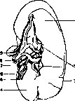
II. Anatomy. The spleen (Latin: lien, syn. splen) is an unpaired organ of soft consistency (elastic), of a dark bluish-red color (after death, the color changes rapidly, and the more so, the more blood is in the organ at the moment of death). The dimensions and weight of the spleen in an adult human fluctuate within the limits of 12–14 x 7–10 x 3–4 cm and 140–200 g. A spleen weight above 200 g is considered a pathological sign. In a newborn, the spleen is on average 5 cm in length and 10 g in weight. There are indications (Neugarten) that the development of the lymphatic apparatus plays a significant role in the weight of the spleen (in cases of status thymico-lymphaticus, the average figures reach up to 255 g). The outer surface of the organ is smooth and shiny during moments of blood engorgement and slightly wrinkled in the stage of contraction. The configuration of the spleen changes depending on the state of neighboring organs; thus, it has the shape of a triangular pyramid when the stomach is contracted and the colon is dilated (one side of the pyramid adjoins the stomach, another the diaphragm, the third the kidney, and the base rests on the colon); when the stomach is full and the intestine is empty, the shape resembles an orange segment (according to Braus). Between these two extreme forms, there are many intermediate ones (an exact idea of them can be obtained by fixing the organ in situ in each individual case). Some compare the shape of the spleen to a coffee bean. In the spleen, 4 surfaces are distinguished: diaphragmatic, renal, gastric, and basal (facies diaphragmatica, renalis, gastrica et basalis, s. colica); in accordance with this, there are 4 edges: the gastric and diaphragmatic surfaces form the anterior edge of the spleen (margo anterior), the diaphragmatic and renal surfaces form the posterior edge (margo posterior), the renal and gastric form the intermediate edge (margo intermedius), and finally, the basal and diaphragmatic surfaces form the lower edge (margo inferior). On the anterior edge, as a rule, and sometimes on the posterior edge of the spleen, there are notches (incisurae). On the anterior edge, there are usually 2 notches, sometimes their number reaches 6–7, which is why this edge is sometimes called serrated (margo crenatus). In exceptional cases, the notches extend along the entire diaphragmatic surface of the spleen. On the gastric surface, there is an unevenly formed slit-like depression—the hilus of the spleen (hilus) (Figure 1), through which the vessels of the spleen enter and exit. Figure 1. Spleen: 1-facies gastrica; 2-hilus; 3-margo ant.; 4-facies renalis; 5-a. gastro-epiploica sin.; 6-margo post.; 7-v. lienalis; 8-a. lienalis; 9-lien accessorius.
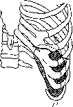



Topography and syntopy. The spleen lies in the abdominal cavity in the depth of the left hypochondrium, adjoining with its diaphragmatic surface to the upper outer half of the diaphragm at the level of the IX–XI ribs. In this place, the spleen is separated from the chest wall only by the diaphragm and the reserve pulmonary space (sinus costo-diaphragmaticus), and during inspiration, also by the lower section of the left lung. From the outer lateral surface of the chest, the spleen is projected in the region between the IX and XI ribs obliquely from behind and above to the front and down; at the same time, it does not reach the spine from behind, and from the front, it does not cross the costal-articular line. The long axis of the spleen corresponds to the course of the X rib in a lying person and is in an almost vertical position in a standing person (especially in an adult woman—Braus). The position of the spleen changes depending on the type of chest structure (Sozon-Yaroshevich) (Figure 2). The spleen is held in its place by the ligamentous apparatus and surrounding organs. The ligamentous apparatus is formed by folds of the peritoneum, which covers the spleen from all sides, leaving only its hilus free; from here, the peritoneum passes into folds: anteriorly, to the greater curvature and fundus of the stomach—lig. gastro-lienale, which passes below into the lig. gastro-colicum, then to the place of attachment of the bursae omentalis—lig. phrenico-lienale, and finally the lig. phrenico-colicum, running from the flexura coli sinistra to the lower surface of the diaphragm. The main significance in terms of the fixation apparatus belongs precisely to this last ligament, which in some cases forms something like a bag in which the organ is suspended. Blood supply to the spleen occurs through the splenic artery (a. lienalis), which is the largest branch of the a. coeliacae; the a. lienalis runs behind the upper edge of the pancreas, located above the splenic vein. On its way, the splenic artery gives off branches to the pancreas and stomach (with the magistral type of vessels, the short arteries of the fundus of the stomach—aa. gastricae breves—sometimes depart from the upper branch of the divided artery at the very hilus of the spleen). The splenic vein (v. lienalis) of large caliber usually begins with several roots from the hilus of the spleen. The splenic vein runs behind the pancreas under the artery of the same name and is one of the 3 roots of the v. portae. The vv. gastricae breves, v. gastro-epiploica sin., and vv. pancreatico-duodenales flow into the v. lienalis. Lymphatic vessels from the spleen (according to Braus, they are present only in the outer layers of the spleen) collect at its hilus into larger trunks and flow into the lymphoglandulae lienales. The latter are located along the entire path of the a. lienalis up to the lgl. coeliacae. Nerves of the spleen (non-medullated) accompany the artery and vein in the form of a plexus (in large animals, they are connected into a thick non-medullated nerve), penetrating the organ. Figure 2. Types of spleen position: a and b-low position; c and d-high position.
...along with them deep into the organ and branching widely there. They originate from the solar plexus and from the right vagus nerve. III. Histology. General structure. The Spleen consists of 3 different basic formations: 1) a capsule with trabeculae extending from it and reticular fibers (reticulum), analogs of the lattice fibers of the liver and other organs; 2) lymphatic nodules (follicles, splenic nodules, Malpighian nodules, Malpighian corpuscles, corpusculum lienis Malpighi, noduli lymphatici lienis Malpighi); 3) the parenchyma of the Spleen itself (red pulp, Spleen pulp, Spleen marrow, substantia medullaris). If one mashes a piece of the Spleen in water (in the stage of incipient decay), the elements constituting the red pulp fall out, and a whitish fibrous mass with small nodules in it (follicles) remains; some call this mass, consisting of the capsule, trabeculae, and follicles, white pulp (others call only the follicles white pulp). In the red pulp, cavities predominate, which are particularly striking in their emptiness in a bloodless Spleen. Similar to the cavities of lymphatic glands, they are called sinuses (sinus lienis), but in the Spleen, the sinuses are filled with blood, not lymph. The sinuses are of various sizes and shapes. As in the corpora cavernosa penis, there are no capillaries in the Spleen; arteries pass directly into the sinuses (arteriovenous anastomoses). Between the sinuses lies the tissue of the Spleen itself—the pulp cords; the sinuses and pulp cords form the red pulp; since both are packed with erythrocytes in a blood-rich Spleen, it is difficult to distinguish them from each other under a microscope. In general, the Spleen consists of elements (sinuses, follicles, cords, trabeculae, and capsule) that also make up lymphatic glands, but the interrelation of these elements and their arrangement is different. Although Malpighian corpuscles represent true follicles with germinal centers, unlike the lymphatic follicles of lymphatic glands, they are not located only on the periphery (in the cortical substance of the gland) and do not penetrate the entire parenchyma. Lymphocytes are formed and enter the bloodstream directly, not by circuitous routes through the lymph. But the main thing here is that the blood flow is constructed completely differently, and lymph flow is absent (according to Braus, lymphatic vessels are present only directly under the capsule of the Spleen and in the tissue areas adjacent to it; inside the organ, they are absent). In the embryo, the Spleen initially resembles a lymphatic gland beginning to form, however, as soon as sinuses and trabeculae (which grow not into the sinuses, but into the parenchyma) begin to form, it begins to take on the character of an organ sui generis. The capsule of the Spleen is covered with a single-layered flat epithelium of the peritoneum, similar to the serous membrane of the stomach; beneath it lies a thick layer of connective tissue—tunica fibrosa, s. albuginea (fibrous or white coat). This fibrous coat is rich in elastic fibers, and in animals, also in numerous smooth muscle cells (in humans, there are few of them). Under the influence of blood filling, the capsule is capable of expanding so as to squeeze blood into the vein in an effort to return to its original position; in humans, this occurs passively under the influence of the tone of elastic fibers, in animals actively—with the help of muscle fibers. The trabeculae behave similarly, being especially densely located closer to the hilum of the Spleen, from where they penetrate into the pulp, forming a system of trabeculae. Elastic fibers (and muscle ones) enable the capsule and trabeculae to withstand significant blood pressure and prevent overstretching of this supporting apparatus; by means of the latter, the Spleen performs the function of a valve for the vessels of the abdominal cavity (see below). Significant enlargement of the Spleen is possible only as a result of pathological changes in the capsule and trabeculae. From the latter, collagen and elastic fibers continue into the pulp; they accompany the artery into the pulp with a dense network. In addition to collagen and elastic fibers, the Spleen also contains a reticulum (reticulum), corresponding to the reticular tissue of lymphatic glands. Similar to the reticular fibers in the liver, they form a network here of thin fibers with the same tinctorial properties. In the nodal places of the reticulum network, there are stellate cells; the latter, together with the endothelium of the sinuses, are a common matrix for the lattice fibers; both pass directly into each other, forming a reticulo-endothelial apparatus. In the loops of the reticulum, lymphocytes and other blood cells are embedded. Lymphatic follicles are formed as a result of the accumulation of lymphatic cells in the outer layer of the tunicae externae of the artery. The follicles of the Spleen look like round disks on sections and give the impression that they are all spheres, like the cortical nodules of lymphatic glands. In reality, the nodules accompany the artery along its length (sometimes even after its division) and resemble more the shape of a cucumber (spherical ones are, of course, also encountered). Germinal centers are often found inside; the latter are sometimes absent because the entire follicle consists entirely of lymphoblasts, or sometimes, conversely, the section passes exclusively through the cortical layer, where there are only mature lymphocytes. The artery is usually pushed away from the germinal center and lies eccentrically. Inside the follicle, a network of capillaries extends from the artery. The lymphoblasts of the germinal center are often in a state of mitotic division. Finished lymphocytes from the cortical layer enter the pulp cords, from there into the sinuses and into the vein. The sinuses are therefore particularly rich in white blood cells. The artery, dividing in the hilum of the Spleen, penetrates into the depth with the trabeculae, in which it lies centrally, while the veins for a long time only lie adjacent to the trabeculae, to then finally penetrate into them and lie next to the artery. The blood flow in the pulp on the segment after the artery has left the trabecula and before the vein has returned to it has been studied only partially. The scheme is presented in Fig. 3. After the artery has given off a network of capillaries in the follicle, it exits it and lies freely in the red pulp; here it forms a brush of small arteries—the arterial brushes of the Spleen (penicilli). These branches do not anastomose with others—they are terminal arteries. Soon, the branches of the arterial brush turn into arterial sheaths (Hülsenarterien-Schweigger-Seidel), which apparently regulate blood flow (sinuses can be filled by injection from a vein, but this cannot be done from an artery, at least on a cadaver). The system of sinuses resembles cavernous tissue; the mechanism of stasis is different, but the effect is the same; blood in the sinus can accumulate (be dammed up) and empty again. The difference from cavernous tissue is that there, diapedesis does not occur in the wall of the cavern, whereas in the Spleen, an exchange of blood elements between the bloodstream and the parenchyma of the Spleen itself necessarily occurs somewhere. In the parenchyma of a normal organ, red blood cells are constantly present; in some diseases, it is sometimes overflowing with erythrocytes. A whole series of questions arises: is the path from the artery to the sinus and to the vein continuous or discontinuous; is there a path through the sinuses into the parenchyma (back and forth); is there another path from the artery to the vein, other than through the arterial sheath. Many understand the arterial sheath as a capillary with a thickened wall consisting of a syncytial complex with many nuclei; the sinuses, as "anschliessende Venen" (capillary "veins"). It is more probable that the arterial sheath is something completely specific to the Spleen, and capillaries are completely absent here. How arterial sheaths regulate blood flow—under the influence of nerve impulses or as a result of chemotaxis—is unknown; there are indications of the presence of non-myelinated nerve fibers in the wall of the arterial sheath. 3 possible blood flows in the Spleen are assumed: 1) blood from the capillaries of the nodule and from the arterial brush through the arterial sheaths all pours into the parenchyma, and from there it is collected into the sinuses—open blood flow; 2) from the arterial brush
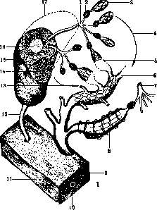
Fig. 3. Blood flow in the spleen pulp: 1 and 7—arterial brushes; 2—arterial sheaths; 3—fenestrated oval terminal chamber of the artery; 4, 5, and 13—possible forms of open blood flow; 6—tube-like primordium of a sinus; 8—venous sinus with transverse hoop-like fibers and fenestrated syncytium (closed blood flow); 9—vein in the trabecula; 10—artery in the trabecula; 11—trabecula; 12—artery inside the pulp; 14—capillaries inside the follicle; 15—follicle; 16—germinal center; 17—contour of the entire follicle.
directly passes into the sinus—closed blood flow; 3) from the capillaries of the nodule—into the parenchyma, from the arterial brush directly into the sinus, whereby the arterial sheath regulates the open and closed blood flow. There is still no consensus regarding the structure of the s i n u s e s; most researchers envision the wall of the sinus as the wall of a barrel (Fig. 4), where the staves correspond to the endothelium (elongated elements with oval nuclei protruding into the lumen), and the hoops correspond to fibers encircling the sinus transversely or obliquely (of the same origin as the reticular fibers of the Spleen). A third element in the architecture of the sinus is also hypothesized—a structureless membrane situated between the endothelium and the encircling fibers (something like a basement membrane between the epithelium and connective tissue). The wall of the sinus is considered permeable to the formed elements of the blood (from the parenchyma into the sinus and back). The main part of the red Spleen in these
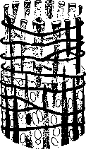
Figure 4. Schematic representation of the spleen sinus: transverse fibers, endothelium with oval nuclei, and endings-
The pulp (along with the sinuses) consists of pulp cords (Billroth's cords, Billrothsche Stränge), which form a complex plexus filling the spaces between the sinuses. In the cords there are many erythrocytes, lymphocytes, and sometimes granulocytes (brought by the blood flow), but there are also cells of local origin, derivatives of the reticulum, called pulp cells (C.-Virchow cells, splenocytes-Türk). These are large mononuclear cells with macrophage properties (erythrophages, pigmentophages, etc.). By means of a microchemical reaction for iron, it is possible to establish that these cells are located mainly at the wall of the sinuses: inside and outside of it; therefore, macrophagia is attributed to the entire reticulo-endothelial system. Pulp cells (pulp cells) are formed by the swelling and release of reticular cells; the transport of iron from the Spleen to the bone marrow is also attributed to them (see below, physiology of the Spleen). Blood platelets are also often found in the pulp cords. IV. Physiology and Pathophysiology. The functions of the Spleen and its role in the organism are still insufficiently studied. The experimental method of research, with the help of which Claude Bernard brilliantly reformed all of physiology in his time, brought many disappointments in the study of the functions of the Spleen. In experiments, the method of excision dominated, but animals tolerated the removal of the Spleen so well that the opinion naturally formed that its role in the organism was insignificant. Numerous reports on the functions of the Spleen often contradicted each other, because the difference in its activity in various animals, depending on the degree of their phylogenetic development, which is known to us now, was not taken into account. It is now known that in the process of ontogenesis the functions of the Spleen also change, and even more than that—within the limits of one and the same animal species, the functions of the Spleen can be different (Eppinger) depending on its individual structure. Our modern knowledge about the functions of the Spleen has been greatly contributed to by clinical studies of recent decades. In particular, the surgical clinic has contributed much of value to this field through its observations and studies of people before and after the operation of removing the Spleen. The functions of the Spleen can be judged, first of all, by studying its physical and histological structure. The characteristic features of the construction of its bloodstream justify the witty remark of Helly: 'The spleen is a regional lymphatic gland of the blood'. The role of the Spleen as a blood filter is now undoubted. It has long been called the 'graveyard' of erythrocytes, but it is now known (Bieling, Ribbert, Kiyono) that the Spleen eliminates from the bloodstream not only damaged erythrocytes, but also various other elements foreign to the blood (coloring substances, bacteria, foreign erythrocytes, etc.). The filtration process in the Spleen is based not only on physical factors (open blood flow through the pulp, slowed blood flow, etc.), but also on the biological activity of its parenchyma. The particular richness of the Spleen in reticulo-endothelium provides it with the cellular macrophagia necessary for filtration, and the abundant accumulation of leukocytes (in the slowed blood flow)—microphagia. Closely connected with macro- and microphagia in the Spleen is another of its functions—immunobiological (production of antibacterial bodies, hemolysins). Bieling verified and confirmed the opinion of Pfeiffer and Marx regarding the richness of the Spleen in antibodies in the first days after immunization. True, the removal of the Spleen shortly before immunization with a cellular antigen, especially in animals used for the first time, usually insignificantly lowers the formation of antibodies (compensatory activity of the rest of the reticulo-endothelium), but there are also indications of special functions of the splenic reticulo-endothelium. Thus, filling the entire reticulo-endothelium of the organism with iron has no effect on the formation of antibodies, while this same method (blockade of the reticulo-endothelium) in previously splenectomized mice completely or almost completely stops the formation of antibodies; this difference in the activity of the splenic and extra-splenic reticulo-endothelium in relation to the production of agglutinins and lysins is manifested especially clearly. Many authors, led by Lubarsch, however, deny these special properties of the Spleen, and some (Blumreich, Jacoby) even speak of greater endurance of splenectomized animals in relation to infections. The long-term results of removing the Spleen in humans (Faerman) also convince us that even without a Spleen, people very well resist many infections. One must think that the richness of the organism in reticulo-endothelium ensures, through adaptation and compensatory activity, this lost function of the spleen. Speaking about the immunobiological functions of the Spleen, one cannot help but touch upon another of its peculiarities: both primary and secondary tumors are extremely rare in the Spleen. True, there are other organs that are rarely subject to metastasis (skin, thyroid gland, stomach, musculature, mammary gland, etc.), but the Spleen, which is a blood filter and so intensively absorbs foreign elements that have penetrated into the blood or lymph, somehow remains, inexplicably, usually free from metastases even in generalized processes. In those cases where metastases in the Spleen do form, they are found in the form of nodular, strictly delimited formations. These observations prompted many to experimental verification: Braunstein notes that neoplasms are easily inoculated into splenectomized animals. Biach and Weltmann prove that sarcomas mixed with a pulp of splenic tissue and inoculated into mice are clearly delayed in their development. These and numerous other similar observations force one to think about some special properties of splenic tissue capable of resisting neoplasms. Spleen and hemolysis. The hemolyzing function is also connected with the filtering role of the Spleen. The indications of Ecker and Kölliker (1847) regarding splenic cells filled with blood corpuscles have found full confirmation in recent reports on erythrophagia in the spleen (Eppinger, Kiyono, Faerman). Some researchers (Malinin, Preyer) observed under a microscope the very process of erythrophagia when they examined completely fresh material. Intracellular hemolysis (erythrophagia) is now disputed by no one; similar functions are manifested by the reticulo-endothelium of both the liver and bone marrow (Aschoff). But the function of the Spleen is apparently not exhausted by intracellular (active) hemolysis alone; there also exists extracellular (passive) hemolysis. The best proof of the latter is the absence of an increase in the number of macrophages in aplastic anemia and in hemolytic jaundice, while there are all signs of significant destruction of erythrocytes. Gauckler considers intracellular hemolysis to be normal, and extracellular to be abnormal, and thinks that the latter leads to the deposition of massive pigment in the Spleen and to pigment overload of the liver. A mixed type of hemolysis indicates hemolytic intoxication; the latter, being sharply pronounced, leads to extracellular hemolysis alone. Therefore, even significant hemolysis, if it is intracellular, is less dangerous than insignificant extracellular hemolysis. The question arises—do all erythrocytes undergo hemolysis in the Spleen, or is it a matter of erythrocytes that are damaged in one way or another. A sharp increase in erythrophages after the introduction of distilled water into the blood and the absence of such an increase in hemolytic jaundice confirm the generally accepted opinion that it is precisely damaged erythrocytes that are hemolyzed in the Spleen. But erythrocytes can be damaged not only outside the Spleen, but also within it; the decrease in the resistance of erythrocytes in the splenic vein compared to the corresponding artery confirms this; this is also confirmed by the increase in the resistance of erythrocytes in the peripheral blood after splenectomy (Pehl, Faerman, et al.). Apparently, the presence in the Spleen of a double blood flow (open through the pulp and closed in the bloodstream) ensures a safe passage for a portion of the erythrocytes; it is unlikely that the Spleen could sort erythrocytes into those that must pass through the pulp and those that can bypass it; however, the function of the Schweigger-Seidel arterial sheaths is capable of significantly influencing the quantitative ratios of erythrocytes in the closed and open blood flow of the organ and thereby actively increasing or decreasing the hemolyzing properties of the Spleen. Along with erythrocytes, the destruction of other formed elements of the blood also occurs in the Spleen, but in significantly smaller quantities. Lepehne discovered phagocytosis of leukocytes in infectious jaundice, Kaznelson and Eppinger observed the destruction of blood platelets in essential thrombopenia (see Surgery of the Spleen). One must think that under normal conditions, the phagocytosis of these formed elements occupies a small place in the activity of the spleen. Questions of the relationship of the Spleen with the liver are also closely connected with its hemolyzing activity. The anatomical relationships of these two organs already predetermine their functional connection. If the Spleen is a filter for blood from its artery, then the liver is the same for blood from the splenic vein.
Studies by Asher and Ebnother found that spleen extract does not always cause hemolysis, but the addition of splenic extract to liver extract constantly and sharply increases the hemolytic properties of the latter. Consequently, there is something in the spleen tissue that activates the hemolytic function of the liver. Especially important are the observations by these same authors on the increase in the hemolytic properties of liver extracts in hypotonic salt solutions. It is obvious that the decrease in the resistance of erythrocytes in the spleen is an important preparatory factor for more effective cooperative activity of the liver. Experiments by Pugliese note a decrease in the amount of bile in animals after the removal of the spleen. However, this is apparently connected not only with a decrease in blood flow to the liver after splenectomy, but also with the loss of the spleen's function of lowering the resistance of erythrocytes and activating the hemolytic properties of the liver. There are also indications of the participation of the spleen in the process of hemoglobin breakdown. Extracts from the spleen activate the breakdown of hemoglobin in the liver; it turns out that this property is preserved in the extract even after boiling. The latter circumstance proves with certainty that the substances in the spleen that break down hemoglobin are completely different from those that create hemolysis (the hemolyzing properties of spleen extracts disappear after boiling). The clinical picture of hemolytic jaundice and its successful treatment by splenectomy, the successful treatment by splenectomy of recurrent attacks of cholelithiasis in hemolytic jaundice (recurrences despite the removal of the gallbladder, Moynihan)—all this vividly characterizes the participation of the spleen in the formation of bile pigment. The question of the direct site of bilirubin formation still remains controversial, but the possibility of its formation in the reticulo-endothelium of the spleen is not excluded (Aschoff, McNee). The close connection in the activity of the spleen and liver is also illustrated by the experiments of Schmidt and others, who observed hyperplasia of the stellate cells of the liver in splenectomized mice; in places, the cells form clusters resembling in shape the (lymphatic) nodules in the spleen; this newly formed tissue was named by Aschoff "Milzgewebe in der Leber" (splenic tissue in the liver). Similar formations were discovered in the liver of a person who died a year after splenectomy (Faerman). Hematopoietic activity of the spleen. The hematopoietic activity of the spleen begins already in the embryonic period (see Hematopoiesis). In the middle of the embryonic period, vigorous erythropoiesis appears in the spleen, which continues until the end of embryonic life and disappears completely after birth. Isolated remarks (Virchow, Bizzozero, Koelliker) about erythropoiesis in the post-embryonic period have not found confirmation. Under normal conditions, the spleen definitely does not produce red blood cells, however, erythropoietic potencies are preserved in it, and bursts of erythropoiesis are observed in some pathological states. Extramedullary erythropoiesis is especially common in various anemias, and sometimes in infections. The spleen does not produce granulocytes in either the embryonic or post-embryonic period. Indications that the cells of the spleen pulp give a positive reaction to oxidase should perhaps be attributed to granulocytes brought here by the bloodstream. It is more likely, however, that the oxidase reaction in splenic tissue is given by Pappenheim's splenocytes, which are ultimately identical to Kiyono's histiocytes and monocytes (according to Naegeli, monocytes constantly give a well-defined reaction to oxidase). This thereby establishes a hematopoietic function for the spleen in the sense of monocyte production. There are even indications that azurophilic granulation is found in the mother cells of the spleen (reticular cells) (Paremuzov), which further reinforces the fact of monocytopoiesis in the spleen, since azurophilic granulation in monocytes is almost constant. There is no doubt about the hematopoietic function of the spleen in relation to the formation of lymphocytes. Lymphatic nodules are the centers of their formation. It is not surprising, therefore, that in the veins of the trabeculae, one counts almost 70 times more white blood cells than in the arteries. Some attempt to explain this phenomenon by the destruction of erythrocytes in the spleen, as a result of which a large number of surviving blood elements accumulate in the veins. In the splenic vein itself, such a large increase in leukocytes compared to the artery is no longer detected (1.8) (just as after the entry of the thoracic duct into the anonymous vein, numerous lymphocytes are quickly lost). It should also be noted that in relation to lymphocytopoiesis in general, the spleen apparently takes part not only by the production of lymphocytes, which is essentially insignificant, but also by an indirect influence on the rest of the lymphatic tissue of the organism. The lymphocytosis that regularly occurs after the removal of the spleen in both humans and animals was explained simply by excessive compensatory activity of the lymphatic tissue as a reaction to the lost function of the spleen. In the old literature (Simon, Hegar; 1847) there are even indications of simultaneously appearing hyperplasia of the lymph glands. However, newer observations completely deny this, and since lymphocytosis after splenectomy turns out to be a long-term phenomenon, the idea of compensatory causes for it turns out to be untenable. At the present time, great importance is attached to the hormonal influences of the spleen on lymphocytopoiesis through the autonomic nervous system (Falta, Bertelli, and others). Fluctuations of lymphocytes in the blood largely depend on the state of the vegetative nervous system (lymphocytosis of vagotonics, lymphocytosis after the introduction of vagotropic substances), and it was natural, of course, to assume the loss of some influence of the spleen on the autonomic nervous system as the cause of lymphocytosis after splenectomy. Berezov considers the main cause of lymphocytosis after removal of the spleen to be a gradual increase in the tone of the autonomic nervous system due to the loss of a "paralyzing hormone" (hormone-inhibitor according to Schultze). It is possible that the observed increase in intestinal peristalsis after splenectomy (Bayer, Faerman, Berezov) is also connected with a change in the vagotonia of splenectomized individuals. The hormonal activity of the spleen in its interrelations with the bone marrow turns out to be especially important. Hirschfeld, Asher, and Frank insist that the spleen suppresses the activity of the bone marrow. In their opinion, the bone marrow after splenectomy becomes more sensitive and capable. These observations formed the basis for indications for the removal of the spleen in various forms of anemia. After the removal of the spleen, the number of red blood cells increases. However, it is necessary to distinguish short-term polycythemia from long-term. The origin of these polycythemias is different. Short-term polycythemia is apparently the result of the loss of the hemolytic function of the spleen, since ligation of the vascular pedicle alone, even without removal of the spleen, leads to short-term polycythemia. As for long-term polycythemia, opinions on its origin are divided. Some believe that increased erythropoiesis is stimulated by the products of erythrocyte breakdown in the bloodstream (Stradomsky, Lepehne), others attribute its origin to the loss of a hormone that suppresses bone marrow activity. Eppinger, Krumbhaar, and others attribute the opposite role to the spleen—stimulating the hematopoiesis of the bone marrow. Through intravital examination of the bone marrow with the help of puncture (Faerman), it has recently been possible to establish new moments in the interrelations of the spleen and bone marrow; in particular, the origin of polyglobulia is being clarified. It turns out that oligocythemia in some diseases of the spleen depends not on anatomical changes in the bone marrow, but on the inhibition of the process of washing out formed elements from the bone marrow into the blood. The picture of bone marrow punctates before and after the removal of the spleen convinces one that hematopoiesis in it in the corresponding cases does not weaken in the least; on the contrary, the bone marrow is rich in cellular elements before the removal of the spleen and poor in them after splenectomy. This fact is reflected in the peripheral blood in that the picture of aregenerative anemia before splenectomy is replaced after it by an increase in the total number of erythrocytes with the simultaneous appearance of a large number of nucleated, young forms of erythrocytes; one cannot help but attribute all this precisely to the inhibitory influence of the spleen on the very process of washing out formed elements from the bone marrow, and not to anatomical changes in the hematopoietic tissue. It must be added that the inhibitory function of the spleen concerns not only erythrocytes, but also all other formed elements of the bone marrow. The number of leukocytes after splenectomy also increases significantly, and not only at the expense of lymphocytes (which was indicated above), but also at the expense of granulocytes. However, in some special pathological states of the spleen, it is possible for it to release some toxins (myelotoxins—Frank) that suppress the very production of granulocytes (typhoid fever) and platelets, due to changes in megakaryocytes (see Giant cells). Opposed to Frank's point of view is the above-cited opinion of Katsnelson, Eppinger, and others (see Hemolysis) regarding the importance of the spleen in the destruction of platelets. One more phenomenon in the picture of peripheral blood after splenectomy was linked to the loss of the spleen's function—the appearance of a large number of erythrocytes with Jolly bodies.
Since these corpuscles are considered as remnants of the erythrocyte nucleus, the function of the spleen was also linked to the process of erythrocyte denucleation. Participation of the spleen in metabolism. The role of the spleen in metabolism is most pronounced in iron metabolism. The spleen in the guinea pig and the rabbit is generally the organ richest in iron (Tedeschi); the iron of the spleen constitutes 5% of the entire dry residue of iron (Oidtmann). The richness of the spleen in iron is explained not only by the reserves formed during blood destruction; experiments by Asher and Sollberger on animals and Bayer on humans are convincing regarding the accumulating properties of the spleen in relation to iron released during metabolism. Schmidt proved that in animals fed iron-free food, iron is preserved only in the spleen, while it completely disappears from other organs. Vogel discovered a significant drop in the number of erythrocytes and hemoglobin in a splenectomized dog when fed iron-free food, whereas the control did not show changes. Thus, the spleen is a storage depot for iron of both exogenous and endogenous origin; apparently, the transport functions of the reticulocytes of the spleen (see anatomy of the spleen) carry out further mobilization of iron to the bone marrow, and this ensures the renewal of erythrocytes. After removal of the spleen, iron is found mainly in the liver, which apparently takes upon itself the hemolytic functions of the spleen. Regarding the role of the spleen in general metabolism, there is insignificant and contradictory information. According to some (Asher, Bernet), nitrogen excretion in splenectomized rabbits increases, while others (Umber-clinical observations, Palladin-experimental) assert the opposite. Clinically valuable data were obtained in experiments by Hashimoto and Pick: a sharp increase in enzymatic protein autolysis in the liver in guinea pigs pre-treated with horse serum; after removal of the spleen, autolysis decreases, and in splenectomized and subsequently sensitized animals, it does not occur at all. One must think that the spleen of sensitized animals plays a role in the intravital wear and tear of the liver, since substances are formed in it that cause proteolytic breakdown in the liver. It is unknown whether the spleen generates any autolysis in the liver under normal conditions, however, for clinical practice, it is more important to consider the significance of the spleen in the pathogenesis of organic liver lesions under certain pathological conditions and, if possible, to prevent them in a timely manner by splenectomy. The participation of the spleen in fat and lipoid metabolism has been studied histologically, experimentally, and chemically. Kusunaki observed an increase in lipoid deposition in the spleen with an increase in lipoids in the circulating blood. Anichkov observed the deposition of lipoids in the pulp with reduced cholesterol feeding of rabbits. Eppinger, King, and others observed the opposite phenomenon—the accumulation of lipoids in the blood after removal of the spleen; irradiation of the spleen with mesothorium was not accompanied by an increase in lipoids in the blood. The role of the spleen in carbohydrate metabolism has not been studied at all. According to Togawa, the spleen is also interested in this metabolism; he also refers to other researchers who discovered a diastatic enzyme in the spleen. There are still many reports illustrating other poorly verified functions of the spleen. Among them, one should note the very old works of Lauret and Lassaigne (1825), Dobson (1847), and others, who pointed to the ability of the spleen to swell during digestion from the absorption of large quantities of blood, thereby protecting the gastrointestinal tract from its excess. It is especially interesting to note that these thoughts, expressed 100 years ago and subsequently forgotten, about the ability of the spleen to absorb and store reserves of blood, now find confirmation in the latest works of Barcroft, devoted to the relationships between the spleen and the total amount of blood in the organism. Barcroft thinks that the spleen is a reserve reservoir for blood and hemoglobin (the spleen can absorb up to 1/5 of the total blood volume of a dog); according to the author, blood entering the pulp is retained there and is released into the general blood flow only in cases of corresponding need for it. Blood deposited in this way turns out to be so isolated from the circulating blood that it remains undamaged even upon the penetration of carbon monoxide into the general bloodstream (carbon monoxide poisoning—Barcroft, Heyer). The absorption and release of blood reserves by the spleen are a function of its neuromuscular apparatus. This activity of the spleen has been studied almost exclusively in animals (dog), and, of course, all materials can only be relatively used for concepts about the functions of the human spleen. Complete denervation of the spleen leads to an increase in the organ, but after some time the tone is restored and the spleen assumes its previous size. Irritation of the nervus splanchnicus major leads to contraction of the spleen. The role of the vagus nerve is not entirely clear. By ordinary irritation of the peripheral vagus nerve, no changes in the spleen are detected. Only after cutting both nervi splanchnici does irritation of the vagus nerve lead to an increase in the spleen. Correspondingly, the pharmacological influences of vago- and sympathicotropic substances on the spleen are manifested. Adrenaline causes a pronounced contraction of the spleen. The role of the spleen in the endocrine system was noted by Lampe in the form of its cooperation with the thymus in the matter of binding acids and neutralizing poisons; Bayer, on the contrary, considers them antagonists. Opinions about the relationships between the spleen and the thyroid gland are also contradictory. Inhibitory influences of a changed spleen on the sex glands are very likely (Faerman). Women with splenomegaly very often stop menstruating, and removal of the spleen in these cases entails the return of menses. But the influence of splenomegaly on the growth and development of the sex glands in the pre-pubertal period is especially pronounced. There are indications of the participation of the spleen in the process of callus formation (Schonbauer) and, finally, references to the special properties of the spleen in protecting the organism from tuberculosis (Schroder, Kaufmann, and others). Bayle supposedly obtained good results in the treatment of tuberculosis with splenic extracts. In general, it should be noted that the activity of the spleen is difficult to account for accurately, since the spleen, on the one hand, functions as part of the large reticulo-endothelial apparatus of the entire organism, and on the other, is in close correlative relationships with many other organs. The best proof of the possibility of complete replacement of the spleen's activity by other organs is the well-known fact of its sometimes complete absence in humans (Titov, McLean and Craig)—alienia. Of course, this does not qualify the spleen as a superfluous or unnecessary organ. With congenital absence of the spleen, the process of development of vicarious functions in other organs proceeds gradually and imperceptibly. When a functioning spleen is removed, we can observe how the organism compensates for its functions by hyperplasia of the reticulo-endothelium and lymphatic glands or even by the regeneration of new organs similar to the spleen. V. Pathological anatomy. The spleen very soon undergoes cadaveric changes. First of all, a change in volume occurs; in humans, the decrease in the spleen is inconsistent (fewer muscle fibers) and depends on the intravital blood volume and rigor mortis. Changes in the pulp are more significant; the proximity of the large intestine very soon causes putrefaction. Autolysis and putrefaction are more strongly expressed the higher the temperature was at the moment of death and the richer the spleen was in blood and infectious material. Cadaveric changes lead to the relaxation of the supporting elements of the organ and cause protrusion of the pulp, which is easily scraped off with a knife; all this can somewhat complicate the distinction between cadaveric changes and intravital ones. Recent studies (the spleen was examined 20-30 minutes after death) suggest that the generally accepted ideas about the 'septic' swelling of the spleen are partially related to cadaveric changes. On the other hand, the early onset of these changes is characteristic of septic diseases, since in them, the richness of the splenic pulp in proteolytic enzymes is noted. Histologically, cadaveric changes are expressed by swelling of the cell body (in the cells of the pulp and reticulum), its clouding, and partial disintegration of the nucleus, as well as the precipitation of hemoglobin from erythrocytes (erythrocyte shadows and the accumulation of formalin pigment along the edges of these shadows). However, the precipitation of formalin pigment, being a post-mortem phenomenon, indicates at the same time increased enzymatic and autolytic processes, which, for example, is characteristic of infection, especially sepsis. Follicles and trabeculae show significant resistance; they can sometimes be detected even 8-14 days after death. Anomalies of position and development. The most frequent anomaly of the spleen boils down to its displacement (see surgical diseases of the spleen); among other anomalies of the position of the spleen, its retroperitoneal ectopia is encountered. In this latter case, one can assume a congenital anomaly of position, but usually, it is a matter of acquired forms. Among developmental anomalies, remnants of deep embryonic notches are most often encountered, sometimes multiple, both along the edges of the spleen and on its surface. The extreme degree of this same anomaly is expressed not only in the lobulation of the organ but also in the formation of accessory spleens. The presence of one small accessory spleen at the hilum of the organ is such a frequent phenomenon that it is not classified as pathology.
As for the larger number of accessory spleens, they are divided into several subgroups depending on their origin. Thus, Oltmanns speaks of 5 different types of accessory spleens. 1. Formed as a result of an extreme degree of lobulation of the organ itself—the spleen as such is absent. 2. As a result of delayed development—accessory spleens are located directly near the large one. 3. Atavistic formations—when the location of the accessory spleens corresponds to phylogenetic sites (greater omentum, peritoneum, vascular pedicle, pancreas, etc.). 4. Intrauterine cellular dystopia. 5. Auto-implantation due to post-fetal trauma—dissemination throughout the abdominal cavity. In this case, the 4th group differs from the 5th in its structure. The former are typical spleens, the latter are splenoids, i.e., not entirely typical in structure. There are other classifications (K. Schilling, Haberer), however, they do not present any essential difference. To avoid misunderstandings, it should be noted that since the appearance of Haberer's works (1901), various names for accessory (supplementary) spleens have appeared in the literature: lien accessories and lien succenturiatus. Haberer insisted on the necessity of distinguishing them from each other, since the former, in his opinion, are completely independent and have no relation to the main organ, while the latter are part of it and develop as a result of deeply penetrating notches. Practically, it is better to use Oltmanns' classification, and in terminology—the name accessory spleen (lien accessorius). Among other developmental anomalies, one should note the complete congenital absence of the spleen and its insufficient development. The former occurs more often and yet belongs to the rarest anomalies. Lubarsch did not encounter a single case of asplenia in 30,000 autopsies. A total of 30 cases have been collected in the literature. In almost all cases, the vascular pedicle was also absent. In most cases, there are no indications of any compensatory changes in other organs; only Birch-Hirschfeld mentions an enlargement of the liver and portal vein, Hodenpyl—of hyperplasia of the lymph nodes, and Faerman—of hyperplasia of the stellate cells of the liver (stuffed with malarial pigment in a patient who died of malaria). Cases of splenic hypoplasia are even rarer; at the same time, it is difficult to decide whether it is a congenital anomaly or an acquired condition. Inclusions of tissues of other organs into the spleen tissue should also be attributed to developmental anomalies: islets of pancreatic tissue were discovered 3 times and liver tissue 1 time. Circulatory disorders. The peculiarities of the structure of the blood channel of the spleen also determine certain deviations from the picture of circulatory disorders characteristic of other organs. Wolff's experiments prove that only the prefollicular zones of the pulp (the region of closed blood flow) react uniformly with other organs to circulatory disorders; the rest of the pulp reacts to circulatory disorders differently depending on the state of the organ at a given moment (blood filling, contractility, etc.). Therefore, splenic anemia is encountered only in cases of sudden or rapidly occurring death from hemorrhage. In cases of slowly developing general anemia (cancer, pernicious anemia, etc.), the spleen turns out to be less bloodless than other organs, and most importantly, an uneven distribution of the remaining blood reserves is discovered in it. Previously, many authors (Schridde, Kaufmann, Orth) noted individual darker areas of the pulp on cross-section in such cases, but interpreted them as hemorrhages; now, however, Lubarsch insists that we are dealing here with blood reserve reservoirs in the sense of Barcroft's doctrine (see Spleen physiology). Active hyperemia has to be observed only as the initial stage of acute inflammation of the spleen (Kaufmann); therefore, splenic hyperemia has been studied only in its congestive form, and the latter, in turn, is more familiar in its chronic form. In cases of acute congestion (during rapidly progressing pneumonia, death from asphyxia, rapidly developed thrombus of the splenic vein), the spleen turns out to be somewhat enlarged, the capsule tense and transparent, and a large amount of dark, red-blue blood exudes from the pulp (of the same color) on cross-section; the trabeculae are barely noticeable. Chronic congestive hyperemia of the spleen is observed: 1) in all lung diseases accompanied by an increase in blood pressure in the lesser circulation; 2) in diseases of the heart valves or muscle that impede the outflow of blood from the lungs; 3) in disorders of portal circulation (and in diseases of the liver and the portal system itself). In these cases, a picture of so-called cyanotic induration is observed: the spleen is enlarged, the capsule is tense but cloudy and thickened (unlike acute congestion), the spleen on cross-section is purple- or cherry-red, dense and somewhat dry; the trabeculae and vessels are thickened; the lymph nodules stand out with their gray color against a dark background. The sinuses are sometimes cavernously dilated, the pulp is rich in erythrocytes, and the reticulum and reticular fibers are hyperplastic. There are indications that the picture of congestive hyperemia of the spleen is different depending on whether there is congestion of central or portal origin (Kaufmann, Nishikawa). Hemorrhages into the spleen tissue are very rare (it may simply be difficult to distinguish them from blood engorgement); occasionally, hemorrhages are observed into the thickness of the trabeculae. Thrombi in the vessels of the splenic pedicle are also rarely encountered. Lubarsch cites 4 cases of arterial thrombosis per 11,627 autopsies (1—pancreatic cancer, 3—arteriosclerosis) and 65 venous thromboses per 30,064 autopsies. Lubarsch divides the causes of venous thrombosis into mechanical (trauma, torsion of the vascular pedicle, etc.), infectious, and toxic. The consequences of splenic vessel thrombosis are the most diverse. Changes in the spleen depend on the characteristics of the thrombus, on the duration and completeness of the vessel occlusion, as well as on changes in the arteries themselves (thrombosis of the latter leads to anemic infarcts (see below)). Usually, venous thrombosis leads to congestive phenomena in the spleen and to enlargement of the organ; however, in a whole series of cases, the spleen not only did not turn out to be enlarged, but atrophy of the organ was even observed; these data to a certain extent contradict the ideas of Eppinger, Kleinschmidt, and others about the pathogenesis of so-called splenomegalia splenothrombotica (see Splenomegaly); it is especially emphasized by Lubarsch that in none of the 65 cases of splenic vein thrombosis were gastric hemorrhages noted either clinically or anatomically. Infarcts of the spleen are encountered as both anemic and hemorrhagic. The former are the result of occlusion of arterial trunks, the latter—venous. In the majority, they have the shape of cones with their apex turned toward the hilum of the spleen. The dimensions of the infarct are greater the more neutral the occlusion of the vessel. Embolisms are encountered in cases of valve lesions and thrombosis of the left heart. Embolisms by cellular elements (megakaryocytes in septic processes and endocarditis lenta) and bacterial embolisms have also been described. The pathogenesis of acute purulent inflammations of the spleen is also closely connected with embolisms and infarcts. There are, however, reports of cryptogenetic abscesses in the spleen, but they rightly arouse doubts. The most frequent cause is, of course, metastatic infection (apparently it settles and develops especially easily during simultaneous embolic occlusion of a vessel), less often—as a result of the spread of inflammation from adjacent organs and tissues (paranephritis, tumor growth of the intestine and stomach), and even less often—the cause is trauma. Usually, single or multiple abscesses of the spleen are described, however, acute purulent inflammations do not always form closed purulent cavities; purulent infiltrates without sharply defined edges can also form (splenites suppuratives—Lancereaux). The "sequestrating" abscesses of the spleen described in German literature by Küttner, and in Russian by Pikin, apparently develop as a result of the fusion of multiple small abscesses or as a result of massive hemorrhages into the organ tissue (trauma). In these cases, the spleen represents a thick-walled sac (thickened capsule) filled with pus with "sequestra" floating in it—spleen tissue that has not yet completely melted (Faerman). A whole series of infections (necrotic pneumonia, typhus, and especially relapsing fever) predisposes to the formation of abscesses. Atrophy of the spleen develops as a result of exogenous and endogenous causes. Typical for the first group is atrophy of the spleen during starvation or in certain avitaminoses; atrophy of the follicles is especially pronounced, the germinal centers disappear almost completely (unlike senile atrophy of the spleen, where the germinal centers are small but do not disappear completely). Erythrophagia is encountered more often in avitaminoses than in simple starvation. The second group (endogenous) includes atrophy as a result of insufficient nutrition caused by damage to the organ itself. These nutritional disorders can be primary (senile atrophy) and secondary due to mechanical, infectious, or toxic factors (e.g., congestive atrophy or atrophy in malarial cachexia). In these cases, the development of cicatricial connective tissue prevails, the lymphatic apparatus is less affected. Arteriosclerotic atrophy of the spleen is also encountered.
(dense, reduced, shrunken Spleen). A sharp reduction of the organ occurs as a result of the scarring of multiple infarcts in the presence of pronounced arterio- and arteriolosclerosis. Sometimes, in arteriolosclerosis of the Spleen, the picture of the so-called spotted Spleen (Fleckenmilz of German authors) develops, based on uneven filling, atrophy, and sclerosis of individual sections of the organ. Necroses in the Spleen develop directly (infectious or toxic) or secondarily—in infarcts. The former appear as multiple, small, yellowish-gray or gray-red foci of disintegration (karyolysis and karyorrhexis prevail over the phenomena of coagulation necrosis). They are most often encountered in acute and subacute infections (in typhus, especially relapsing fever, in endocarditis, in diphtheria, and scarlet fever). Large foci of primary necroses are sometimes encountered after unsuccessfully concluded blood transfusions. Secondary necroses, which have developed in infarcts, are distinguished primarily by the character of the latter (shape, size—see above, Spleen infarcts). The fate of these necroses depends primarily on whether we are dealing with septic or aseptic infarcts. Above, the possibility of the development of abscesses in septic infarcts was already indicated; in the others, the matter usually ends with a rust-brown or (in the absence of hemorrhages) colorless scar. Anemic infarcts are most often encountered; indications of a high frequency of hemorrhagic infarcts must apparently be explained by the erroneous inclusion among the latter of septic infarcts, which, with their dirty-red color, resemble a hemorrhagic infarct. Necrotic processes in infarcts are accompanied by corresponding changes in the capsule as well. True, the necrosis often does not reach the capsule, and the latter is separated from the mass of the infarct by a narrow strip of unchanged pulp. Necrosis of the entire Spleen is also encountered (thrombosis of the splenic pedicle, its ligation, twisting of the pedicle); the Spleen turns into a cyst with liquid contents of a brown-red color. Autolytic processes rapidly turn the parenchyma of the Spleen into a liquid mush (liquefactive necrosis). An analogous picture is observed during the prolapse of the Spleen into the stomach in cancers of the latter. Amyloid of the Spleen and hyaline deposits in it are encountered very often. Among other organs, the Spleen occupies first place in the frequency of amyloid deposition. They distinguish: 1) diffuse deposition of amyloid in the Spleen—in these cases the organ is somewhat enlarged, very dense, on cross-section the tissue looks bloodless (even under pressure blood is not released), shiny, light-red in color; in this form of amyloid, the Spleen resembles ham on cross-section, and such a Spleen is also called a "ham" Spleen (if the anemia is especially pronounced, then the Spleen on cross-section rather resembles lard—"lardaceous" Spleen); 2) focal deposits of amyloid in the follicles (sago Spleen)—in the majority, the organ is not enlarged and not bloodless, on cross-section the follicles stand out, resembling boiled, swollen grains of sago; 3) mixed forms. Hyaline deposits are encountered in the Spleen in the arteries of the trabeculae and follicles in the form of cords in the follicles and in the form of nodules throughout the pulp. Hyaline deposits are observed mainly along the course of vessels in old age and senility, as well as in many diseases of the blood. Calcareous deposits in the Spleen are rare; they are encountered in the form of calcified abscesses and necrotic foci (in Gandy-Gamna nodules; see Mycotic splenomegaly). They are more often encountered in the form of phleboliths, which are apparently deposits of lime in the thrombi of small veins (on cross-section they look like coarse brownish-yellowish sand embedded in the pulp here and there). Among pigment deposits in the Spleen, one must note hemoglobin-derived (hemosiderin, hematoidin, malarial pigment, and bilirubin) and non-hemoglobin-derived (brown and melanotic pigments). The significance of the former is sufficiently noted in the chapter on the participation of the Spleen in iron metabolism [see separate table (to the article Scarlet fever), figs. 5 and 6] (see also Malaria); non-hemoglobin-derived pigments are encountered in senile atrophy of the Spleen (brown pigment—see Lipofuscin) and in disintegrating melanotic tumors. Exogenous pigmentations are also encountered (anthracosis, argyrosis). Regarding lipoid deposits, it must be noted that they are the result of adsorption processes, and not degenerative ones. In childhood, they are discovered more often in the follicles, in adults—in the pulp. In significant quantities, they are discovered in acute infections (typhus, sepsis), as well as in the vicinity of necrotic foci, gummas, tuberculous and leukemic foci. In cells, lipoids are deposited in the form of droplets, while uniform impregnation is encountered in the fibers and in the spaces between them. Regarding the state of the Spleen in infectious diseases—see Infectious diseases, as well as individual infections. The very fact of the enlargement of the Spleen in infections has still not been precisely explained. The enlargement is not exhausted by "splenitis" alone (as a result of the entry of infectious agents), especially since in some cases the infection is not discovered in the Spleen itself. The old theory of Botkin regarding the paralytic influence of infection on the nerves of the Spleen still finds confirmation today in the experiments of Wolf: significant enlargement of the Spleen under the influence of irrigation with salt solutions up to 60°; such an enlargement cannot be obtained by stopping the outflow of blood—the tone of the trabeculae and capsule proves to be sufficiently high to withstand blood pressure, but as soon as the paralyzing influence of heat manifests itself, the Spleen increases twofold. Thus, active hyperemia is the main factor in the enlargement of the "septic" Spleen. Along with it go exudative and productive processes. Jawein attempts to substantiate septic splenic tumor by an increase in the destruction of blood in infections. In his opinion, it is precisely the hemolyzing properties of the streptococcus that explain the constant enlargement of the Spleen in streptococcemia; by this, the theory of Ponfick regarding a "spodogenous tumor of the spleen," i.e., a Spleen stuffed with slag, is put forward again. The Spleen eliminates slag from the blood, however, this cannot explain a large septic Spleen; in anaerobic sepsis, accompanied by severe hemolysis, the Spleen does not enlarge. Morphologically, in a septic Spleen, along with active hyperemia, the following changes are observed: 1) large accumulations of leukocytes (positive reaction to oxidase) in the pulp of the Spleen and the outer zone of the follicles; in severe cases of pyemia, the follicles also turn out to be permeated with leukocytes; in some infections, accumulations of plasma cells are described, predominantly around the trabeculae and vessels. 2) Hyperplasia of the cellular elements of the pulp itself (Orth considers this hyperplasia the basis of septic splenitis). 3) Significant deposits of iron-containing pigment throughout the pulp and in the outer zone of the follicles, inside which it is never present. In far-advanced septic cases, changes in the Spleen are very difficult to trace, because as a result of significant processes of destruction, both the structural and cellular clarity of the tissue is lost. Specific changes in the Spleen in tuberculosis are encountered either: 1) in the form of miliary tuberculosis [see separate table (to the article Streptococci), fig. 4], 2) in a chronically caseous nodular form, and 3) in the form of tuberculous splenomegaly. Miliary tuberculosis of the Spleen can be a manifestation of general miliary-disseminated tuberculosis (a colossal number of nodules of identical size), but it is also encountered independently in more favorably proceeding forms (a significantly smaller number of nodules of varying size). Tuberculous splenomegaly can develop both in miliary tuberculosis of the Spleen and in the caseous-nodular form. In these cases, it is a matter of significant diffuse development of connective tissue, which causes the enlargement of the organ. As for the tuberculous changes themselves in the Spleen, in all cases, the most diverse nodules in their structure are encountered (epithelioid, giant-cell, caseous, hyaline, etc.). The location of tuberculous nodules is fixed by almost all authors predominantly behind the follicles. Isolated tuberculosis of the Spleen is encountered extremely rarely; one can speak of this form only when no other, even if long-dormant, focus is truly discovered in other organs. The forms of isolated tuberculosis can be various, starting from single foci without enlargement of the organ to large splenomegalies with multiple foci. Changes in the Spleen in syphilis are described mainly in congenital forms, but even in these cases, specific gummatous changes are very rare (see Syphilis). Regarding the Spleen in various systemic diseases of the hematopoietic organs, see Leukemia. Changes in the Spleen in pernicious anemia, see Anemia, pernicious anemia. In burns and blood-destroying poisonings (potassium chlorate, toluylenediamine, morels), congestion with blood and products of its destruction are constantly noted in the Spleen. In burns, hyperplasia of the follicles with significant deposition of lipoids is also pronounced. In cirrhosis of the liver, the Spleen is often significantly enlarged, however, the old theory that in these cases it is a matter of congestive Spleen is now supported by no one anymore (see Cirrhosis of the liver). Tumors in the Spleen are very rare (both primary and metastases). Among benign tumors, vascular tumors (hemangiomas and lymphangiomas) are described mainly; the latter can reach gigantic sizes.
Malignant tumors are represented by sarcomas (primary carcinoma of the spleen has apparently not yet been proven by anyone) - lymphosarcomas, large round-cell sarcomas, spindle-cell sarcomas, and angiosarcomas. More often, these are diffuse tumors of the organ, leading to its great enlargement and metastatic involvement of the retroperitoneal lymph nodes, liver, and lungs. Among parasitic cysts of the spleen, only echinococcus is encountered. Solitary cysts in the upper half of the spleen are more common. There are no particular changes in the rest of the organ (except for compression of the pulp and some fibro-adenia). Non-parasitic cysts of the spleen occur in the form of large solitary cysts (serous or bloody-serous) or multiple small ones (from detached epithelium of lymph vessels or from tears in the capsule with prolapse of the parenchyma). Two cases of dermoid cyst of the spleen are known. VI. Methods of examination of the spleen. The main goal of examining the spleen is to determine its size as accurately as possible, and in cases of enlargement, to distinguish it from other organs or neoplasms. Recently, attempts have also been made to create functional examinations of the spleen: direct—by introducing sympathicotropic and vagotropic substances, and indirect—by means of organ puncture. For the examination of the spleen, the following are used: palpation, percussion, fluoroscopy and radiography, puncture (in case of significant enlargement), and injection of adrenaline and pilocarpine. In a normal state, the spleen can be determined only by percussion, while palpation is the main method for examining an enlarged organ. Usually, palpation can be performed with the patient in a supine position: the hand lies flat on the abdomen at a right angle to the edge of the left false ribs so that the fingertips are in the angle between the 10th rib and the free end of the 11th rib at a distance of about 1 cm from the lower edge of the 10th rib; during a deep breath, the spleen itself descends toward the fingers; with slight enlargement of the organ, it is necessary to penetrate the fingers into the hypochondrium. It is generally accepted that an unenlarged spleen cannot be palpated; however, if it is not possible to palpate the spleen in the patient's usual position, it is necessary for control to perform palpation in a diagonal position (in the right semi-lateral position—at an angle of 45°). In cases of great enlargement of the spleen, its lower edge descends freely into the abdominal cavity, and then it is also possible to palpate the anterior edge of the organ with its characteristic notches, as well as the anterior (thoraco-abdominal) surface of the spleen. Palpation determines not only the enlargement of the spleen but also its consistency and tenderness. Percussion. The spleen, i.e., its parietal part, is percussed in the standing or sitting position of the subject or, even better, in the right lateral position; the dull splenic sound is delimited along the axillary line at the top from the clear pulmonary sound, and at the bottom from the tympanic sound of the stomach (usually not reaching 2 cm from the costal arch); the upper border runs almost horizontally, the lower one—from back to front and from top to bottom. The anterior edge of the spleen in its lower part forms the outer border of Traube's semilunar space; the posterior border of the spleen cannot be determined by percussion, as the dullness here extends to the spine and kidney. The height of the splenic dullness (the distance between the upper and lower borders) is usually equal to 5–7 cm (Fig. 1) (see also topography of the spleen). Percussion of the spleen does not always give an accurate idea of its true size, since the stomach and intestines are in such intimate contact with it that their contents (gases or solid masses) can simulate an enlargement or reduction of the organ. Leube advises considering only the spleen that is palpable as enlarged; however, it is also necessary to remember the possibility of displacement of an unenlarged spleen (displacement by a tumor or a low-standing diaphragm). Fluoroscopy and radiography of the spleen: in the presence of gases in the colon, the contours of the spleen are visible during fluoroscopy; favorable conditions for examining the spleen under a screen can be created by artificial inflation of the stomach and colon. The spleen is revealed particularly clearly radiographically after the introduction of oxygen into the abdominal cavity (pneumoperitoneum) or into the perirenal tissue (pneumolien, Faerman) (Fig. 2). The latter method (technique as for pneumorenal) is decisive in difficult cases of differential diagnosis. There are reports of lienography after the introduction of contrast agents into the blood (thorotrast), but there is no certainty that this preparation is not devoid of harmful side effects on the body. Puncture of the spleen is used only in cases of significant enlargement of the organ for differential diagnosis (aleukemic leukemias, lymphogranulomatosis, tuberculosis, myelo- and hepato-lienal forms of splenomegaly, etc.). Reports of a large number of successfully performed spleen punctures (Day, Ferguson, Faerman) confirm Nagy's opinion that bleeding after spleen punctures (Körte, Kehr, and others) is the result of technical errors. The administration of morphine to the patient before the puncture and holding the breath during the puncture do not prevent the peritoneal reflex (gasping inspiration) at the moment of piercing the peritoneum; since this inspiration coincides with the moment the needle penetrates the spleen, the latter, shifting downward, tears its capsule against the needle fixed in the abdominal wall. The correct technique for spleen puncture basically boils down to forcing the patient to inhale as deeply as possible before piercing the peritoneum and holding their breath; then the peritoneal reflex at the moment of puncture is unable to cause an inspiration, the spleen remains motionless, and the puncture leaves a negligible hole in the capsule, from which there is no bleeding. The puncture is performed with a thin and dry needle, but with a powerful syringe (20 g). A few drops of splenic blood, which end up in the needle as a result of the puncture, are quite sufficient to prepare smears, which, after standard staining (Giemsa), give a picture of one or another structure of the spleen, reflecting to a significant extent the function of the organ (hemopoiesis, leukemias, erythrophagia, etc.). The reaction of the spleen to the injection (intramuscular or intravenous) of adrenaline is sometimes expressed by its significant contraction, which determines the absence of adhesions with the parietal peritoneum, as well as the absence of cirrhotic changes in the spleen. Frey's attempt to build a functional examination of the spleen on this did not prove justified (Berezov, Faerman): it turned out that the appearance of lymphocytosis in the blood after the injection of adrenaline depends not only on the state of the follicular apparatus of the spleen but also on a whole series of other causes. Regarding the significance of pilocarpine in determining the hormonal activity of the spleen—see the section on the physiology of the spleen. VII. Surgical diseases of the spleen. Surgical interventions on the spleen are performed both for its isolated diseases and in connection with systemic diseases of the hematopoietic apparatus. Among isolated diseases, traumatic injuries of the spleen are most often the reason for intervention. These injuries are divided into open wounds and subcutaneous ruptures. Wounds are divided into gunshot, stab, and stab-cut wounds. Located deep under the diaphragm, the spleen is rarely wounded in isolation; gunshot wounds of the spleen are almost as a rule accompanied by wounds of other organs (stomach, pancreas, liver, lung). The shape and size of the damage to the spleen in gunshot wounds depend on the projectile (kinetic energy, size), the place of impact (center, periphery), and the state of blood engorgement of the organ. In most cases, these wounds lead to great destruction of the organ (hydrodynamic effect of the projectile). In the case of stab and stab-cut wounds, the spleen is more often the only damaged organ. In these cases, the range of damage is also smaller. However, even in these cases, as with gunshot injuries, wounds of the spleen are accompanied by significant bleeding, requiring immediate surgical intervention. Accurate diagnosis of a spleen wound is not always easy; one has to be guided by more or less probable assumptions (topography of the wound canal, signs of bleeding, etc.); the indication for surgical intervention is the presence of a penetrating wound of the abdominal cavity. Often the spleen partially or completely prolapses into the wound canal (to the surface of the abdominal wall or into the pleural cavity). The method of choice in the treatment of spleen injuries is splenectomy; suturing the spleen wound or tamponade are appropriate only in exceptional cases (insignificant damage to the organ or extremely serious condition of the patient). A very significant place in the surgery of the spleen is occupied by subcutaneous rupture of the organ. When the abdominal wall is intact and it is only a matter of a significant contusion of the abdomen or left hypochondrium, it is always necessary to remember the possibility of a rupture of the spleen. Even in cases of significant hemorrhage into the free abdominal cavity, the traumatized patients sometimes do not give the impression of being seriously injured. Ruptures of the spleen in children proceed sometimes particularly insidiously: with slight tension of the abdominal wall, some pallor, and a completely satisfactory pulse, they readily fall asleep and look quite well. However, soon, formidable signs of severe anemia appear, and laparotomy reveals the abdominal cavity filled with blood.
Sometimes an injury does not immediately lead to a complete rupture of the organ, i.e., only the pulp tears, while the capsule remains intact; in this case, initially only a subcapsular hematoma forms, and the injured person feels only some soreness in the region of the spleen; there are no signs of bleeding. But after a certain interval of time (from several hours to several days), if the hematoma grows, the elasticity of the capsule is exhausted and its rupture occurs with all the formidable consequences—a two-stage rupture of the spleen. These latent periods of a two-stage rupture of the spleen must be known and kept in mind when establishing a regimen for an injured person with a suspected contusion of the spleen. Prolonged rest, cold on the region of the spleen, and hemostatic agents (serum, calcium) can prevent a catastrophe. Cases of self-healing of a splenic rupture are known (Selbsttamponade—the rupture is covered by the omentum). A pathologically enlarged spleen ruptures particularly easily; the frequency of splenic ruptures in malarial areas is well known (inhabitants of tropical countries willingly use a blow to the left hypochondrium to more surely strike down an enemy). Spontaneous ruptures of the spleen are also possible. Usually, this happens during infectious diseases (typhus, malaria); cases of spontaneous rupture of the spleen in syphilitics treated with malaria inoculation have been described. Signs of a splenic rupture are: abdominal pain, tension of the abdominal muscles, pallor of the skin, rapid pulse, and an early rise in temperature when measured in the rectum; muscle tension may be absent in the case of very large bleeding (loss of reflexes as a result of anemia of the nerve centers). Treatment in all cases is surgical—splenectomy. Infarcts of the spleen can be a reason for seeking medical help. Sometimes a rapidly occurring large infarct of the spleen leads to an acutely developing perisplenitis (see); sometimes a phrenicus phenomenon on the left is expressed. One must remember about infarcts during differential diagnosis with a spontaneous rupture of the spleen. Observation of the patient very soon convinces one of the absence of signs of bleeding and the prevalence of symptoms of local irritation of the peritoneum (perisplenitis). Therapy is reduced to rest and painkillers. Sometimes an infarct is complicated by suppuration and a splenic abscess forms. In these cases, diagnosis is not difficult. When, however, an abscess forms gradually (splenitis suppurativus), as always happens in the process of an acute infectious disease, diagnosis is exceptionally difficult. Only when the inflammation spreads to the capsule do painful sensations and other above-mentioned symptoms of perisplenitis appear, facilitating the possibility of establishing a diagnosis. "Sequestrating abscesses" of the spleen are diagnosed more easily (see above, pathological anatomy of the spleen); in these cases, it is a matter of a significant enlargement of the organ and a rapidly developing perisplenitis with local peritonitis. Sometimes the process proceeds so quickly that from the very beginning one has to diagnose a subdiaphragmatic left-sided abscess, and only large pieces of tissue, discovered after incision in the hemorrhagic-purulent contents of the cavity, force one to think of a sequestrating spleen. Treatment of a splenic abscess consists of a single-stage opening of the abscess. Depending on the localization of the abscess, one has to go either transpleurally or through the abdominal wall. In cases of multiple splenic abscesses, complete removal of it is indicated (if there are no large adhesions). The prognosis, even with surgical treatment, is serious (23% mortality). Displaced spleen (syn.: wandering spleen, floating spleen) and torsion of the splenic pedicle. Under normal conditions, the spleen is so well fixed in the left hypochondrium under the diaphragm that its movements are limited only to displacement downward and upward in accordance with the movements of the diaphragm during the act of breathing. However, the spleen is fixed in its place not only by the ligamentous apparatus but also by the pressure of surrounding organs on it, as well as by intra-abdominal pressure. Thus, both relaxation of the abdominal wall, and displacement of other organs, and of course relaxation of the ligamentous apparatus can play a role in the pathogenesis of splenic displacement. Displacement can be congenital and acquired. Many think that without a congenital predisposition, displacement of the spleen does not occur at all (the particular mobility of the organ in these cases is caused by the absence of the fusion of the mesogastrium with the posterior wall of the peritoneum, which usually occurs in the third month of intrauterine life). The immediate cause of splenic displacement, however, in the majority of cases, is its enlargement. In acquired forms, the predisposing moment must be considered to be relaxation of the abdominal press (childbirth, general exhaustion); women suffer from splenic displacement significantly more often (up to 80%). The symptomatology of splenic displacement depends on the degree of its displacement. Cases of displacement of the spleen into the small pelvis have been described (tumors of the appendages were diagnosed); with less significant displacements, the spleen was mistaken for a kidney. A correct diagnosis of the disease is based on a careful questioning of the patients—usually there are indications of a gradual displacement of the tumor, which from the very beginning is noticed by the patients under the left hypochondrium. Careful palpation of the tumor also helps diagnosis—it is possible to palpate the shape characteristic of the spleen with notches on its anterior edge. One should not overlook the percussion determination of the spleen in its normal place. Differential diagnosis with the kidney is facilitated by simple inflation of the intestine, pyelography, and lienography. Subjective phenomena in patients with splenic displacement are very diverse: sometimes simply dyspeptic phenomena (pulling of the bottom of the stomach by short vessels), in other cases—pain in the region of the spleen. The most dangerous complication of splenic displacement is the torsion of its pedicle. An acutely proceeding torsion is accompanied by phenomena of an "acute abdomen"; slowly developing torsion of the pedicle proceeds unnoticed by the patients; subsequently, a peculiar necrosis of the organ develops (colliquative necrosis), when the capsule turns into a thickened sac, inside of which is a brown, liquid mush (autolytically melted pulp). Such a displaced and necrotic spleen quickly forms adhesions with surrounding loops of the intestines (due to which the capsule is preserved) and causes severe suffering with painful sensations and disorders of bowel movements up to obstruction. Therapy in cases of insignificant splenic displacement is conservative: general strengthening treatment, a truss, prohibition of heavy work. In more severe cases—splenectomy (splenopexy is a complex and unreliable operation). Torsion of the splenic pedicle always indicates removal of the spleen. Tumors of the spleen occupy a small place in the surgery of this organ. Usually, it is a matter of rare cases of primary sarcomas. Recognition of these tumors succeeds only when there are already foci in the nearby retroperitoneal lymph nodes or in other organs (liver, lungs). In the early diagnosis of splenic tumors, puncture of the spleen can play a significant role (see above). Surgical interventions for splenic cysts are more successful. Parasitic cysts of the spleen—echinococcus—have practical significance (their frequency according to Aleksinsky is 3.12%, according to Trinkler 3.2%). Isolated echinococcus of the spleen is encountered more often (it is also encountered in echinococcosis of the abdominal cavity). Isolated echinococci of the spleen are difficult to diagnose. Enlargements of the spleen in this disease do not differ for a long time from splenomegaly. Only in cases where the echinococcus is located in the lower pole of the spleen, closer to the capsule, is it possible to determine the cystic nature of the tumor (and very rarely the thrill of hydatids). The blood picture is without changes (in particular, there is no eosinophilia). For differential diagnosis with other diseases, one has to use mainly the data of the immunobiological Casoni reaction. When suspecting an echinococcus, one should never use puncture of the spleen, as a result of which seeding of the abdominal cavity can easily occur. As in other organs, echinococcal cysts of the spleen can suppurate. In the clinical picture, corresponding phenomena then appear (pain, rise in temperature, neutrophilic leukocytosis). Regarding therapy, one has to choose between opening the cyst and splenectomy. Single-stage opening of the cyst gives excellent results (0% mortality). The worst is marsupialization (22% mortality). Enucleation and splenectomy give 16% mortality. The choice of one or the other method depends mainly on the possibilities of access both to the spleen itself and to the echinococcal cyst (adhesions). Non-parasitic cysts of the spleen are rarely the object of surgical intervention. Therapy—splenectomy. In difficult cases (adhesions), subcapsular splenectomy (see) has been described. To the isolated diseases of the spleen, one must also add aneurysm of its artery [see separate table (to the article Sycosis), fig. 2]. This disease is encountered rarely (it has been confirmed 7 times on the operating table), but it has practical significance, as an aneurysm, being overlooked during an operation on the spleen, can cause fatal bleeding in the postoperative period. It is necessary to ligate the vessel centrally from the aneurysm. At the beginning of the current century, removal of the spleen began to be widely used in a whole series of cases of systemic lesions of the hematopoietic apparatus.
Surgery, in its victorious march since Lister, stumbled precisely on the hematopoietic system (surgeons did not know how to distinguish leukemic spleen from other diseases and suffered failure after failure, operating specifically in cases of leukemia). At the present time, the indications for surgical intervention in systemic splenopathies have become more or less clarified. Thus, in leukemias (myelosis and lymphadenosis), the removal of the spleen has been abandoned by almost everyone. Indications that splenectomy favorably influences the erythroid germ of the bone marrow and thereby eliminates anemia, which greatly aggravates the course of leukemia, reports of improvements in the patients' well-being in connection with the removal of a large tumor from the abdominal cavity, and even reports of improvement in the immediate outcomes of splenectomies after preliminary blood transfusion and X-ray irradiation (this prevents the postoperative hemorrhages common in leukemias)—all this should no longer entice the surgeon to operate. Therapy for leukemias must be conservative (X-ray). A special place may be occupied by lymphogranulomatosis in cases of isolated involvement of the spleen. Cases of successful removal of the spleen in isolated involvement by lymphogranulomatosis have been described. Indications for splenectomy are fully justified, just like other surgical interventions in any isolated forms of lymphogranulomatosis (glands of the stomach, intestines). It is possible that in these cases, diagnostic difficulties can be alleviated by a successful puncture of the spleen. Indications for the removal of the spleen in systemic diseases of the organs of erythropoiesis and hemolysis have also narrowed at the present time. Thus, the frequently applied splenectomy for pernicious anemia has now been completely abandoned (successes of conservative treatment with liver and stomach preparations). Splenectomy for anaemia pseudoleucaemica infantum (Jaksch) has also been abandoned, since the clarification of the etiological causes of this symptom complex makes causal therapy possible (rickets, syphilis, etc.). One must completely refuse splenectomy in erythremia: the removal of the spleen in this disease places the organism in a catastrophic position, since a powerful reservoir for excess blood and the most powerful blood-destroying organ disappears. The vascular plethora occurring in these cases after splenectomy with a sharp increase in blood viscosity leads to multiple thromboses and the rapid death of the patient. When comparing the clinical picture and the pathological changes in the spleen in erythremia (see pathological anatomy) and in hemolytic jaundice (see), the success of removing the spleen in the latter becomes understandable. The splenectomy proposed by Micheli (1911) for hemolytic jaundice has found many followers. At the present time, several hundred cases of curing this disease by splenectomy are known. But it is especially important to note that in some cases, splenectomy is the only measure capable of saving a life. The chronic course of hemolytic jaundice with remissions sometimes does not require surgical intervention, but in other cases, the anemia reaches an extreme degree, and only the removal of the spleen is capable of eliminating it. It is important to note that these brilliant results of surgical treatment of hemolytic jaundice are noted only in its congenital form (Minkowski type); in its other form—acquired (Hayem type)—the etiology can be different, and in connection with this, splenectomy may even be inappropriate. The isolated reports of the failure of surgical treatment must be explained by insufficient attention to the differentiation of these two forms of hemolytic jaundice and the incorrectly established indications for splenectomy in connection with this. The removal of the spleen is successfully applied in essential thrombocytopenia—in chronic essential thrombocytopenia, "benign," according to Frank's classification. The latter must be emphasized because some surgeons, in search of the etiological factor of thrombocytopenia, stop at its infectious origin (bacteriological studies of the blood, anatomical changes in the spleen) and in connection with this, confuse this form with septic forms. Naturally, the personal experience of these surgeons (severe postoperative complications, lethal outcomes of operations, relapses) forces them to speak of contraindications to splenectomy in Werlhof's disease (see). However, this point of view stands in contradiction to the opinion of a large number of surgeons and therapists both of our Union and foreign ones. No matter how unsuccessful the name "essential thrombocytopenia" may be in the classification of Werlhof's purpura, the disease, isolated under this name into a separate group, is clearly distinguished from other forms of thrombocytopenia in various infections, agranulocytosis, etc. The good results of splenectomy in it highlight it even more brightly among the other above-named forms, in which splenectomy was previously considered contraindicated. As for the infectious theory of thrombocytopenia itself, it has long been established for a whole series of cases. Whether there is an infection in the etiology of "chronic, benign essential thrombocytopenia" is difficult to say. Much speaks against such an assumption: the fact that it is predominantly women who fall ill, the coincidence of bleeding with the menstrual period, the disappearance of bleeding during pregnancy ("ovarian rest"), recovery after ligation of the splenic artery, remissions after irradiation of the spleen, ovaries, etc. In any case, during the course of the disease, there is a clear change in the endocrine profile; and the presence of an undoubted close connection in the activity of the spleen and the sex glands explains the favorable results of splenectomy. No one has refuted with certainty the theories of Frank and Kaznelson on the pathogenesis of thrombocytopenia (the spleen suppresses platelet formation in the bone marrow—the spleen excessively destroys platelets). In any case, along with symptomatic thrombocytopenia in the most diverse infections, we know of chronic, benign essential (to date) thrombocytopenia. One cannot categorically exclude some infection from its etiology, but in the clinical syndrome, the usual signs of infection are absent: the temperature does not rise, thrombi do not form, the anemia is always of a pseudo-aregenerative character (bone marrow puncture). Sometimes, in sections from the bone marrow punctate, altered megakaryocytes are found. In general, the disease lasts a long time, the intensity and volume of blood loss slowly increase; and finally, when conservative therapy no longer provides remissions, splenectomy successfully eliminates the bleeding and the patients recover. Rapidly (acutely) progressing forms of thrombocytopenia, accompanied by septic phenomena, severe degenerative anemia, etc., are not subject to splenectomy. Surgical methods of treatment are applied especially widely and with the greatest success in splenomegalies. At the present time, there is no longer any need to prove the belonging of this form of spleen disease to the group of systemic lesions of the hematopoietic organs. Even if it is not yet possible to completely eliminate the concept of "idiopathic" splenomegaly, there is, in any case, sufficient experience to clarify in each individual case in which organs the changes associated with the lesion of the spleen are nested. From a practical point of view, from the point of view of questions of therapy and, mainly, indications for surgical intervention, there is not even a particular need to look for any of the "labels" in the form of, for example, Banti's disease, splenic anemia, splenomegalic cirrhosis, malarial splenomegaly, mycotic, splenothrombotic, idiopathic, etc. All searches in the field of clarifying the diagnosis are needed for the final goal—an exact understanding of the pathogenesis of each splenomegaly and perhaps the clarification of methods for the prevention of this disease. But along with research work, it is necessary to provide help to the patient; it is necessary to know that in this area, "precise" diagnostics practically yield nothing (one cannot speak of any causal therapy anyway), because it does not resolve the main question—the indications and contraindications for surgical intervention. The study of the question in the practice of a large amount of surgical material convinces one that the indications can be determined more simply and clearly. The spleen, regardless of what cause (whether thrombosis of its vein, malaria, or some unknown virus) brought it into a state of splenomegaly, undergoes various morphological changes, and depending on this, its functions are distorted in various ways (either in the form of the development of hypersplenism or, conversely, the loss of some functions). As a result, in the general syndrome in splenomegalies, associated changes are suppressed sometimes in one organ and sometimes in others. One of the most striking facts in surgical practice is the observations of how easily very emaciated, anemic people often tolerate the removal of the spleen (in splenomegalies) and, conversely, how blooming, strong people with a good blood composition perish without a visible reason. Observations and studies in this direction convince one that in splenomegalies of the most diverse origin, the changes in other organs are localized sometimes more in the bone marrow and sometimes more in the liver. In the first case, splenomegaly is accompanied to a greater or lesser degree by anemia, and accordingly, the patients often look very emaciated and weak; in the second, there is no anemia, and the patients look good.
A careful study of these forms reveals two major syndromes in the most diverse splenomegalies. Group 1. In the spleen, there is significant hyperplasia of the reticulo-endothelium; the bone marrow is functionally impaired; with its satisfactory anatomical state, the peripheral blood presents a picture of one or another degree of aregenerative anemia; the liver is often significantly enlarged, but without any signs of cirrhotic changes; the frequently encountered ascites is the result of excessive functional load on the liver from the enlarged spleen. The ascites disappears immediately after splenectomy. Patients recover quickly after splenectomy. Group 2. In the spleen, there is a picture of one or another degree of sclerosis; the bone marrow and peripheral blood are without changes; sometimes there are signs of increased blood regeneration (red bone marrow in the long bones); the liver, as a rule, presents a picture of atrophic cirrhosis. In this case, the presence or absence of ascites is not always an indicator of the degree of cirrhotic changes in the liver. Ascites is observed even in the initial stages of liver cirrhosis and is sometimes absent in cases where the macroscopic and microscopic picture leaves no doubt that the liver could in no way cope with the colossal flow of blood. Those of the patients in this group who had ascites receive some relief after splenectomy (the ascites decreases somewhat, but never disappears completely). Others (without ascites) invariably perish immediately after splenectomy. Thus, removal of the spleen is indicated in the case of the 1st group, where the changes associated with the spleen are nested in the bone marrow (myelo-lienal form of splenomegaly), and is contraindicated in the 2nd group, where, simultaneously with the spleen, the liver is mainly changed (hepato-lienal form of splenomegaly). The question of which of these two groups to assign one or another splenomegaly to is decided by studying a triad of indicators: 1) the picture of the peripheral blood (anemia—1st group, good composition—2nd group); 2) the spleen punctate (reticular cells, extramedullary hematopoiesis, erythrophagia—1st group, solid small lymphocytes—2nd group); 3) the bone marrow punctate (bone marrow rich in cellular elements, many regenerative forms—1st group, poor in cellular elements and regenerative elements—2nd group). Gaucher-type splenomegaly—see Gaucher's disease.
I. Faerman. VIII. Spleen in children. Data on the weight of a normal child's spleen are contradictory. Thus, for a newborn, the following figures are cited: 7-8 g (Maslov), 8-10 g (Masse, Zasukhin), 13.5 g (Helmreich; range from 5 to 20 g). There are also disagreements regarding weight changes in relation to age: while according to the figures cited by Maslov, the weight of the spleen doubles by 5 months (16 g), triples by 1 year (approx. 25 g), and increases tenfold by 10 years (approx. 65 g), Helmreich speaks of a doubling of weight by 1 year and a tripling by 3 years. True, all figures are established on a limited number of cases. The linear dimensions of the spleen, according to Zasukhin, are as follows (in cm) (see table on p. 60). Here, too, the measurements were made only on a few corpses of children who died suddenly. It is this last circumstance that harbors the possibility that these spleens were also not normal (pathological constitution). Furthermore, according to Stricker, the dimensions of the spleen in children who died from respiratory tract diseases or gastrointestinal disorders do not differ from the normal dimensions given above. In young children, accessory spleens are quite often found (according to Helmreich in 14-25% of cases); their sizes range from a millet seed to a walnut. In individual cases, up to 40 accessory spleens are found (Otto). By means of percussion, the normal spleen can usually be determined between the 9th and 11th rib. For newborns, Fleury considers the upper border to be the upper edge of the 8th rib. Percussion of a normal spleen in an infant is an unreliable matter and presents difficulties due to the fact that it is partly covered by the left lobe of the liver, the fundus of the stomach, and the transverse part of the colon. In addition, percussion can be hindered by meteorism, the child's crying, rapid breathing, etc. According to Brüning, it is best to percuss along the scapular line; according to Filatov, between the posterior and middle axillary line. The method of palpation appears more reliable. Palpation is most conveniently performed in the supine position and slightly on the right side. Probing should be done with a warm hand, placing the hand flat on the abdominal wall and waiting until the patient is not contracting the abdominal muscles. A firm, although very slightly enlarged spleen is easy to palpate, but a spleen of soft consistency is difficult to palpate even with significant enlargement; the most common error is searching for the lower edge of the organ higher than it actually is. It is considered that the spleen normally does not protrude from under the costal margin and is not palpable. However, Sternberg, having examined 200 healthy children under the age of 1 year, found that in 58% of cases it protrudes from under the costal margin. Zamkin, on the basis of observations of 2,100 healthy children aged from 10 days to 12 years, asserts that up to one year the spleen is palpable in 53% of cases, protruding from under the costal margin by 1-3 cm. At the age of 2 to 4 years, the spleen was palpable in 30% of cases, and at a later age, even less frequently. At the same time, the type of food and nutritional status played no role. Thus, protrusion of the spleen from under the costal margin cannot in all cases be considered a pathological phenomenon, for this does not always indicate its enlargement. Pathological changes of the spleen in childhood are observed more often than in adults. An increase in its size, or its palpability, is great even in healthy children. Almost all disease processes can lead to the spleen becoming palpable. In this case, one must distinguish between an increase in its size and a hardening of its consistency, because of which even a normal spleen often yields to palpation, especially with a flaccid abdominal wall. Dulitsky rarely found the upper border of the spleen by percussion at the 9th rib in sick infants: more often the spleen is percussed from above in the 7th-8th intercostal spaces. In rickets, enlargement of the spleen is inconstant, but in severe forms, it is almost always present. It is unlikely, however, that rickets itself is the cause of splenic enlargement. Congenital syphilis is the most frequent cause of splenic enlargement, especially in the first months of life. Its density in this case is usually significant. According to Reitz, the spleen is enlarged in 80% of children with congenital syphilis. Even in newborn syphilitics, the spleen on average reaches 0.76% of body weight, whereas in non-syphilitic children this average figure is equal to 0.53% (pathological-anatomical data of Birch-Hirschfeld). In late syphilis, the spleen is usually not enlarged. In young children, a fairly firm enlargement of the spleen is observed in miliary tuberculosis. If in the first semester of the first year of life syphilis is the most frequent cause of a large and firm spleen, then after 6 months and up to 2 years of age, such a spleen can be encountered in Jaksch-Hayem anemia. Jaksch-Hayem disease (anaemia pseudoleucaemica infantum) is characterized hematologically by a sharp decrease in erythrocytes and hemoglobin with a color index greater than one, the appearance of normoblasts, and significant leukocytosis. Clinically, the following are noted: sharp pallor, decline in nutrition, enlargement of the liver, and a very large spleen, sometimes reaching the small pelvis. The disease is distinguished by a prolonged course. This disease was etiologically linked to rickets and syphilis, however, this is not so; in the presence of a predisposition, this disease form can apparently develop as a result of various harmful factors of both an infectious and other nature. The enlargement of the spleen noted by many in status thymico-lymphaticus is apparently inconstant and uncharacteristic, however, in pasty, excessively well-nourished children, the spleen is often palpable. In uncomplicated bronchopneumonia in infants, according to Dulitsky's data, the spleen is enlarged in approximately 70% of cases, and, conversely, in toxic dyspepsia and colitis, the spleen is rarely palpable. Besides this, almost all acute infections proceed with enlargement of the spleen. A peculiar firm enlargement of the spleen and its periodic decrease after gastrointestinal bleeding occurs in thrombosis of the splenic veins. Furthermore, the spleen often enlarges due to circulatory disorders (certain pulmonary and cardiac diseases), leukemia, constitutional hemolytic anemia; in lymphogranulomatosis, the spleen is distinguished by a nodular surface. Göppert asserts that in nasopharyngitis in young children, it is possible to determine an enlargement of the spleen. The temporary swelling of the spleen in certain acute infections and other diseases, which bears a symptomatic character, should be distinguished from that complex and diverse symptom complex which bears the name of hepatolienal syndrome. This syndrome, characteristic mainly of early childhood, is characterized by the fact that a massive enlargement of the spleen and simultaneously the liver develops gradually, along with a whole series of other disorders (physical and mental development, nutritional disorders, hematopoietic system, etc.). At the basis lie, apparently, constitutional peculiarities of the organism, conditioning a peculiar reaction of the liver and spleen to various, often still unknown, harmful factors. The essence of the disease is likely an affection of the entire mesenchyme. Classifying these diseases, Maslov subdivides them into: 1) fibrocytic type (this includes, for example, cirrhotic enlargement of the liver and spleen in congenital atresia of the bile ducts); 2) reticulo-endothelial type: hemolytic splenomegaly, Banti's disease, Gaucher's, Niemann-Pick; 3) fibrocytic-reticulo-endothelial type (this can include, for example, syphilitic splenomegaly, splenomegaly in leishmaniasis, chronic malaria, etc.); 4) hematopoietic type: splenomegaly in leukemia, thrombocytopenia, splenic anemia; 5) degenerative-lenticular type (for example, Wilson's disease, etc.) and 6) cardio-articular type (for example, Still's disease, splenomegaly in endocarditis lenta, etc.). All these forms have not yet been sufficiently studied.
Related articles
Mentioned in
Cite this page
“Spleen.” Soviet Medical Encyclopedia. English translation of Bolshaya Meditsinskaya Entsiklopediya, 1st ed. (Moscow, 1928–1936), ed. N. A. Semashko. https://sovietmedicalencyclopedia.pages.dev/article/spleen/