Splenectomy
Historical document, translated for reference. It reflects medical knowledge of the 1920s–30s and is not medical advice.
Summary
Splenectomy is the surgical removal of the spleen, first performed successfully in 1867 by Pean for a splenic cyst. The article details the historical development of the procedure, surgical techniques, approaches, and complications management.
Encyclopedia article (1928–1936)
Splenectomy, splenectomia (from Greek splen- spleen and ectomia- excision, resection), an operation currently used in a number of cases of isolated diseases of the spleen and in certain diseases of the hematopoietic apparatus (see Spleen). The first S. was performed in 1826 (if we do not consider less reliable cases of spleen removal: Zaccarelli in 1549, Viard in 1581, Clarkes in 1676) by the German surgeon Quittenbaum from Rostock; it concerned a patient with an enlarged spleen due to cirrhosis of the liver - the patient died shortly after the operation. The next S. was performed only 30 years later (apparently for malarial splenomegaly) by another German surgeon Kuchler; and this time the operation ended fatally (bleeding from an unnoticed accessory vessel of the splenic pedicle). The third S. (Spencer Wells, 1865), performed for leukemia, also ended fatally.7 The first successful splenectomy was performed by Pean (in 1867) for a splenic cyst. In the following years, removal of the spleen was used more and more frequently with varying success: Ceci reported in 1894 a mortality rate after S. of 51.6%, Vulpius (1895) 49.6%, Bessel-Hagen (1900) 38.3%, Carstens (1905) 27.4%. At present, experience in this field is so great that the results of the intervention are no longer judged indiscriminately (see Spleen, surgical diseases), and the immediate and long-term results of spleen removal in certain diseases turn out to be brilliant: 33 S. for splenic cysts without a single death, 108 for hemolytic jaundice with 4 fatal outcomes (Lecene, Deniker), etc. The data presented from 100 years of experience with S. determine the different attitude toward it at different times; in 1857 a monograph by Simon G. of Darmstadt appeared, aimed at preventing further 'tödterische Heilversuche' (murderous treatment trials), and in the first decades of the current century, persistent voices were raised about the need to expand indications for S. in a number of diseases (pernicious anemia-Er-pinger, leukemia-HirscMeld, etc.). At present, the line is leveling out, indications and contraindications for S. are clearer and the operation is technically developed in all its details.-The main task in S. is hemostasis, which depends on good access to the splenic pedicle and careful ligation of its vessels. Access to the spleen can be achieved by the abdominal route or transpleurally. The latter route is not used for S. (except-only in cases where as a result of injury the spleen falls into the pleural cavity and one has to resort to thoracolaparotomy-Ceidler, Oppel), it is used only for splenotomy for abscesses of the upper pole of the spleen. Opening the abdominal cavity for S. must be done by various methods depending on the size of the organ, its location and the expected changes in it. Many incisions of the abdominal wall for S. have been proposed. The simplest and most convenient is the left oblique incision parallel to the costal arch and 1-2 cm away from it (similar to the right-sided one for operations on the bile ducts, proposed by Czerny-Kehr); this incision can be extended at both ends as much as the circumstances require (size of the organ, adhesions, etc.). In particularly difficult cases (high location, need to ligate vessels in situ) one can resect the costal arch itself (Vanverts, Auvray) or only temporarily transect it according to the Marvedel method from this same incision (Lawen makes it at the level of the costal arch itself). Sprengel considers more physiological an incision starting somewhat higher than the middle of the distance between the xiphoid process and the navel and going outward obliquely, parallel to the costal arch, to the outer edge of the left rectus muscle (cutting through it), from where, forming an obtuse angle, it bends upward and outward parallel to and through the fibers of the external oblique muscle. Lecene and Deniker, seeking to preserve innervation, propose an incision parallel to the course of the intercostal nerves: the incision begins at the level of the IX costal cartilage and along the arch, convex downward and to the left, it goes somewhat beyond the midline, passing above the navel (Fig. 1). Less convenient is the median incision; usually from this incision S. is performed only in cases of acute abdominal injuries when there is no precise diagnosis of the injury before the operation. Most often it is necessary to add to the median incision another transverse incision through the left rectus muscle. In very large splenomegalies (reaching the pelvis with the lower pole) one has to resort to a large longitudinal incision along the outer edge of the left rectus muscle; Lejar adds to this incision an additional one

Figure 1,
outward. Good access to the splenic vessels along with the appropriate incision must also be ensured by the position of the patient on the operating table: a high roll under the lumbar region facilitates access under the diaphragm and raises the vascular pedicle. Good anesthesia (general ether anesthesia or spinal anesthesia and only in very weakened patients\ local) is an essential condition. The position of the surgeon in relation to the patient varies depending on the type of incision and at different moments of the operation. Longitudinal incisions and the Lecene incision are easier to perform while standing on the right side of the patient, oblique incisions-on the left. Ligation of the vascular pedicle is best done while standing on the right side only with a mobile and small spleen, otherwise, when the organ is significantly enlarged and adherent by its posterior edge to the abdominal wall, it is better to stand on the left, then during mobilization of the spleen it is easier to control at all times both in front and behind the dilated vessels of the splenic pedicle and the gastric fundus. Ligation of the vascular pedicle of the spleen in cases of acute trauma does not present great difficulties; when significant bleeding is involved, one cannot engage in the usual search for vessels in the depth of the lesser omentum after incision of the lig. gastro-lienalis; it is quicker and simpler to expose the pedicle by turning the posterior edge of the spleen forward (after rupture of the lig. phrenico-lienalis); together with the spleen, the tail of the pancreas is also pulled, and at its upper edge the splenic vessels are grasped. In cases where the spleen is significantly enlarged, this method (proposed by Yasenetsky-Voinov) is inappropriate, as pulling the spleen forward and to the right can cause rupture of the organ itself or of the dilated veins. In both cases, abundant and difficult to stop bleeding occurs. In cases of significant enlargement of the spleen, as well as in the presence of significant adhesions, the ideal is ligation of the vessels in situ. With good access to the splenic hilum, the anterior edge of the spleen is pulled outward (sometimes it is better to bring the lower pole of a large spleen into the wound, which can almost always be done easily together with the splenic flexure of the colon) and one proceeds to open the lesser omentum, between two ligatures the vessels of the lig. phrenico-colici and gastro-lienalis. In the depth under the posterior leaf of the bursa omentalis, the elevation of the pancreas and the vessels going inward from it are visible, which are then sectioned after careful isolation and ligation (Fig. 2). Special attention must be paid to the short vessels of the gastric fundus and the tail of the pancreas; inclusion of the latter in the ligature of the splenic vessels is accompanied in the postoperative period by significant and prolonged elevation of temperature (necrosis of pancreatic tissue). And yet in some cases one has to prefer such mass ligation to attempts at all costs to isolate the vessels and ligate them separately; the latter is accompanied by cutting through the ligatures and repeated bleeding in altered vessels, which ultimately forces leaving a clamp a demeure (an entirely acceptable procedure in extreme cases, as in nephrectomy-Faerman). The short vessels of the gastric fundus should be ligated when the gastrosplenic ligament is incised and then rechecked when the splenic pedicle itself is ligated, since these short vessels arise both from the main trunk of the splenic artery and from its branches, almost directly from the hilus of the spleen. Bleeding from the short vessels of the gastric fundus is especially dangerous when a large spleen is initially brought into the wound by its upper pole. After the surgeon's hand, introduced blindly into the hypochondrium, separates the lig. phrenico-lienale, and sometimes also adhesions with the diaphragm, the spleen, suddenly freed from fixation above, so forcefully protrudes into the wound that the short vessels of the gastric fundus, unnoticed, rupture, which arise from the upper branches of the splenic artery directly from the upper half of the splenic hilum. This dangerous complication is not initially noticed by the surgeon, as the gastric fundus remains in the depth under the dome of the diaphragm; the bleeding is discovered only when the large spleen, after tedious and sometimes very prolonged ligation
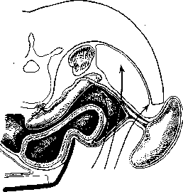
Figure 2. Approaches to the splenic pedicle.
the main vessels are removed from the operative field. A much greater danger threatens when the intima of these vessels, after rupture, does not cause bleeding at all, and the surgeon sutures the abdominal cavity without noticing the complication, and in the postoperative period, when blood pressure is restored or accidentally increases with a coughing thrust or vomiting, patients die from sudden profuse bleeding into the free abdominal cavity. It is necessary to emphasize that this complication is also observed in cases of technically very simple splenectomies. When dealing with a completely mobile spleen with a magistral type of its pedicle, the entire operation comes down to ligation between two ligatures of the main trunks of the splenic artery and vein. By thus eliminating the blood supply to the organ, it is boldly severed (like a kidney) from the remaining connections, and the short vessels of the gastric fundus give abundant retrograde bleeding. Sometimes the adhesions of the spleen to surrounding organs and tissues are so abundant and massive that its removal does not seem possible; in such cases, it is recommended to limit oneself to ligation of the splenic artery, expecting that atrophy of the organ will follow [Lotsch, Blain, v. Stubenrauch propose this operation instead of S. in case of organ dysfunction, Lotsch, Brzhozovsky-as a preliminary act in splenectomies to preserve blood reserves in the body]. However, there are reports indicating the danger of organ necrosis developing in these cases (see Spleen, colliquative necrosis). Lesen, referring to Lombard and Deboucher, recommends using the subcapsular S. in cases of significant adhesions. After the spleen has been removed, it is necessary to recheck the reliability of all ligatures on the pedicle, carefully examine the wall of the gastric fundus (in search of retracted vessels), wipe the posterior abdominal wall and diaphragm with a swab (in search of temporarily thrombosed vessels), and only after this, peritonize the operative bed with the remnants of the ligaments (lig. phrenico-colicum, gastro-lienale) and the parietal peritoneum of the posterior abdominal wall. The abdominal cavity should always be sought to be sutured tightly. Leaving tampons is extremely dangerous due to the subsequent suppurations that develop, causing subdiaphragmatic abscesses with all the ensuing consequences. It is better to spend time on more careful hemostasis than to be tempted to leave tampons to stop 'parenchymal bleeding'.
Related articles
Mentioned in
Cite this page
“Splenectomy.” Soviet Medical Encyclopedia. English translation of Bolshaya Meditsinskaya Entsiklopediya, 1st ed. (Moscow, 1928–1936), ed. N. A. Semashko. https://sovietmedicalencyclopedia.pages.dev/article/splenectomy/