Leukemia
Historical document, translated for reference. It reflects medical knowledge of the 1920s–30s and is not medical advice.
Summary
Leukemia is a systemic disease of the blood-forming apparatus characterized by hyperplastic growth of lymphoid or myeloid tissue or reticulo-endothelial tissue, accompanied by an increase in white blood cells in the blood. The article discusses its classification, clinical manifestations, etiology, and pathogenesis.
Encyclopedia article (1928–1936)
Leukemia, (leukaemia; Virchow, 1845), a systemic disease of the blood-forming apparatus, based on the hyperplastic growth of lymphoid or myeloid tissue or reticulo-endothelial tissue, accompanied by an increase in the blood of the quantity of white blood cells. In one or another form of the leukemic process, the entire blood-forming system is affected, the part of the blood-forming tissue opposite to this form of L. also undergoes hyperplasia and begins to produce cells corresponding to the nature of the leukemic process; thus, in lymphoid L., the bone marrow undergoes lymphoplastic metaplasia, in myeloid L., lymphatic tissue undergoes myeloplastic metaplasia. The growth of blood-forming tissue in L. can also have an aggressive character, so that in the nosological system of diseases, L. is located between hyperplastic processes and true tumors and is considered as neoplastic growths of blood-forming tissue (hemoblastoses), in which they constitute the group of "leukemias"-lymphadenoses, myeloses, and reticulo-endothelioses. As for the increase in white blood cells in the blood, i.e., true white blood disease, in contrast to leukocytosis, this increase is not only very significant and persistent, but usually also progressive; moreover, the increase itself occurs at the expense of young, immature forms of white blood cells, which are either not observed at all in normal blood or are noted in single instances, for example, as a transient phenomenon in leukocytosis with the so-called shift to the left. In individual cases, the increase in white blood cells can be so significant that their ratio to red blood cells becomes 1:1, and the blood itself becomes whitish, resembling a mixture of cranberry juice with milk. There are, however, cases of L. in which the absolute number of leukocytes is within normal limits, and only upon examination of the leukocyte formula is the characteristic symptom of a significant rejuvenation of leukocytes or an unusual distortion of the percentage relationships of individual forms immediately apparent, for example, in the form of a sharp predominance of lymphocytes. As a general rule, the change in blood intensifies toward the end of the disease, reaching the highest degrees in the atonal period. L. can have chronic and acute courses; chronic course can have an acute ending; finally, the course of L. can be subacute. In addition, the leukemic process in blood-forming tissue is not always expressed clinically by a full-fledged leukemic hematological syndrome; the latter can also be completely absent, accordingly subleukemic and aleukemic forms of the disease arise. Sometimes these forms follow one another as stages of the same process; thus, aleukemic periods can precede leukemic ones. Leukemic shifts in blood are generally not persistent; thus, if leukemics get any infection (pneumonia, erysipelas) or exacerbation of a past infection (tuberculosis), the leukemic composition of their blood often undergoes significant changes and, more often, in the sense of a return to normal, which simulates recovery or improvement. Etiology and pathogenesis of chronic L. are not clarified. There is a theory that considers leukemic growths as true neoplasms (Ribbert, Banti). The possibility of tumor-like growths in L. and the possibility of malignant growth of these growths, i.e., growth that destroys adjacent tissues, bring these diseases closer to malignant tumors. However, L. are systemic diseases, while foci of heterotopic origin of leukemic tissue should be considered not true metastases, but phenomena of autochthonous blood formation in the connective tissue of organs and tissues. The participation of the vascular system in the blood-forming function acquires special significance. Proliferative and metaplastic processes on the part of "coastal cells", i.e., endothelium, as well as adventitial cells, lead to the complete closure of white blood cells in the lumens of vessels, especially capillaries [see separate table (pp. 543-544), Fig. 6], turning entire systems of vascular branches from blood vessels into blood-forming ones (Davydovsky); naturally, destructive changes in the vascular walls are also observed in this case, which contributes to the development of hemorrhages, i.e., the appearance of the so-called hemorrhagic diathesis, this usual companion of L., especially acute ones. The anatomical manifestations of hemorrhagic diathesis are hemorrhages in the skin, mucous membranes, serous membranes; it is also characteristic to appear with fibrinohemorrhagic deposits on the inner surface of the dura mater. To the same category of phenomena should also be attributed hemorrhages in the brain, sometimes symmetrically located. It is clear that such changes in capillaries and small vessels must be accompanied by a violation of tissue nutrition: ulcerative-gangrenous processes are a very frequent phenomenon in leukemics. Blood-forming processes along the course of the connective tissue stroma of organs and tissues are expressed morphologically either in the appearance of grayish, gray-greenish rounded nests or in the diffuse enlargement of the organ while completely preserving its inherent contours. Thus, sometimes "large leukemic kidneys", large ovaries, uterus, appendix are found, and the microscope reveals that the main mass of the organ consists of newly formed cellular elements, while the parenchymal elements are strongly pushed apart, thinned out. It is important to note that such diffuse leukemic hyperplasias and infiltrates do not manifest themselves clinically in any special way: for example, urine in "large leukemic kidneys" may not show special changes. Relatively often, diffuse mesenchymal blood formation leads to tumor-like formations (see below). There is an attempt to explain the leukemic process as a violation of correlation in blood formation (Ziegler). For example, X-rays cause atrophy of the spleen follicles, and as a result of their death, as a violation of correlation between lymphatic and myeloid systems in the body, myeloid tissue develops in the place of the lymphatic apparatus of this organ. One can assume the occurrence of myeloid L. on the same principle. The possibility of endocrine influences on the correlation between lymphatic and myeloid systems is permissible, and a correlation disturbed in this way can manifest itself by proliferation of one or another blood-forming tissue (Naegeli). The frequency of L. diseases among radiologists is noted, and X-rays can play the role of a disruptor of this correlation in blood formation. At present, the theory of infectious origin of chronic L. attracts the most attention. The universality of the lesion of the blood-forming system, the occurrence of autochthonous foci of leukemic tissue in organs indicate the generalization of the pathogenic agent, similar to what is observed in infectious diseases. From this point of view, it is quite permissible to assume the possibility of the existence of a virus in L., primarily localized in the blood-forming system and giving a tissue reaction of the leukemia type. However, its existence has not yet been proven. The finding of unknown Protozoa in leukemic blood has significance only for the history of the question. The finding of various bacteria in the blood of leukemics speaks only of accidental infection, to which leukemics are so prone. Attempts to transmit the disease by inoculating the blood of a sick person to a healthy one gave negative results in humans. Only in birds is such transmission from individual to individual possible. There are indications in the literature on focal and familial occurrence of the disease. However, all these data have not been verified and do not yet make it possible to resolve the question of the infectious nature of chronic L. In relation to acute L., there are more reasons to assume an infectious origin, taking into account the acute course of the disease, quite high temperature and the general condition of the patient, resembling a severe infection. There is a definite opinion (Sternberg) that distinguishes acute myeloid L. into a special group and considers it a streptococcal disease, since streptococcal diseases usually give a picture of significant irritation and proliferation of myeloid tissue. In infants with various septic processes, a very high leukocytosis (for example, 50-60 thousand) is often observed, which borders on that observed in L.; at the same time, the leukocyte formula can give pictures close to leukemic (for example 80% neutrophils or as many lymphocytes). However, even here one cannot deny the possibility of secondary infection with leukemic blood. The same must be said in relation to other bacteriological findings in acute L. On the other hand, chronic forms can take an "acute course." Therefore, such a separation of some acute L. into a special group as simply infectious diseases cannot be accepted. Whether constitutional predisposition plays a role in the development of L. after any infection (status lymphaticus) cannot be said definitely. Thus, the available data are insufficient to resolve the question of the infectious origin of acute leukemia. In connection with the question of the etiology of L., the existence of the so-called leukemoid reactions, observed in some local and general suffering, essentially different from L., should be noted.
Thus, there are known cases of cancers (stomach, lungs) with a leukemic reaction from the blood and hematopoietic organs, or cases of widespread tuberculosis of the lymph glands with similar blood pictures. It is hardly possible to speak of a simple coincidence of two (leukemia + cancer, resp. + tuberculosis) diseases in such cases; it is more correct to assume that here we have a special reaction from the blood and hematopoietic organs to the corresponding factors underlying or accompanying various neoplastic, resp. inflammatory processes. In other words, there is a strong tendency not to base the development of L. on any specific etiological causes (e.g. one or another pathogen), but to consider them as a special (leukemic) type of reaction of the organism to diverse and essentially different ailments associated with significant metabolic disorders in some respect. Moreover, it should be pointed out that even in the anat.-hist. relation the hematopoietic organs (and probably all mesenchyme) in different subjects show some and sometimes significant deviations (e.g. the amount of myeloid tissue in the long bones, inclusions of lymph follicles in the bone marrow). It is natural to expect, therefore, that functionally these organs will also differ in a certain peculiarity; the extreme variants of such peculiarity are apparently the leukemic processes as a whole. To what extent these structural and reaction peculiarities are conditioned by genotypical factors is a question for the future. At present, however, there are some facts speaking in favor of this assumption, e.g. several cases of L. in one family or the development in one member of a typical L., in another--a leukemoid reaction. Some objection to the genotypical nature of L. may be the fact of the transmissibility of the disease, e.g. in chickens. Mice, dogs, cats, and pigs may also suffer from L. L. is widespread everywhere. It is difficult to establish in what regions this disease occurs more frequently. There are some indications of a greater frequency of this disease in Poland among the poor Jewish population.- All ages are capable of contracting L., most often middle age; up to 4 years of age L. is encountered exceptionally rarely. In general, L. is a rare disease: in Moscow for 1923-27, out of 33,609 autopsies (including newborns), 51 cases of chronic L. and 35 cases of acute were noted; consequently L. constitute 0.25% of autopsies. However, cases of congenital L. have been described repeatedly. Boys get sick more often than girls; for the lymphadenoic form of L. the ratio is 2:1. Chronic leukemias. Clinically, chronic lymphadenoic, or lymphatic (1. lympho-ides) leukemia (chronic lymphadenosis) first of all manifests itself with symptoms from the lymphatic apparatus, namely enlargement of the lymph glands. The development of the disease is usually slow, gradual. Painless enlargement of the glands either begins in some one area, or the disease at once becomes generalized, or in some one area the enlargement of the glands predominates over other places. Similarly, the lymphatic apparatus of internal organs and the lymph glands located inside the body become hyperplastic: intestinal follicles, mesenteric glands, mediastinal glands, etc. In individual cases of the disease, such selective and predominant affection of this lymphatic apparatus leads to the formation of tumor-like growths, dominating in their symptoms in the picture of the disease and giving it a quite peculiar character (tumor of the mediastinum, of the abdominal cavity, etc.). The onset of the disease with the lymphatic apparatus of the pharynx (tonsils), Meniere's symptom complex on the basis of infiltration in the inner ear, protrusion of the eyeball in infiltrates at the bottom of the eye, priapism from affection of the cavernous bodies of the penis--all such symptoms can greatly vary the picture of the disease and complicate the diagnosis. Enlargement of the liver due to the development of lymphatic tissue in the connective tissue of the organ is a common symptom of L. The spleen always participates in the process, but not always to the same degree; sometimes splenomegaly suppresses other manifestations from the lymphatic apparatus. Affection of the skin also does not constitute a great rarity (see below). Skin hemorrhages are not uncommon. In general, the hemorrhagic diathesis in L. manifests itself with various hemorrhages, especially nasal, but also in other organs (in the stomach from leukemic ulcers, in the nervous system, etc.). Their cause is the destructive processes accompanying the proliferation and metaplasia of the vascular endothelium and adventitial cells. Good health is long preserved in patients with chronic lymphadenosis, although painful sensations in the bones as a clinical expression of affection of the bone marrow often trouble the patients. Gradually, however, exhaustion, cardiac weakness, edema and inevitable death develop. The duration of the disease is on average up to 8-14 years. In childhood the course of the diseases is usually more rapid, and their duration is limited to months. Fever is usually absent or gives insignificant fluctuations of temperature; greater temperature movements occur only in some complications, as well as in childhood. In the absence of complications with nephritis the urine remains unchanged; in rare cases in the urine is excreted the Bence-Jones protein; often large amounts of uric acid are washed out with the urine as a result of the intensified breakdown of the corresponding blood cells.-The most demonstrative changes are those from the blood, namely the white part of it (see separate table, fig. 1). They consist in an increase in the number of leukocytes to hundreds of thousands, sometimes to a million, while in the leukocytic formula small lymphocytes predominate in enormous quantities (usually up to 90%). They most often appear in the form of narrow-protoplasmic forms, almost naked nuclei, especially with large numbers of leukocytes; the nuclei often show phenomena of pyknosis, the chromatin collecting in clumps, leaving light intervals between them, whence the comparison of the structure of the nucleus with a wheel (see separate table, fig. 6 and 6a). In old people with great chronicity of the disease wide-protoplasmic lymphocytes usually predominate, with a large content of azurophilic granularity. Much more rarely in the blood are lymphoblasts encountered as a preceding stage of the small lymphocyte, even more rarely--lymphoidocytes as the universal mother cell. Mitoses in lymphocytes are not frequent. By chance neutrophilic myelocytes are also found in the blood as products of myelocytosis from irritation around leukemic lymphomas in the bone marrow. In general other kinds of leukocytes are present in the blood in scanty amounts. The amount of Hb and erythrocytes can for a long time remain normal, but at the end of the disease usually a severe anemia develops with normoblasts and megaloblasts in the blood.-- The diagnosis of the disease follows from its clinical picture and the changes in the blood.-The prognosis is hopeless even with treatment, which can only prolong the life of the patient. Pathological anatomy. Characteristic is the significant enlargement of the lymph glands, sometimes of all the glands of the body, sometimes only of certain regions, e.g. the neck, mediastinum, abdomen. The glands are dense, pale-gray in color, succulent, not adherent to the skin, usually movable, but may also be immovable when the process involves the capsule of the glands and the adjacent cellular tissue. On microscopic examination--phenomena of diffuse hyperplasia of lymphoid cells with complete obliteration of the gland structure; at the beginning of the process the usual structure may be observed with sharp hyperplasia of the follicles. The spleen is always greatly enlarged, dense, at the beginning of the disease soft, gray-red, with clearly noticeable round or angular Figure 1. Chronic myeloid leukemia. Figure 1a. Chronic myeloid leukemia: a-lymphoidocyte; b-leukoblast; c-eosinophilic promyelocyte; d-neutrophilic promyelocyte; e-basophilic promyelocyte. Figure 2. Acute myeloid leukemia. Figure 2a. Acute myeloid leukemia. Erythroblastic series: a-megaloblasts; b-macroblasts; c-normoblast; d, e-karyorrhexis in normoblasts; f-normoblast with basophilic granularity. Figure 3. Acute myeloid leukemia (macromyeloblastic=macrolimphocytic type): a-macroblast; b-basophilic normoblast; c-Humprecht's body; d-mesomyeloblasts (=mesolymphoidocytes); e-macromyeloblasts (=macrolymphoidocytes). Figure 3a. Acute myeloid leukemia (macromyeloblastic=macrolimphocytic type): a-macromyeloblast (=macrolymphoidocyte); b-the same with azurophilic granularity (transition into leukoblast); c, d-strongly vacuolated myeloblasts. Figure 4. Acute myeloid leukemia: a-normoblast; b-lymphocyte; c-mesolymphoidocytes with vacuoles containing azurophilic grains; the other cells are mesomyeloblasts and micromyeloblasts (=meso- and microlymphoidocytes). Figure 4a. Acute myeloid leukemia (mesomyeloblastic and micromyeloblastic = meso- and microlimphocytic types). ! Figure 5. Acute myeloblastic leukemia (from a case of chloroma; predominance of the so-called Rider forms). Figure 5a. Various forms of myeloblasts and leukoblasts with strongly polymorphous nuclei. Figure 6. Chronic lymphatic leukemia; a-Humprecht's body. Figure 6a. Chronic lymphatic leukemia; a-mature lymphocytes. Figure 7. Acute lymphatic leukemia; a-Humprecht's body. Figure 7a.
Acute lymphatic leukemia; lymphoblasts.
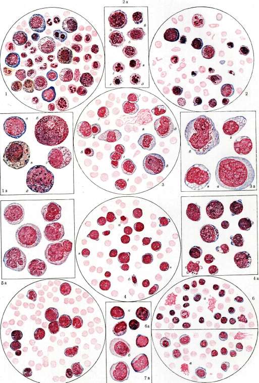
See Leukemia.
with light gray follicles; the capsule is significantly tense; tears in it are possible; in places, gray or yellowish infarcts are noted. Later the organ increases even more, its density increases; the color of the cut becomes dark brown, and the pattern itself is mottled due to thrombosis of vessels, infarcts, hemorrhages, and focal scleroses. The capsule is unevenly thickened; adhesions with surrounding organs often occur. On microscopic examination - marked hyperplasia of Malpighian bodies, abundance of lymphoid cells in the pulp; in necrotic areas, sometimes (in contrast to myeloid leukemia - rarely!) crystals of Charcot-Leyden are found (leucine and tyrosine as products of protein breakdown); in late periods - significant depletion of cells, diffuse sclerosis of the pulp, especially with intensive X-ray treatment. - Bone marrow: universal hyperplasia with transformation of the red-gray (in the epiphyses) and yellow (in the diaphyses) bone marrow into raspberry-red. Microscopically - diffuse hyperplasia of lymphoid cells with more or less significant, almost complete displacement of myeloid hematopoiesis. In some cases, a pale color of the bone cut is observed, disappearance of the succulence of the marrow - a phenomenon of leukemic osteosclerosis: thickening of the trabeculae of the spongy substance, some sclerosis of the bone marrow tissue. Cases where the bone marrow has no special changes (aplastic leukemias) are very rare. - The liver is sometimes enlarged 2-3 times, pale, rather firm; on the surface and in the cut, small grayish nodules, so-called lymphomas, i.e., rounded foci of cellular hyperplasia along the course of Glisson's capsule, are often visible; in the parenchyma, deposits of fine-grained hemosiderin are common (also observed in the spleen and bone marrow). - The blood of the corpse is liquid, light, with jelly-like gray clots. The heart is very flabby, the muscle is clay-like, usually with clearly expressed phenomena of fatty degeneration; its cavities are dilated. A significant tendency to diffuse hyperplastic processes in various organs (kidneys, prostate, testes) [see separate table (art. 543-544), fig. 5] with their enlargement (see above). A tumor-like enlargement of the thyroid gland, of the lymphatic apparatus of the root of the tongue, often strikes the eye. It is even considered that the pronounced involvement of the thymus in the process is a reliable sign of lymphatic leukemia. Hemorrhagic diathesis, necrotic changes of the mucous membranes are usually absent or weakly expressed. Aleukemic lymphadenosis (the name 'pseudoleukemia', according to Congheim, is now abandoned). The clinical picture exactly corresponds to the picture of chronic lymphemia, but without the hematological symptom of leukemia. The red part of the blood remains normal for a long time; later anemia is discovered. The number of leukocytes is normal, subnormal or slightly increased. The leukocyte formula rarely remains normal; usually a relative lymphocytosis is noted, and among the lymphocytes, lymphoblasts may also be found. Between aleukemic and leukemic forms of lymphadenosis, all transitional forms exist ('subleukemic' lymphadenosis). - Diagnosis of those cases where there is a relative lymphocytosis and precursors of lymphocytes in the blood is not difficult, but is difficult in the absence of this symptom. Differentiation from other lymphomatoses - lymphogranulomatosis, tuberculous and syphilitic lymphomas, and malignant diseases of the lymph glands - is most confidently resolved only by biopsy of the gland, more difficult - by the cellular punctate of the gland, easier - by puncture of the bone marrow, showing the lymphocytic cellular composition of the smear. Puncture of the bone marrow is absolutely necessary for those rare cases of aleukemic lymphadenosis where the process is localized only in the bone marrow and the clinical disease is expressed only by anemia, as well as when the involvement of the glands remains clinically unnoticeable, but in the picture of the disease the enlarged spleen predominates as a symptom-complex of anaemiasplenica. - The prognosis of the aleukemic form does not differ from the prognosis in leukemic form. Myeloses. In the clinical picture of chronic myeloid leukemia or chronic leukemic myelosis, the enlargement of the spleen stands out especially sharply despite the fact that the basis of the disease here is the lesion of the bone marrow. Previously this form of leukemia was called splenic or splenogenic and was opposed to myelogenous. At present this division is considered obsolete. Corresponding to the presence of splenomegaly, the first sensations of the patients refer to the splenic area, where there may be a feeling of pain and heaviness, a feeling of a foreign body; then phenomena of progressive weakness, dizziness, bone pain, etc., appear. The lymph glands, metaplastically myeloplastic, rarely enlarge to such sizes as in lymphatic leukemia. Sometimes their enlargement is completely unnoticeable despite the presence of histological changes. In some cases, even in myeloses, tumor-like growths of myeloid tissue in the mediastinum, abdominal cavity, etc., are formed. The liver is always enlarged due to heterotopic neoplasm in it of hematopoietic tissue. Ascites may also be observed. Lesions of the skin are rare. When infiltrates are located anywhere in organs vital for life, a special clinical picture can develop, as in lymphemia. The Bence-Jones body is rarely found in the urine. Hemorrhagic diathesis is common by the end of the disease. The blood picture is always characterized by various degrees of anemia with normoblasts. The number of leukocytes is increased to hundreds of thousands, with a mixed cellular composition and varying degrees of rejuvenation of the leukocyte formula of granulocytes: metamyelocytes, myelocytes, promyelocytes, lymphoidocytes (myeloblasts, myelogenies). In the blood there are young cells of all types of granularity, but predominantly of the neutrophilic series, however, sometimes the number of basophilic myelocytes and eosinophils increases greatly [see separate table, fig. 1a]. A shift of the nuclear formula to the left in granulocytes is noted simultaneously with the appearance of giant forms with polysegmented nuclei. In very chronic forms of the disease in old people, as well as in periods of remission of the disease, the number of polymorphonuclear neutrophils greatly predominates over the young forms. During exacerbation of the disease, the number of maternal cells increases significantly. Monocytes and small lymphocytes are always in small numbers. The number of platelets increases. - The course of the disease is progressive, leading to cachexia. The duration of the disease is several years, rarely - up to ten years. Death can occur quickly due to various complicating diseases. The transition of chronic myelosis into an acute course is observed more often than in lymphemia. - Diagnosis of the disease presents no difficulties. Only rare cases of large leukocytosis (up to hundreds of thousands) can cause doubt, as well as the picture of leukemoid blood with myelocytic elements. However, in myeloses there is a reaction from the entire granulocytic system, and not only from neutrophils, as in cases simulating myelosis. On patho-anatomical examination, diffuse hyperplasia of the entire bone marrow is found with a change in its color to gray, gray-green, and green; in the latter cases, they speak of 'pyoid' (i.e., resembling pus) bone marrow or of chloromyelosis; the change in color is due to the abundant proliferation of myeloid forms (myelocytes, myeloblasts). In very rare cases, the bone marrow is macroscopically unchanged (aplastic forms). - The spleen is greatly enlarged (sometimes 10-20 times) and its lower border almost enters the pelvis; on average, the enlargement is more constant and significant than in lymphatic leukemia. The follicles of the organ are greatly reduced, mostly completely unnoticeable: the reaction of the pulp cells to oxidase is sharply positive due to diffuse myelosis of the pulp; sometimes there is some sclerosis of the organ; infarcts and scars with deposits of iron-containing pigment are often observed; the capsule is often strongly and unevenly sclerosed; adhesions with surrounding organs are common. - The lymph glands are either not enlarged at all or the enlargement is small and usually not universal. The color of the glands is reddish; microscopically - proliferation of myeloid forms in the sinuses and medullary cords, as a result of which the pattern of the gland is strongly erased. Blood clots of significant size, their color is gray, brown-chocolate or gray-green. As in the blood, so in the hematopoietic organs, the precipitation of needle-shaped (less often - rhombic) Charcot-Leyden crystals is often observed, especially in leukemia with abundant content of eosinophilic forms. It is considered that the appearance of protein crystals (see) is a fairly reliable sign of myeloid leukemia. It should be borne in mind that neither significant enlargement of the spleen nor diffuse myelosis of it (by themselves and taken together) are specific for myeloid leukemia; they can also be observed in malignant anemia, in diffuse metastatic carcinoma of the bone marrow, in leishmaniasis, and in some other splenomegalies. There is a tendency to myeloid proliferation along the lymphatic apparatus of the digestive tract, sometimes with the formation of ulcers simulating typhoid fever. - The liver is always greatly enlarged, smooth, pale on the cut with a grayish tint. - Microscopically - more or less abundant proliferation of myeloid elements and moreover not along the periphery of the lobules, which is characteristic for lymph.
L., and along the course of the capillaries of the lobules (intraacinous), sometimes with sharp atrophy of the liver parenchyma itself [see separate table (pp. 463-464), Fig. 1]. A leukemic myelosis has the same picture, but without the pronounced hematological leukemic symptom. Individual myelocytes, as well as promyelocytes and myeloblasts, are still found in the blood. These young forms are very characteristic where the number of leukocytes is small. But the leukocyte formula can also be normal. An enlarged spleen and anemia in such patients prevail in the picture of the disease. - The diagnosis here appears to be very difficult, as it requires differentiation between other forms of splenomegaly with anemia - Banti's disease, malignant granuloma of the spleen, hemolytic jaundice, etc. Biopsy or puncture of the bone marrow helps in the diagnosis, during which a clearly expressed myeloid proliferation is found with a return of bone marrow tissue to an undifferentiated state and with the production of early-mother cells. Puncture of the spleen is not conclusive, as myeloid metaplasia of the spleen can also be observed in other diseases. Acute leukemias. The clinical picture of acute leukemias in lymphadenoses and myeloses shows no differences. Acute myeloses are most common. Acute L. usually begins abruptly, with high temperature, severe general condition, and hemorrhagic diathesis. Sometimes there is a prodromal stage of malaise and general weakness for some time. Often the disease begins with profuse nosebleeds and gangrenous-inflammatory changes in the oral cavity, pharynx, with extreme pallor of the patient (gangrenous leukemic angina). Bleeding can occur in the skin and in the most diverse organs and can become fatal for the patient's life. The lymph glands show enlargement, but slight, and besides leukemic glands, there can be also inflamed glands as a result of a reaction to inflammatory and gangrenous processes in the patient. The spleen increases in size, but significantly less than in chronic L. The liver also swells when hematopoietic tissue develops in it. Leukemic periostitis is very common. In acute L., tumor-like growths of leukemic tissue in the mediastinum and other parts of the body are also observed. Secondary infection is extremely easily added, which in some cases can favorably influence the leukemic process, which is expressed by a decrease in the number of leukocytes in the blood and improvement of the formula, and in others accelerates the fatal outcome (pneumonia, erysipelas, etc.). - Duration of the disease - from several days and weeks to a few months. The fatal outcome should be considered the rule, exceptions to it are questionable. Clinically distinctive is the form of acute L., known under the name of chloroma (see separate table, Figs. 5 and 5a). For this form, tumor-like periosteal growth and a greenish tint of hyperplastic formations are characteristic. Chloroma is rarely found in infants, but most often it is found in preschool and school-age children. In general, half of all cases of chloroma fall on childhood. All other symptoms of acute L. are usually expressed in chloroma as well, which connects these two diseases. In typical cases of chloroma, patients have an extremely characteristic appearance due to the change in the configuration of the skull as a result of tumor-like growths in the temporal regions, protrusio bulborum with chemosis of the eyelids and waxy pallor of the skin. Blood picture. On the side of the red elements, rapid development of severe anemia is found, usually of hyperchromic type with degenerative and regenerative signs (anisocytosis, poikilocytosis, micro-macrocytosis, anisochromia, polychromasia, basophilic stippling, erythroblasts with various pictures of nuclear metamorphosis) (see separate table, Figs. 2 and 2a). The picture of the white part of the blood is diverse. Here there can be a mixed-cell leukocyte formula, as in chronic forms of myeloses, but more often an increase in basophilic leukocytes and eosinophils in the blood is absent, while myeloblasts can be in varying amounts (acute myeloid L.). In some cases, a clear predominance of erythroblastic forms is noted (erythroleukemia). In other cases, the disease proceeds as acute L. from mother cells of the leukocyte series (myeloblasts, lymphoidocytes), in the greatest percentage or exclusively constituting the leukocyte formula (acute myeloblastic L.). Mother cells can be represented in macro- and microgenerations, which correspond to macro- and microlymphoidocytic L. (see separate table, Figs. 3, 3a, 4, 4a). The same blood picture can also be in acute lymphadenoses, which creates difficulties in recognizing the disease during the patient's life. Indeed, the diagnosis of myelosis can be facilitated by the presence of a positive oxidase reaction in lymphoidocytes in myeloses, but this sign is not decisive, as it can also be absent in myeloid nature of the disease. And since the cellular structure of mother cells in myeloid and lymphoid L. does not show bright differences, the diagnosis is established only pathoanatomically and not always. In cases where both myelocytes and of the same size as lymphoidocytes are present in the blood, one can think of the myeloid nature of the disease. Individual myelocytes can be in the blood also in acute lymphemia as a reflection of the so-called 'myelocytosis from irritation' around the proliferating lymphadenoic tissue in the bone marrow. In such cases, however, their size is arbitrary and does not necessarily harmonize with the size of the lymphoidocytes of this case of L. Leukemia from microlymphoidocytes differs from acute L. with predominance of microlymphocytes by the structure of the cell nuclei, revealing a delicate chromatin network, although dense, but not collecting into chromatin lumps, as in microlymphocytes. The total number of leukocytes in acute myeloses varies, as in chronic leukemias. Sometimes acute myeloses proceed without an increase in the number of leukocytes (acute aleukemic myelosis) and even with leukopenia, and in some cases myelocytes are present in the blood, in others - only myeloblasts. If there is no simultaneous increase in lymph glands and spleen, it is very difficult to distinguish such cases from infectious diseases; hence the tendency of some to isolate acute myeloses from the concept of L. (see also Myeloses). In acute lymphadenoses, all transitions from lymphoidocytes and lymphoblasts to microlymphocytes are usually present in the blood (see separate table, Figs. 7 and 7a). Sometimes, however, only lymphoidocytes are present in the blood, and then the nature of the disease is determined only pathoanatomically. Tumor-like lesion of the thymus, if present, can also speak for the lymphadenic nature of the disease (not absolutely). Differentiation from septic processes proceeding with suppression of the granulocytic apparatus is very difficult here and can be done only pathoanatomically - by finding signs of hyperplasia somewhere in the lymph tissue. A certain part of the so-called agranulocytic anginas also belongs to these diseases. Mother cells in acute lymphadenoses often appear in the form of Rieder forms with irregular lobed nuclei and in some cases show various inclusions in the protoplasm, more often of azurophilic nature. In cases with clearly expressed clinical symptoms, the diagnosis of acute L. is not difficult; in other cases it is almost impossible during life and is resolved pathoanatomically. - The classical symptoms of acute L. on autopsy are hemorrhagic diathesis (bleeding into the skin, mucous membranes, serous membranes), gangrenous-ulcerative changes in the area of the pharynx, gums, mouth (angino-scorbutic form of acute L.), and sometimes in other parts of the gastrointestinal tract; in particular, leukemic hyperplasia and ulcerations of the lymph apparatus of the intestine can simulate typhoid fever, gangrenous processes of the pharynx - diphtheria, and bleeding from ulcerated gums and cheeks - scurvy. The spleen is enlarged slightly or strongly; in the abundantly scraping pulp, many young myeloid forms are found. The bone marrow is diffusely hyperplastic, red or gray-red, often gray-green (acute chloroleukemia). The lymph glands are mostly enlarged, but often their size is usual, and only microscopic examination reveals diffuse myelosis of them and blurring of the organ pattern; sometimes the glands also have a greenish color. Proliferation of myeloid forms (mainly myeloblasts) is sometimes found everywhere (in the brain, stomach, tongue, intestine, kidneys, liver, testes, etc.), accompanied by hemorrhages; significant bleeding can occur, for example, in the brain (leukemic apoplexies). The blood and blood clots in acute L. attract attention with their gray, and sometimes gray-green color. The nature of the substance itself that gives the tissues a green color remains unknown; on freshly cut objects, the green color gradually fades, acquiring a grayish tint; when exposed to ammonia vapors, the color can be restored and maintained for a considerable time.
The green coloration of pathologically enlarged glands in itself is not specific for Leukemia and can be observed, for example, in myelomas, in tuberculous lymphadenitis (without the simultaneous occurrence of caseous-purulent necrosis).- Therapy for acute leukemias is hopeless and can only be symptomatic. Both chronically and acutely, leukemic and aleukemic forms of so-called plasma-cellular leukemias can occur, in which the hematopoietic tissue shows proliferation of plasma cells, found in varying quantities in the blood. This rare form of leukemia may be related to both the lymphoid and myeloid systems, since the origin of plasma cells is possibly from agranulocytic elements in both.- In some cases, the leukemic process, running acutely or chronically, shows aggressive growth with infiltration of surrounding organs and the formation of tumor-like growths that exert pressure on neighboring organs. These leukemias constitute a group of sarcoleukemias (leukosarcomatoses-see), aleukemic and leukemic; in some cases the leukemic tissue remains unstained, while in others it contains a green pigment (chloromas). Furthermore, sarcoleukemias can be of lymphoid and myeloid type, showing the same variations in the leukocyte composition of the blood as is characteristic of other forms of Leukemia. Unstained sarcoleukemias most often have a lymphoid character and lead to excessive proliferation of some group of lymph glands, usually in the mediastinum, sometimes in the skin, etc. Chloromas, either of myeloid or lymphoid type, most often manifest as tumors of the orbit with protrusion of the eyeball, along the spine, etc.- Diagnosis can be established by biopsy. In rare cases, even chronic Leukemias have a colored pathological-anatomical substrate. Sometimes only part of the leukemic tissue is colored green. The prognosis is hopeless. The question of mixed Leukemias remains unresolved. In the blood in such cases, alongside lymphocytosis, myelocytosis is also noted. Some cases of mixed Leukemias should be interpreted as micromyeloblastic Leukemias, where it is easy to confuse microgenerations of myeloblasts with microlymphocytes. In general, it should be emphasized that significant difficulties often arise in the hematological classification of Leukemias (especially acute), and mainly because to this day there are no absolutely precise criteria for differentiation of the youngest forms of red and white blood cells (lymphoblasts, myeloblasts, erythrogonia), which differ at certain stages of development by extremely insignificant and inconsistent details, not to mention that the fundamental questions about the genesis of the formed elements of the blood, in particular the question of the special origin of monocytes, remain controversial (see Hematopoiesis). The introduction into hematological practice of the oxidase reaction [see separate table (pp. 463-464), fig. 2] although it has brought some clarity to certain cases, but in the most obscure cases (with the least mature, entirely 'lymphoid' elements) this reaction does not give definite results, being for example negative or doubtful in undoubtedly myeloid forms of Leukemia. If this reaction comes out weakly positive, then the interpretation of the case is difficult on the other hand, namely that the most sensitive reagents used for this reaction (dopa-reaction with dihydroxyphenylalanine) give positive results with almost any cell regardless of its origin. On the basis of all these difficulties, the proposal has long existed to designate all Leukemias with a sharp rejuvenation of the formed elements with the indifferent term 'hemocytoblastic' Leukemias (or hemohistiocytoblastic; Stammzellenleukämie of German authors), i.e., Leukemias from blood-forming forms, which will apparently be the most rational solution at our current level of knowledge. All leukemic processes occur with anemias, and the involvement of the red part of the bone marrow in these diseases is undeniable. Sometimes anemia reaches a great degree. In such cases, one can speak of leukoanemia (see). In some cases, Leukemia as the final chord of the disease process is characterized by the extreme degree of anemia with the disappearance of leukemic features in the blood. There is no transition of Leukemia into pernicious anemia, but only the extreme exhaustion of the leukoplastic ability in the hematopoietic tissue. In Leukemia, inflammatory and atrophic changes of the tongue type of Hunter's glossitis, atrophy of the gastric mucosa (clinically manifesting as achylia), combined scleroses along the conducting systems of the spinal cord can be observed. All this (see also above on the spleen in myeloid Leukemia) allows one to see in Leukemia, as it were, a certain degree of kinship with malignant anemia. The question of monocytic Leukemias awaits its resolution. However, facts are accumulating that testify to the possibility of proliferation of the reticulo-endothelial system as a substrate for the occurrence of monocytic-leukemic or aleukemic (leukopenic) hematological syndrome, 'reticulo-endotheliosis' with acute or chronic course. In the blood in such cases, leukocytes of the monocyte type are found. Some of the diseases belonging here are undoubtedly myeloblastic Leukemias with broad-protoplasmic old atypical myeloblasts and pathological-anatomical changes of myeloid Leukemia. Whether another part can be considered true leukemias is premature to decide, but one cannot fundamentally object to this, since at least some monocytes of normal blood are ascribed to histiogenic origin. The practical differentiation of individual forms of leukemic, aleukemic processes, as well as adjacent forms such as lymphosarcomatosis, anaemia pseudoleukaemica infantum, etc., can sometimes encounter significant and even insurmountable difficulties due (as one could a priori expect) to the abundance of 'transitional', 'mixed', 'atypical' forms, etc. These difficulties are especially great when examining biopsy material. It is rightly considered that the main mass of errors of pathologists falls on the life studies of lymph glands. Indeed, very similar pictures can be for example in lymph. Leukemia, pseudoleukemia, lymphosarcomatosis, and in the initial stages of so-called lymphogranulomatosis (Hodgkin's disease). This must be remembered by both the therapist and the surgeon, who often resort to such a diagnostic method. Finally, when examining both biopsy and autopsy material, it is important to consider whether treatment with radiant energy took place: the latter can introduce into the histological and microscopic pictures very essential features that strongly distort the basic pictures (necroses, scleroses, inflammatory infiltrates, etc.) or exacerbate some of them (e.g., hemorrhagic diathesis). I. Davydovsky, A. Kryukov. Skin manifestations of Leukemia.The skin can be affected in Leukemia in two ways: 1) in it, as in all other organs, leukemic, histologically specific infiltrates can develop; 2) it can react to the pathogenic cause or to the secondary toxic products caused by it by the development of morphologically different rashes with a histological structure not specific for Leukemia. At the suggestion of Audry, non-specific rashes are called leukemides. Specific skin lesions are observed in both lymphatic and myeloid Leukemia [see separate table (for the article Lichen), fig. 1]. Leukemides are characteristic mainly of chronic lymph. Leukemia. All these rashes can be observed both in pronounced Leukemias and in their sub- and aleukemic forms.-- Skin lesions in myeloid Leukemia are rare and little studied. It has only been established that acute myeloid and myeloblastic Leukemia is often accompanied by more or less extensive hemorrhages into the skin and mucous membranes. Much more characteristic are the specific infiltrates (leukaemia cutis myeloides). Histologically they consist mainly of cells of the myeloid series, which give (although not always) a positive reaction to oxidase. Along with them, lymphoid elements and erythrocytes are encountered in smaller quantities. The infiltrates are located in the dermis and in the upper layers of the subcutaneous tissue, mechanically smoothing out the papillae [see separate table (pp. 543-544), fig. 4]. The starting point for the development of infiltrates is apparently the area around the sweat and sebaceous glands. Most often the infiltrates are localized on the trunk and either barely protrude above the level of the skin or appear as rather sharply limited tumor-like formations up to the size of a walnut or larger. Their color is varied (reddish, brownish, ashen-gray), but always with a bluish tint. Subjectively, the nodes cause no discomfort, but sometimes they are painful on pressure. There is no close connection between the degree of blood changes and the number of nodes. Sometimes nodes develop before changes in the blood occur. Skin lesions in lymph. Leukemia are better studied than in myeloid, as they occur more frequently.
Specific manifestations are expressed: a) by limited nodular formations, characteristic both clinically and histologically (leukaemia lymphatica cutis circumscripta); b) by diffuse reddening and thickening of the skin of the erythrodermia type (erythrodermia leukaemica or leukaemia lymph, cutis diffusa and closely related to it lymphodermia perniciosa Kaposi). The nodules of the first type consist of a dense collection of uniform small lymphocytes and are rich in newly formed dilated blood vessels. Inflammatory phenomena in the nodules are almost completely absent. Starting around the vessels of the deep dermal network, as well as around follicles and glands, the infiltrate is then located in the lower and middle thirds of the dermis and in the subcutaneous tissue. The papillary layer of the dermis is smoothed out but not infiltrated and forms, although narrow, a characteristic sharp boundary between the infiltrate and the epithelium. The latter often undergoes secondary changes. In leukaemia cutis diffusa, the histological picture is much more varied, both in terms of the greater polymorphism of the cellular composition of the infiltrate and in regard to its localization (diffuse infiltration of the papillae). The pathogenesis of specific and non-specific skin lesions is apparently different. In specific lesions, the skin reacts to the pathogenic cause of L. in the same way as all other organs, i.e., with a hematopoietic function. The view of the metastatic genesis of leukemic infiltrates, i.e., the transition of the cells composing them into the skin from the blood, has been almost abandoned by all authors at the present time. The pathogenesis of leukemids is most likely reduced to toxic or autotoxic factors. It is possible that products of the breakdown of nuclear substance of leukocytes or other substances entering the blood from lymph glands are of importance. Sometimes specific leukemic infiltrates can indirectly be the cause of leukemids (development of herpes zoster due to specific infiltration of the intervertebral nodes, skin hemorrhages due to destruction of vessel walls by specific infiltrate, etc.). Clinically, leukemids can manifest as skin hemorrhages, universal itching, urticaria, prurigo (prurigo lymphatica), herpesiform dermatitis, pemphigus, shingles, etc., simulating similar diseases of non-leukemic origin. The favorite localization of leukemic infiltrates is the face, especially the nose and adjacent parts of the cheeks, the area of the eyelids and eyebrows, the chin, the earlobes; sometimes significant disfigurement occurs, and the face acquires the characteristic facies leontina. They are less frequently observed on the skin of the extremities (extensor surfaces) and trunk. Along with elements of a spotted nature, flat-elevated plaques and semi-spherical nodular formations protruding significantly above the level of the skin with a smooth shiny surface on which dilated vessels are visible are encountered. The color of the nodules is usually yellowish-brown-red, less often bluish-red. The consistency of the infiltrates is dense-elastic, sometimes very soft. They usually do not adhere to the underlying tissues. In rare cases, when the infiltrates are located only in the subcutaneous tissue, the skin can move freely over them. Having reached a certain size, the nodules and infiltrates remain stationary sometimes for several years. Phenomena of regression in the form of softening, ulceration or scarring are not characteristic of them. Diffuse lymph. L. of the skin proceeds as a generalized exfoliative erythrodermia (see) and is observed mainly in elderly men. It ends fatally within a few years. In this disease, generalized hyperplasia of all lymphatic tissues of the body is usually absent, for which reason the connection with true lymph. L. is doubtful. Recognition of leukemids and leukemic erythrodermia is impossible without detailed examination of the blood picture. Relative leukocytosis is more characteristic of the so-called 'pseudoleukemic' skin diseases, which can however take the course of typical 'leukemia'; the latter can in turn take in some cases a 'pseudoleukemic course'. Blood examination is of decisive importance for the recognition of pseudoleukemias, since changes in the skin can be the same as in true leukemia. Leukemic infiltrates can be recognized on the basis of localization, color, consistency and persistence of the nodules. Hematological and histological examinations finally decide the diagnosis. - Prognosis. Leukemids (with the exception of unfavorable extensive hemorrhages) and skin infiltrates have no special prognostic significance in lymph.. L. The abundant appearance of nodules in myeloid L. is very unfavorable. Cases of leukemic erythrodermia with extensive weeping have a shortened course. - Treatment of leukemids coincides with the treatment of the general disease. Local treatment is carried out purely symptomatically. X-ray irradiation gave temporary improvement. The safest remedy in all types of L. of the skin is arsenic in ascending doses. Several poorly traced observations (Heinrich and others) of L. of the skin in syphilitics are known, in which specific treatment had a favorable effect.
S. Rosenthal. Treatment of L. With the exception of radiotherapy for chronic L. (see below), all methods proposed and used for the treatment of L. have proven powerless. Among symptomatic measures that either reduce the number of leukocytes in the peripheral blood or generally improve the patient's condition, benzene and arsenic must be mentioned. Benzene with olive oil is prescribed in gelatin capsules (Benzol + Olivarum aa 0.5 three to four times a day). Its use for a month or more leads, it is true, to a reduction in the number of leukocytes in the peripheral blood, but at the same time, anemia is sometimes observed to worsen, and sometimes also leukemic cachexia. At the same time, the effect of treatment with benzene is highly transient, which is why the prescription of this remedy is hardly justified. Injections of arsenic give a symptomatic effect in some cases, just like bed rest and enhanced nutrition. All three remedies can assist in the most important method of treating L.—radiotherapy. All other methods of treating L., tested by various authors (iron, phosphorus, organotherapy, blood transfusion, inhalation of oxygen, and many others), are not justified and do not deserve attention. Treatment of acute L. is completely useless, as it never has an effect, and the disease as a rule quickly ends fatally. Radiotherapy of L. X-rays among other methods of treating L. occupy first place. Radiotherapy is based on the high sensitivity of hematopoietic organs to X-rays. In animals, after irradiation of the spleen with X-rays, degenerative changes are observed histologically in the follicles; nuclei of lymphocytes disintegrate, phagocytes consume the products of disintegration. In place of the disappearing lymphoid tissue, the number of reticulo-endothelial cells increases. The bone marrow is also drawn into the process of degeneration, and together with it, multinucleated leukocytes die. With a significant disappearance of follicular tissue, the number of trabeculae increases, and in some animals, a pigment accumulation is observed. Lymph nodes also decrease in size, and in them, as in the spleen, the death of leukocytes is observed. In the bone marrow, the effect of the rays is reflected first of all on its lymphocytic elements. However, degenerative changes of the myeloid tissue soon appear. Young cells are more sensitive to X-rays, so first myeloblasts and myelocytes die, then polymorphonuclear cells are affected. In the marrow, a sharp impoverishment of cellular elements occurs. Cells of the erythropoietic system react much more weakly to irradiation with X-rays. Some authors completely deny the effect of X-rays on these cells. If the animal does not die, regeneration of lymphoid tissue can soon be observed, which in animals can occur as early as a week after irradiation. Along with changes in hematopoietic organs, the following changes in the blood picture are observed in the peripheral blood: leukopenia occurs with the disappearance of leukocytes. All this gave the basis for using X-rays and radium preparations for the treatment of L., the action of which is largely identical with the action of X-rays. L. is the first of the diseases of internal organs in which as early as 1901 Senn (Serm, America) and Schütze (Schütze; Germany) successfully applied radiotherapy. Numerous observations of recent years have made it possible to establish to a certain extent the limits of what can be achieved in the treatment of L. with X-rays and to determine which of its forms gives a better prognosis. Myeloid L. responds more favorably to treatment. The favorable effect is manifested first of all in the spleen. Even with extremely large sizes, it decreases quite quickly, often reaching normal size. Enlarged lymph nodes can also decrease at this time, not only with their direct irradiation, but also as a result of the remote action of rays when the spleen is irradiated. In favorable cases, the changes in the blood picture are most pronounced. After irradiation, the number of leukocytes often shows a short-term increase, and then their number slowly begins to decrease, and at the same time the composition of the blood approaches normal, and the number of immature forms decreases significantly. However, the composition of the blood very rarely returns completely to normal. With careful examination, pathological forms of leukocytes can almost always be found: myelocytes and promyelocytes. Hemoglobin and the number of erythrocytes increase. During radiotherapy, during the period of decrease in the number of leukocytes and the size of the spleen, the excretion of uric acid in the urine increases, due to the enhanced breakdown of leukocytes (resp. their nuclei). In the future, the excretion of uric acid decreases and with a normal number of leukocytes returns to normal. The curve of uric acid excretion can serve as a relative indicator of the course of treatment and to a certain extent have prognostic significance. The influence of radiotherapy on the general condition and well-being of patients is most pronounced. The temperature drops already in the first days after treatment. Weakness, apathy, and night sweats disappear. The feeling of pressure in the abdomen also decreases as the spleen shrinks. Work capacity increases: patients often soon return to work and perform it quite satisfactorily for quite a long time. In lymphatic L., radiotherapy also sometimes gives good results, although not as durable as in the myeloid form. The patients' condition improves. The tumor of the nodes decreases. The composition of the blood approaches normal. Compared with myeloid L., the change in the composition of the blood toward normal occurs more slowly and less frequently. Questions of dosage and timing of radiotherapy in L. are of paramount importance. The degree of sensitivity of each patient to X-rays varies; in each patient, their effect in most cases decreases the more often they are used and the longer the disease lasts. In some patients, even after the first irradiation, a significant decrease in the spleen, lymph nodes, and a sharp decrease in the number of white blood cells is obtained. However, one must avoid applying such doses that cause these changes in one or two days. As is known, the criterion of recovery is not these changes, but the improvement in the patient's general condition and the improvement in the composition of the blood toward normal. A significant breakdown of leukocytes in a short time can only cause general intoxication. Therefore, doses should be small at first. Treatment is carried out under strict control of both the patient's general condition and the composition of the blood. If no favorable effect is obtained in the first days after irradiation, it is necessary to wait some time, as sometimes a favorable result occurs only after several weeks. One must be very cautious about increasing the dose immediately after the first irradiation without a waiting control period. A slow and gradual improvement in the blood picture is the best guarantee (relatively) of a good prognosis. It is not possible to determine exactly (in numbers) at what number of leukocytes treatment should be stopped. It is extremely harmful to reduce the number of white blood cells to leukopenia: this leads to a worsening of the patient's condition and often to death*. Treatment begins with relatively small doses—15-25% H.E.D. per field. Depending on the size of the spleen, it is divided into a larger or smaller number of fields 10 X 10 cm. Blood examination during treatment is carried out systematically, no less often than every 3-4 days. At the beginning of the disease and in those patients who undergo radiotherapy for the first time, the favorable effect occurs quickly. After several courses, it is already necessary to increase the dose or also irradiate the liver, bone marrow. The favorable effect of treatment with X-rays is still transient, and patients sooner or later die from their suffering. It is important to resolve the question of whether the duration of life is prolonged in patients with L. under radiotherapy. Statistics, based on a large amount of material (Klewitz, Schuster, Minot), cannot note a sharp difference between patients who underwent radiotherapy and those not treated by this method. However, cases have been published where the disease, thanks to radiotherapy, lasted for 10 or more years. The statistics of authors who deny prolongation of life have one major drawback: they have no data at all on whether the therapy was correctly conducted in all the cases they cite. Therefore, there are all reasons to believe that radiotherapy is also a favorable factor in terms of prolonging life. Intravenous application of thorium has been abandoned by many. The application of radium to the area of the spleen—alternately on different fields—gives satisfactory results and should be recommended where X-rays no longer work. As for the patho-anatomical changes caused in leukemics by X-rays, there are very few studies in this direction*. One of the serious complications of irrational use of radiotherapy is hemorrhagic phenomena.
Prym studied all published autopsies performed shortly after radiotherapy, and all changes reduce to the following: proliferation of connective tissue, adhesion of the spleen capsule to surrounding organs, and (in one case of Warthin) limited necrotic foci in the spleen, lymph glands, and BONE marrow.
Related articles
Mentioned in
Cite this page
“Leukemia.” Soviet Medical Encyclopedia. English translation of Bolshaya Meditsinskaya Entsiklopediya, 1st ed. (Moscow, 1928–1936), ed. N. A. Semashko. https://sovietmedicalencyclopedia.pages.dev/article/leukemia/