Hematopoiesis
Historical document, translated for reference. It reflects medical knowledge of the 1920s–30s and is not medical advice.
Summary
Hematopoiesis refers to the processes of formation of blood elements, occurring in hematopoietic organs. This article details embryonic and post-embryonic hematopoiesis across different vertebrate species, describing the roles of various organs and tissues in blood cell development.
Encyclopedia article (1928–1936)
HEMATOPOIESIS, HEMATOPOIETIC ORGANS. By hematopoiesis, or hemo-poiesis (from Greek haima-blood and poesis-formation), one understands the processes of formation of the formed elements of blood. H. occurs in the so-called hematopoietic organs and consists in the development, multiplication and maturation of blood cells. It proceeds continuously, since the formed elements of blood, after a brief period of functioning, undergo wear and disintegration. In differentiated H., one can separate the processes of development of red blood cells (erythropoiesis) from the development of granular white blood cells (granulopoiesis) and non-granular white blood cells (lymphopoiesis). Embryonic hematopoiesis. Blood cells of the embryo are derivatives of mesenchyme, simultaneously with the development of which H. begins. The first hematopoietic organ is the blood vessels in the vascular field (see Area vasculosa), consequently outside the body of the embryo-period of extra-embryonic H.; then the liver becomes the main hematopoietic organ-period of hepatic H., or pre-cerebral period, and finally somewhat later the bone marrow begins to function-period of bone marrow H. In addition, mesenchyme of various organs, the spleen and lymphoid tissue participate in H. of the embryo. Non-embryonic H. begins in the very earliest period of embryo development, simultaneously with the development of mesenchyme: in the area of the yolk sac in mammals, birds, reptiles and selachians, separate clusters of mesenchymal cells form, so-called blood islands of Wolff, the central cells of which round up, while the outer cells flatten, forming an endothelial lining. The number and volume of blood islands rapidly increase; they surround the developing embryo on all sides, located in the so-called vascular field (area vasculosa). Cellular strands, by which the blood islands connect with each other, transform into thin-walled tubes, due to which primary blood vessels arise with round cells floating in the fluid-primary blood cells (Maximow). In human embryos, according to investigations by Schridde, first empty rudiments of blood vessels appear, from the walls of which secondarily arise free cells floating in the fluid. Primary blood cells remain unchanged for the shortest time, and then differentiate into large hemoglobin-containing cells-primary erythroblasts and further into erythrocytes. However, according to Maximow, part of them remains in an undifferentiated state, functioning as the first colorless blood corpuscles of the embryo. Primary erythroblasts and erythrocytes serve as oxygen carriers in the early period of embryonic development and then gradually die out (in the human embryo at the end of the 3rd month).-The second hematopoietic organ is the liver, in which hematopoiesis begins in humans at the end of the 2nd month of embryonic life. According to research by Saxer, Mollier, Neumann, Maximow, from the perivascular mesenchyme of the embryonic liver, hemocytoblasts form, which extravascularly give rise to secondary erythroblasts and megakaryocytes. Secondary erythroblasts fully correspond to erythroblasts of the adult organism and, maturing, give rise to secondary erythrocytes. Granulocytes in the liver also develop extravascularly, more often from small forms of wandering cells without the typical stage of hemocytoblast and myelocyte. During the middle period of pregnancy, blood formation in the liver reaches its greatest degree, and the larger part of the organ's mass consists of blood-forming tissue; in the newborn, the liver still contains foci of embryonic hematopoiesis. Bone marrow as a hematopoietic organ begins to function later than the liver. Mesenchyme of the body already from the very earliest stages of embryo development serves as the site of H. Fixed mesenchymal cells and endothelium of blood vessels round up, become isolated and give rise to two kinds of wandering cells: of the hemocytoblast type and histiocytes (Maximow). Both are capable of giving rise in the process of further development to identical cells and can probably pass into each other. Saxer called them primary wandering cells. From them develop scattered in the mesenchyme hematopoietic foci, containing erythroblasts, granulocytes and megakaryocytes. The first lymph nodes appear in the wall of the cervical lymph sac in the human embryo 30 mm long; later they develop in other lymph sacs, as well as along the course of lymphatic vessels. From the mesenchyme of the lymph sac wall, polymorphic primary wandering cells form, among which from the very beginning large numbers of small lymphocytes develop, partly by the direct rounding of small mesenchymal cells, partly by the division of larger wandering cells. The assertion of Schridde, Turk and Naegeli about the development of lymphocytes from the endothelium of lymphatic vessels is not confirmed by Maximow. In addition, in the rudiments of lymph nodes, from primary wandering cells granulocytes can develop, and in later stages-individual megakaryocytes and small islands of erythroblasts. True follicles with germinal centers develop only in post-embryonic life.-The embryonic spleen is the site of significant erythropoiesis, continuing until the end of embryonic life, while granulopoiesis is absent. Here also, mesenchymal cells give rise to blood elements. In other vertebrates, embryonic H. proceeds in general according to the same plan as in mammals. In birds, in the vessels of the area vasculosa, primary blood cells for the most part differentiate into primary erythroblasts (Danchakova), and part of the cells remains undifferentiated and later functions as hemocytoblasts. From them at the end of the 4th day of incubation, secondary erythroblasts begin to develop, maturing into nucleated erythrocytes. Unlike mammals, in birds in the vascular field, very numerous granulocytes develop extravascularly from mesenchymal cells through the stage of hemocytoblast (see.). The liver does not play a significant role in H. In the embryonic bone marrow, erythropoiesis proceeds intravascularly, while granulopoiesis is extravascular, which is preserved throughout life. Instead of blood platelets of mammals, in all other vertebrates, small cells-thrombocytes-circulate in the blood, which in birds develop in the vessels of the bone marrow from small lymphocytes through the stage of thromboblast (megakaryocytes are absent here). While embryonic H. in reptiles stands very close to birds, in amphibians it presents certain peculiarities: in the embryos of frog and axolotl, the circulating blood initially contains only large, yolk- and pigment-rich cells, the larger part of which transform into primary erythroblasts. In tailless amphibians (frog), an important hematopoietic organ in the embryonic period is the mesenchyme in the area of the pronephros, primary kidney and in the sheath of the mesenteric artery, where lympho- and granulopoiesis occurs. In all blood vessels of the embryo, from small lymphocytes thrombocytes develop, and from large ones-secondary erythroblasts; somewhat later erythropoiesis concentrates in the vessels of the liver and finally is permanently established throughout life in the lumen of the vessels of the bone marrow, while lympho- and granulopoiesis proceed extravascularly. In tailed amphibians (axolotl), lympho- and granulopoiesis occur in the liver, while erythropoiesis occurs in the spleen. The primary kidneys do not play a role here. Among fish, embryonic hematopoiesis has been more thoroughly studied in selachians (Maximow), in which it shows great similarity with the hematopoiesis of birds and reptiles. In addition to H. in the wall of the yolk sac, in certain places of the body's mesenchyme, namely-in the wall of the esophagus (Leydig's organ) and in close contact with the tubules of the primary kidney and gonads, foci of lympho- and granulopoiesis develop, functioning throughout life. The spleen is the final erythro- and thrombocytopoietic organ, and in later stages of embryonic development, around its arteries, large accumulations of lymphocytes develop. Hematopoietic organs. H. in the post-embryonic period occurs in myeloid tissue, lymphoid tissue and the ret.-end. apparatus. Myeloid tissue of the bone marrow under normal conditions is the only place of development of granulocytes, erythrocytes and blood platelets (see Bone marrow). In the loops of the reticular tissue of the bone marrow lie freely cellular elements, forming the parenchyma (fig. 1). These include erythroblasts, erythrocytes, granulocytes and their young forms, hemocytoblasts and megakaryocytes. It is debatable whether small lymphocytes and monocytes are present in the parenchyma. In 1 mm3 of bone marrow, expressed from the rib of an adult dog, there are about 500,000 erythroblasts and 1,200,000 leukocytes (Timofeyevsky). Lymphadenoid tissue is the site of formation of lymphocytes. It consists of lymph nodes, white pulp of the spleen and lymph follicles of mucous membranes. Its connective tissue framework consists of reticular tissue, in the loops of which lie lymphocytes (fig. 7).-The ret.-end. system of the body is by many authors considered as an independent hematopoietic organ.
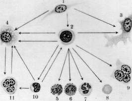
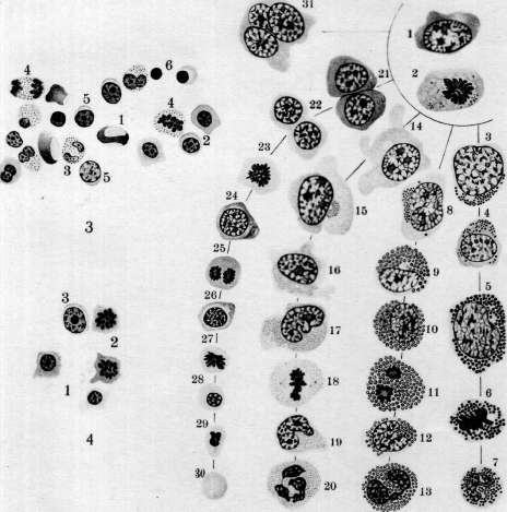
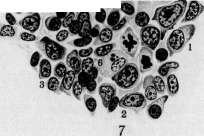
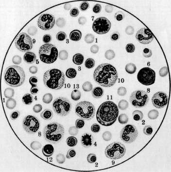
It includes wandering cells of connective tissue at rest, reticular cells of bone marrow, spleen, lymph nodes, including here the endothelium covering lymph sinuses and venous sinuses of bone marrow and spleen, further Kupffer cells of the liver, part of the endothelium of venous capillaries of the adrenal gland and pituitary gland. The participation of the reticulo-endothelium in hematopoiesis cannot be considered fully clarified. According to the research of Ferrata, Marchand, Herzog, Chlopin, Sysoev and others, under certain conditions, elements of myeloid tissue can develop from it; others (Maksimov) deny its ability to hematopoiesis. The research of Aschoff, Kiyono and Chashin showed that with sufficiently strong vital staining of animals with lithium carmine, individual mobilized ret.-end. cells or histiocytes, vital stained, appear in the blood, especially in the right heart. In connection with these data and in view of the close similarity of blood histiocytes with monocytes, the doctrine of the origin of blood monocytes from cells of the reticulo-endothelial apparatus arose (Aschoff, Kiyono, Schilling). Hematopoiesis at different periods of human life has certain peculiarities. The intensity of hematopoiesis characteristic of childhood is gradually replaced by a slower rate of blood regeneration in adults and a significant weakening of the functions of hematopoietic organs in the aged organism. In the newborn, hematopoietic organs still bear some features of the embryonic period: the bone marrow is rich in hemocytoblasts, the liver has remnants of embryonic hematopoiesis, lymph nodes contain many large lymphocytes, and are still devoid of Flemming's multiplication centers. Lymph nodes in early childhood are relatively large, poor in follicles, the multiplication centers in which begin to be marked at 2 months, but clearly appear only by the age of two. The increased function of hematopoietic organs in childhood is reflected in the composition of peripheral blood: in it we find young forms of red and white blood cells - erythroblasts, polychromatophilic erythrocytes, large lymphocytes. This period of life is also characterized by frequent and abundant development of extramedullary hematopoiesis, ease of disturbance of the functions of hematopoietic organs and more frequent megaloblastic transformation of the bone marrow under the influence of various harmful influences than in adults. In the aging organism, the intensity of hematopoiesis decreases, the red bone marrow becomes poorer in parenchymal elements, in place of which fat cells develop; the area of its distribution decreases; erythropoiesis is especially weakened; atrophic changes occur in the lymph nodes, part of the lymph nodes undergoes complete depletion and fibrosis. The ability of the hematopoietic organs of an old person to respond with an increase in their function to the enhanced breakdown of blood elements is significantly weakened. Erythropoiesis (normal and pathological). In normal conditions, erythroblasts develop in the bone marrow mainly by the homoplastic method, i.e. by the multiplication of erythroblasts already present there. In their development into erythrocytes, the eryroblast passes through several stages, namely - proerythroblast, polychromatophilic erythroblast, normoblast and finally normocyte (figure 5). At this time, the basophilia of the protoplasm, sharply expressed in proerythroblasts, gradually weakens, Hb accumulates in the protoplasm, the nucleus decreases in size, loses nucleoli, and acquires a coarser structure. Finally, the nucleus shrinks, becomes pycnotic and then, according to some authors, is expelled from the normoblast, while according to others, it undergoes intracellular dissolution. The processes of division by karyokinesis play an important role in such gradual maturation of the erythroblast. At this time, the size of cells of subsequent generations becomes smaller and smaller, approaching the size of an erythrocyte (fig. 5). The loss of erythroblasts, due to their maturation into erythrocytes, is replenished by the multiplication mainly of already hemoglobin-containing cells, while proerythroblasts are as if in reserve and only with increased erythropoiesis begin to produce Hb in their protoplasm and multiply intensively. Polychromatophilic erythroblasts, developing directly from proerythro- Figure 1. Section of rabbit bone marrow. Thrombocytopoiesis: 1-megakaryocyte with finely granular protoplasm; 2-lumen of bone marrow sinusoid; 3-Bizzozero plates formed from the protoplasm of the megakaryocyte; 4-endothelium of the sinusoid. (From a preparation from an unpublished work by Chasovnikov.) Figure 2. Scheme of genetic relationships of connective tissue elements of blood (arrows indicate the direction of development): 1-undifferentiated mesenchymal cell; 2-hemocytoblast (large lymphocyte); 3-fibrocyte; 4-histiocyte; 5-basophilic leukocyte; 6-eosinophilic leukocyte; 7-neutrophilic leukocyte; 8-erythrocyte; 9-megakaryocyte; 10-small lymphocyte; 11-monocyte. (According to Maksimov.) Figure 3. 20-hour culture of hemocytoblasts from the blood of a patient with acute myeloid leukemia. Hematopoiesis in vitro: 1-erythrocyte; 2-erythroblasts; 3-segmented neutrophils; 4-division figures of neutrophilic myelocytes; 5-hemocytoblast; 6-nucleus of a degenerated cell. Figure 4. Multiplication of erythroblasts in the same culture: 1-erythroblasts; 2-division figures of polychromatophilic erythroblasts; 3-hemocytoblast. Figure 5. Development of myeloid elements of human bone marrow from a hemocytoblast: 1-hemocytoblast; 2-karyokinesis figure of a hemocytoblast; 3 and 4-basophilic promyelocytes; 5-basophilic myelocyte; 6-karyokinesis figure of a basophilic myelocyte; 7-polymorphonuclear basophil; 8-eosinophilic promyelocyte; 9 and 10-eosinophilic myelocytes; 11-division figure of an eosinophilic myelocyte; 12-eosinophilic metamyelocyte; 13-polymorphonuclear eosinophil; 14 and 15-neutrophilic promyelocytes; 16 and 17-neutrophilic myelocytes; 18-karyokinesis figure of a neutrophilic myelocyte; 19-neutrophilic metamyelocyte; 20-polymorphonuclear neutrophil; 21-proerythroblasts originating from a divided hemocytoblast; 22-26-polychromatophilic erythroblasts gradually enriching with hemoglobin; 27-division figure of a normoblast; 28-mature normoblast; 29-normoblast with pycnotic nucleus; nearby - a free expelled nucleus; 30-erythrocyte; 31-megakaryocyte. (According to Maksimov.) Figure 6. Section through rabbit bone marrow after intravenous injection of lithium carmine and ink: 1-normoblasts; 2-polychromatophilic erythroblasts in a state of karyokinesis; 3-proerythroblasts; 4-eosinophilic myelocytes; 5-special myelocytes; 6-division figure of a special myelocyte; 7-hemocytoblast; 8-megakaryocyte; 9-reticulo-endothelial cells with protoplasm stuffed with ink clots and lithium carmine grains; 10-lumen of sinusoids; 11-fat cell; 12-artery; 13-erythrocytes in the lumen of the sinusoid. (According to Maksimov.) Figure 7. Section from a lymph node of a rabbit. Part of a follicle with a multiplication center in an active stage: 1-large lymphocytes; 2-medium lymphocytes; 3-small lymphocytes; 4-division figures of medium lymphocytes; 5-indifferent mesenchymal cell; 6-macrophage. Figure 8. Smear of bone marrow from the rib of an anemic dog. Expressed erythropoiesis: 1-erythrocytes; 2-normoblasts; 3-polychromatophilic erythroblasts; 4-division figures of normoblasts; 5-division figure of a polychromatophilic erythroblast; 6-proerythroblast; 7-division figure of a proerythroblast; 8-band neutrophils; 9-young neutrophils; 10-neutrophilic myelocytes; 11-hemocytoblast; 12-lymphocyte; 13-erythrocyte with Jolly body. (To illustrate the article Hematopoiesis.)

To the article Hematopoiesis.

To the article Hematopoiesis.
Blasts have larger sizes than erythrocytes, and their protoplasm is stained by a mixture of acidic and basic dyes to an intermediate tone. Due to their large size, they are sometimes called macroblasts. Erythroblasts lie in the parenchyma of the bone marrow in small clusters of cells that are in the same stages of maturation (fig. 6). The mechanism of penetration of erythrocytes from the parenchyma of the bone marrow into the lumen of the sinuses is not fully elucidated. Heteroplastic erythropoiesis, i.e., the development of proerythroblasts from an indifferent cell, is not yet fully understood. According to the view of the unitarists (Maximov), the hemocytoblast of the bone marrow can differentiate in various directions, including into proerythroblasts (fig. 5). In this case, karyokinetic division is necessary, as during it the internal restructuring of the cell nucleus occurs, which becomes capable of developing only into an erythrocyte. According to the teaching of the dualists (Naegeli), the young erythroblast (pronormoblast) develops from an indifferent mesenchymal cell, while Schridde produces it from the endothelium of blood vessels. Ellermann speaks of a special maternal cell of the erythroblastic series, the so-called erythrogony. Experiments with the explantation of leukemic blood prove that the hemocytoblast is capable in some cases in vitro to differentiate in the direction of erythroblasts (Timofeevsky and Benevolenskaya).- Pathological increase in erythropoiesis is observed mainly in anemias (fig. 8). At this time, the content of erythroblasts in the bone marrow increases, the number of mitoses increases, heteroplastic erythropoiesis intensifies, and fatty bone marrow is replaced by red. In some particularly severe anemias, for example, malignant anemia, extramedullary hematopoiesis develops (see below heterotopic hematopoiesis). With severe intoxications and infections, suppression of erythropoiesis can occur: the number of erythroblasts decreases, and their division figures disappear (aregenerative-aplastic anemias). Experimentally, suppression of erythropoiesis can be caused by repeated bloodlettings. A decrease in erythropoiesis is also observed in leukemias, especially acute ones. In malignant anemia, the development in the bone marrow, along with normoblasts, of very large hemoglobin-containing cells—megaloblasts and megalocytes—is observed. Ehrlich expressed the view that the megaloblastic transformation of the bone marrow is a return to embryonic hematopoiesis, and megaloblasts are identical to the primary erythroblasts. This hypothesis found support among many hematologists. Heteroplastic development of megaloblasts cannot be considered fully elucidated. According to Naegeli, they develop from mesenchymal cells, according to Ferrata—from hemohistioblasts (histiocytes), according to Lambin—from reticulo-endothelium. In addition, the disturbance of normal erythropoiesis can manifest itself: 1) in the breakdown of the erythroblast nucleus into individual segments; 2) in the preservation of remnants of nuclear substance in the erythroblast (Jolly bodies, Cabot rings); 3) in polychromasia and basophilic "dotting" of erythrocytes and the presence in them of a reticulo-filamentous substance stained by basic dyes in unfixed preparations; 4) in insufficient production of Hb. Some of these deviations are sometimes found in normal blood, but they are especially pronounced in anemias. Leukopoiesis (normal and pathological). Granular leukocytes develop normally exclusively in the bone marrow, mainly by the homoplastic method, i.e., by the reproduction and maturation of young granular cells, the so-called myelocytes (fig. 5, 6 and 8). These large cells have a round or kidney-shaped nucleus and granular protoplasm. Depending on the nature of the granularity, neutrophilic, eosinophilic, and basophilic myelocytes are distinguished. The maturation of myelocytes into the corresponding segmented leukocytes passes through several stages, more thoroughly studied for neutrophils. The cell nucleus first becomes horseshoe-shaped (metamyelocyte of Pappenheim or juvenile neutrophil, according to Schilling), then it acquires a rod-shaped form in the form of a curved, densely staining cord (rod-nucleated neutrophil, according to Schilling) and finally breaks down into several (2-5 or more) segments connected by narrow bridges (segmented neutrophil). Starting from the metamyelocyte stage, cell multiplication ceases. Heteroplastic granulopoiesis, which is usual in embryonic hematopoiesis, is rarely encountered in the adult organism under normal conditions. It is considered proven that all 3 types of myelocytes originate from a common ancestral cell—the hemocytoblast (myeloblast). Other, less common names for this cell: lymphoidocyte (Pappenheim), basophilic myelocyte (Dominici), lymphoid bone marrow cell (Türk). Unitarists (Maximov), in addition to the name "hemocytoblast," also designate it as a large lymphocyte, whereas dualists (Naegeli) adhere to the name "myeloblast." The hemocytoblast is a large cell with a round or slightly kidney-shaped nucleus, the chromatin of which in dry stained smears has the appearance of a thin uniform meshwork with small nodular thickenings. The nucleus contains several nucleoli. The protoplasm is basophilic, non-granular, and its quantity is small. The hemocytoblast gives a positive reaction to the proteolytic enzyme, oxidase, and peroxidase—in this they see its difference from the large lymphocyte or lymphoblast, which is morphologically close to it but does not give these reactions. In the adult organism, the number of hemocytoblasts is small, in newborns they are significantly more numerous, and in the embryonic period they predominate over other cells. According to Maximov, the hemocytoblast, in maturing into a myelocyte, must undergo mitosis, during which the differentiation of daughter cells in one direction or another occurs (fig. 5). At this time, the basophilia of the protoplasm decreases, the amount of the latter increases, and in it sometimes a dense azurophilic granularity develops, the chromatin clumps of the nucleus become coarser, and the nucleoli become less distinct. Then the production of one or another type of specific granularity begins, first in a small amount, usually around the nucleus at the site of the kidney-shaped depression. Such transitional forms from hemocytoblasts to myelocytes are distinguished under the name promyelocytes. With further maturation of the promyelocyte, the azurophilic granularity, if it was present, disappears, the basophilia of the protoplasm gradually weakens, the specific granularity increases, and in eosinophils, along with oxyphilic grains, individual basophilic, non-metachromatic grains often appear. In addition, the development of myelocytes directly from an indifferent mesenchymal cell (hemohistioblast of Ferrata, ret.-end. cell) without the intermediate stage of hemocytoblast is admitted, especially in leukemias. On the other hand, the hemocytoblast, like in the embryo, can also develop from an indifferent mesenchymal cell in the adult organism (see below heterotopic hematopoiesis). The exit of mature granulocytes into the lumen of the bone marrow sinuses is explained by their ameboid mobility; here, a change in blood pressure in the sinuses, and consequently their lumen, may also play a role. An increase in the intensity of granulopoiesis is observed in many infections and especially in myeloses. With neutrophilic hyperleukocytosis, the production of neutrophils increases, and a transformation of fatty bone marrow into red may occur. Depending on the strength and duration of the causing factor, the cellular composition of the bone marrow can change sharply, and heteroplastic granulopoiesis significantly intensifies. Enhanced reproduction of eosinophilic myelocytes and increased formation of them from hemocytoblasts are observed in eosinophilic hyperleukocytosis. In myeloses, a transformation of fatty bone marrow into myeloid tissue occurs with the development of younger granulocytes in its parenchyma, and in acute cases—hemocytoblasts. In chronic infections, intoxications, and especially in myeloses, along with the transformation of fatty bone marrow into red, the development of islands of myeloid tissue occurs in other places of the body (see below heterotopic hematopoiesis). Suppression of granulopoiesis is observed in many body intoxications, in some severe infections, in malignant anemia. The bone marrow in this case becomes poor in granulocytes, and the hemocytoblast may be the predominant element in it. This is usually accompanied by degenerative changes in granulated leukocytes and foci of necrosis of the bone marrow parenchyma. Deviations from the norm in the development of granulocytes can consist of: 1) insufficient production of specific granularity or even its complete absence; 2) uneven maturation of the nucleus and protoplasm; 3) abnormal lobulation of the nuclei of hemocytoblasts (Rieder's forms); 4) the development of the so-called neutrophilic twins, i.e., neutrophils that have two nuclei, constructed identically. A particularly strong deviation from the norm in granulopoiesis is observed in acute myeloses: almost complete absence of granulopoiesis, with the hemocytoblast being the predominant element in the parenchyma of the bone marrow.
Sometimes it has small sizes, being close in its morphology to a small lymphocyte (micromyeloblast). Among the degenerative forms of granulocytes, the degenerative forms of band neutrophils, appearing in the bone marrow in the so-called degenerative nuclear shift (Schilling), deserve attention. The formation of blood lymphocytes occurs in lymphoid tissue, mainly in lymph nodes and spleen, predominantly by the homoplastic route (fig. 7). It is usually believed that small blood lymphocytes are formed by the division of large lymphocytes, which are therefore often called lymphoblasts (Naegeli), more rarely - lymphogonia by Benda. However, according to Maximow, multiplication of medium mesolymphocytes is much more frequently observed. The division of lymphocytes occurs in lymph nodes, mainly in the center of multiplication of follicles (fig. 7). Two phases of the follicles of the lymph node are distinguished: active and resting. During the first phase, the follicle of large size contains many large and medium lymphocytes with numerous mitoses; during the second phase, there are few cellular elements in it, and small lymphocytes predominate. Heteroplastic formation of lymphocytes occurs from indifferent mesenchymal cells. In this case, a fixed mesenchymal cell divides mitotically, giving rise to round cells with light nuclei and weakly basophilic protoplasm. With further multiplication of the latter, typical medium lymphocytes are obtained. This process is particularly pronounced when a lymph node follicle transitions from the resting phase to the active phase (Maximow). In the development of new lymphatic follicles, lymphocytes are formed from the same indifferent mesenchymal cells. This formation occurs mainly in the place of former fat lobules. The genetic relationships of fixed and free cells of lymphoid tissue, according to Maximow, can be schematically expressed as follows: ^ Medium and -> Small ^--' large "*~ lympho-Undifferentiated '
"""-"-». Fixed macro-phages stioctites Under pathological conditions, Maximov also allows for the possibility of development from large and medium lymphocytes of elements of myeloid tissue, from small lymphocytes-micromyelocytes, and from both-monocytes and macrophages. The possibility of the origin of monocytes from free histiocytes (macrophages) is also allowed by Maximov. Pathological lymphopoiesis, observed in some infections, intoxications, and especially lymphadenoses, may consist in the excessive formation of large lymphocytes-lymphoblasts, not normally encountered in the adult blood, further-in the abnormal lobulation of the nuclei of large lymphocytes (Rieder's forms), in the development of lymphocytes almost devoid of protoplasm or, conversely, with an abundant amount of the latter, in the absence of azurophilic granules normally found in one third of lymphocytes. Enhanced lymphopoiesis is accompanied by hyperplasia of lymphoid tissue with enlargement of lymph nodes, spleen, follicles of mucous membranes, thyroid gland, and with the development of new lymphomas in various places of the body. Inhibition of lymphopoiesis occurs due to the destruction of lymphoid tissue of lymph nodes; at this time the blood becomes poor in lymphocytes (lymphogranuloma, lymphosarcomatosis). The origin of blood monocytes is not fully elucidated. There are three main views: 1) monocytes originate from the reticulo-endothelial apparatus by mobilization of ret.-endo. cells (Aschoff, Kiyono, Schilling); 2) monocytes develop in the myeloid system from myeloblasts (Naegeli); 3) monocytes are formed from lymphocytes by hypertrophy of their nuclei and protoplasm and represent a transitional form from lymphocyte to macrophage or polyblast (Maximov, Bloom). Such hypertrophy of a lymphocyte occurs in blood vessels with slowed blood flow. However, Maximov does not deny the origin of monocytes from reticulo-endothelium. Blood monocytosis is usually accompanied by hyperplasia of the reticulo-endothelial apparatus and mobilization of reticulo-endothelial cells, which may appear in the blood, especially in some chronic sepses. These cells bear all the signs of histiocytes or macrophages and give all possible transitions to ordinary monocytes. All this speaks for the role of reticulo-endothelium in the origin of monocytes; at the same time a lymphocyte, as shown by implantation experiments, can in the shortest time turn into a monocyte. Thrombocytopoiesis (normal and pathological). In lower vertebrates and in birds thrombocytes are true cells, provided with nuclei. In mammals and man blood plates are anuclear formations, which according to Wright's research originate from separated pieces of protoplasm of megakaryocytes of the bone marrow. Thrombocytopoiesis apparently occurs in such a way that the megakaryocyte sends into the lumen of the bone marrow sinusoids amoeboid processes of azurophilic protoplasm; from them small particles detach, which are carried by the blood current in the form of blood plates (fig. 1) (see Vizzocero plates). However, some authors to this day adhere to the old views on the origin of blood plates, deriving them either from the protoplasm of leukocytes and erythrocytes or from the nuclei of leukocytes. According to Schilling, a blood plate is a modified nucleus of an erythroblast, being expelled from a young erythrocyte already in the bloodstream. Increased development of megakaryocytes from hemocytoblasts and their enhanced functioning is accompanied by an increase in the number of blood plates in the blood and the appearance of giant forms (chlorosis, myeloses). Sometimes parts of megakaryocytes with fragments of nuclei enter the blood. Some pathological processes are accompanied by thrombocytopenia and the appearance of pathological plates with abnormal granulation or absence of the latter or with a strongly expressed basophilia of the marginal zone or plates of abnormal size (malignant anemia, aplastic anemias, "essential thrombopenia"). Relationships between hematopoietic organs and blood. The morphological composition of blood depends on two factors: on the production of formed elements and on their consumption, and both these processes are in the closest mutual connection and mutually condition each other. An increase in erythropoiesis is reflected in the composition of blood by the appearance of regenerative forms of erythrocytes and erythroblasts. But with strong and rapid irritations of the bone marrow, the appearance of erythroblasts in the blood can be caused by their simple washing out from the myeloid tissue due to disturbance of blood circulation in it. In megaloblastosis of the bone marrow, megalocytes and megaloblasts appear in the blood. The absence in the blood of regenerative forms of erythrocytes in the presence of severe anemia allows one to assume the absence of erythropoiesis in the bone marrow (see also Anemia). The relationships existing between leukopoiesis and the composition of blood leukocytes are more complex, since the distribution of leukocytes in the vascular bed and the processes of consumption of leukocytes can proceed independently for each type. In rapidly developing neutrophilic leukocytosis, mobile neutrophilic leukocytes can in the shortest time emigrate from the parenchyma of the bone marrow into the blood vessels. In chronic inflammatory processes without complications, occurring with almost complete absence of nuclear shift, the bone marrow is rich in mature and young neutrophils, and in severe acute infections with strong nuclear shift it turns out to consist mainly of promyelocytes (Schilling). In leukemias, phenomena of hyperplasia of hematopoietic organs in most cases find reflection in the leukemic composition of blood, and from the morphology of the latter one can judge the morphological composition of hematopoietic organs, and vice versa. In general, however, it should be said that the composition of blood by no means always reflects the processes taking place in hematopoietic organs; there exists some mechanism, not yet elucidated, which, depending on the circumstances, facilitates or hinders the transition of formed elements from the bone marrow into the peripheral blood (bone marrow barrier). (See also Leukocytoses, Leukemia, Leukocyte formula.) Heterotopic H. Under pathological conditions (infections, intoxications, severe anemias, myeloses) the appearance of myeloid tissue in different places of the body is observed, so-called extramedullary myelopoiesis. Myeloid metaplasia as a rule arises first in the spleen, then in the liver, lymph nodes, adrenal glands and other places of the body. At first myelocytes appear, then megakaryocytes, later and not always-erythroblasts. The extramedullary origin of myeloid elements at present is explained by the majority as corresponding differentiation of local elements (autochthonous theory). The colonization theory, according to which myeloid metaplasia arises from cells carried by the blood current to various places of the body, is at present rejected by the majority. The question of from which local cells the elements of myeloid tissue develop is one of the most controversial in hematology. Part of the authors (Schridde, Herzog, Naegeli) derive them from cells of the vascular wall. Close to this stands the teaching of Ferrata, Hoff, Sysoev and others, attributing hematopoietic ability to the reticulo-endothelium. According to Ferrata, elements of myeloid tissue develop from the reticulo-endothelium, passing through the stages of hemohistioblast and hemocytoblast. Mollendorff considers that granular leukocytes can develop from fibroblasts; this opinion however meets with objections from the majority of scientists. Dominici, Downey, Weidenreich, Maximov endow lymphocytes with hematopoietic abilities. According to Maximov, histiocytes and vascular endothelium do not possess the ability for hematopoiesis, and extramedullary myelopoiesis occurs either at the expense of blood and tissue lymphocytes or from indifferent mesenchymal cells. Theories of hematopoiesis. The question of the genetic relationships of the formed elements of blood to each other and to the cellular forms of connective tissue is to this day one of the most controversial in hematology. Here one can distinguish four main directions, or four theories of hematopoiesis: 1) unitarian direction. 2) moderately-unitarian, 3) dualistic and 4) trialistic. The unitarian doctrine (Grawitz, Maximov, Weidenreich, Chlopin, Myasoyedov) holds that all non-granular elements of blood and hematopoietic organs, i.e. small lymphocytes, large lymphocytes, hemocytoblasts, as well as (according to some) and monocytes, are undifferentiated cells capable, under certain conditions, of giving rise to granular leukocytes, erythrocytes and megakaryocytes. According to this doctrine the hemocytoblast (myeloblast) and large lymphocyte are capable of giving rise to identical cells, and the existing biochemical and morphological differences between them are not constant and depend exclusively on the temporary conditions of existence. The unitarian doctrine is based mainly on experimental research, especially of Maximov and his students.
According to this doctrine, in the early embryonic period, all mesenchymal cells possess full hematopoietic capabilities, whereas in the adult organism this ability is retained by certain undifferentiated mesenchymal cells, to which, according to some, the reticulo-endothelium also belongs. According to Maximow, part of the connective tissue cells are unilaterally differentiated (fibroblasts, endothelium of blood and lymph vessels), part (cells of the reticulo-endothelial apparatus and generally histiocytes) have retained only certain abilities (the ability to develop into fibroblasts and blood monocytes) and finally part have retained all embryonic properties with wide hematopoietic possibilities (Figure 2). The doctrine of moderate unitarists (Pappenheim) produces all blood elements from a common progenitor - the lymphocyte (hemocytoblast). Blood and lymphatic tissue lymphocytes are already differentiated elements incapable of hematopoiesis. According to the dualist doctrine (Ehrlich, Naegeli, Schridde), lymphocytes and granulocytes have two progenitors - the lymphoblast and the myeloblast, which under no conditions can transform into each other and differ in morphological and biochemical features. According to Naegeli, the indifferent mesenchymal cell is the connecting link for blood elements, which possesses the full potential of hematopoietic capabilities. According to the trinitarian doctrine, histiocytes, both free and fixed, constitute the 3rd independent hematopoietic organ, similar to the myeloid and lymphatic systems of the organism. This organ produces blood monocytes, which thus in their origin are independent of lymphocytes and granulocytes (Aschoff). Culture of normal and pathological blood. The culture of blood leukocytes in vitro, first performed by Avrorov and Timofeevsky, made it possible to clarify some controversial issues in hematology. Granulocytes of normal blood, as cells with completed development, undergo disintegration in vitro within a few days, whereas lymphocytes and monocytes develop in a short time into phagocytic polyblasts or macrophages. This confirms Maximow's doctrine on the role of non-granular leukocytes in the origin of cellular forms in inflamed tissue. In older cultures, macrophages elongate and gradually transform into typical fibrocytes producing tonofibrils and collagen fibers - true connective tissue develops (Maximow). In their development into a polyblast, the blood lymphocyte I passes through the monocyte stage, which indicates the possible genesis of monocytes from lymphocytes (Maximow, Blum, Timofeevsky and Benevolenskaya). The manifestation of hematopoietic ability by lymphocytes and monocytes of normal blood in vitro does not occur. In this respect, these cells differ from the hemocytoblast (myeloblast) of leukemic blood: hemocytoblasts differentiate in vitro into granulocytes in the shortest time, more rarely into erythroblasts [see separate table (pp. 531-532), Figures 3 and 4, and separate table (pp. 659-660), Figure 15] (Timofeevsky and Benevolenskaya). Comparison of these two factors speaks against the unitarian doctrine. Lit.-see lit. to article Hematology and Bone marrow.
Related articles
Mentioned in
Cite this page
“Hematopoiesis.” Soviet Medical Encyclopedia. English translation of Bolshaya Meditsinskaya Entsiklopediya, 1st ed. (Moscow, 1928–1936), ed. N. A. Semashko. https://sovietmedicalencyclopedia.pages.dev/article/hematopoiesis/