Fibrous Reticula
Historical document, translated for reference. It reflects medical knowledge of the 1920s–30s and is not medical advice.
Summary
Fibrous reticula are a type of connective tissue fiber that forms networks within the body, particularly in lymphatic organs, bone marrow, and the liver. These fibers, composed of reticulin, have unique staining properties and serve as a supporting framework for various tissues.
Encyclopedia article (1928–1936)
FIBROUS RETICULA (Gitterlasern, reticulum, reticular, argyrophilic fibers, "third type" of fibrous connective tissue), do not exist independently, but are part of the richly cellular reticular tissue. The latter consists of cells (reticular cells, Kupffer's cells) connected to each other in a syncytium, for which they form a supporting framework [see separate table (pp. 27-28), fig. 5]. Reticular tissue even in the mature organism has the properties of young connective tissue (mesenchyme); it forms the basis of lymph glands (adenoid, lymphoid tissue), bone marrow (myeloid tissue), spleen, intestinal mucosa, forms the ret.-end. apparatus both in the walls of liver capillaries and in other places and, generally, is widely distributed in the body.-Properties of fibers. In form, reticular fibers resemble elastic fibers, but consist of a special substance, reticulin, and have been classified as a special type based on their reactions; they do not swell from weak acids (unlike collagen fibers), are not digested by trypsin (difference from elastic fibers), are impregnated with silver by the Oppel and Bielschowsky method, with silver tannin of Rio Hortega, are stained with methylene blue (Timofeev). They are homogeneous, have a sinuous course, give off branches, which, interconnecting, form networks. The fibers are closely connected with cells, passing through their cytoplasm. In the composition of networks, thicker and thinner fibers can be distinguished; the former serve as main fibers, giving off smaller branches along their course. In some places, the fibrous formations have the character of a three-dimensional network, in the loops of which tissue fluid flows; thus, they replace lymphatic vessels (in the liver). But in other places, the fibrous network is stretched in one plane in the form of a mesh membrane. In this case, there is a special homogeneous substance impregnating the spaces between the fibers. Such mesh membranes form, for example, boundary membranes in glandular organs.-Cells of reticular fibers (reticular cells of hematopoietic organs, Kupffer's cells of the liver, etc.) are characterized by broad prospective potency, being the basic cellular stock for cells of the lymphoid, myeloid series, histiocytes and fibrocytes. In the mature organism, in some organs (lymph glands, in bone marrow, in spleen, ret.-end. apparatus) they are constantly active; they behave as phagocytes and easily transform into macrophages, polyblasts (histiocytes). Relation of reticular fibers to collagen fibers. Reticular fibers in the mature organism in places directly pass into collagen fibers; hence the view that these are not fully developed, immature collagen fibers. According to Laguesse, the thinnest fibers of the fibrous reticula correspond to pre-collagen fibers, the thick fibers-pre-collagen fibers (poor in collagen)-directly pass into collagen fibers. However, this view is not generally accepted. According to another view, reticular fibers are an integral part of one biological system, while collagen fibers belong to a completely different one-the fibroblastic one.--Regarding the development of reticular fibers, there is no unified view. Some authors find the primary rudiments of them in the cell itself (in the endo- or ectoplasm or on its surface, epicytellarly), others-in the intercellular amorphous substance. Some try to connect their appearance with the transformation of protoplasmic elements (mitochondria, etc.) into them; for others, this is a purely physicochemical process of gelatinization, i.e., the precipitation of a special substance which precipitates first in the form of grains or rods in the cytoplasm or outside it, and then grows into fibers. Some pathological processes are accompanied by the appearance of young connective tissue. Thus, in the formation of granulomas, epithelioid cells arise, connected to each other syncytially and having a supporting apparatus of reticular fibers. The latter are so stable that they remain in tissue necrosis (caseous degeneration). Scar formation or the appearance of collagen fibers depends on the appearance of other, more differentiated cells-fibroblasts. However, under pathological conditions
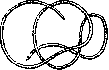
reticular fibers undergo changes: they thicken and become impregnated with collagen-collagenize.
Related articles
Mentioned in
Cite this page
“Fibrous Reticula.” Soviet Medical Encyclopedia. English translation of Bolshaya Meditsinskaya Entsiklopediya, 1st ed. (Moscow, 1928–1936), ed. N. A. Semashko. https://sovietmedicalencyclopedia.pages.dev/article/fibrous-reticula/