Mesenchyme
Historical document, translated for reference. It reflects medical knowledge of the 1920s–30s and is not medical advice.
Summary
Mesenchyme is embryonic connective tissue that arises from the mesoderm in vertebrates, and sometimes from ectoderm and endoderm in invertebrates. It consists of spindle-shaped and star-shaped cells forming a sponge-like or reticular framework filled with intercellular substance.
Encyclopedia article (1928–1936)
Mesenchyme (from Greek mesos-middle and echeo-I pour, I fill), embryonic connective tissue; arises in vertebrates from different areas of the mesoderm. Along with this, some authors also allow for the participation of ecto- and endoderm in the formation of mesenchyme. Its formation from all three embryonic germ layers (mainly from meso- and ectoderm, rarely from endoderm) occurs in many invertebrates. The concept of mesenchyme was formulated by the Hertwig brothers (O. and R. Hertwig, 1881) and distinguished by them from the concept of "mesoderm." Mesenchyme is characterized by its composition of isolated or connected by their processes cells of spindle-shaped and star-shaped form, forming a sponge-like or reticular framework, the loops of which are filled with liquid or semi-liquid, gelatinous, amorphous intercellular substance. The mesenchymal primordia formed by one means or another (see Mesoderm), growing, spread and fill all the spaces between the embryonic germ layers (see separate table, fig. 2). The increase in the mass of the embryo's body is largely associated with the enhanced multiplication of mesenchymal elements. Mesenchyme formed from sclerotomes, mainly accumulating around the chorda and neural tube (axial mesenchyme), gives rise to the entire axial skeleton. The cutaneous plate of myotomes forms part of the cutaneous connective tissue. The other part of the connective tissue of the skin, the connective tissue and skeleton of the limbs, as well as the connective tissue and smooth muscles of internal organs are formed from mesenchymal primordia of the parietal and visceral plates of the splanchnomes (see Mesoderm). In recent years, mainly by Hueck, new views on the development of mesenchyme have been expressed, which have found particular sympathy among pathologists. According to Hueck's conception, the cells of the embryonic germ layers are connected to each other by protoplasmic processes into a continuous syncytial mass. Such processes also connect to each other the individual embryonic germ layers, so that the spaces between them are filled not with amorphous ground substance, but with a dense reticular or spongy protoplasmic mass. The cell nuclei shift from the embryonic germ layers into this mass, and thus the initial form of embryonic mesenchyme arises, in which the ground substance and fibers are secondarily formed. The development of the ground substance occurs by the type of compaction of the surface layer of the protoplasmic spongy mass, bordering on the pores embedded in it, similar to how the formation of film surfaces occurs in a colloidal solution. Repeated many times, the process of compaction of protoplasmic surface layers leads to the filling of the pores of the syncytial spongy mass with ground substance, which appears partly in the form of membranes and fibers. The syncytial mass itself, being consumed in this process, remains only in the form of remnants among the ground substance and then appears as a cellular network, in the nodal parts of which the nuclei are located. Subsequently, the impregnation of the films and fibers of the ground substance with collagen or elastin occurs, as a result of which the corresponding fibers of connective tissue are formed. However, part of the ground substance remains in its original form between the fibers and membranes, and from it new layers of fibrous connective tissue can subsequently develop without the participation of cells. Based on these concepts, Hueck comes to the conclusion that a sharp boundary cannot be established between cellular protoplasm and ground substance. The latter, representing only a modification of protoplasm, is not, as was previously thought, lifeless, performing only a supporting function, but is also endowed with life properties (a view first expressed by Lukyanov in 1894). The views presented by Hueck particularly well explain the pathological development of the intermediate substance in the sclerosis of various organs (e.g., in atherosclerosis of arteries, cirrhosis of the liver), when the participation of cellular forms in the sclerotic process completely recedes to the background. Simultaneously with the appearance of mesenchyme during embryonic development, the first vascular-blood primordium also arises. In selachians and most amniotes, the edges of the proliferating extra-embryonic mesoderm, which later becomes part of the wall of the yolk sac, lose their character of continuous, epithelium-like plates and completely transform into a loose mesenchymal cellular mass (Ruckert and Mollier, Maximov). In the part of the yolk sac wall called the vascular field, in direct proximity to the endoderm, blood islands-irregularly shaped dense cellular aggregations-arise from this cellular mass, connected to each other by thin cellular strands. The mesodermal cellular elements that did not enter the composition of the blood islands and mesenchymal primordium, densely connect to each other like an epithelium and transform into the final extra-embryonic mesoderm, which then splits due to the formation of the extra-embryonic coelom into two layers. The internal cells of the islands round off, and
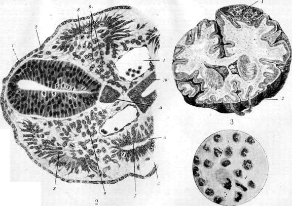
Int. I. flonept.....jfi cross-section of a human embryo: 1 - neural tube; 2 - ectoderm; 3 - notochord; 4 - intraembryonic coelom; 5 - parietal mesoderm; 6 - splanchnic mesoderm; 7 - intermediate cell mass of mesoderm; 8 - somite; 9 - lateral plate mesoderm; 10 - amnion. Figure 1. Cross-section of the trunk of a sea lamprey embryo (12 somite stage) according to Maximov: 1 - neural tube; 2 - ectoderm; 3 - notochord; 4 - aorta; 5 - coelomic cavity; 6 - parietal mesoderm; 7 - visceral mesoderm; 8 - somite; 9 - mesenchyme; 10 - intestine. Fig. 2. Meningococci. Figure 3. Lower surface of the frontal lobes. Figure 4. Meningococci. They separate from each other due to the accumulation of fluid between them and transform into primary blood cells (Maximov) or mesamoeboid cells (Minot) of the embryo. The edge cells of the islands and the cells connecting them, by flattening, come close to each other and transform into the endothelium of the first blood vessels. In the human embryo, blood islands develop in the wall of the yolk sac, and also, according to Bremer, in the mesentery and allantois from extra-embryonic mesoderm or mesenchyme, which arises very early and independently of its embryonic part. In other areas of the embryo, tubular, liquid-filled rudiments of blood vessels, consisting initially also of a single endothelium, are formed independently of the first rudiment described and somewhat later by the fusion of flattened mesenchymal, so-called vascular cells with each other. The endothelium of the cardiac rudiment apparently forms in the same way from mesenchymal elements called cardiac cells. After the fusion of different rudiments of the circulatory system and the beginning of blood circulation, the primary blood cells formed in the manner described above and the products of their differentiation are evenly distributed throughout the circulatory system. In different areas of the mesenchyme of embryos (first of all in the head region), it is possible to observe the rounding and transformation of part of the branched mesenchymal elements into free wandering cells of various types - initially hemocytoblasts (see), and then so-called histoid wandering cells, similar in a number of properties to histiocytes of the connective tissue of the adult organism (Maximov). In rabbits, the formation of separate blood islands in certain areas of intra-embryonic mesenchyme has also been described (Maximov). (For further development and fate of embryonic blood elements - see Hematopoiesis.) The early isolation of the first rudiment of blood and blood vessels from certain areas of mesoderm gave reason to consider it completely isolated, independent of mesenchyme, an embryonic rudiment, so-called angioblast (His, 1900; Minot, 1911, etc.). However, the close genetic relationships that subsequently exist between mesenchyme and blood elements in embryos, as well as between blood and connective tissue in the adult organism, speak against the theory of angioblast as an independent rudiment. According to the prevailing views at the present time, the first vascular-blood rudiment is considered to belong to the common mesenchymal rudiment. Likewise, the participation of entodermal elements along with mesodermal elements in the formation of blood islands is not confirmed. On the other hand, some facts obtained recently by methods of experimental embryology indicate that at very early stages of development of amphibian embryos, for example, at the very beginning of gastrulation (see Gastrula), in the so-called presumptive epidermis, i.e., in the part of the cellular material from which the skin epithelium subsequently develops, mesenchymal rudiments of connective tissue are also contained (Holtfreter). This makes it probable the presence of mesenchymal rudiments not only as part of mesoderm, but also in other embryonic germ layers. According to some data (Stone), in the head region of the ganglionic plate in a frog embryo, there is also a mesenchymal rudiment of part of the skull skeleton (so-called mesectoderm). Then mesenchyme and the products of its differentiation turn out to be strictly delimited from all other embryonic rudiments and tissues of the organism. Further transformations of mesenchyme come down to the differentiation of its initially equivalent elements into various cellular forms: connective tissue, blood, smooth muscle, and to the production of different types of intercellular substances. At the present time, however, it must be admitted that in certain types of connective tissue of the adult organism, undifferentiated mesenchymal, or so-called cambial elements remain as a reserve cellular material, having outstanding significance in the phenomena of regeneration. Their existence is revealed with particular persuasiveness under experimental conditions, as well as in many pathological processes. Such areas of embryonic or little differentiated mesenchyme in the adult human are the milk spots of the omentum, hematopoietic organs (cells of the reticular stroma of lymph nodes, bone marrow and spleen), and finally the connective tissue cells around capillaries (pericytes). Under various irritations, these elements can give various pictures of differentiation up to foci of hematopoiesis, foci of ossification, etc. On the other hand, part of the mesenchymal elements that subsequently form cartilage or bone already at very early stages of development acquire some specific differences. Experiments with tissue cultures (Fischer and Parker) showed that elements of the skeletogenic areas of mesenchyme, differentiating outside the organism, can form intercellular substance resembling cartilage or bone. Under such conditions, the mesenchyme of the skin differentiates into fibrous connective tissue. Thus, mesenchyme represents the common rudiment of all types of connective, cartilaginous and bony tissues, blood, lymph, as well as visceral and vascular musculature of the future organism. Undifferentiated mesenchyme, preserved in a certain amount even in the adult organism, is recently attributed enormous importance in the processes of inflammation, hematopoiesis, immunobiological reactions, etc. Due to the diversity of functions and special sensitivity of mesenchymal elements of the adult organism to various irritants, they are combined into a single system of particularly actively reacting cells under the name "active mesenchyme" (Leopold, 1912). The cells of the ret.-end. system are also included here, although the latter are still more differentiated than the proper mesenchymal cells described, for example, by Maximov everywhere in connective tissue. Thus, mesenchymal cells do not have the ability to accumulate particles of colloidal solutions and suspensions in their protoplasm, while the cells of the ret.-end. system exhibit this property to a high degree. True, transitional forms exist between those and other cells, so that the cells of the ret.-end. system are the closest derivatives of indifferent mesenchymal cells and are constantly formed anew from them. The diverse functions attributed to "active" mesenchyme, in general, coincide with the functions of the reticulo-endothelial apparatus (see).
Related articles
Mentioned in
Cite this page
“Mesenchyme.” Soviet Medical Encyclopedia. English translation of Bolshaya Meditsinskaya Entsiklopediya, 1st ed. (Moscow, 1928–1936), ed. N. A. Semashko. https://sovietmedicalencyclopedia.pages.dev/article/mesenchyme/