Hydatidiform Mole
Historical document, translated for reference. It reflects medical knowledge of the 1920s–30s and is not medical advice.
Summary
Hydatidiform mole refers to various formations resulting from the death of a developing egg, either in the uterus or fallopian tube. The most significant type is the vesicular mole, characterized by the transformation of chorionic villi into transparent, vesicular formations.
Encyclopedia article (1928–1936)
HYDATIDIFORM MOLE (vesicular, bloody, fleshy), a term denoting a series of formations of different origins that result from the death of a developing egg, either inside the uterus or in the fallopian tube. Among these various formations, the vesicular mole (mola hydatidosa) deserves the greatest attention due to its consequences - the transformation of chorionic villi into transparent, vesicular formations, thanks to which the fertilized egg turns into a conglomerate of small, uneven-sized vesicles, generally resembling clusters of small grapes (Figure 1). However, alongside the branched arrangement of vesicles, sometimes they are connected to each other and by the thinnest threads resembling beads. The threads connecting individual vesicles to each other have varying lengths. In places between groups of vesicles, there are loose masses of blood, clots of fibrin, and decidual tissue. The size of the expelled vesicular mole can vary greatly: sometimes it does not exceed the size of a chicken egg, while at other times it represents a large (as large as an adult's head) crimson mass that easily breaks down into formless areas of various sizes and individual vesicles. The entire egg is often affected, while in other cases only individual areas of the fetal membranes are affected. Often in the center of the mole, a cavity lined with amnion can be found, in which the fetus is usually absent. Only in rare cases can a fetus be found, usually markedly retarded in its development. The smaller vesicles of the mole usually do not contain a cavity, while in the larger ones there is a cavity in the center, filled with a clear watery fluid that easily flows out when punctured. The frequency of vesicular mole, according to various authors, is extremely different. On average, it can be considered that approximately 1 vesicular mole occurs per 3,000 pregnancies. However, there are other figures; for example, Brindeau gives a figure of 1 mole per 529 pregnancies. The influence of age apparently does not affect the appearance of mola: vesicular mole has been observed in both young 20-year-old women and women over 55 years old. However, taking into account the circumstance that women in later years find pregnancy more difficult, Hitschmann believes that mola has a greater tendency to develop in older women. A case of vesicular mole has been described in a 9-year-old girl. Histogenesis. Kossmann points out that mola was already known to Hippocrates. What is surprising is that he already understood mola as a myxomatous degeneration of the chorion. According to Virchow, each vesicle of the mole consists of an epithelial covering and the basic embryonic connective tissue, a continuation of Wharton's jelly of the umbilical cord. The center of changes, in Virchow's view, lies in the myxomatous change of this connective tissue base of the villus, and he considers mola a true neoplasm. Virchow's view was initially accepted by most researchers. However, soon it met with serious objections. A number of researchers (Essen-Moller) considers the vesicular mole not a neoplasm, but a simple hyperplasia with subsequent cystic degeneration of the connective tissue base of the villi. Kerer takes a further step in the study of the vesicular mole and considers it as a derivative of normal villi, which throughout their development retain the character of early embryonic development of the first 2-3 months of pregnancy (character of the trophoblast). Finally, Marchand and almost simultaneously with him L. Frankel laid the foundation for that view of the structure of the vesicular mole which can now be considered generally accepted. They see the main cause of the occurrence of mola not in the connective tissue base of the vesicle, but in its covering epithelium (syncytium and Langhans cells). The epithelium of the villi multiplies, maintaining its youthful character (Figures 2 and 3), and at the same time undergoes degeneration (vacuolization). The changes in the stroma of the villus are already secondary. The peripheral part of this stroma is preserved, while the central part undergoes liquefaction, which leads to the formation
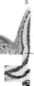
Figure 1. Chorionic villi, transformed into transparent vesicular formations. Figure 2. Mucous degeneration of the stroma (b) of a villus in hydatidiform mole; a-epithelium; c-vessels. Figure 3. Normal villus: a - layers of villus epithelium; c - vessels. Figure 4. Stagnant papilla: a - optic nerve; b - edematous optic papilla; dilation of the venous network; c - retina. Figure 5. Calcified masses (a) in the protoplasm of the epithelium of renal tubules; b - part of a glomerulus (case of poisoning with corrosive sublimate). Figure 6. Deposition of lime (o) in the artery wall (case of osteomalacia). See also: Hydatidiform Mole, Stagnant Papilla, Lime Deposits. The main significance of the covering epithelium of the vesicles, besides everything else, also follows from the connection of the hydatidiform mole with the subsequent epithelial disease observed in some cases - chorion-epithelioma (chorion-epithelioma). In recent years, a number of researchers (Freund, Langhans, Durante and especially Hinselmann) have drawn attention to the special condition of the blood vessels of the hydatidiform mole - their complete or partial impassability for blood. This occurs either due to the congenital underdevelopment of the vessels (Hinselmann) or is the result of obliterating endarteritis (Durante). From this arose a new theory of the origin of the mole. The edematous state of the villi on the basis of this theory is a consequence not of stagnation, but of the accumulation of nutritional material, which is absorbed by the covering epithelium of the villus, but cannot, due to the obliteration of the vessels, flow away from here to the fetus and be assimilated by it (A. Brindeau, C. Jeannin). In approximately half of the cases of hydatidiform mole, as well as in chorion-epitheliomas, special cysts can be observed in the ovaries (most often on both sides), reaching the size of an adult's fist. Steckel (Stockel) was the first to point out that these cysts can be interpreted as cysts of the corpus luteum; further research showed that in this case it is not a matter of cysts of the corpus luteum, but of cysts of an atretic follicle. Etiology. To the present time, there is no established view on the etiology of hydatidiform mole. According to some researchers, its occurrence depends on changes in maternal tissues: deciduous endometritis, cystic degeneration of the ovaries, general diseases of the mother. According to another view, the cause of the mole lies in the egg itself, namely in the above-mentioned condition of its vascular system and its covering epithelium. A number of considerations speak in favor of this theory: 1) cases of twins, in which one egg develops normally, while the other turns into a mole; 2) cases in which neither before the disease nor after it were pathological processes observed in the mother's sexual sphere, and finally 3) cases in which both before the birth of the mole and after it, the woman had completely normal deliveries of a healthy fetus. Symptoms. At first, pregnancy develops completely normally. The first symptom of the mole is bleeding, which usually occurs for the first time during the second month of pregnancy. This bleeding can quickly stop, only to recur again and again later. In most cases, the uterus, due to its large size, does not correspond to the duration of pregnancy. Some authors note in the mole a more frequent and pronounced appearance of albuminuria and edema and point to a more frequent than in normal pregnancy development of intractable vomiting. Diagnosis is made on the basis of: 1) bleeding in the early periods of pregnancy, 2) abnormally rapid growth of the pregnant uterus, 3) absence of fetal parts and fetal heartbeat and 4) presence of protein in the urine and edema of the lower extremities. The diagnosis becomes certain if a vesicle of the mole is found in the bloody discharge. The prognosis in hydatidiform mole is by no means as favorable as was thought earlier. The patient is threatened by a number of complications: 1) death from bleeding before delivery, during delivery and in the postpartum period; 2) severe, often fatal postpartum infection due to the absence of good contraction of the puerperal uterus and 3) the danger of an extremely malignant new formation at various times in the postpartum period - chorion-epithelioma. Therapy. If there are no indications for artificial termination of pregnancy, it is better to avoid any intervention and wait for the onset of spontaneous labor. If the woman is bleeding or begins to have a fever, it is necessary to proceed with careful emptying of the uterus by dilating the cervix and digitally removing the degenerated egg. It is necessary to avoid instrumental emptying, since in not infrequent cases of penetration of the uterine walls by altered villi (destructive mole), perforation of the uterine walls can easily occur. After emptying the uterus, it is necessary to prescribe pituitrin and ergot preparations. Curettage sometimes has to be resorted to in cases in which, despite emptying the uterus, bleeding does not stop for 10-14 days (Steckel). A woman who has given birth to a mole must after childbirth always remain under medical supervision, because if after a short or long period bleeding recurs again in her, in most cases it is a fatal disease - chorion-epithelioma. In recent years, a number of cases of so-called destructive mole (destruirende Blasenmole) have been published. The histogenesis of this condition, clinical picture, treatment and consequences do not differ from the ordinary mole. The special feature of this form of mole is that the chorionic villi, turned into vesicles, continuing to penetrate into the underlying tissue, can grow through the entire wall of the uterus, penetrate into the parametrial veins and even, spreading further, reach the vena cava. From the veins, particles of such a mole are carried by the blood current even to the lungs. However, these particles never give metastases, and therefore are not a consequence of a malignant process. In some cases, the proliferating villi can destroy the uterine wall and rupture into the abdominal cavity. Despite the possibility of perforation and widespread dissemination, this form of mole by most authors who observed it, according to its anatomical and histological structure, is not classified as a malignant new formation (Marchand, R. Meyer). The diagnosis of destructive mole presents great difficulties. It is clinically impossible to distinguish it from the ordinary mole. Usually this form is diagnosed only after some time, and then only accidentally, when perforation of the uterus occurs and as a result - intraperitoneal bleeding, which under the wrong diagnosis of ectopic pregnancy brings the patient to the operating table. In a number of other cases, the destructive mole was also recognized during an operation for tumors in the parametrium, which were interpreted as chorion-epitheliomas. Bloody mole, fleshy mole. The death of the fetus does not always lead to contraction of the uterus and miscarriage. After the death of the fetus, if the membranes usually do not continue their development, then at least they can receive from the maternal tissues sufficient nutritional material to continue their existence. The fetal villi are absorbed, the fetal membranes collapse and, lying in folds, separate from the underlying uterine wall. As a result of this, hemorrhages occur between the uterine wall and the membranes, which can penetrate all the fetal membranes and, after organization, turn them into voluminous blood masses narrowing the cavity of the egg. Sometimes blood also penetrates into the egg cavity. Such an egg, turned into a massive blood clot - a bloody mole (haematomola), can form both in the uterus and in the tube in ectopic pregnancy. However, in the latter case, blood penetrates into the egg from the maternal vessels due to the corrosion of the muscular wall of the tube by the villi of the egg growing in the tube. Sometimes a mole can form not due to rupture of vessels, but due to excessive filling of the intervillous spaces with blood (mola aneurysmatica). In the center of a bloody mole, a preserved cavity can be found with sometimes a still preserved fetus. The organizing blood mass can gradually lose its blood color due to the breakdown of blood cells and absorption of the blood coloring substance. In such cases, the mole gradually discolors and turns into a fleshy mole (mola carnosa). For bloody and fleshy mole, see also Abortion and Pregnancy.
Related articles
Mentioned in
Cite this page
“Hydatidiform Mole.” Soviet Medical Encyclopedia. English translation of Bolshaya Meditsinskaya Entsiklopediya, 1st ed. (Moscow, 1928–1936), ed. N. A. Semashko. https://sovietmedicalencyclopedia.pages.dev/article/hydatidiform-mole/