Fallopian Tubes
Historical document, translated for reference. It reflects medical knowledge of the 1920s–30s and is not medical advice.
Summary
The Fallopian tubes are paired muscular tubes extending from the uterus to the ovaries, responsible for transporting eggs after ovulation and serving as the site of fertilization. This article describes their anatomical structure, histological composition, and physiological functions.
Encyclopedia article (1928–1936)
FALLOPIAN TUBES (uterine) (tubae uterinae Fallopii, s. salpinges), or oviducts (oviducti), are paired muscular tubes, 10-12 cm in length (minimum 7 cm, maximum 19.5 cm, according to Bischoff), of approximately cylindrical shape, extending directly from the angles of the uterus and lying in the upper edge of its broad ligament (fig. 1). Anatomically, the following parts are distinguished in the F. t.: 1) the interstitial part (pars interstitialis), or the intramural part, passing through the wall of the uterus (para intramuralis) and communicating with it through a very narrow opening (not wider than 1 mm) (ostium uterinum tubae); this part is delimited from the uterine musculature in the form of a special annular formation (in which is located the aforementioned hair-thin canal), which some (Reinberg, Arnstam) consider to be a sphincter
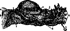
Figure 1. Longitudinal section of the tube (according to Sappey): 1-lig. utero-ovaricum; 2-lig. tubo-ovaricum; 3-fimbria ovarica; 4-fimbriae; 5-hydatis Morgagni; 6-plicae longitudinales; 7-lig. rotundum; 8-cavum uteri; 9-ostium uterinum.
of the tube; 2) the isthmus or isthmic part (pars isthmica, s. isthmus tubae), narrow, short, straight, but with thicker walls; 3) the ampullary part (pars ampullaris, s. ampulla), longer (2/3 of the entire length of the tube), wider, very tortuous, ending in a funnel-shaped expansion, the so-called infundibulum of the tube. The infundibulum is surrounded by fimbriae, or fimbriae tubae, formed by the splitting of the wall of the infundibulum into separate lobes. Each fimbria bears small notches along its edge, which makes the entire opening of the tube appear as if bitten, hence the old name-"devil's bite", "morsus diaboli" (a popular botanical term for the plant Scabiosa, whose roots resemble the free end of the F. t.). One of the fimbriae, longer, in the form of a tongue, extends to the tubal pole of the ovary (extremitas tubaria ovarii), where it attaches. It is called the ovarian fimbria (fimbria ovarica). Under it lies the tubo-ovarian ligament (lig. tubo-ovariale). On the free abdominal end of the tube, a small appendage, a remnant of the Wolffian duct, is often found, which hangs freely in the form of a polyp on a long stalk-Morgagni's hydatid (hydatis Morgagni). In structure, the F. t. resemble the uterus. Their walls consist of: 1) the serous coat (tunica serosa), 2) the muscular layer (t. muscularis), and 3) the mucous membrane (t. mucosa). The peritoneum covers the tube from above and from the sides, leaving uncovered that part of the tubal wall that faces the cavity of the broad ligament. Here the anterior and posterior leaves of lig. lati connect with each other, forming the mesentery of the tube (mesosalpinx). Through the mesosalpinx, vessels and nerves go to the tube. The serous coat is closely fused with the underlying layer of the tubal wall. The peritoneum is abundantly supplied with a dense network of lymphatic vessels. Under the peritoneum lies a layer of loose connective tissue, which surrounds the oviduct like the adventitia of vessels. Tunica muscularis of the F. t. consists of smooth muscle fibers arranged here in three layers (Gruzdev): the outer (subserous)-longitudinal, the middle-circular, and the inner (submucosal)-also longitudinal. The latter is usually well expressed only in the interstitial and isthmic parts of the F. t. A characteristic feature of the tubal mucosa is the longitudinal folds (plicae) running throughout its entire length. The folds begin in the interstitial part of the F. t., where they (usually 4) are barely expressed; the further toward the abdominal opening, the higher the folds become, the main folds begin to divide (into secondary, tertiary, etc.); in the ampullary part, the entire lumen of the tube is filled with folds. A real labyrinth of folds is formed (fig. 2); through this labyrinth the egg cell released from the ovary must pass. In cross-section, the tubal lumen has a star-shaped shape. The mucous membrane of the tube is lined with a single-layered ciliated cylindrical epithelium, the cilia of which beat toward the uterine end of the tube. In addition to cells with ciliated cilia, the tubal mucosa contains secretory elements-special glandular cells, without cilia, located mainly in the interstitial part of the tube, in the depressions between the folds. In the stage of secretion, these cells appear club-shaped swollen; after being freed from secretion, they transform into narrow cells with an elongated nucleus occupying most of the cytoplasm. These cells do not give reactions to mucus. Accumulating in certain parts of the F. t., the secretory cells form structures resembling primitive glands. However, according to the generally accepted view, the mucous membrane of the tube does not have true glands. The stroma of the mucosa consists of thin, vessel-rich connective tissue with a large number of elastic fibers. In the loops of the stromal layer, round and spindle-shaped cells are embedded; in places (closer to the uterus), fat and plasma cells, as well as lymphocytes, are also found. In the isthmic and interstitial parts of the F. t., the stroma (submucosal layer) is almost completely absent, so that the elements of the muscularis come into direct contact with the epithelial covering of the mucosa. The latter circumstance plays a significant role in the pathogenesis of tubal rupture in ectopic pregnancy.
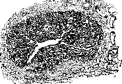
Figure 2. Cross-section through the Fallopian tube in pars ampullaris.
The blood vessels for the F. t. pass in their mesentery (mesosalpinx). Nutrient blood is supplied by arterial branches of the ovarian artery (a. ovarica, s. spermatica int.) and the ascending segment of the uterine artery (ramus ascendens a. uterinae). Venous blood is drained from the tubes into the plexus pampiniformis, 4ff
FALLOSHN
(a dense venous plexus in the area of the mesentery of the F. t.) and into the plexus utero-vaginalis (in the thickness of lig. lati laterally to each side of the uterus). Lymphatic vessels go in the infundibulo-pelvic ligament (lig. infundibulo-pelvicum, s. suspensorium ovarii) and are directed to the lymph glands of the corresponding lumbar region (gland. lumbales).-Nerves of the F. t.: branches of plexus spermatici int.-The anatomical structure of the F. t. changes with age. In sexually mature women, the tubes have the structure described above. In embryonic life, they are strongly tortuous, their shape resembles a corkscrew, and they lie very high, significantly above the upper edge of the bony pelvis. By the end of intrauterine life, the epithelium is provided with ciliated cilia, and an unusual abundance of folds is noted in the tubes, which in general appearance already resemble the tube of an adult woman. The F. t. undergo significant structural changes during the climacteric. The fibrous degeneration, characteristic of the senile involution of the entire female genital apparatus, gives a picture of gradual morphological withering of the F. t. They decrease in volume, wrinkling and reduction of mucosal folds occur, the covering epithelium becomes lower, the cilia disappear completely, in the muscular layer there is a striking abundance of fibrous connective tissue growth, blood vessels sclerose, undergo hyaline degeneration, etc. The F. t. can be conditionally considered as the excretory duct of the ovary. Their purpose is to transport the egg cell to the uterus after ovulation. In the tube, in addition, the meeting of the egg with spermatozoa occurs (see Fertilization). The meeting occurs in the ampullary part of the F. t. (the so-called "receptaculum seminis", where in the labyrinth of folds the seminal threads are detained. The F. t. are not only a passive excretory duct, but also possess a number of active independent functions. They are primarily characterized by a secretory function. The secretion of the F. t. is a cyclic process, which in some animals (rabbit, dog, pig, bat) occurs in the form of regularly and sequentially alternating phases (Moreaux), each of which is characterized by certain changes in the epithelium of the pre-uterine (isthmic) part of the tube, where the secretory apparatus of the F. t. is actually concentrated in these animals. The cyclic phases: the ciliated phase (ciliated cells predominate), the secretion phase (the epithelium of the pre-uterine part of the F. t. shows the greatest secretory activity), the excretory phase (excretion of the produced product) and the indifferent phase (reverse development toward the first phase). In women, the question of the secretory activity of the mucosa of the F. t. cannot yet be considered fully resolved. According to observations by a number of authors (Snyder, Iwata, Cahen), throughout the menstrual cycle, we have in the mucosa of the F. t. a cyclic alternation of phases of secretion and rest, during which ciliated cells transform into secretory cells and vice versa. In the middle of the intermenstrual period, the tubal epithelium is high, with predominance of ciliated cells; in the pregravid phase, non-ciliated cells, lower in number, appear in significant quantities, from which part of the cytoplasm is detached (secretory TUBES
V.
.
cells); in the first half of the menstrual cycle, these cells again reach their former size. Some (Schridde), however, deny the secretory function of the tube, not allowing for the possibility of ciliated cells transforming into non-ciliated cells. The majority, however, based on thorough research (Moreaux, Chasovnikov, Gurevich), considers it an established fact that ciliated cells transform into glandular and goblet cells, secreting mucus.-In addition to the secretory function, the F. t. have an active contractile function. In animals, rhythmic contractions of the F. t. have long been established, quite frequent (15 per minute) in the days preceding follicle rupture, and slow (5 per minute) at the time of corpus luteum maturation. Research by Cahen (after injections of lipiodol into the tube cavity) showed similar phenomena in women as well. The contractile (peristaltic) function of the tubes, according to modern views (Sobotta, Mikulicz-Radecki and others), is the main factor in the movement of the egg through the tube to the uterus. The ciliary theory, which considered the oscillating movements of the cilia as the main transport medium for the egg, is now abandoned by the majority. The tube apparently also has the ability to antiperistaltic movements, which can arise in it as a result of mechanical irritation. Developmental anomalies of the F. t. The oviducts develop from the cranial part of the Müllerian ducts. The latter are laid in the embryo in the form of a funnel consisting of cylindrical epithelium, outward from the primary kidney (Wolffian body) and laterally from its ureter (Wolffian duct). A hole (future abdominal opening of the tube) soon forms in the funnel, next to which folds (future fimbriae) with additional openings (5-6 in number) begin to form from the epithelium, which communicate with the funnel. The Müllerian cords, initially solid, later obtain a lumen by the dissolution of epithelial cells. At 3-4 months of intrauterine life, the first longitudinal folds can be noted in the tube, from which secondary and tertiary folds arise. The cylindrical epithelium develops ciliated hairs at the 10th month of pregnancy. Developmental anomalies of the F. t. include: 1. Aplasia (agenesis) of the tubes, complete absence of them-a rarely encountered anomaly, more often unilateral (with uterus unicornis), much less often bilateral (with absence of the uterus). 2. Accessory tubes (tubae accessoriae) are observed far from rarely. They are either solid (without a lumen) or hollow with a well-developed fimbrial rosette. Accessory tubes can serve as the starting point for the development of tubal pregnancy. Valdgard described a young egg in the mesentery (mesosalpinx) of an accessory tube. 3. Doubling of the tube (doubling of the Müllerian ducts) was described in the human embryo by Nagel. In adult women, doubling of the tube (on one side) has been described. 4. Accessory openings (ostia accessorla). The multiplicity of tube openings Holzbaeh considers as an atavistic phenomenon. Richard described accessory tube openings as early as 1851. Frankl has in his collection two specimens of F. t. with additional (3-4) openings. 5. Rudimentary tubes have been repeatedly described as rudimentary formations extending from one or the other corner of the uterus. Sachs described a case where the tube, in the absence of ovaries, had a length of only 5 cm, without a lumen and without a fimbrial end. In Spencer's case, in the area of both uterine horns, there were only hints of tubes in the form of button-like formations. 6. Infantile tubes-long (even Winckel pointed to this sign as congenital), tortuous, with numerous folds. Infantile tubes play a role in the etiology of ectopic pregnancy (Freund's theory, see Pregnancy, extrauterine pregnancy). Positional anomalies of the F. t. The long, tortuous tubes in infantilism have already been mentioned. Excessive length of the tubes is also noted in ovarian and parovarian tumors. A more substantial practical interest is the twisting of the tubes (torsio). The tubes can twist secondarily together with the twisting of the stalk of ovarian tumors or independently, more often on the basis of inflammatory processes. Usually the tube twists in its middle or lateral part. In some cases (extremely rarely), a twisted tube can completely untwist from the uterus and turn into a free body (corpus liberum) lying in the abdominal cavity. More often, the tubes change their position due to incorrect positions of the uterus, as well as on the basis of inflammatory processes in the peritoneum surrounding them (fixing adhesions). Dislocation of the tubes on the basis of congenital causes is rarely encountered. Inflammatory processes-see Salpingitis. The Fallopian tubes are a favorite site for the development of foci of tuberculosis (see below). Tumors of the tubes. Among neoplasms of the tubes, retention cysts occupy one of the first places in frequency, so-called salpingocele or hydrosalpinx. These formations can be classified as tumors only formally, in essence they are closer to inflammatory processes (non-proliferating tumors). From a practical point of view, salpingoceles are of great interest (see Hydrosalpinx, Haematosalpinx, Pyosalpinx). True neoplasms of the F. t. (blastomas), if not counting tube cancer, on the contrary, are encountered extremely rarely. In most cases, they have only a purely casuistic or path-anatomical interest. Among benign connective tissue tumors (mature, differentiated forms) in the tubes, the following are encountered: 1. Myomas and fibromyomas. In the literature, together with doubtful cases, there are only about 39 cases in total (Dietrich). The favorite localization of tube myomas is the uterine segment of the tube. There are no reliable cases of myomas of the ampullary part of the tube. The size of tube myomas is usually small, rarely do they reach the size of an apple (Lindquist observed a tube myoma of 43/4 kg). Histologically, tube myomas, resp. fibromyomas, do not differ from uterine fibromyomas. 2. Lymphangiomas of the F. t. (7 cases in the entire world literature)-tumors the size of 'a pea or cherry, with well-defined contours, are encountered mainly in the isthmic part of the tube. On section, the tumor consists of cavities of various sizes and shapes, lined with endothelium (dilated lymph capillaries); the endothelium is in a state of proliferation. Lymphangiomas of the F. t. should be interpreted as tumors suspicious of malignancy. 3. Lipomas of the F. t.-only individual cases have been described (Lefort and Durand, Pape, Parona). 4. Chondromas of the F. t. A case of chondrofibroma of the tube was described by Outerbridge. 5. Osteomas. True osteomas of the tubes have not been described. In most described cases, it is a matter of metaplastic or heteroplastic bone formations; more often on the basis of inflammatory processes. Among malignant stromal tumors of the F. t., the following have been described: 1. Sarcomas. Primary sarcomas of the tube-very rare neoplasms, even compared to the relatively rarely observed cases of primary tube cancer (out of 320 cases of primary tube cancer, only 12 cases of primary tube sarcoma have been described). The starting point for the tumor is the mucous membrane of the tube, sometimes the tube wall itself. In structure (papillary nature), tube sarcomas resemble carcinoma. Under the microscope-the usual picture for sarcoma. The tubes can sometimes serve as the site of development of metastatic sarcomas that had primary localization in the cervix, in the axillary lymph glands, etc. Clinically, sarcoma of the F. t. cannot be recognized, it is usually diagnosed only under the microscope. Clinical symptomatology-general with tube cancer. The prognosis is more unfavorable, even compared with cancer. According to Gosset's data, out of 7 radically operated sarcomatous patients, only one patient had clinical recovery. 2. Riemann described a case of endothelioma of the tube. Macroscopically, the tumor represented an ordinary salpingocele. Only under the microscope was the correct diagnosis made (a picture similar to sarcoma). Endotheliomas of the tubes-formations highly malignant. Franque described a case of a mixed tumor-carcino-sarco-endothelioma, which developed partially from the endothelium of blood vessels. Among benign tumors of the F. t. of epithelial type, individual cases of polyps, papillomas, adenomas, dermoids have been described. Tube polyps are especially rarely encountered. A reliable case can be considered the polypous adenoma described by Zweifel (in the isthmic part of the tube a polyp the size of a cherry). Lahm described one case of a tube polyp. Usually, however, tumors described as tube polyps were in reality beginning adenocarcinomas, inflammatory neoplasms, sometimes decidual growths, etc. Dermoid cysts of the tubes are also rarely encountered (about 20 cases described by Orthmann). Macro- and microscopic picture of tube dermoids presents no peculiarities compared with dermoids of other organs. Recently Aschheim described a case of a tube teratoma (with cartilage, fat, smooth muscle, glands). Among tumors of the tubes, tube carcinomas are the most frequently encountered and have the greatest practical and clinical significance.
Primary, secondary, and metastatic cancers are encountered in the tubes. - P r i m a r y c a n c e r o f F. t. At present, about 320 cases have been described in the literature (cancer of the tubes was first described by Orthmann in 1886). In Russian literature, there are about 15 cases of primary tubular carcinoma. The clinical picture and pathological anatomy of this neoplasm can be considered sufficiently clarified. The etiology and pathogenesis, as in cancer in general, remain dark and unexplained. Some (Sanger and Barth, Martin, Fromme and Heynemann) believe that the basis for the development of tubal cancer lies in chronic inflammatory processes, in particular gonorrhea and tbc. Others (Stolz, Kehrer, Zangemeister, Fischer) dispute this, pointing to the discrepancy between the frequency of salpingitis on the one hand, and the rarity of tubular carcinoma on the other. In addition, inflammatory processes and tbc as a rule affect both tubes, while tubular carcinoma, on the contrary, is most often unilateral (according to Levitsky in 81%). Among the predisposing factors for primary tubal cancer, the climacteric period should be taken into account, although cases of primary tubular carcinoma have been described in young women. The soil for the development of cancer in the tube can be the formations found in the thickness of the tubal wall, which are included in the concept of salpingitis isthmica nodosa (adenomyosis according to Frankl', adenomyositis according to R. Meyer) (see Salpingitis). There seems to be a certain predisposition to cancer of F. t. from childless or women who have given birth once. Tubular carcinomas are most often encountered at the age of 40 - 55 years. Primary cancer of the tube more often localizes in the middle third of the tube and its abdominal segment, while the uterine end is affected much less frequently. Macroscopically, the cancer-affected tube (Fig. 3) changes its shape
Figure 3. Carcinoma tubae dextrae (d): a-left tube; b-left ovary; c-metastasis to the right ovary. (retort-shaped with sausage-like swelling at the abdominal end); often has a sealed fimbrial end; the size of the tumor varies from the thickness of a little finger to the size of a child's head; in individual cases, the F. t. affected by cancer reached the size of an adult's head; the tumor has a firm elastic consistency, sometimes somewhat soft (medullary cancer), a smooth surface (until papillae have grown through the peritoneal covering of the tube); when the wall of the tube is opened up by growing papillae, the cancerous process spreads not only
Figure 4. Carcinoma tubae: a-tunica muscularis; b-primary tubal epithelium; c-carcinomatous proliferations in the wall of the tube; d-papillary proliferations protruding into the lumen of the tube.


along the surface of the tube, but also per continuitatem to adjacent tissues; moreover, in these cases, the tube becomes adherent to the pelvic peritoneum and neighboring organs; inflammatory adhesions are present at the periphery of the tumor. The content of tubular carcinoma is a whitish papillary mass, in some areas of a brain-like consistency, closely adherent to the walls of the tube. Microscopically, among tubular carcinomas, the following are distinguished: 1) papillary form (more common) - in the form of multiple nodular proliferations in the dilated lumen of the tube (Fig. 4), 2) alveolar - in the form of large cellular fields, separating the connective tissue and giving peculiar protrusions to the side, and 3) mixed (carcinoma mixtum) - papillary-alveolar. The sclerosing type of tubal cancer is encountered much less frequently - with marked development of connective tissue and small-alveolar proliferations. In sclerosing carcinoma, hemorrhages and tissue necrosis are often observed. Orthmann (1903) and Amreich (1922) described (one case each) primary cancer of the F. t. with stratified squamous epithelium, which developed as a result of metaplasia (of the accommodative type) of the cylindrical epithelium into squamous. The most characteristic form of primary tubal cancer is nevertheless the papillary structure of the tumor. Usually, the cylindrical epithelium (tumoral) lacks cilia. Polymorphism is clearly expressed. The clinical picture of primary tubal cancer, in terms of symptomatology, presents nothing characteristic. The disease proceeds with the same symptoms as are observed in inflammatory tumors of the tubes. The symptoms to which attention should be paid are as follows: 1) Vaginal discharge - watery, yellowish-amber colored, more often with an admixture of blood (always a suspicious sign in terms of malignancy of the neoplasm). According to Ruge, in tubular carcinoma, we have profuse watery discharge in 25% of cases. Relatively frequently in tubal cancer, hydrops tubae profluens, s. hydrosalpinx profluens (periodic emptying of the accumulated contents of the tube into the uterus and out through the vagina with preceding colicky pains) is observed, which, as is known, is occasionally also encountered in simple hydrosalpinx (see Hydrosalpinx). Zweifel, in 121 cases of tubal cancer, noted the presence of hydrops tubae profluens in 20 cases. In two of his own cases, Zweifel made the correct diagnosis based solely on the described symptom. 2) Early onset of colicky pains (in contrast to cancer of the uterus and ovaries, in which pains appear in the late stages of the disease). This symptom occurs in tubal cancer in approximately 3/4 of all cases. The intensity of the pains varies. Sometimes they take on a convulsive character. The pains are localized in the lower abdomen, in the sacrum, and often radiate to one or both legs. The pains are caused by stretching of the tubal wall, peristaltic contractions of its musculature, pressure of the tumor on neighboring organs and nerve plexuses, as well as the development of inflammatory phenomena around the neoplasm. 3) Presence of atypical uterine bleeding, not decreasing after curettage. 4) Absence of ascites (occurs very rarely and in small amounts). 5) Absence of elevated temperature and in the history of inflammatory processes. The diagnosis of primary tubal cancer is so difficult that even at laparotomy, a correct diagnosis is not always possible. According to Fonio's data, the correct diagnosis of tubal cancer was made in no more than 6.5% of cases. In differential diagnosis, the possibility of confusion with inflammatory pyosalpinx, tubal pregnancy, ovarian tumors, and even fibroid of the uterus (on a stalk) should be considered. A presumptive diagnosis can be made based on the clinical signs mentioned. Recently, the Zondek-Aschheim reaction has been used for diagnosis (positive result).-The prognosis in cancer of the F. t. is extremely poor, mainly due to late diagnosis. In the opinion of some clinicians, tubular carcinoma is more malignant than cancer of the uterus. Here, the anatomical features of the tubes play a role - their thin walls, which cause more rapid and early spread of the cancerous process beyond the primary localization. Metastases in tubal cancer spread by all three routes: hematogenous, lymphogenous, and canalicular. The uterus, ovaries (37 cases according to Zweifel), and the surrounding peritoneum are most frequently affected.-Treatment-radical operation followed by radiotherapy. The results of surgical intervention are unsatisfactory (recurrences and metastases within the nearest year). Long-term cure (more than 3 years without recurrence) is noted in 4% (Beck, Stanca) and in 6% (Franke, Zweifel). During the operation, the uterus must necessarily be removed, since in primary cancer of the F. t. it is affected in 12% (Ruge). Cases of metastases to the cervix, resp. portio vaginalis (Kundrat, Hofbauer, Schafer) have been described. The results of subsequent X-ray irradiation cannot be assessed due to the small number and briefness of observations (Amreich, Thaler, H. Kustner). To improve the results of surgical treatment of primary tubal cancer, it is necessary to operate more extensively on any suspicious neoplasm in the tubes. Secondary cancer of the F. t. arises on the basis of cancerous transformation of benign epithelial tumors, mainly papillary blastomas. These secondary carcinomas have no clinical significance due to their extreme rarity.-Metastatic cancer of the tubes occurs either by direct transition of the neoplasm from the ovaries (more often) and uterus, resp. cervix (rarely), or it is carried here via lymphatic routes, as a true metastasis, from some other source in the body. From neighboring organs (from the uterine cavity or through the fimbrial end of the tube in ovarian cancer), the spread of the cancerous process can occur via the canalicular route. Cancer can also be carried into the tubes through blood vessels (extremely rarely). Cancerous metastases to the tube are in general an extremely rare phenomenon. Incidentally, true Krukenberg tumors have also been described in the tubes. In the entire world literature, only 11 such tumors are counted. One must think that they are not as rare as can be judged from the literary data. In all cases of ovarian Krukenberg tumor, the tubes must be carefully examined.-Chorio-epithelioma of the tubes-see Chorioepithelioma. Actinomycosis of the tubes. Extremely rare cases of infection of the tubes with the ray fungus, according to the clinical and pathoanatomical picture, present nothing special compared to actinomycosis in other organs. Infection apparently occurs through the intestine (a controversial question). The tubes are more often involved in the process together with other organs. Diagnosis is possible only by histological and bacteriological examinations.
M. Malinovsky. Tuberculosis of the Fallopian tubes (salpingitis tuberculosa), an inflammatory disease of the tubes caused by infection with tuberculous bacilli and characterized by the development of specific tubercles. Tuberculosis of the tubes and uterus was first described by Morgagni in 1744. From this time, the study of tuberculosis of both the tubes and other parts of the female genital sphere began. Only since the time of the research of Virchow and R. Koch (1882), which clarified the pathological anatomy and etiology of tuberculosis in general, did the study of tuberculosis of the Fallopian tubes begin on a solid scientific path. Steven in 1883 first discovered Koch's bacillus in tuberculous tubes. The frequency of tuberculosis of the female genital organs among gynecological diseases in the clinic, according to Williams, Polano, Schroeder, Kiparsky, Melnikov, and Morozova and others, ranges from 1% to 7.7%. All parts of the female genital apparatus can be affected by tuberculosis, but the tubes are most frequently affected, mainly their ampullar segment. The frequency of tuberculous salpingitis according to Kronig, P. Schroeder, Gorizontov, and others reaches 85-90% of all tuberculous diseases of the female genital organs. The reason for such frequency and predisposition of the tubes to tuberculosis has not yet been fully elucidated, but apparently it is due, on the one hand, to the abundant blood supply of these organs, and on the other hand, to the slowed blood flow in them, promoting the settling of circulating tuberculous bacilli. After the tubes, the uterus is more frequently affected (47-70%), followed by the ovary (Fig. 5) (15-44%), the vagina (6.7-9%), and finally the external genital organs (1%). Tuberculous salpingitis is usually observed in both tubes simultaneously (Fig. 6).
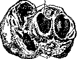
Figure 5. View of a sectioned ovary affected by the caseous form of tuberculosis. Several tuberculous cavities are visible (1); 2-wall of the cavity.
In addition, it often combines with tuberculosis of other parts of the female genital sphere; especially often it is combined with tuberculosis of the uterus (according to Gorizontov, Kronig, Schroeder, Simmonds)-in 32.9-60-70%, with tuberculosis of the ovaries and peritoneum-52-68.5% and more. conglomerate inflammations

Figure 6. Tuberculosis of the endometrium, Fallopian tubes, and perimetrium.
Tumors of the appendages of tuberculous character, which are based on markedly changed Fallopian tubes, occur quite frequently among inflammatory tumors of the appendages; according to R. Schröder, they occur in 10%, and according to Pankow, even in 22%. Age and predisposing factors. Tuberculous salpingitis, like tuberculosis of other parts of the female genital sphere, is mostly a disease of young age - 20-30 years. Predisposing factors for the development of tuberculous tubes, as well as other parts of the female genital sphere, are the puerperal state, insufficient development of the sexual organs, and previous lesions by inflammatory processes, especially gonorrhea. However, there is no unanimity among authors regarding the predisposing influence of the latter; for example, Sellheim and Pankow completely deny it. Mode of infection and routes of spread. A number of pathologists (Bollinger, Schmorl, Albrecht, Aschoff, etc.) have recently established that tuberculous salpingitis, like tuberculosis of other parts of the female genital sphere, is almost exclusively of secondary nature. It most often arises from obvious or hidden tuberculous foci in the lungs (89.5%), in lymph glands, intestines, peritoneum, and other organs. Regarding the importance of tuberculous peritoneum in the development of tuberculous salpingitis, the views of researchers differ sharply. According to some (Albrecht, Baumgarten, etc.), tuberculous peritoneum very rarely serves as a source of the disease of the tubes. Baumgarten was unable to establish the transition of the process from the peritoneum to the mucous membrane of the tubes experimentally (on rabbits). According to others (Krenig, Ghon, Kafka, Sellheim, Gorizontov), such a transition, on the contrary, plays a major role in the development of genital tuberculosis and often occurs. From the above-mentioned tuberculous foci, the process spreads mainly into the tubes through blood and lymph vessels and then per continuitatem. The latter method of spread of tuberculosis has significance in the transition of the process from the intestine, peritoneum, and other neighboring organs. From distant foci, infection is carried into the tubes mainly through blood vessels. Here, tuberculous bacilli, due to the slowed blood flow (Amann), settle in the tissues and cause the tuberculous process. Tuberculous tubes very often serve as a source for the development of tuberculosis in other sexual organs. The ascending spread of tuberculosis from below upward along the mucous membranes from the sleeve into the uterus, etc., is theoretically quite permissible, but in reality in women with genital tuberculosis it occurs very rarely and only under especially favorable conditions such as stagnation of secretion, antiperistalsis, etc. (Jung, Baumgarten). Primary tuberculous salpingitis in the true sense of the word has not yet been established patho-anatomically to this day, therefore the question of it has only purely theoretical interest. Primary tuberculous tubes is theoretically just as permissible as primary tuberculosis of bones. The rarity of primary tuberculosis of the lower part of the genital canal suggests that infection of the female genital organs through the semen of a tuberculous man, in which, by the way, only a small number of tuberculous bacilli could be found, if it can occur, is very rare and does not have the practical significance that was attributed to it earlier. Pathological anatomy. The diseased tuberculous tube has macroscopically in general the same changes as in septic and gonorrheal inflammation, i.e. it is thickened, hardened, and twisted. The thickening is more strongly developed in the ampullary part and often in the isthmic and interstitial parts. Here we quite often encounter so-called nodular salpingitis (salpingitis isthmica nodosa), which was mistakenly considered by Hegar and his disciples to be exclusively characteristic of tuberculous salpingitis. Besides sometimes noticeable to the naked eye, tubercles, a characteristic feature of tuberculous salpingitis is the accumulation in the tubal canal of caseous masses, which in some cases protrude as plugs from the abdominal openings. In tuberculous salpingitis, the abdominal ends of the tube remain unsealed in approximately half of the cases (Neupemann). In case of sealing of the tubes, they often turn into so-called tuberculous pyosalpinges (pyosalpinx tuberculosa), containing in their dilated canal much caseous pus and sometimes reaching considerable sizes (up to a fist and more). The basis of the described changes in the tubes is the development in them of round-cell infiltration, tubercles, and their further changes. Depending on the development of these changes either in the mucous membrane, or in the muscular layer, or in the serous coat - 1) tuberculous endosalpingitis (endosalpingitis tuberculosa); 2) tuberculous mesosalpingitis (mesosalpingitis tuberculosa) and 3) tuberculous perisalpingitis (perisalpingitis tuberculosa) are distinguished. Sometimes the disease of the tubes may be limited to only the serous coat (perisalpingitis tuberculosa), which occurs when tuberculous peritonitis spreads to the peritoneum of the tubes. Most often the mucous membrane alone is affected by tuberculosis, or the mucous membrane together with the muscular layer. In the mucous membrane under the epithelium, usually a small or significant number of miliary tubercles are found, and in places desquamation and death of the epithelium and fusion of the folds of the mucous membrane occur, while in other places, on the contrary, proliferation of it and formation of gland-like passages (Kundrat, Polano, Franke). These proliferative processes in the epithelium are in some cases very strongly expressed, giving some resemblance to the beginning of cancer. With further development and fusion of tubercles, as well as with the occurrence of caseous degeneration in them, the mucous membrane of the tube gradually necrotizes and turns into caseous masses. The same thing happens in the muscular layer. The main constituent part of the above-mentioned conglomerate tuberculous tumors of the appendages are the changed tubes. Besides them, the intestine, most often the flexura sigmoidea or rectum, closely fused with the tube and sometimes communicating with its canal, and finally the ovary, which is often affected by tuberculosis in this case, enter into the composition of these tumors. All the described changes in the tubes occur in the chronic course of the process. In the acute course, which occurs in general acute miliary tuberculosis, strong round-cell infiltration, a small number of giant cells, many tuberculous bacilli, and abundant caseous necrosis of the mucous membrane are noted in the tubes. No special changes in the external appearance of the tube occur in the acute form (Orthmann). In spontaneous healing of tuberculous salpingitis, which can sometimes occur besides in the tubes also in other parts of the genital sphere (uterus, ovaries), fibrous change, caseation and calcification of tubercles and other tuberculous products, as well as sealing of the tubal openings by abundant peritoneal adhesions, occur. Course and symptoms. The acute course of tuberculosis of F.t. occurs less frequently, is observed in acute general miliary tuberculosis and does not cause noticeable gynecological symptoms. Chronic tuberculous salpingitis, as well as chronic tuberculosis of the ovaries, uterus, and other sexual organs, is more frequently observed and proceeds with the same symptoms as occur in chronic gonorrheal or septic inflammations of the female genital sphere. These symptoms consist mainly of leucorrhea, disorders of menstruation, pains, and infertility. They depend partly on the salpingitis itself, partly on accompanying diseases of other sexual organs and the pelvic peritoneum. Leucorrhea in tuberculous salpingitis is rarely of tubal origin due to emptying of the contents into the uterine cavity (sactosalpinx tuberculosa profluens), but most often depends on the accompanying tuberculous endometritis. Disorders of menstruation, according to Schröder and Martin, occur only in 50% and manifest as dysmenorrhea, amenorrhea, and rarely as metrorrhagia and menorrhagia (hypermenorrhoea). These disorders depend both on the general effect on the body of tuberculous infection, and on the often accompanying lesion of the ovaries and uterus by the same process. Infertility, being a very frequent companion of tuberculous salpingitis, depends on the change in the mucous membrane of the tubes (endosalpingitis tuberculosa), on the sealing of their abdominal openings, as well as on the accompanying endometritis, but sometimes in tuberculous salpingitis uterine or extrauterine pregnancy can occur. The first is often prematurely interrupted, the second is mostly localized in the funnel of the tubes (graviditas infundibularis) and has the usual outcomes. According to Hepner (Hörpner), only 8 cases of tubal pregnancy in tuberculous tubes are known. Pains are mostly localized in the lower abdomen and back with irradiation into the legs and are not particularly intense. They are caused by contraction of the tube, stretching of the serous coat, and development of an inflammatory process in the peritoneum around the appendages with formation of adhesions or liquid exudate. Temperature is within normal limits or subfebrile. From the side of the abdominal walls, persistent tension, as well as retraction, and sometimes, on the contrary, protrusion and enlargement of the abdomen due to the exudative process in the peritoneum are often observed. When a considerable conglomerate tumor of the appendages develops, a diffuse or clearly delimited hardening can be palpated through the abdominal covers in one or the other hypogastrium.
During gynecological examination, significantly thickened and altered tubes are found on one or both sides of the uterus, which may be either mobile or adhered to the ovaries, forming the picture of typical chronic salpingo-oophoritis with periadnexitis; sometimes in the area of the appendages, large-sized tubo-ovarian cysts or conglomerate tumors are found, characterized by an abundance of dense, slightly painful adhesions. In the presence of the listed symptoms and objective data, the process in the tubes has a slow, progressive course. In most cases, patients with tubal salpingitis die from tuberculosis, which has flared up either in the primary foci (lungs or other organs) or, which occurs much less frequently, in the sexual organs. Sometimes tubal salpingitis completely subsides, and the previously enlarged appendages significantly decrease and become dense, which indicates self-healing (Hegar, A. Martin). Prognosis. The prognosis of tubal salpingitis, as well as tuberculosis of other sexual organs, is generally serious. Although cases of benign course and sometimes self-healing are not so rare, patients suffering from this disease are constantly under the threat of exacerbation and secondary septic infection. With the introduction into therapy of tuberculosis of the sexual organs of X-ray rays and other physical methods of treatment, as well as with the limitation of the scope of application of major operations, mortality has decreased, and the chances of cure have increased. Diagnosis. The diagnosis of sexual tuberculosis and in particular tuberculosis of the uterine appendages is made on the basis of history, general objective and gynecological examination, as well as on the basis of auxiliary methods, such as examination with mirrors, curettage of the uterus, and trial excision of ulcers of the vaginal canal with subsequent microscopic examination of the obtained tissues, examination for tubercle bacilli of the sexual secretions and fluids obtained by trial puncture. The presence of tuberculosis of the lungs in the past and present, phthisic habitus and infantilism of the sexual sphere are factors speaking in favor of tubercular disease of the appendages, but of course only when excluding septic and gonorrheal diseases. The presence of chronic tubo-ovarian cysts in virgins and children also indicates with greater probability their tubercular nature. Detection of tubercular ulcers in the lower parts of the vaginal canal or tubercular endometritis, as well as finding tubercle bacilli in the secretions and fluids of the vaginal canal makes the diagnosis of tuberculosis of the appendages, in particular the tubes, almost certain. In general, up to now at our disposal there is not a single clinical sign which could be reliably relied upon in recognizing tuberculosis of the appendages. The development of nodular thickenings in the tubes, nodules in the posterior Douglas space and along the course of the sacro-uterine ligaments, the detection of which Hegar and his students (Selheim, Bulius and others) attributed pathognomonic significance to tuberculosis of the tubes and pelvic peritoneum, can occur not only on the basis of tuberculosis, but also on the basis of septic, gonorrheal inflammation, and can also be in papillary cysts of the ovary, carcinomatosis of the peritoneum and endometriosis. For the diagnosis of tuberculosis of the appendages (in particular the tubes), the examination of blood for leukocytosis and the rate of erythrocyte sedimentation has a certain auxiliary value. According to Krenig, the absence of leukocytosis, and according to Gragert, sharply expressed erythrocyte sedimentation and a small leukocytosis most frequently accompany chronic tuberculosis of the appendages. Since in the overwhelming majority of cases tuberculosis of the appendages is secondary, diagnostic methods with tuberculin (ophthalmoreaction, Pirke reaction and its modifications on the portio vaginalis of H. Freund, subcutaneous injections of tuberculin according to Birnbaum) have proved to have no diagnostic value for tuberculosis of the sexual organs in general and the tubes in particular. Having received a general reaction to tuberculin, one cannot be sure that the organism is reacting precisely to the process in the sexual sphere. As for the local reaction in the sexual organs, it is little reliable due to the possibility of the appearance of local reactive phenomena to tuberculin also in inflammatory diseases of clearly non-tubercular nature. The latter give, according to Borrelly, a local reaction to tuberculin in 37-65%. In such a situation, the most reliable method of recognizing tuberculosis of the appendages is trial laparotomy, to which one has to resort in some cases. The diagnosis of tuberculosis of the uterine appendages (tubes) is definitely clarified in most cases either during the operation or only by microscopic examination of the removed tubes and ovaries. In recent times, for the purpose of greater clarification and giving greater reliability to the presumptive diagnosis of tuberculosis of the tubes (uterine appendages), Yagunov, Mandelstam and Teverovsky began to resort to the method of vaccinediagnosis according to Burlakov, consisting in injections in a known sequence separately intradermally and into the thickness of the mucosa of the vaginal sleeve, cervix uteri and wall of the rectum of four different vaccines (gono-, staphylo-, strepto- and colivaccines) and allowing to definitely exclude septic, gonorrheal and colibacillary diseases of the vaginal canal. These authors have indeed, so far on a small material, obtained good diagnostic results. Treatment. Radical treatment of sexual tuberculosis in general and of the uterine appendages in particular is possible only in those cases when these organs are affected in the form of more or less isolated foci with latent or slightly active state of other tubercular foci of the organism. At present there exist two basic methods of radical treatment of tuberculosis of the appendages and uterus - surgical and non-surgical, conservative. The latter method includes treatment with a quartz lamp (Yagunov) and mainly X-ray rays. One surgical treatment, which was carried out rather widely and radically in the past, in the form for example of complete removal of the uterus with appendages, gave a high primary mortality of 10-25% (Krenig, Bumm, Thaler). X-ray therapy of sexual tuberculosis, being a young method, dating from 1909 (Spaeth, Wetterer), has no direct primary mortality and gives favorable results, but is characterized by duration (up to a year or more) and requires an absolutely exact diagnosis, which can be established only by operative intervention. Therefore, most modern gynecologists adhere to combined treatment, consisting in the combination of operation and X-ray therapy, and the operations mostly have a conservative or diagnostic character and consist in laparotomy. In easily movable or slightly adhered appendages in the presence or absence of ascites, laparotomy is performed, the fluid is released, obviously diseased tissue is removed without touching the uterus, if there are no sharp changes in it, and then after a certain time X-ray therapy or treatment with a Bach quartz lamp should follow. In case of finding abundant adhesions in the area of the altered appendages or finding the dry form of tubercular peritonitis during laparotomy, one should refrain from removal of the appendages and separation of adhesions (due to the danger of great trauma and fecal fistulas), and, limiting oneself to trial excision, close the abdominal cavity and in the future apply X-ray rays. In the presence of abundant adhesions and tubercular pyosalpinxes or abscess of the ovary, it is better first of all to carry out X-ray therapy. If the latter does not help, then a radical operation (removal of appendages, and if necessary also of the uterus, by means of laparotomy) is performed. Incision of tubercular purulent appendages is performed in the presence of vital indications. The incision most often has to be made in the posterior fornix (posterior colpotomy). The need for such incisions is rare. An unfavorable consequence of them is the possibility of formation of chronic purulent fistula. In cases of tuberculosis of the appendages accompanied by acute phenomena, the usual anti-inflammatory treatment is applied, i.e., rest, ice, narcotics. Often the accompanying tubercular endometritis is best treated, according to the view of some recent authors (Weibel), conservatively - with X-rays, quartz lamp, etc., and not actively, by means of curettage. In clinically isolated disease of the endometrium, curettage gives quite satisfactory results (Veit, P. Schroder). For X-ray therapy, small doses of rays (from 1/4 to 1/48 HED) are used, regarding the magnitude of which unanimity has not yet been achieved among authors. Weibel recommends doses of 1/8-1/10 HED from two large fields in front and behind, with a filter of 3 mm aluminum, with intervals between irradiations of 4-8 weeks. Stephan, Keller, Uter, Pankov and others recommend doses of 1/8-1/12 HED. For treatment with such doses, one can use small and inexpensive X-ray apparatus. Along with the described treatment, it is necessary to prescribe the usual measures aimed at raising the general condition of the organism, i.e., enhanced nutrition, climate treatment, sun baths, rest, arsenic, iron, and at the same time treatment of the existing tubercular foci of other organs. All this treatment is most conveniently carried out in special sanatoriums. According to P. Schumacher (P.
Schumacher), by means of the described radical and auxiliary therapy, it is possible to achieve up to 70-80% clinical cures and up to 20-30% improvements. 513 Prevention. General prevention-see Tuberculosis. In relation to the sexual organs, preventive measures are the observance of cleanliness and rules of sexual hygiene, as well as abstaining from coitus with a tuberculous man, especially one suffering from tuberculosis of the urinary system. If this is not possible, then precautions should be taken, such as coitus condomatosus or disinfecting douches (with sublimate) etc.
N. Goriantov. Insufflation of the Fallopian Tubes. Insufflation of the F. t. (pertubatio, insufflatio) is used in female infertility for diagnostic purposes, to determine the patency of the tubes. Diagnosis of tubal infertility can also be carried out by a number of other methods. The most objective of them is metiosalpingographia (see), in which it is possible not only to see on the X-ray whether the tubes are patent or not, but also to determine which part of the tube is obstructed. According to various statistical data, tubal infertility is observed in 21% to 90% of all cases of female infertility. It is most often associated with obstruction of the tubes due to past infections, both gonorrheal and septic. Other causes of tubal obstruction are less common; these should include intrauterine injections of iodine, operations on the tubes, congenital anomalies, etc. Insufflation of the tubes was first proposed by Rubin in 1919; he introduced oxygen through the uterus and tubes into the abdominal cavity and determined the presence of gas in the abdominal cavity by X-ray. His original technique was very complex; the amount of gas introduced reached up to 1 liter (averaging 300 cm3). The equipment was not very portable and expensive; it consisted of a metal oxygen bomb, a two-necked flask with an antiseptic liquid for filtering the oxygen; on the way to the uterus there were a manometer and gas meters to determine the pressure and amount of gas introduced. Over time, this equipment was significantly simplified. Simplified apparatuses have been described and proposed by Selheim and Engelmann in 1922 and in our USSR by Mandelstam. Selheim's apparatus consists of a syringe with a capacity of 150 cm3, the syringe is connected to a manometer showing the pressure in the uterus and tubes (fig. 7). Selheim based the diagnosis of patency on listening to the sounds of passing air, the drop in pressure in the manometer, and the absence of resistance to the movement of air in the syringe when pressing on the piston. Fig. 7. Selheim's apparatus for insufflation of the Fallopian tubes. In the apparatus, air is forced into the uterus by a rubber bulb, the air introduced passes through hydrogen peroxide in a two-necked flask, and the pressure in the system is controlled by a manometer (fig. 8). With patent tubes, the pressure drops in the manometer during insufflation, and bubbles of air pass through the liquid in the Wulf flask,
Figure 8. Apparatus for insufflation of the Fallopian tubes according to Mandelstam.
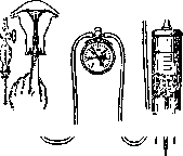
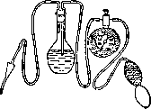
spirit. The Mandelstam apparatus has become widely used in the USSR both in its original form and in various modifications. Subsequently, insufflation of the tubes with a 10-gram Laënnec syringe was proposed, with pressure not being measured in this case. Such simplified apparatus was described by Legechenko (1925). With the simplification of the apparatus, various modifications of the tips introduced into the uterine cavity also appeared. The simplest model is a Braun syringe tip with a rubber cone from a Tarnovsky syringe fitted onto it. Instead of the Braun tip, a metal female catheter can also be used. Mandelstam proposed a metal conical cannula that hermetically seals the external opening of the cervical canal. Zelgeim and Stiasny propose instead another method: with one hand, the tip is fixed to the external cervical os, and at the same time, bullet forceps applied to the anterior lip of the vaginal part pull it forward toward the surgeon. In addition, a whole series of self-retaining tips for fixing the cannula and cervix has been proposed. Among Soviet self-retaining tips, the Zhivatov cannula should be mentioned. Insufflation of the tubes is performed in the intermenstrual interval, preferably on the second week after menstruation. Insufflation of the tubes is contraindicated in the presence of acute and subacute inflammatory processes of the pelvic organs, in erosions, endocervicitis, in bloody, purulent, and seropurulent leukorrhea; in pregnancy, in malignant neoplasms, and in the presence of large anatomical changes in the appendages of the uterus. General diseases of the body, high temperature, diseases of the kidneys, heart, endocrine system, etc. are also contraindications to insufflation of the tubes. Where there are doubts about the correctness of the diagnosis, it is necessary before insufflation to subject patients to blood examination, erythrocyte sedimentation rate, and to determine the degree of purity of the vaginal discharge in order to exclude the presence of infection and to avoid complications associated with exacerbation of the inflammatory process or the introduction of infection by the air current from the lower parts of the sexual tract into the uterus, tubes, or peritoneum. Insufflation of the tubes is a serious procedure. Therefore, some clinics perform it only in a hospital setting. The patient is placed in a gynecological chair, and the vagina is opened with specula. The vagina and vaginal part are wiped with alcohol or tincture of iodine. The anterior lip of the cervix is grasped with bullet forceps or Muse forceps. A tip connected by a rubber tube to an air insufflation apparatus is introduced into the uterus. The tip should tightly close the opening of the cervical canal, so that air does not escape from the uterus into the vagina. Air is slowly introduced into the uterus with a rubber balloon, with pressure in the system being measured by a manometer. Air pressure above 150-180 mm of mercury should not be used during insufflation of the tubes, as at higher pressure there is a danger of rupture of the tube. Based on the drop in pressure, one can judge how much air has passed into the abdominal cavity; thus, if pressure from 150 mm dropped to 50 mm, then it should be considered that 30 cm³ of air has passed, if pressure from 100 dropped to 50, then the amount of air introduced is equal to 15 cm³. Based on pressure, Mandelstam judges the degree of patency of the tubes. Thus, if symptoms of patency appear at a pressure below 75 mm Hg, then the tubes are easily patent; at a pressure from 75 to 125 mm they are satisfactorily patent, and above 125 mm the tubes are stenosed. During air insufflation, the surgeon or assistant auscultates sounds from the abdominal wall indicating patency of the tubes. At the end of insufflation, the patient should be laid down. Symptoms indicating patency of the tubes are as follows: 1. On auscultation, a clear tubal sound is heard, resembling the whistling of wind in a chimney pipe. These sounds can be heard on one or both sides. Based on a unilateral tubal sound, one can speak with very little probability of unilateral patency of the tube. On auscultation, other sounds unrelated to tubal patency can sometimes be heard - peristalsis of the intestine, escape of air from the cervix into the vagina when the external opening of the cervical canal is not tightly closed. In case of obstruction of the tube in the ampullary part, sounds related to the penetration of air into the dilated end of the tube can also sometimes be heard. 2. With patent tubes, the pressure in the manometer drops during insufflation, because air from the closed system penetrates into the abdominal cavity. 3. With patent tubes, air bubbles pass through the antiseptic liquid in the two-necked flask. 4. Air introduced in large quantities into the abdominal cavity, pressing on the diaphragm, raises the liver dullness above the costal margin. 5. With patent tubes, pains in the shoulder and scapula (Phrenicussymptom) are observed due to pressure of air on the diaphragm. This symptom is clearly expressed when large quantities of air are introduced. When a small amount of air penetrates into the abdominal cavity, this symptom may also be absent. 6. With patent tubes, air can be determined by radiography in the peritoneal cavity. The most reliable symptoms indicating patency of the tubes are as follows: auscultation of tubal sounds, drop in pressure in the manometer during insufflation, and Phrenicussymptom; the latter may be absent when a small amount of air is introduced into the abdominal cavity. Where there is any doubt about the correctness of the diagnosis, insufflation of the tubes should be repeated after some time, preferably in the next intermenstrual interval. For clarification of the diagnosis of patency, metro-salpingography is also indicated. An incorrect diagnosis may depend on spasm of the tube; to avoid this, belladonna or morphine preparations can be administered. Complications observed during insufflation are most often related to errors in technique (too much pressure and forced introduction of air) or to insufficiently careful selection of patients for insufflation of the tubes (possibility of exacerbation of a previous inflammatory process or introduction of infection into the higher genital organs). Cases of death after insufflation of the tubes have been described, with mortality being 1:1,000 and related to incorrect selection of patients for insufflation. According to some authors, insufflation of the tubes, in addition to valuable diagnostic data, sometimes also has a therapeutic effect. These authors claim that as a result of insufflation, pregnancy occurs in 8-12% of cases. The onset of pregnancy can be explained by both the straightening of the folds of the tube and the stretching of light adhesions and fusions (tubal gymnastics according to Zelgeim). Besides uterine pregnancy, individual cases of ectopic pregnancy have also been described after insufflation. Insufflation of the tubes is also performed with an open abdominal cavity during operations on the tubes; in this case, air can be introduced both from the vaginal and uterine side and from the abdominal end of the tube. For insufflation from the abdominal end, a syringe is most often used. After salpingostomy, insufflation of the tubes is also recommended in order to avoid the formation of adhesions and to check the results of the operation. It deserves attention that even in the hands of very experienced doctors (e.g. Kustner from Zelgeim's clinic), insufflation of the tubes sometimes leads to incorrect conclusions, and therefore the results obtained from insufflation should be subjected to a more critical evaluation than is usually the case. It is very possible that the method of insufflation of the tubes will acquire greater value in the future if we can use the new apparatus proposed by Rubin, which makes it possible to graphically record fluctuations in pressure in the uterus, depending on the contractile ability of the tubes. With the introduction into practice of insufflation of the tubes, no operation undertaken for infertility can be performed without prior verification of tubal patency.
M. Pobedinsky.. Transplantation of Fallopian Tubes into the Uterus. In cases of infertility caused by obstruction of the F. t. in the interstitial or isthmic part, it is sometimes necessary to resort to the operation of removing the affected part of the tube and transplanting the remaining part into the uterus in order to restore the patency of the tubal canal. Such an operation was first published in 1899 by Ries and Weitkins; they applied it to a woman in whom the right appendages had previously been removed; as a result, pregnancy occurred, which ended in miscarriage. However, at that time the operation was not yet recognized by gynecologists and began to be applied only after the case published in 1921 by Cullen and Shaw, where after transplantation of the tube the patient became pregnant, with the first pregnancy ending in miscarriage and the second in normal delivery. Strassmann in 1924 was already able to publish several cases and proposed his own method of operating. Since then, a large number of authors have published their cases and developed the technique of the operation. If initially transplantation of the F. t. was performed incidentally in cases where the affected tube on one side was removed, while the second tube was affected in the isthmic part, at present transplantation is applied both incidentally and as an independent operation in the presence of infertility. When applying the operation of transplantation of the F. t., a number of authors (Mandelstam, Unterberger, Michaelis, etc.) indicate that as a result of the operation patency and normal physiological function of the tubes are restored; thus, Reiprich, on the basis of data from world literature, calculates cases where pregnancy subsequently occurred at 10-15%; according to Sedyukov's data, out of 72 published cases of tube transplantation, pregnancy was observed in 23%, according to the material of Mandelstam and Kiparsky, pregnancy occurred twice out of 21 cases. From the indications available in the literature, pregnancy occurs from 3 months to 3 years after the operation. Among the proposed methods, it is necessary to dwell on the operations of Strassmann, Unterberger, Mandelstam, Kiparsky, and Sedyukov. Strassmann's operation is performed as follows: the uterine cavity at the apex of the angle is opened with a transverse incision in such a way that the tube can be passed into the uterine cavity through the resulting opening; then two catgut sutures are placed on the serous covering of the internal end of the tube and passed through the entire thickness of the uterine wall, making an incision on the mucous membrane and an exit on its serous covering, with one end of each thread being brought out on the posterior and the other on the anterior surface of the uterus; before immersing the end of the tube into the uterine cavity, a thin probe is introduced through the ampullary end; pulling on the ligatures and guiding with the probe, the tube is immersed into the uterine cavity, after which the catgut sutures are tied; in the interval between the two fixing sutures, several nodal catgut sutures are applied. It is necessary here to adhere to the rule of not pulling the sutures too tightly and thus not compressing the tube.--Unterberger proposes to make a sagittal incision in the area of the uterine angle, and the internal end of the tube after resection of the affected part to be split into two lips and then fix them to the mucous membrane of the uterus with two catgut sutures, in the manner of implanting the ureters into the bladder, brought through the entire thickness of the uterine wall; on the uterine wall incision, submerged muscle and superficial sero-serous catgut sutures are applied. Mandelstam to eliminate compression of the tube by the uterine wall proposes in the uterine angle, at the location of the interstitial part, to cut out a conical canal from the uterine wall with opening of its cavity, into which the tube is immersed. After the canal is ready, on a needle through the entire uterus from front to back, a long ligature is passed; the thread is grasped with forceps from the formed canal and brought out in the form of a loop, the thread is cut, needles are placed on its ends, with which both lips of the longitudinally incised internal segment of the tube are grasped 2-3 mm from the edge; these threads are tied in knots, then a probe is introduced through the ampullary end of the tube and with its help and by pulling on the external threads the tube is immersed. By tying the ends of these threads, the tube is fixed to the uterine wall and the tube's mucosa to the uterine mucosa; with nodal sutures the serous membrane of the tube is sutured to the uterus. Kiparsky instead of excising the uterine angle with a scalpel uses a special instrument in the form of a cylinder with a cutting edge—a uterine trepan, which, passing through the uterine wall, removes the corresponding piece of tissue. The disadvantages of this method, in the opinion of Sedyukov, Unterberger, and others, is that here one has to operate blindly, due to which it is not visible how the implanted tube fits to the uterine cavity; this can also explain the absence in the literature of any indication where after transplantation by Kiparsky's method pregnancy was subsequently observed.-In 1932 Sedyukov published 10 cases of transplantation of F. t.; he finds that in case of obstruction of both tubes it is necessary to perform bilateral transplantation. He performs the operation as follows: the uterine cavity is opened with a transverse incision along the uterine fundus (fig. 9); the affected part of the tube in the isthmic or interstitial part of the tube is resected (fig. 10); to stop bleeding one catgut suture is applied along the edge of the uterus behind and slightly below the attachment of the round ligament of the uterus, the second suture—on the tubal artery at the site of the tube incision, on its mesentery (fig. 11); then needles are placed on the ends of this suture and one thread is passed through the entire thickness of the uterine wall, bringing it out to the anterior surface (fig. 12), and the second—to the posterior, after which the ends of the thread are tied; thus the tube is fixed, and its incision edges are connected. The operation ends with first fixing the tube with several thin sutures in the area of its mesentery on the anterior and posterior surfaces of the uterus, and then closing the 17 uterine opening with thicker nodal sutures (fig. 13), over which Minin considers it necessary to apply an encircling catgut continuous seromuscular suture.-Okintchits considers that there is no need to cut the entire uterine fundus, but it is sufficient to limit oneself to one of its angles, making an incision of such a size that the transplantation of the tube can be performed under visual control. During the transplantation operation, it is necessary that the isthmic part is not completely removed, and the longer the ampullary end of the tube is left, the more data there is for the possibility of obtaining persistent patency in the postoperative period. According to Mikulicz-Radecki's research, the entire tube, both its ampullar and isthmic parts, represents one physiological whole, the unity of which is of great importance for the successful functioning of the tube. The loss of the ampullar part in terms of violation of muscular activity (peristalsis) has less significance than the loss of the isthmic part. Therefore, when transplanting the tube into the uterus, it is desirable to preserve a part of the isthmus, which, having more powerful musculature, will better resist the uterine wall than the weaker musculature of the ampullar end of the tube.-The question arises, is it necessary in case of obstruction of both tubes to perform bilateral transplantation, as Sedyukov proposes, or can one limit oneself to transplantation on one side? Minin agrees with the opinion of Douay, who advises with the aim of limiting trauma to perform unilateral transplantation, and in his two cases applied Strassmann's operation with unilateral transplantation. Gorizontov considers that for pregnancy to occur it is not necessary to necessarily transplant both tubes; thus, he cites 10 cases of timely delivery after transplantation, with bilateral transplantation in 5 cases and unilateral in 5 cases. The operation of transplantation of the F. t. must be approached with particular seriousness, one must take into account that often the obstruction, for which the operation was performed, occurs again, and sometimes in the presence of a hidden infection in the postoperative period, serious complications can be expected. Before the operation, it is necessary to observe the temperature for a long time (3-4 weeks); an increase in temperature should be a contraindication; before the operation it is necessary to examine the blood picture, the rate of erythrocyte sedimentation reaction, and also to accurately establish the site of obliteration using metro-salpingography. Consequently, the operation of transplantation of the Fallopian tubes can be performed only in cases where there is a completely subsided process that has not given exacerbations for a long time; special caution is necessary if there is a history of gonorrhea.
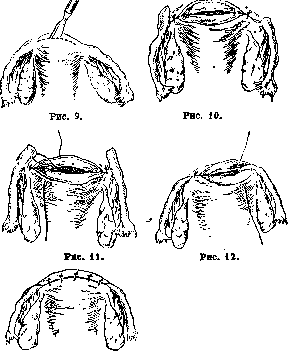
In cases of infertility caused by obstruction of the Fallopian tubes in the interstitial or isthmic part, it is sometimes necessary to resort to the operation of removing the affected part of the tube and transplanting the remaining part into the uterus in order to restore the patency of the tubal canal. Such an operation was first published in 1899 by Ries and Weitkins; they applied it to a woman in whom the right appendages had previously been removed; as a result, pregnancy occurred, which ended in miscarriage. However, at that time the operation was not yet recognized by gynecologists and began to be applied only after the case published in 1921 by Cullen and Shaw, where after transplantation of the tube the patient became pregnant, with the first pregnancy ending in miscarriage and the second in normal delivery. Strassmann in 1924 was already able to publish several cases and proposed his own method of operating. Since then, a large number of authors have published their cases and developed the technique of the operation. If initially transplantation of the Fallopian tubes was performed incidentally in cases where the affected tube on one side was removed, while the second tube was affected in the isthmic part, at present transplantation is applied both incidentally and as an independent operation in the presence of infertility. When applying the operation of transplantation of the Fallopian tubes, a number of authors (Mandelstam, Unterberger, Michaelis, etc.) indicate that as a result of the operation patency and normal physiological function of the tubes are restored; thus, Reiprich, on the basis of data from world literature, calculates cases where pregnancy subsequently occurred at 10-15%; according to Sedyukov's data, out of 72 published cases of tube transplantation, pregnancy was observed in 23%, according to the material of Mandelstam and Kiparsky, pregnancy occurred twice out of 21 cases. From the indications available in the literature, pregnancy occurs from 3 months to 3 years after the operation. Among the proposed methods, it is necessary to dwell on the operations of Strassmann, Unterberger, Mandelstam, Kiparsky, and Sedyukov. Strassmann's operation is performed as follows: the uterine cavity at the apex of the angle is opened with a transverse incision in such a way that the tube can be passed into the uterine cavity through the resulting opening; then two catgut sutures are placed on the serous covering of the internal end of the tube and passed through the entire thickness of the uterine wall, making an incision on the mucous membrane and an exit on its serous covering, with one end of each thread being brought out on the posterior and the other on the anterior surface of the uterus; before immersing the end of the tube into the uterine cavity, a thin probe is introduced through the ampullary end; pulling on the ligatures and guiding with the probe, the tube is immersed into the uterine cavity, after which the catgut sutures are tied; in the interval between the two fixing sutures, several nodal catgut sutures are applied. It is necessary here to adhere to the rule of not pulling the sutures too tightly and thus not compressing the tube.--Unterberger proposes to make a sagittal incision in the area of the uterine angle, and the internal end of the tube after resection of the affected part to be split into two lips and then fix them to the mucous membrane of the uterus with two catgut sutures, in the manner of implanting the ureters into the bladder, brought through the entire thickness of the uterine wall; on the uterine wall incision, submerged muscle and superficial sero-serous catgut sutures are applied. Mandelstam to eliminate compression of the tube by the uterine wall proposes in the uterine angle, at the location of the interstitial part, to cut out a conical canal from the uterine wall with opening of its cavity, into which the tube is immersed. After the canal is ready, on a needle through the entire uterus from front to back, a long ligature is passed; the thread is grasped with forceps from the formed canal and brought out in the form of a loop, the thread is cut, needles are placed on its ends, with which both lips of the longitudinally incised internal segment of the tube are grasped 2-3 mm from the edge; these threads are tied in knots, then a probe is introduced through the ampullary end of the tube and with its help and by pulling on the external threads the tube is immersed. By tying the ends of these threads, the tube is fixed to the uterine wall and the tube's mucosa to the uterine mucosa; with nodal sutures the serous membrane of the tube is sutured to the uterus. Kiparsky instead of excising the uterine angle with a scalpel uses a special instrument in the form of a cylinder with a cutting edge—a uterine trepan, which, passing through the uterine wall, removes the corresponding piece of tissue. The disadvantages of this method, in the opinion of Sedyukov, Unterberger, and others, is that here one has to operate blindly, due to which it is not visible how the implanted tube fits to the uterine cavity; this can also explain the absence in the literature of any indication where after transplantation by Kiparsky's method pregnancy was subsequently observed.-In 1932 Sedyukov published 10 cases of transplantation of Fallopian tubes; he finds that in case of obstruction of both tubes it is necessary to perform bilateral transplantation. He performs the operation as follows: the uterine cavity is opened with a transverse incision along the uterine fundus (fig. 9); the affected part of the tube in the isthmic or interstitial part of the tube is resected (fig. 10); to stop bleeding one catgut suture is applied along the edge of the uterus behind and slightly below the attachment of the round ligament of the uterus, the second suture—on the tubal artery at the site of the tube incision, on its mesentery (fig. 11); then needles are placed on the ends of this suture and one thread is passed through the entire thickness of the uterine wall, bringing it out to the anterior surface (fig. 12), and the second—to the posterior, after which the ends of the thread are tied; thus the tube is fixed, and its incision edges are connected. The operation ends with first fixing the tube with several thin sutures in the area of its mesentery on the anterior and posterior surfaces of the uterus, and then closing the 17 uterine opening with thicker nodal sutures (fig. 13), over which Minin considers it necessary to apply an encircling catgut continuous seromuscular suture.-Okintchits considers that there is no need to cut the entire uterine fundus, but it is sufficient to limit oneself to one of its angles, making an incision of such a size that the transplantation of the tube can be performed under visual control. During the transplantation operation, it is necessary that the isthmic part is not completely removed, and the longer the ampullary end of the tube is left, the more data there is for the possibility of obtaining persistent patency in the postoperative period. According to Mikulicz-Radecki's research, the entire tube, both its ampullar and isthmic parts, represents one physiological whole, the unity of which is of great importance for the successful functioning of the tube. The loss of the ampullar part in terms of violation of muscular activity (peristalsis) has less significance than the loss of the isthmic part. Therefore, when transplanting the tube into the uterus, it is desirable to preserve a part of the isthmus, which, having more powerful musculature, will better resist the uterine wall than the weaker musculature of the ampullar end of the tube.-The question arises, is it necessary in case of obstruction of both tubes to perform bilateral transplantation, as Sedyukov proposes, or can one limit oneself to transplantation on one side? Minin agrees with the opinion of Douay, who advises with the aim of limiting trauma to perform unilateral transplantation, and in his two cases applied Strassmann's operation with unilateral transplantation. Gorizontov considers that for pregnancy to occur it is not necessary to necessarily transplant both tubes; thus, he cites 10 cases of timely delivery after transplantation, with bilateral transplantation in 5 cases and unilateral in 5 cases. The operation of transplantation of the Fallopian tubes must be approached with particular seriousness, one must take into account that often the obstruction, for which the operation was performed, occurs again, and sometimes in the presence of a hidden infection in the postoperative period, serious complications can be expected. Before the operation, it is necessary to observe the temperature for a long time (3-4 weeks); an increase in temperature should be a contraindication; before the operation it is necessary to examine the blood picture, the rate of erythrocyte sedimentation reaction, and also to accurately establish the site of obliteration using metro-salpingography. Consequently, the operation of transplantation of the Fallopian tubes can be performed only in cases where there is a completely subsided process that has not given exacerbations for a long time; special caution is necessary if there is a history of gonorrhea.
N. Minin. Lit.: Berezon L., Air test of patency of the Fallopian tubes, Journal of Obstetrics and Women's Diseases, vol. XXXIX, book 1, 1918; Bykov S., On the diagnostic and therapeutic significance of insufflation of the Fallopian tubes in infertility, ibid., vol. XLII, book 2, 1919; Gorizontov N., Implantation of the Fallopian tubes into the uterus, ibid., 1934, no. 5; Zaretsky S., Papillary tumors of the Fallopian tubes, ibid., 1907, no. 9; Levitsky M., On the question of primary cancer of the Fallopian tubes, Journal of Obstetrics and Women's Diseases, 1913, no. 12; Mikhnov S., On the question of diseases of the Fallopian tubes and ovaries in pathological-anatomical and clinical aspects, dissertation, St. Petersburg, 1889; Popov D., On the changes in the Fallopian tubes in uterine fibromas, Doctor, 1890, no. 51; Serdyukov M., Critical evaluation of modern methods of diagnosis of tubal infertility, Journal of Obstetrics and Women's Diseases, vol. XXXVIII, book 1, 1927; Cahen, Les modifications de la trompe uterine chez la femme, Archives internationales de médecine expérimentale, vol. IV, 1928; Condamine F., Du traitement chirurgical de la tuberculose annexielle, Gynécologie et obstétrique, vol. XIX, 1929; Dietrich H., Neue Neubildungen der Eileiter (Biologie und Pathologie des Weibes, hrsg. v. J. Halban u. L. Seitz, B. V, T. 1, Wien-Berlin, 1928, lit.); Grusdew F., Zur Histologie der Fallopischen Tuben, Zentralblatt für Gynäkologie, 1897, no. 10; Ka both G., Eileiterdurchblasung, Berichte über die gesamte Gynäkologie und Geburtshilfe, B. XII, H. 3, p. 129, 1927 (lit.); Pop off D., Zur Morphologie u. Histologie der Tuben und des Parovariums beim Menschen während des intra- und extrauterinen Lebens bis zur Pubertät, Archiv für Gynäkologie, B. LXIV, H. 2, 1893; Serdyukov M., La chirurgie restauratrice des trompes, ses méthodes et ses résultats, Gynécologie, vol. XXXI, 1932; Weible W., Tuberkulose des weiblichen Genitalapparates (Biologie und Pathologie des Weibes, hrsg. v. J. Halban u. L. Seitz, B. V, T. 1, Wien-Berlin, 1928, lit.). See also the corresponding chapters of the main guides indicated in the literature to the article Gynecology.
Related articles
Mentioned in
Cite this page
“Fallopian Tubes.” Soviet Medical Encyclopedia. English translation of Bolshaya Meditsinskaya Entsiklopediya, 1st ed. (Moscow, 1928–1936), ed. N. A. Semashko. https://sovietmedicalencyclopedia.pages.dev/article/fallopian-tubes/