Infundibulum
Historical document, translated for reference. It reflects medical knowledge of the 1920s–30s and is not medical advice.
Summary
This article describes the infundibulum as a funnel-shaped structure located at the base of the brain, connecting the tuber cinereum to the posterior lobe of the pituitary gland. It discusses its anatomical position, its development from the diencephalon, and its histological composition, noting that its physiological function and pathology remain largely unclear.
Encyclopedia article (1928–1936)
INFUNDIBULUM (Latin for funnel; French: tige pituitaire), a formation located at the base of the brain; it has the shape of a funnel with a cavity inside; above, the tuber cinereum continues into this funnel; downward, the infundibulum transitions into the posterior lobe of the pituitary gland (Figures 1 and 2); behind it are the corpora mamillaria, in front of it is the chiasma of the optic nerves, and it is surrounded by a ring of dura mater in the form of the diaphragma sellae turcicae. The cavity of the infundibulum continues into the third ventricle, forming the recessus infundibuli.
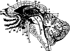
Figure 1. Internal surface of the brain. Floor of the III ventricle: 1—splenium corp. callosi; 2—pulvinar; 3—gl. pinealis; 4 and 5—corpora quadrigemina; 6—tegment. of the cerebral peduncle; 7—velum medullar. ant.; 8—IV ventricle; 9—spinal canal; 10—pons Varolii; 11—basis of the cerebral peduncle; 12—corpora mamill.; 13—tuber cinereum; 14—recessus infundib.; 15—infundibul.; 16—posterior lobe of the pituitary; 17—anterior lobe of the pituitary; 18—n. opt. et recessus supraoptici; 19—lamina supraoptica; 20—commissura alba ant.; 21—cingulum; 22—rostrum corp. callosi; 23—genu corp. call.; 24—sept. pellucid.; 25—lamina terminal.; 26—columna fornicis; 27—for. Monroi; 28—fornix; 29—commissura grisea; 30—habenula; 31—psalterium.
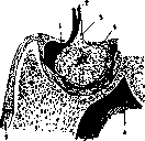
Figure 2. Pituitary gland, consisting of 2 lobes: 1—anterior; 2—posterior (black), continuing into the infundibulum; 3—cavity in the pituitary; 4—diaphragma sellae turc.; 5—sin. cavern.; 6—sin. sphenoid.; 7—dura mater. (From Poirier.)
The infundibulum develops from the primary anterior vesicle together with the thalamus opticus and the corpora mamillaria; it belongs to the diencephalon.
Located between the tuber cinereum and the posterior lobe of the pituitary gland, it is formed by the same gray lamina, consisting of glial tissue; no ganglionic cells have been found there; thin nerve fibers pass near it, heading toward the pituitary gland.
The physiological significance of this formation has not been clarified; its pathology is also unclear; the literature cites isolated cases of tumors originating from the infundibulum, with a clinical picture of dystrophia adiposogenitalis. Histologically, such tumors either turn out to be gliomas or belong to tumors of the pituitary duct.
E. Kononova.
Related articles
Mentioned in
Cite this page
“Infundibulum.” Soviet Medical Encyclopedia. English translation of Bolshaya Meditsinskaya Entsiklopediya, 1st ed. (Moscow, 1928–1936), ed. N. A. Semashko. https://sovietmedicalencyclopedia.pages.dev/article/infundibulum/