Occipital Lobe
Historical document, translated for reference. It reflects medical knowledge of the 1920s–30s and is not medical advice.
Summary
This article describes the anatomy, structure, and functions of the occipital lobe of the human brain, detailing its surfaces, gyri, and connections to the thalamus and other brain regions.
Encyclopedia article (1928–1936)
OCCIPITAL LOBE, the posterior lobe of the brain, is located at its posterior pole. In humans it is of small size, has the shape of a triangular pyramid with the apex directed backward and the base, without sharp boundaries, passing into the convolutions lying in front. The occipital lobe has three surfaces: external, internal, and inferior. The external surface (Fig. 2) lies on the convex part of the hemisphere (convexitas), posterior to the imaginary line drawn from the upper edge of the fissure parieto-occipitalis to the incisura praeoccipitalis; by three sulci it is divided into three occipital convolutions (details see Brain). On the internal surface (Fig. 1) the fissura parieto-occipitalis separates the occipital lobe from the parietal lobe; behind this sulcus lies the cuneus, bounded below by the fissure calcarina. Then comes the inferior surface, on which there are only two convolutions — gyrus lingualis and gyrus fusiformis, separated by the fissure collateralis. Inside the occipital lobe passes the posterior horn of the lateral ventricle (see Ventriculi cerebralis), which does not reach 2–3 cm to the occipital pole; along its floor and outer wall run three eminences: bulbus cornu posterioris, calcar avis, and eminentia collateralis, corresponding to the indentations of the three sulci — fissura parieto-occipitalis, calcarina, and collateralis. The white matter constituting the occipital lobe surrounds the posterior horn and consists of associative, commissural, and projection fibers. Long associative and projection fibers form two layers in the outer wall of the posterior horn — stratum sagittale externum and internum; short associative fibers connect all three surfaces of the occipital lobe, as well as the convolutions of adjacent lobes; among them are known stratum proprium fissurae calcarinae, cunei, convexitatis, fasciculus transversi, lobi lingualis et cunei, fasciculus occipito-verticalis. From long associative fibers, the fasciculus longitudinalis inferior connects the occipital lobe with the temporal lobe, the cingulum, and the fasciculus subcallosus with the frontal lobe. Commissural fibers connecting the occipital lobes of both hemispheres pass through the splenium and the posterior parts of the trunk of the corpus callosum, forming the forceps major and minor, which together with their continuation, the tapetum, directly surround the posterior horn; in addition to the corresponding lobes, fibers of the corpus callosum connect the occipital lobe with the convolutions of other lobes of the opposite hemisphere. Projection fibers connect the occipital lobe with the primary visual centers (pulvinar thalami optici, corpus geniculatum laterale, and anterior quadrigeminal body), as well as probably with the brainstem and the pons (occipital-pontine system). Fibers coming from subcortical visual centers terminate in both lips of the fissure calcarina, i.e., in the cuneus and in gyrus lingualis, and partly also in other convolutions of the occipital lobe. The cortex of the visual area is characterized by the presence of the V-shaped line of Vicq d'Azyr, is called the area striata, and forms area 17 of Brodmann; in addition, area 18 and 19 of Brodmann are located in the occipital lobe (see Architectonics of the Cerebral Cortex). Due to the termination of visual fibers in the occipital lobe, its main function is visual (see Visual pathways, centers). Unilateral damage to the occipital lobe causes hemianopia (see), and bilateral damage causes cortical blindness. As for the convolutions of the external surface, some authors point to their dependence on visual representations and visual memory; their disease in the left hemisphere causes mental blindness. In the occipital lobe there are also areas whose irritation causes combined movement of the eyes, pupils, and the accommodation apparatus. Besides changes on the side of vision, disease of the occipital lobe can give some disturbance of the psyche (decline of intellect, change of character), but these disturbances are characteristic not of the occipital lobe, but of the cerebral cortex in general; hallucinations in the form of light sensations, etc., are also observed.
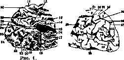
Fig. 2. Fig. 1. Internal surface of the posterior parts of the left hemisphere of the brain: 1- paracentral lobule; 2 and 27- gyrus lingualis; 3- corpus callosum; 4- gyrus hippocampi; 5 and 22- gyrus fusiformis; e- fissura temporalis interna; 7- inferior temporal gyrus; 8- lingula; 9- cuneus; 10- praecuneus; 11- fissura Rolandi; 12- splenium corporis callosi; 13- trunk of corpus callosum; 14- fornix (crus posterior); 15- fissura calcarina et parieto-occipitalis; 16- fissura hippocampi; 17, 20, and 23- fissura collateralis; 18- gyrus hippocampi; 19- fissura temporalis inferior; 21- incisura praeoccipitalis; 24 and 25- fissura calcarina; 26- fissura propria cunei; 28- fissura parieto-occipitalis; 29- fissura supraparietalis; 30- fissura calloso-marginalis (According to Dejerine.) Fig. 2. External surface of the posterior parts of the left hemisphere of the brain: 1- anterior central gyrus; 2- posterior central gyrus; 3 and 23- superior parietal gyrus; 4- angular gyrus; 5, 6, and 7- supra-marginal gyrus; 8- fissura Sylvii; 9- superior temporal gyrus; 10 and 12- superior temporal fissure; 11- middle temporal gyrus; 13- middle temporal fissure; 14- superior occipital gyrus; 15- middle occipital gyrus; 16- superior occipital fissure; 17- middle occipital fissure; 18- gyrus descendens; 19- inferior occipital gyrus; 20- inferior temporal gyrus; 21- fissura Rolandi; 22, 25, and 26- interparietal fissure; 24- superior parietal gyrus; 27- fissura parieto-occipitalis; 28- incisura praeoccipitalis; 29- postcentral fissure.
E. Kononova,
Related articles
Mentioned in
Cite this page
“Occipital Lobe.” Soviet Medical Encyclopedia. English translation of Bolshaya Meditsinskaya Entsiklopediya, 1st ed. (Moscow, 1928–1936), ed. N. A. Semashko. https://sovietmedicalencyclopedia.pages.dev/article/occipital-lobe/