Hemianopsia
Historical document, translated for reference. It reflects medical knowledge of the 1920s–30s and is not medical advice.
Summary
A definition and clinical overview of hemianopsia, a condition characterized by the loss of half of the visual field in both eyes. The article details the anatomical pathways, the distinction between homonymous and heteronymous forms, and the diagnostic significance of pupillary reactions.
Encyclopedia article (1928–1936)
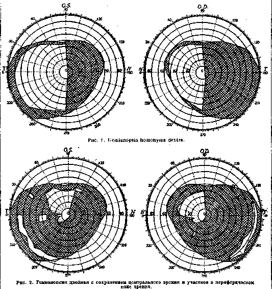
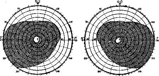
HEMIANOPSIA, HEMIANOPIA, hemianopsia, hemianopia (from Greek hemi-half, a-negative particle, and opsis-vision), half-blindness, loss of one half of the visual field in each eye. Hemianopsia is divided into hemianopsia homonyma and hemianopsia heteronyma. The word homonyma means "on the same side," so that in hemianopsia homonyma, the same sides of the visual field are lost in both eyes; in right-sided hemianopsia, the patient does not see objects located in the right half of his visual field, and in left-sided, those on the left. When reading, a patient with hemianopsia must move the book or turn his head to be able to follow the lines. The line separating the blind part of the visual field from the seeing part runs either directly along the midline through the fixation point or deviates by several degrees toward the blind side, bypassing the fixation point in the form of a semicircle. In most cases, the entire half of the visual field is blind in both one and the other eye (see figure 1); sometimes, however, the lost area occupies only part of the half of the visual field in the form of a sector, quadrant (hemianopsie en quadrant, en secteur, see figure 2) or, finally, symmetrical parts in different places of the half of the visual field of both eyes are lost; such losses are called "scotomas." Central vision in hemianopsia is for the most part preserved. Hemianopsia can be observed in both halves of the visual field in both eyes—double hemianopsia (see figure 3), with preservation or loss of the central visual field; in the latter case, there is complete blindness—cortical blindness. Hemianopsia also differs in intensity: for the most part, the lost visual field is blind to all types of visual sensations, but sometimes a dissociation of visual perceptions is observed: vision for form and color is lost, while perception of light and movement is preserved in the same visual field.—Hemianopsia heteronyma is characterized by the loss of either the inner halves of the visual field—bilateral nasal hemianopsia (h. binasalis, see figure 4)—or the outer ones—bilateral temporal hemianopsia (h. bitemporalis, see figure 5). Hemianopsia is observed both in functional diseases of the nervous system (hysteria) and in organic ones. Among organic diseases, hemianopsia occurs with lesions of the visual pathways, starting from the chiasma opticum and up to the cortex of the visual region in the occipital lobe: chiasma opticum, tractus opticus, subcortical visual centers (corpus geniculatum laterale, pulvinar, corpus quadrigeminum anter.), radiatio optica, and the cortex of the occipital lobe, mainly around the fissura calcarina. Hemianopsia heteronyma occurs with a lesion of the chiasm, and hemianopsia homonyma—with a lesion of all other parts of the visual pathway. The origin of h. heteronyma is explained by the arrangement of the crossed fibers of the optic nerve. The crossed fibers, coming from the inner halves of the retinas, occupy a central position in the chiasm; a lesion of only the central part entails the loss of the outer halves of the visual field (since the inner parts of the retina perceive stimuli from the outer halves of the visual field), i.e., h. bi
HEMIANOPSIA, HEMIANOPIA temporalis. Uncrossed fibers occupy an outer position in the chiasm. If one assumes the existence of such a disease in which both outer parts of the retina would suffer, then only the uncrossed fibers of the optic nerve, coming from the outer parts of the retina, would suffer, fibers from the outer parts of the retina of their own side and from the inner—of the opposite side; a lesion of this pathway entails the loss on one's own side of the outer half of the retina (consequently, the inner visual field) and the inner half of the retina on the opposite side (con
270 270
Figure 3. Double hemianopsia with preservation of central vision.
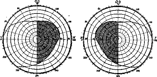
Figure 4. Hemianopsia binasalis.
receiving visual sensations from the inner parts of the visual field—then h. binasalis will result; in clinical practice, however, only a unilateral disease of the nerve fibers coming from the outer parts of the retina is observed, and therefore nasal hemianopsia is encountered only on one side—h. nasalis. The origin of h. homonyma is fully explained by the composition and course of the visual fibers (see figure 6) from the retina to the cortex (see Visual pathways, centers). The composition of the visual pathways, posterior to the chiasm, includes fibers from the outer parts of the retina of their own side and from the inner—of the opposite side; a lesion of this pathway entails the loss on one's own side of the outer half of the retina (consequently, the inner visual field) and the inner half of the retina on the opposite side (consequently, the outer visual field); thus, in each eye there is a loss of the homonymous visual field (right or left), and it will always be opposite to the affected hemisphere due to the refraction of the light ray occurring in the lens; h. homonyma dextra is observed in diseases of the left visual pathway and vice versa. Upon destruction of the cerebral cortex within the visual center, the perceiving apparatus related to the homonymous halves of the hemianopsia of both retinas is affected.—Double hemianopsia is observed with simultaneous lesion of the visual pathway on both sides. H. homonyma,
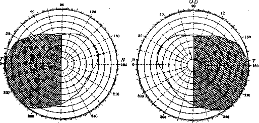
Figure 5. Hemianopsia bitemporalis.
caused by a lesion of the tractus optici or subcortical visual centers, is differentiated from hemianopsia that occurred due to a disease of the radiatio optica or the cortex of the visual region, by the presence in the first case of the hemianopic pupil reaction, whereas in the second case this pupil reaction is absent.
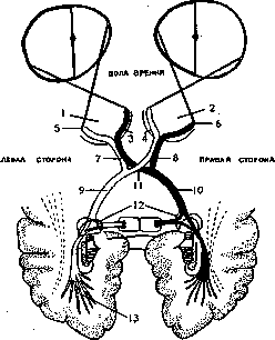
Figure 6. Diagram of the visual pathways according to Kno-blanch: 1 and 2—left and right eye; 3 and 4—nasal side on the left and right retina; 5 and 6—temporal side on the left and right retina; 7 and 8—n. opticus sin. and dext.; 9 and 10—tractus opticus sin. and dext.; 11—chiasma; 12—primary visual center; 13—radiating bundle of Gratiolet.
Related articles
Mentioned in
Cite this page
“Hemianopsia.” Soviet Medical Encyclopedia. English translation of Bolshaya Meditsinskaya Entsiklopediya, 1st ed. (Moscow, 1928–1936), ed. N. A. Semashko. https://sovietmedicalencyclopedia.pages.dev/article/hemianopia/