Vision
Historical document, translated for reference. It reflects medical knowledge of the 1920s–30s and is not medical advice.
Summary
This article from the 1928–1936 Soviet Great Medical Encyclopedia explains the physiological and physicochemical processes of vision in insects and vertebrates. It details the mechanics of light perception, the role of retinal pigments, and the trichromatic theory of color vision.
Encyclopedia article (1928–1936)
VISION is a complex physical and physicochemical process by which humans and animals obtain a representation of the size, distance, mutual arrangement, and color of individual objects in the surrounding world. Physiology of vision. The visual apparatus in insects and vertebrates is constructed according to two different types. Vision in insects. Sensations in the insect eye are connected with the appearance of a direct image of luminous objects on the retina (Fig. 1). In the insect eye, there is a series of prismatic formations, which, on the side facing the light source, have a rounded shape. A ray of light, represented in the figure as segments with an arrow, penetrates along the axis of the prism to its end, where it causes irritation in the nerve endings. If the ray falls obliquely (ray AB), then, upon hitting the side surface of the prism, it is absorbed there and does not reach the terminal nerve endings. Thus, only those rays that travel along the prism can reach the nerve endings and produce an image. If we have a series of points in space A, M, then these points, sending rays An and Mm parallel to the axes of the prism, will produce a direct image at the bottom of the eye.
Vision of vertebrate animals. The eye of vertebrate animals is constructed according to the type of a camera obscura, or photographic apparatus, and contains a series of refracting media that allow rays falling on these media to refract and produce an inverted real image at the bottom of the eye. The rays reaching the bottom of the eye cause physicochemical processes in the apparatuses connected with the terminal nerve endings, specifically in the rods and cones, which release ions (see Ionic theory of excitation). These latter create excitation in the visual cells and nerves and carry this excitation to the brain. The physicochemical processes occurring at the bottom of the eye must be considered photochemical processes; moreover, in color vision in the fovea centralis, one must assume the existence of 3 different pigments, of which one (G) is the most sensitive to irritation by green rays and possesses a maximum absorption in this region of the spectrum; the maximum absorption of another pigment, R, is shifted into the region of red rays; the maximum absorption of the third pigment, V, is toward the violet rays. The three absorption curves, which must be assumed in three different elements in the eye, can have the form shown in Figure 2 (pigments R, G, V). Their properties will be understood from the following. Along the abscissa axis, we plot the colors of the spectrum, marked below with the corresponding names, and along the ordinate axis, the degrees of decomposition of the pigments under the influence of light and the associated magnitudes of nerve excitation. If red light acts on the retina, it decomposes
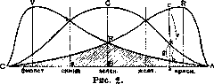
most strongly the pigment whose maximum decomposition lies in the red-orange part of the spectrum; the pigment with a maximum in the green part decomposes more weakly, and the pigment with a maximum decomposition in the violet part of the spectrum decomposes the least. For brevity, we will call the corresponding pigments red-sensitive, green-sensitive, and violet-sensitive. Upon the action of various rays of the spectrum, as can be seen from the table, there are the following relationships:
the fact that with any irritation caused by any ray, there is always a decomposition of three different pigments, and therefore if we take the region from A to B (Fig. 2), then in the shaded part AFB we have an equal decomposition of red-, green-, and violet-sensitive pigments. We therefore have a sensation of white color, upon which color sensations are superimposed. These color sensations are determined by the ratio of the quantities of pigments R and G, sensitive to red and green color; for the red-green part of the spectrum, these values are r and g, and since r equals the difference in decomposition of the red-sensitive pigment R, represented by the ordinate ac, and the violet-sensitive pigment V (ordinate ab), and g equals the difference in the quantity of decomposition of the green-sensitive pigment G (ordinate ak) and the violet-sensitive pigment V (ordinate ab), then we have for chromaticity F in the region AB the general formula: Fx = In the space from B to C, the degree of irritation of the fibers, the color of monochromatic light (red, yellow, green, blue, violet). Red-sensitive, Green-sensitive, Violet-sensitive. Strong, Weak, Weak, Moderate, Moderate, Weak, Weak, Strong, Weak, Weak, Moderate, Moderate, Weak, Weak, Strong. Sensation of color: Red, Yellow, Green, Blue, Violet. The action of all rays of the spectrum on the eye gives a sensation of white color, corresponding to which the areas bounded by the absorption curve and the abscissa axis are the same for all curves. If there are rays in the spectrum which, acting simultaneously, give equal decomposition for red-sensitive, green-sensitive, and violet-sensitive pigments, then a sensation of white color of a certain intensity will also be obtained; these rays are called complementary rays. Complementary rays, as both theory and experience show, are as follows: for red, the complementary rays are blue-violet; for orange, blue; for green, a mixture of red and violet (purple color); for blue, orange; and for blue-violet, red. One can simply explain the existence of complementary rays, proceeding from the ionic theory of excitation. On the basis of the previous data, one can determine in the following way two qualities of color that are obtained upon the simultaneous action of different colors on the eye. All colors have a certain chromaticity of the ray and saturation, which is determined by the admixture of the sensation of white color; this admixture exists in all color sensations, even those caused by a monochromatic ray. The sensation of white color depends on how easily one can understand, there is the following relationship for chromaticity: ^2 = m^r/q. Thus, different parts of the spectrum—from red to green and from green to violet—bear a different character, since chromaticity is determined by different values of i?! and jP2, and this corresponds to different physiological sensations obtained from two different parts of the spectrum. In the red-green part, sensations are characterized, according to Goethe, as warm sensations, and the green-violet as cold sensations. Finally, a third combination of R, G, and V is possible, a combination which is expressed in this way: .F3= -y~o. This third possible group of combinations of R, G, and V corresponds to the sensation of a purple tone.—Thus, the visual apparatus of man is constructed according to the type of an apparatus giving three different kinds of sensations. From the combination of these three different kinds of sensations, one can obtain all the richness of the colors of nature. In the mixing of colors, when two monochromatic rays act simultaneously on the eye, the mixture of rays causes the following sensations: the rays give either a sensation of white color, if these rays are complementary, taken with a certain intensity, or a sensation of a monochromatic color lying between the mixed rays, with the addition of a certain amount of white color. This is obtained in the case where two rays are closer to one another than the complementary rays, e.g., if we mix red and green rays: in this case, the mixture is obtained as yellow (Rayleigh). Or finally, if the rays are located further apart than the complementary ones, then a sensation of purple color will be obtained upon mixing. All these new sensations are always obtained with an admixture of white color, as can be observed with the help of a series of instruments constructed by Helmholtz. The phenomena of color mixing can be described if one constructs a circle, along the periphery of which various colors are located (Newton; Fig. 3). Along the diameters are located those rays, which...
are purple. %: (and green) can Figure 3.
\
s'
according to this scheme can be determined as follows: the quantity of red color can be defined as a certain weight attached to the end of lever AB at the point marked with the letter A. Another weight, corresponding to the quantity of green color, is attached at point B. Then, according to the rules of mechanics, the center of gravity of the weights is found; let this point be point M; then, by drawing a ray through center O and point M to the circumference, one finds exactly that point F which corresponds to the color of the mixture. In this case, a yellow-green color will be obtained. The closer point M lies to the center, the greater the admixture of white color will be. Thus, a simple rule for color mixing is obtained. This diagram was proposed by Newton and more thoroughly developed by Helmholtz and Maxwell. Successive images. If light has acted upon the eye and caused decomposition in the pigments of the eye, then the ions formed at the site of irritation and acting on the optic nerve are gradually removed from the retina and give rise to the phenomena of successive images. Two types of these images can be distinguished. If one takes a bright light source and illuminates the eye briefly, and then enters a dark room, the light source that illuminated the eye will appear on the dark background with all its forms and details as a brilliant bright image. This image will change its brightness and colors, passing sequentially through a series of individual shades. If, after obtaining a successive image in the eye, we look at a brightly illuminated background (e.g., a white wall), then the successive image will appear dark against the bright background. Thus, we obtain either a positive trace (a light image on a dark background) or a negative trace (a dark image on a light background). The ionic theory of excitation allows for a full explanation of the existence
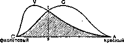
Figure 4.
of both positive and negative light traces. In the absence of one of the pigments, for example, the red-sensitive R, there are, as is easy to see from Fig. 4, simpler relationships. Since, with equal irritation of the nerves connected to the green-sensing and violet-sensing pigments, a sensation of white color always arises, we will obtain a sensation of yellow-green color with an admixture of white in the range from 4 to s, and a sensation of violet color with an admixture of white from s to C. These two colors are connected with the sensation of white color, which is admixed in a greater quantity than is the case in a normal eye; furthermore, an interesting feature of the eye that lacks red sensation (color blindness) is that at a certain point, specifically at point s, the spectral color gives equal decomposition of the two pigments and, consequently, a sensation of white color. This is observed in the green-blue part of the spectrum. Such are the processes occurring in the center of the eye (in fovea centralis retinae). Peripheral vision. At the present time, the processes of human vision must be considered from two points of view. Firstly, there is the above-described apparatus, which, under bright illumination, gives an idea of the color of objects, their shape, and their mutual arrangement. This apparatus is called central, and its perceiving parts are the cones. Along with this first apparatus, the periphery of the field of vision is served mainly by a second apparatus, the so-called peripheral, or dark apparatus of twilight vision. It consists of rods, which give an idea only of the brightness of illumination, without providing any distinction in the sensation of colors. The sensation of the shape of objects with the help of rods under peripheral illumination is completely different than during the irritation of cones, and the reasons for this lie in the fact that each cone is connected to only one nerve fiber, whereas several rods are connected to one nerve fiber. To determine the minimum angle corresponding to visual acuity in the center, it is necessary to irritate two cones separated by one non-irritated cone. In this case, two light points are visible, separated by a certain dark space. For the rods, the relationship is different, and two separated sensations are obtained if two groups of irritated rods are separated by a group of non-irritated ones. As is easy to understand, the angular relationships will be different, and on the periphery, one can determine the shape of objects more crudely than in the center. The irritation received by the cells of the retina is transmitted through the optic nerves to the superior colliculi (corpora quadrigemina) and from there to the occipital lobes of the brain.
P. Lazarev. Visual disorders in diseases of the nervous system are very numerous and diverse and are observed both in organic diseases of the nervous system and in functional ones; they are expressed by: 1) a decrease in visual acuity—amblyopia, or complete loss of vision—amaurosis; 2) a disturbance in color perception; 3) a loss of the visual field; 4) a disorder of the motor apparatus of the eye; and 5) a disorder of sensitivity. Changes in visual acuity, color perception, and the visual field can be observed in diseases of any part of the visual pathway from the retina to the cortex of the occipital lobe; the visual pathway can be directly affected by some pathological process—a neoplasm, hemorrhage, softening, trauma—either in the occipital lobe, or at the base of the brain, or even in the orbit itself. The visual pathway can be affected simultaneously with other parts of the brain by an infectious or toxic process (encephalitis, meningitis, cerebrospinal syphilis, etc.), and finally, the visual pathway, especially its anterior part—the optic nerve—reacts with changes to diseases of even a distant part of the brain, but which are accompanied by an increase in intracranial pressure (brain tumors, abscesses, hydrocephalus, etc.). Changes in the optic nerve can be established during life with the help of an ophthalmoscopic examination of the fundus; in the initial stage, they amount to hyperemia of the optic disc, which can progress either to papilledema, if there is increased intracranial pressure, or to optic neuritis, observed in various diseases of the brain. Any pathological process in the optic nerve (papilledema, optic neuritis) in the progressive course of the disease turns into optic atrophy, which in this case will be secondary; in most cases, based on the state of the fundus, one can say on what grounds it developed. Optic atrophy can also occur primarily; due to the characteristic gray color of the disc, it is called, in addition to primary, gray atrophy; it is observed in tabes dorsalis, general paralysis of the insane; a variety of atrophy—pallor of the temporal halves of the optic discs—is characteristic of multiple sclerosis. A characteristic change in the macula lutea is found exclusively in amaurotic idiocy. Tubercles and cysticerci on the fundus are not uncommon in homogeneous diseases of the brain or its membranes. Sometimes changes in the fundus do not correspond to the intensity of the change in visual acuity—almost complete blindness with a normal or almost normal state of the disc; this is observed in retrobulbar neuritis, when the optic nerve is mainly affected and only later do the changes reach the disc. Clinically, with the above-described changes in the visual pathway, a decrease in visual acuity of various degrees is observed, up to complete blindness. Narrowing of the visual field in all directions is called concentric contraction; it is observed in severe changes in the optic nerve, as well as in functional diseases of the nervous system (hysteria). Loss of vision in one half of the visual field is known as hemianopsia; it changes depending on the location of the lesion of the visual pathway, due to which it plays a large role in the diagnosis of the localization of the process (see Hemianopsia). Bilateral hemianopsia is observed with bilateral lesions of the visual pathways and results in complete blindness. With a lesion of the left angular gyrus (Dejerine's center), word blindness is observed—the inability to read, distinguish, or recognize letters; with a lesion of the occipital lobe, mainly its outer surface, mind blindness (see) develops—the absence of recognition of objects. The loss of small areas in the central visual field is called a scotoma. Along with a decrease in visual acuity, the perception of colors (color sensation) also decreases; color blindness—achromatopsia—is encountered more often as a congenital defect, but can develop in some diseases of the visual pathway or centers, as well as in hysteria. Visual disorders, in addition to diseases of the visual pathways, are also observed in diseases of the motor apparatus of the eye or in disturbances of sensitivity. Various diseases of the nervous system (tabes, general paralysis of the insane, syringobulbia, polioencephalitis superior, cerebrospinal syphilis, myasthenia, thrombosis of the cerebral sinuses, migraine, and others), especially those developing in the region of the brainstem, can cause paralysis of the eye muscles—ophthalmoplegia—due to paralysis of the oculomotor, abducens, and trochlear nerves or diseases of their nuclei; less often, oculomotor disorders are observed with lesions of the conductors or centers. Ophthalmoplegia can be complete or partial in various combinations; in partial cases, the eyeball deviates toward the healthy muscle. Ophthalmoplegia of the external eye muscles can be combined with paralysis of the internal eye muscles (ciliary muscle and sphincter of the pupil), these latter can also be affected independently of other eye muscles and combined with other symptoms from the nervous system; clinically, they manifest themselves as changes in the pupil and its reflexes. Nystagmus is observed in many diseases of the central nervous system (cerebellum, vestibular system), in meningitis, multiple sclerosis, and in children with some congenital diseases of the nervous system and others. Conjugate deviation of the head and eyes is observed with central foci. Sensory disorders observed in the eye in some diseases of the nervous system (tabes, lesion of the trigeminal nerve, etc.) can lead to a decrease in vision or even its complete loss due to the development of neuroparalytic keratitis or other trophic disorders. Prevention and treatment of visual disorders depend entirely on the etiological factor (see the corresponding diseases). E. Konopova. Loss of vision is a decrease or complete loss of the ability to perceive light stimuli from objects of the outside world. Such loss of vision in the sense of weakening of vision or complete blindness can occur as a result of damage to the eyeballs by blunt, sharp-pointed, or firearm weapons and other foreign bodies penetrating the eye, as well as from thermal and chemical influences (hot liquids, caustic substances, etc.). Among the possible consequences of such violence, one can mention: cracks in the cornea (often with the formation of an abscess), hemorrhages into the retina, wounds of the sclera, damage to the lens, ruptures of the membranes of the eye, etc. In addition, cases of blindness after blows to the supraorbital region and concussions of the brain have been described. Finally, vision can suffer from the influence of occupational hazards: amblyopia and neuritis in workers dealing with substances containing lead, night blindness from the action of carbon disulfide vapors during the vulcanization of rubber products, etc. Specifically, it is necessary to clarify the following questions: 1) to what extent is visual acuity reduced, 2) are there any changes leading to disfigurement of the face. Often, an external examination of the eyes is sufficient to establish a persistent visual disorder. Where there are no obvious deviations from the norm, ophthalmoscopy, testing with charts (Snellen's, Kryukov's, etc.), and all special methods of clinical examination are necessary; in this case, one must also keep in mind the possibility of aggravation or simulation. Feigned myopia or weakening of vision can be uncovered by examining vision with various lenses: to detect simulated unilateral blindness, the following method is used: a biconvex lens, approximately 10 diopters in strength, is placed in front of the "healthy" eye, and the patient is asked to read; if the subject succeeds, then simulation is present, since a healthy eye cannot distinguish letters through such a lens. Loss of vision is classified by the legislation of the USSR as a serious injury, and various degrees of its weakening are classified as minor (formerly—as less serious and minor) bodily injuries (Articles 142, 143 of the Criminal Code of the 1927 edition). A sharp decrease in visual ability is equated to complete blindness provided that the victim does not distinguish the outlines of nearby objects, for example, cannot count the fingers of a hand. Loss of vision in one eye is considered a serious injury if it leads to disfigurement of the face due to the absence or deformation of the eyeball, the presence of corneal scars, ectropion of the eyelids, etc. In ophthalmology, charts (Josten's, Katsaurov's, etc.) have long been used, in which the lost working capacity is expressed as a percentage, corresponding to the decrease in visual acuity (visus). Blindness in both eyes, as well as the loss of one eye in the absence of the other, are estimated at 100% loss of working capacity. Loss of vision in one eye—33 1/3–35%. In addition to this, when determining the loss of working capacity, the state of accommodation, eye movements (paralysis of the eye muscles), binocular and eccentric vision, as well as the nature of the victim's occupation (his working capacity) are taken into account. For example, for a watchmaker and an engraver, weakening of vision in one eye has more significance than the loss of an eye for a manual laborer or a carpenter.
Cases involving the loss or weakening of visual capacity may be the subject of criminal and civil proceedings, depending on whether the eye injury was the result of malicious intent, caused by a third party, or occurred during an accident (railway catastrophe, injury by machine parts in production, etc.). At the present time, the degree of disability is established by the group system by social-labor expertise bodies (medical expertise bureaus, control commissions), with six disability groups existing: Group I—100% loss of working capacity, Group II—from 70% to 99%, Group III—from 50% to 69%, Group IV—from 30% to 49%, Group V—from 15% to 29%, Group VI—up to 14% (Collection of Statutes, 1926, No. 86, Art. 627). V. Vladimirsky.
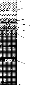
Statistics of diseases of the organs of vision. According to the international nomenclature, diseases of the organs of vision include: 1. Diseases of the eyeball: of the conjunctiva (conjunctivitis is singled out separately when registering patients), cornea, iris, lens (cataract is singled out separately), choroid, retina, and optic nerve. Ophthalmitis. Panophthalmitis. 2. Follicular conjunctivitis. 3. Trachoma. 4. Tumors of the eye (except for cancer of the eyeball, which belongs to the group of 'neoplasms'). 5. Diseases of the eyelids, lacrimal organs, vitreous body, and the muscular apparatus of the eyes. Anomalies of refraction and accommodation. Diseases and tumors (except for cancer) of the orbit. Amblyopia. Night blindness. Absence of the lens (aphakia). Parasites of the eyeball. Introduction of foreign bodies into the ocular region. (Diphtheritic inflammation of the conjunctiva of the eye belongs to the group of epidemic diseases, gonococcal conjunctivitis to the group of infectious non-epidemic diseases, as does tuberculosis of the eye.)
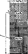
In pre-revolutionary Russia, about 6 million cases of diseases of the organs of vision were registered annually, or about 60 cases per 1,000 of all outpatient patients (in 1911—64, in 1912—62, in 1913—63). Of all eye diseases registered in 1913, 49% were conjunctivitis, 17% were trachoma, and 34% were other eye diseases. For every 1,000 of all inpatient patients, there were about 35 patients with eye diseases annually (in 1911—37, in 1912—37, in 1913—36). For individual provinces, the indicated ratio of the number of patients with eye diseases to the sum of all patients showed fairly large fluctuations. For 9 Zemstvo provinces, where the relevant materials were processed by Zemstvo sanitary bureaus, diseases of the organs of vision constituted the following ratios per 1,000 of all registered patients (Table 1).
Table 1. Province Years Ratio per 1,000 patients: Saratov 1903 77.7; Voronezh 1898-1902 66.6; Kursk 1891-1895 64.5; Kherson 1887-1892 58.6; Samara 1895 58.3; Yekaterinoslav 1898-1901 57.3; Moscow 1898-1902 52.4; Tambov 1898-1900 47.8; Ryazan 1904 42.2.
In one and the same province, the ratio of the number of diseases of the organs of vision to the sum of all patients remained fairly stable over a long period of years. Thus, for the Moscow province, this ratio (per 1,000 patients) for individual periods is: in 1878-82—46.7‰; in 1883-87—46.5‰; in 1888-92—51.1‰; in 1893-97—54.0‰; in 1898-1902—52.5‰; and in 1926—51.7‰. For every 10,000 inhabitants in 1913, 508 patients with eye diseases were registered throughout Russia. This average indicator for the whole country for diseases of the organs of vision increased significantly for those provinces where trachoma was widespread. In the Moscow province, per 10,000 population, patients with eye diseases were registered: in 1906-1908—346 men and 372 women; in 1926—596 men and 563 women. This growth should be attributed mainly to the increased utilization of medical care by the population.
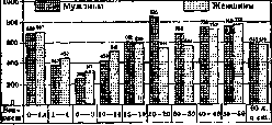
An idea of the composition of diseases of the organs of vision is provided by the following table, which shows the figures for the main forms of eye diseases registered in 1926 in the Moscow province and the city of Moscow (see also Figure 5). Table 2. Form of disease: Moscow province (Total: 147,166; Percentage of total: 100.0), City of Moscow (Total: 34,351; Percentage of total: 100.0). Conjunctivitis: Moscow province 57.1%, Moscow city 23.33%. Myopia: Moscow province 1.6%, Moscow city 27.0%. Other anomalies of refraction: Moscow province 5.2%, Moscow city 7.3%. Introduction of foreign bodies: Moscow province 6.0%, Moscow city 2.7%. Trachoma: Moscow province 2.6%, Moscow city 1.6%. Cataract: Moscow province 1.6%, Moscow city 1.4%. Night blindness: Moscow province 0.2%, Moscow city 0.05%. Nystagmus: Moscow province 0.1%, Moscow city 0.05%. Other diseases of the organs of vision: Moscow province 25.6%, Moscow city 36.57%.
The highest place, both in the province and in the city of Moscow, is occupied by conjunctivitis; in second place are anomalies of refraction. In the province, conjunctivitis and night blindness are higher than in the city of Moscow. Trachoma and cataract are at the same level. In the city of Moscow, anomalies of refraction and the introduction of foreign bodies occupy a higher place. The incidence of diseases of the organs of vision by age groups and sex for the Moscow province and the city of Moscow for 1926 (per 1,000 population of the corresponding age and sex) is presented in Table 3. In the Moscow province, at the age under 14, diseases of the organs of vision are observed somewhat more often among women than among men. At the age of 20 to 39, the incidence among men is significantly higher than among women. In older ages, no large difference in the incidence of both sexes is noted. In the city of Moscow, among children under 14 years of age, the incidence is also somewhat more frequent among girls. In all older age groups, however, men show a significantly higher incidence.
Table. Age Moscow province: Under 1 year, 1-4 years.
Other diseases of the organs of vision.
Figure 5. Composition of diseases of the organs of vision in the Moscow province and the city of Moscow for 1926.
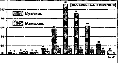
than women. The lowest incidence, both in the city and in the province, is observed in children of both sexes aged 5 to 9 years. Earlier ages show a significantly higher incidence (in the province it is especially high in infants). The maximum incidence among men in the province and the city of Moscow is observed at the age of 20 to 29 years (see also Figure 6). Table 4 shows the incidence by individual forms of diseases of the organs of vision by age and sex for the Moscow province for 1926 (per 1,000 population of the corresponding group). Table 5 shows the incidence by individual forms of diseases of the organs of vision by age and sex for the city of Moscow for 1926 (per 1,000 population of the corresponding group). In the Moscow province, conjunctivitis shows the highest indicators in infants of both sexes, then in men aged 20 to 29 and in elderly women 50-59; in the city of Moscow, it most affects children under 5 years of age, then men 20-49 and women 50-59. Myopia among persons of both sexes and in all age groups in the city of Moscow is expressed significantly more strongly
Table 4. Individual forms of diseases of the organs of vision in the Moscow province (per 1,000 population of the corresponding group): Conjunctivitis, Myopia, Trachoma.
2000 1800 1600 1400 1200 1000 800 600 Moscow Province. Men. Women. Age 0-1, 1-4, 5-9, 10-14, 15-19, 20-29, 30-39, 40-49, 50-59, 60 and older. Figure 6. Diseases of the organs of vision by age and sex in the Moscow province and the city of Moscow for 1926 (per 10,000 population of the corresponding group).
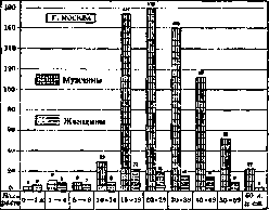
than in the province; its maximum is noted at the age of 20-29, and it decreases thereafter (other anomalies of refraction are noted mainly in old and senile age). Among the population older than 15, it is registered more often in men. The introduction of foreign bodies (Figure 7) among
Figure 7. Introduction of foreign bodies into the organs of vision in the Moscow province and the city of Moscow for 1926 (per 10,000 population of the corresponding group). men is observed significantly more often than among women; this is especially pronounced in the city of Moscow, where the incidence rate is much higher than in the province. The highest indicators are observed at the age of 15 to 49 years. Trachoma is distributed more evenly across age groups; the maximum of diseases is noted at the age of 15 to 19 years. Cataract is observed mainly in old age. Night blindness in the province is observed more often in old age, in the city of Moscow—in young and middle age. Table 6. Percentage ratio of cases and days of disease of the organs of vision to the sum of all cases and days of disease. Branches of labor: Cases of disease (m., f., total), Days of disease (m., f., total). Extraction and manufacturing industry: 2.0, 1.9, 2.1 (cases); 1.7, 1.7, 1.8 (days). Railway transport: 1.1, 1.6, 1.2 (cases); 1.0, 1.2, 1.7 (days). Satisfaction of spiritual needs: 2.0, 2.1, 1.3 (cases); 1.7, 1.1 (days). Table 7. Per 100 insured of the corresponding sex there were: Cases of disease. Branches of labor: Extraction and manufacturing industry: 2.42. Transportation.
(excl. railway) (1.94 Railway transport... 1.79 Supply... 0.66 Healthcare institutions, Soviet, scientific-educational institutions and stage activities... 2.24 1.52 0.71 0.46 0.79 0.38 0.45 Days of Vision 24.4 25.1 23.0 16.5 17.4 14.3 10.1 9.8 8.1 10.9 Average duration of one disease (in days) 10.1 11.2 11.8 10.6 9.7 15.4 9.4 13.8 17.7 13.8 6.3 6.9 16.4 15.2 Diseases of the organs of vision among the insured. Table 6 (according to data from the Central Social Insurance Administration, relating to 1925) shows the percentage of diseases of the organs of vision in the USSR among individual groups of insured persons in relation to all diseases in the corresponding branch of production. The first two groups (Table 6) give a higher percentage than the last. Among men, the percentage is higher than among women in all branches of labor. The greatest number of cases of diseases of the organs of vision per 100 insured persons (Table 7) falls on those working in the mining and manufacturing industries, the smallest—in Soviet institutions. The same applies to days of illness. The duration of one case of illness for the first groups, on the contrary, is less than for the latter. In the industry of the Moscow Governorate (Table 8), diseases of the organs of vision are most often observed
Table 8. Per 100 insured persons in the industry of the Moscow Governorate in 1927, there were cases and days of illness of the organs of vision. Per 100 insured persons there were Average Industry cases days of illness duration of disease Woodworking... Textile... Metal... Glass-porcelain... Printing... Food and flavoring... 2.8 2.5 2.5 2.3 2.3 2.2 1.7 1.7 1.6 1.2 29.7 21.3 15.5 16.8 14.4 17.8 13.0 11.6 10.9 7.5 10.7 8.4 6.3 7.4 6.1 8.0 7.5 6.6 6.7 6.1 All industry 2.6 19.3 7.4
among woodworkers, and the cases of illness among them were the most severe (the greatest duration of one case). The fewest diseases of the organs of vision are noted among those working in the food and flavoring industry. Diseases of the organs of vision and disability. The role of diseases of the organs of vision in the etiology of labor disability for the entire Union is visible from Table 9. Table 9. Per 100 disabled persons of each group and each gender in the USSR in 1925, there were cases of disability from diseases of the organs of vision: Categories Men Women Disabled I group... II group... III group... I-III groups... IV-VI groups... 9.3 4.0 3.7 4.3 7.1 10.4 3.0 3.8 3.3
The average age at the establishment of disability from diseases of the organs of vision was 48.3 years for men and 48.2 years for women. Per 100 labor disabled persons examined by the Bureau of Medical Expertise of the city of Moscow for 1925 and 1926, there were 2.9% disabled persons from diseases of the organs of vision, including 3.2% among workers and 2.6% among employees. By individual types of labor, this percentage was: Table 10. Workers Food and tobacco workers... Tanners... Manual laborers... Local transport... Builders... Printers... Metalworkers... Textile workers... Garment workers... Woodworkers... Chemists... 1925 1926 5.0 6.4 4.4 4.6 4.3 4.4 3.9 2.5 3.9 1.8 2.8 5.0 2.6 2.5 2.5 2.8 2.2 1.9 1.8 2.0 1.8
Employees 1925 1926; Educational workers 3.6
Economic-distribution personnel 2.2 Medical-sanitary personnel 2.3 Communications workers 2.1 Technical personnel 2.3 Labor safety workers
1.6 2.6 2.7 3.2 3.2 4.3 2.3 1.4 2.4 0.7 The highest percentage of disability from diseases of the organs of vision is noted among workers in the food industry, tobacco workers, leather workers, and laborers, and among employees - among workers in education. I. Doreyzer. Professional diseases of the organ of vision. Among the specific professional diseases of the eye, first of all, one should point to the nystagmus (see) of coal miners, which in some countries affects up to 10% of coal miners. Similarly, a specific professional disease of the eye is the cataract (see) of glassblowers ("Glasblaserstar" of the Germans). This suffering has been included in the list of professional diseases and in our modern labor legislation. An extensive group of professional diseases of the eye consists of eye injuries in connection with working conditions. One of the first places in frequency of eye injuries is occupied by the metalworking industry. The frequency of injuries and their severity depend on the detailed working conditions. Thus, according to Glezerov's data, who examined workers at the "Krasnoye Sormovo" plant in 1928, the eyes of metalworkers-pneumatics (chasers, riveters, etc.) are most often injured, to whom fragments or scale flying off when striking steel or iron may more often get. Injuries are very common among grinders (Lazarev, Glezerov, etc.), and then among turners, planers, etc. The vast majority of injuries refer to the entry of foreign bodies into the cornea (according to some authors, up to 80% of all eye injuries), with relatively small percentage falling on severe eye injuries (according to Glezerov, among Sormovo metalworkers - 0.78%). In recent years, attention has been drawn to the fact of decreased hair sensitivity of the cornea among metalworkers, metal turners, chippers, etc. When examining such workers, Samoilov and Freiman discovered a significant decrease in the tactile sensitivity of the cornea, and established that this violation of sensitivity is associated with the frequent entry of small particles of metal dust, grinding dust, etc. into the cornea. This connection is proved by the fact that the violation of sensitivity increases in frequency with the increase in the professional seniority of workers. It is possible that the presence of decreased corneal sensitivity and the resulting weakening of the protective reflex (blinking) explains the particular frequency of traumatic injuries among workers of these professions. In workers in the mining and coal industry, severe eye injuries from flying fragments of rock, coal are common; they are especially common and dangerous during explosions of dynamite, etc. As a result of professional eye injuries, the cornea is often infected, and creeping corneal ulcer often develops. In construction workers, burns from lime and eye injuries from fragments of stone and other building materials are common. Burns of the eyes with lime often lead to blindness as a result of deep changes in the cornea caused by the chemical action of lime, the formation of extensive adhesions of the conjunctiva of the eyelids with the conjunctiva of the sclera up to complete closure of the eyelids (symblepharon, ankiloblepharon, etc.). In the chemical industry, burns of the conjunctiva, cornea, and eyelid skin are especially common as a result of the action of various acids, alkalis, metal salts, various gases, etc. Under the influence of dust, conjunctivitis often occur in workers in various industries. Thus, professional conjunctivitis are common among workers in the flour milling, baking industry, among textile workers, etc. Ioffe, who examined the eyes of textile workers in Ivanovo-Voznesensk factories, found that although among the main groups of workers in spinning and weaving factories diseases of the conjunctiva are not common, they still increase when moving from less dusty to more dusty departments, reaching among combers, for example, 25% of all workers. Markedly expressed conjunctivitis have been noted in workers dealing with various products of the distillation of coal, mainly with tar and hard resin (pitch). These conjunctivitis often proceed acutely, with a severe clinical picture and are due to the chemical action of the substances contained in the pitch (most likely anthracene, acridine) and finally to photodynamic effects (in the light the effect of the pitch is especially sharply manifested). In close relation to professional eye injuries is the disease of the cornea that often develops on the basis of injury in the form of creeping corneal ulcer. Ulcus corneae serpens is often found among the rural population and is treated by some as a professional disease of farmers. Further, it is necessary to point to diseases of the organ of vision on the basis of professional poisonings. Of these, lead poisoning of workers is still practically important. Eye damage in lead poisoning can manifest in various forms, and the most characteristic have long been considered lead amblyopia or amaurosis; in such cases, there is usually a retrobulbar toxic neuritis, which can be observed in its typical form with central scotoma, etc.; in some cases, a picture of papillitis is also found, sometimes even of a choked disk, more rarely - inflammation of the retina. In the presence of brain phenomena from poisoning (encephalopathia saturnina), hemianopsias are observed. Rarely in lead poisoning, there are paralysis of the eye muscles (more often due to paralysis of the abducens nerve and more rarely - of the oculomotorius nerve). Eye damage in professional lead poisoning - usually phenomena of the late period of the general disease. In poisoning with carbon disulfide, used especially in the rubber industry, eye damage is often observed: according to Frost, 72% of all chronic CS2 poisonings consist in visual impairment. Most often, a toxic retrobulbar neuritis with all its characteristic symptoms develops, rarely - retinitis and chorioiditis; all these sufferings have a relatively good prognosis. In poisoning with arsenic, which sometimes occurs in workers dealing with arsenic or its compounds (in the mining industry, wallpaper factories, etc.), eye damage in the form of neuroretinitis, axial neuritis are sometimes found, but especially characteristic is a special conjunctivitis with necrotic round gray-yellow plaques. Mercury poisoning is sometimes accompanied, like lead poisoning, by amblyopia as an expression of toxic neuritis; in some cases, it leads to atrophy of the optic nerve. Poisoning with nitrobenzene sometimes causes hemorrhages in the retina, hyperemia of the disk, retrobulbar neuritis. - Prevention of diseases of the organ of vision on the basis of professional poisonings, naturally, consists in preventing the poisoning itself (see Professional poisonings). Special professional diseases of the organ of vision are observed in persons dealing with artificial, sometimes of exceptional power, sources of radiant energy used in the most diverse branches of modern industry, technology, laboratory research institutes, some branches of modern medicine. Such sources are: various kinds of electric lamps, mercury-quartz and carbon arc lamps, the power of which reaches tens and hundreds of thousands of candles, heated masses of metals and carbon electrodes in autogenous welding of metals, electric steel arc furnaces, molten masses of steel, cast iron, glass, sparks during short circuits of high voltage currents, with which one has to deal in the steel and iron foundry industry, glass production, film production, at iron and rail rolling mills, in persons serving electric motors (trams, subways, electric trains), in the electrical industry, at electric power stations. The main factors of harmful influence on the eye of radiant energy: power of the source, its distance from the eye, duration of irradiation, nature of the rays (wavelength) and the resulting absorbability of them by various eye tissues, as well as individual predisposition. The harmful effect of radiant energy is manifested mainly on the connective membrane and on the anterior part of the eyeball and most often appears in the form of the so-called ophthalmia electrica, o. photoelectrica, photoophthalmia. Ophthalmia electrica was studied and described in detail in the 90s of the 19th century by Professor Maklakov in workers engaged in autogenous welding of metals. The light of the voltaic arc obtained in this process causes peculiar burns of the skin coverings and at the same time is the cause of o. electricae, which is expressed in the following form. 8-10 hours after work, the most severe pains in the eye, sensation of foreign bodies, lacrimation, blepharospasm, photophobia, edema of the conjunctiva of the eyelids, sometimes of the eyeball, hyperemia of the eye, miosis or mydriasis appear. In milder cases, everything passes without a trace in 3-4 days.
A similar picture is also given by professional photo-ophthalmia in cinema workers, and in recent years, accompanying corneal lesions in the form of cloudiness and erosions of its central sections have been increasingly noted in them; in severe cases of ophthalmia electrica, phenomena caused by damage to the retina and optic nerve have been described, which subjectively manifest as decreased vision, the appearance of central and paracentral scotomas, and sometimes complete temporary amaurosis, and objectively as phenomena of hyperemia of the optic nerve head and edema of the retina. Usually all these changes also resolve favorably. Sparks arising from short circuits of high-voltage currents sometimes cause even more severe eye damage. In addition to the symptoms mentioned above, iridocyclitis and changes in the fundus of the eye resembling the picture of specific chorioretinitis are sometimes observed, functionally the eye returns to normal in most cases only within several weeks. In 7% of cases, Terrien observed a persistent decrease in vision within the range from 0.1 to light perception, and Rohmer observed atrophy of the optic nerve, so the prognosis in severe cases of blindness from short circuits of current requires great caution. It is also necessary to mention professional eye diseases in persons who, due to the nature of their profession, are to some extent exposed to radiation by X-rays and radium. Prevention of eye diseases caused by radiant energy is reduced to protecting workers from harmful rays by using appropriate filters placed directly in front of the light source, and to protecting the eyes with glasses that absorb harmful rays; in addition, attempts have been made and are still being made to use certain solutions instilled into the conjunctival sac for preventive purposes (Aesculin, Aqua zeozoni, Corodenin, etc.). Treatment of photo-ophthalmia is carried out according to the general rules of ophthalmology. In conclusion, it should be mentioned that until recently, myopia was also considered a typical professional eye disease, observed in persons who, due to their professional activities, needed to work for long periods at close range; thus, a specific professional myopia of typesetters, watchmakers, mechanics performing fine work, jewelers, etc., was spoken of. At present, the view on the pathogenesis of myopia has changed, and the role of close-up work is recognized by most as secondary; therefore, professional work requiring close-up work is considered only as a factor favoring the development of myopia in eyes that are hereditarily predisposed to it. Such a view, however, should of course not limit the scope of preventive measures to prevent the development of myopia in the corresponding industries (for details, see Myopia). V. Chirkovsky.
Related articles
Mentioned in
Cite this page
“Vision.” Soviet Medical Encyclopedia. English translation of Bolshaya Meditsinskaya Entsiklopediya, 1st ed. (Moscow, 1928–1936), ed. N. A. Semashko. https://sovietmedicalencyclopedia.pages.dev/article/vision/