Nystagmus
Historical document, translated for reference. It reflects medical knowledge of the 1920s–30s and is not medical advice.
Summary
Nystagmus is a rapid, repetitive movement of the eyeballs that can be spontaneous or experimentally induced. This article explains the classification, types, mechanisms, and experimental studies of nystagmus, including the work of Flourens, Ewald, and Barany on labyrinthine stimulation.
Encyclopedia article (1928–1936)
NYSTAGMUS, nystagmus (from Greek nystagmos - blinking), rapidly repeated movement of the eyeballs (tremor of the eyes); occurs both spontaneously and artificially (experimentally induced). By the number of tremors per second, 3 degrees of N. are distinguished - very rapid, moderately rapid, and slow. By the amplitude of movements, N. is distinguished as fine, moderate, and coarse. By the type of movements - pendular (undulating) and jerky, and in pendular N., eye movements originating from one point occur in both directions with equal speed, while in jerky N., eye movements in one direction occur slowly, and in the other direction - quickly. N. can be binocular, less often monocular (in one eye); if the direction of N. is the same in both eyes, it is called conjugate N. (associated), and if different - dissociated (rarely observed). Associated nystagmus is: horizontal, rotatory, horizontal-rotatory, vertical, diagonal, wheel-shaped, and elliptical. Spontaneous N. is usually a pathological phenomenon. N., being predominantly a reflex to irritation, is named after the organ whose irritation causes it. Thus, nystagmus caused by irritation of the semicircular canals is called labyrinthine, caused by irritation of the vestibular nerve outside the labyrinth - vestibular, irritation of the optical apparatus - optical; the latter manifests in the form of optokinetic or fixation N. (see below). Finally, there is positional N., which is a consequence of disruption of the corresponding cortical centers that keep the eyes completely at rest in any of their deviation. The mechanism of N. was first clarified on the basis of the study of experimentally induced N. For the first time, experimental labyrinthine N. was induced by Flourens (1828). After cutting the lower vertical semicircular canal of a pigeon, Flourens noticed that the eyeball and eyelids made continuous, sharp movements. In subsequent years (1870-1880), a number of authors (Hogyes, Breuer, Bornhardt) induced labyrinthine nystagmus in animals, using heat, cold, rotation, electric current as irritants. In 1892, Ewald, experimenting on pigeons, used pressure and rarefaction as irritants. Exposing the right membranous horizontal canal of a pigeon in a small area, Ewald then
Figure 1.
Figure 2.
Figure 1 and 2. Ewald's experiment. Figure 1: 1 - bony wall of the right horizontal semicircular canal; 2 - plug between the bony wall and the membranous wall of the membranous semicircular canal; 3 - direction of pressure on the canal; 4 - direction of displaced endolymph to the left; 5 - slow component to the left, in the direction of endolymph movement (from the ampulla); 6 - direction of the fast component to the right (toward the ampulla). Figure 2: 1 - plug; 2 - direction of the suction action of the piston; 3 - direction of endolymph flow to the right; 4 - slow component to the right - toward the ampulla; 5 - direction of the fast component to the left - away from the ampulla. canal this was hermetically sealed. Pressing with a special piston on the parts of the canal lying between the plug and the ampulla, he thereby caused a flow of endolymph toward the ampulla (Figure 1). As a result of increased pressure, a regular horizontal movement of the head and eyes occurred in the direction of endolymph movement (away from the ampulla), in this case - to the left; immediately following this deviation, the head and eyes returned to the original position by a rapid movement, i.e., in this case to the right (toward the ampulla). When pulling out the piston, suction occurs and as a result, reverse flow of endolymph, causing weaker eye and head movements than when pressing (Fig. 2), again directed in the direction of endolymph movement, i.e., in this case to the right; as in the first case, following this deviation, the eyes and head returned to the initial position by a rapid movement, i.e., to the left (away from the ampulla). The first eye movement, caused by the flow of endolymph, is called the slow, the second - the fast component of N. A similar phenomenon can be reproduced in a patient with a fistula of the semicircular canals (see Labyrinthitis); if the bony wall, for example, of the right horizontal canal is destroyed in some place by caries, then mechanical pressure on this fistula and even an increase in pressure in the right external auditory canal by pressing with a finger on the tragus usually causes lively ("pressor") N. toward the affected fistula labyrinth, i.e., to the right, and a decrease in pressure by suction causes N. toward the healthy side, i.e., to the left. Since it is easier to observe the fast component, the direction of N. is designated by the direction of this latter, i.e., if the fast component is directed to the right, it is customary to denote that N. is present to the right. It is enough to remember Ewald's law to be able in each individual case of labyrinth irritation, caused by the flow of endolymph (during rotation, cooling, heating) to predict in which direction N. will be directed. This law states: the flow of endolymph directed toward the ampullary end causes N. in the direction of this same ampulla, while the flow of endolymph directed away from the ampulla, i.e., toward the smooth end, causes N. in the direction of the non-irritated labyrinth. Similar experiments on the anterior and posterior vertical canals have established that the aforementioned Ewald's law with respect to these canals has the opposite effect, namely: the flow of endolymph toward the ampulla causes N. in the opposite direction, and N. always occurs in the plane of the irritated canal. Thus, when the horizontal semicircular canals are irritated, horizontal nystagmus is obtained, frontal - rotatory, sagittal - vertical, and sometimes diagonal (Fig. 3). To explain these phenomena, it is most probable to assume that in each of these cases different nerve endings in the ampulla are irritated, transmitting impulses along fibers connected with different nerve centers of the oculomotor nerves. The effect of temperature on the labyrinth (temperature below and above the body temperature of the animal) results in the appearance of a similar flow of endolymph, and depending on the direction of the endolymph flow toward the ampulla or away from the ampulla, N. occurs in the corresponding direction according to the regularity resulting from the above-mentioned Ewald experiment. Barany (Barany) first systematically performed thermal (caloric) irritation of the labyrinth in humans and introduced it into the methodology of otoneurological research. It is based on Ewald's law, as well as on the physical law about the change in specific weight of liquids at different temperatures. So, for example, if 20 cm³ of water at 27° is poured into a person's right ear with the head in a vertical position and imagine the parts of the labyrinth subjected to cooling (Fig. 4), it is seen that the anterior, vertical, and horizontal canals fall into the area of the water jet. As a result of cooling the walls, the adjacent lymph will also cool down and as a result move downward, i.e., in the anterior vertical semicircular canal toward the ampulla, and in the horizontal - away from the ampulla. According to Ewald's law, the flow of lymph away from the ampulla in the horizontal semicircular canals causes nystagmus in the direction opposite to the flow of endolymph, i.e., in this example N. will occur to the left and horizontal, since the horizontal semicircular canal was irritated. In the vertical semicircular canal, however, the lymph flow will be directed toward the ampulla, but Ewald's law
Figure 3. Graphic designation of types of nystagmus: 1 - horizontal to the right; 2 - horizontal to the left; 3 - vertical upward; 4 - vertical downward; 5 - rotatory to the left; 6 - rotatory to the right; 7 - horizontal-rotatory to the left with vertical component; 8 - horizontal-rotatory to the right with vertical component; 9 - wheel-shaped nystagmus; 10 - elliptical nystagmus horizontal to the left; 11 - elliptical nystagmus diagonal to the right.
Figure 4. Scheme of caloric nystagmus and technique of its observation: 1 - injection of 20 cm³ of water (27.5°) with a syringe into the external auditory canal along the posterior-superior wall; 2 - direction
of endolymph flow in the right v relation to the frontal semicircular canal; 3 - in the horizontal semicircular canal; 4 - direction of horizontal-rotatory nystagmus. for vertical semicircular canals states that N. is directed in such cases in the opposite direction, i.e., to the left. Thus, caloricization of the right ear with cold water should give horizontal and frontal N. to the left, which is indeed what happens, but the frontal, appearing together with the horizontal, together give horizontal-rotatory nystagmus. However, this view is not universally accepted: phenomena in a living organism are subject not to physical, but to more complex physiological regularities, in particular in this case the main significance belongs to vascular phenomena; when caloricizing the ear with cold water
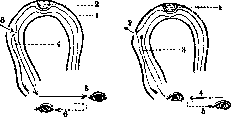
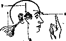
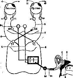
Fig. 5. Diagram of the central mechanism of labyrinthine horizontal nystagmus (nystagmus is caused by cold water): 1-direction of cold water stream; 2-tympanic membrane; 3-tympanic cavity; 4-horizontal semicircular canal; 4a-labyrinthine nerve; 46-gangl. Scarpae; 5-vestibular fibers in the composition of the posterior longitudinal bundle; 5a-nuclei of the posterior longitudinal bundle; e-IV ventricle; 7-posterior quadrigeminal body; 8-p. abducens (VI-its nucleus); 9-p. oculomotorius (III-its nucleus); 10-direction of slow component; 11-direction of fast component; P and T-centers of fast and slow components (vestibular nuclei of the medulla oblongata and pons); re-m. rectus externus; ri-m. rectus int.
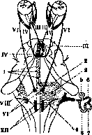
In the area directly adjacent to the irrigated area, superficial ischemia occurs, while in the deep vessels of the labyrinth there is collateral hyperemia; as a result of vasodilation, outflow of endolymph occurs, intralabyrinthine pressure decreases, which serves as the source of N. In other words, temperature acts on the endolabyrinthine environment through the vasomotors. The mechanism of labyrinthine N. appears according to recent research as follows. Under the influence of endolymph current, the freely oscillating hairs of the neuroepithelium (cupula; see Inner ear) are set in motion, which causes irritation of the peripheral ending of the vestibular nerve. Under the influence of endolymph current, the impulse is transmitted along the nerve to its nuclei (see Vestibularis nervus and fig. 4). In the nuclei are located two centers, one of which causes the slow component, the other the fast component. From these centers, the impulse goes along fibers located homolaterally and contralaterally in the composition of the posterior longitudinal bundle to the oculomotor nuclei; from the latter, the impulse is transmitted to the oculomotor muscles, the contraction of which causes corresponding eye movements. The slow component is called first, and then from the center of this latter, the impulse is transmitted to the center of the fast component. Impulses, alternating in one center and then the other, cause alternately the fast and then the slow component (fig. 5).-Mechanism Fig 6- Pathways connecting the nuclei of vestibular N. and oculomotor nerve: 1-arcuate fibers of N. Nystagmus For rotatory, horizontal and vertical movements; 2-area of vertical nystagmus; 3-area of horizontal nystagmus; 4-area of rotatory nystagmus; 5-cochlea
can be caused by irritation of individual areas in the region of the vestibular nerve nuclei in the brainstem: anterior vertical; 7-horizontal; 8-posterior vertical up to the semicircular canals. Horizontal N. is caused mainly from the area between the caudal beginning of Deiters' nuclei, approximately to the knee of the facial nerve, rotatory-mainly from the most caudal parts of this area, while vertical-from the anterior parts. These relationships, established in rabbits, are also observed in humans (fig. 6). Experiments with subcutaneous injection of poisons showed that disorders of labyrinthine function (disorders of statics, nystagmus, dizziness) are due to selective damage to the just mentioned areas (statokinetic centers in the region of the medulla oblongata, pons, and midbrain). The vestibular symptom complex noted in botulism is also due to damage to these places in the brainstem, i.e., labyrinthine-vestibular N. 42 is a reflex consisting of a centripetal segment (membranous labyrinth, vestibular nerve) and a centrifugal one (three pairs of oculomotor nerves with muscles) and of central connections going from the nuclei of the vestibular nerve to the anterior quadrigeminal body (nuclei of the vestibular nerve, abducens, trochlear, and oculomotor) (see Vestibularis nervus). Labyrinthine-vestibular N. has a certain biological significance. If the eyes were at rest when the head turns, the image would shift from the corresponding point on the retina, but head movement causes simultaneous flow of endolymph and consequently corresponding eye movement. Optokinetic N. To understand the essence of the optokinetic reflex, the following must be noted. The eye has 3 accommodative adaptations: 1) accommodation to distance (functions of the lens and convergence of visual axes), 2) accommodation to light intensity (function of the iris) and 3) accommodation to the movement of the fixed object (optokinetic N.). The latter occurs as follows: let us imagine that the eye fixed a certain number of objects moving to the left and followed them, turning in the direction of their movement, i.e., to the left. A moment will come when it is necessary to transfer fixation to the next point of the object approaching from the right, i.e., to turn the eyes to the right. This movement must be fast, otherwise some points will manage to escape fixation. The first eye turn (to the left) corresponds to the slow component of nystagmus, and the second (to the right) to the fast one. By repeating these movements, the eye can examine the largest number of flashing objects (a similar phenomenon occurs when viewing objects from the window of a moving train, which is why this nystagmus was first called railway nystagmus). This is the biological meaning of the optokinetic reflex. Experimentally, optokinetic N. is caused as follows (fig. 7). At a distance of 50 cm from the subject, a rotating cylinder covered with colored stripes is set up. When the cylinder rotates to the right, the subject fixes one of the stripes, and immediately horizontal N. appears in the direction opposite to the rotation of the cylinder, when rotating upward-N. downward, when rotating downward-N. upward. Since fixation is necessary to cause optokinetic N., the latter is essentially a fixation nystagmus. Most likely, the centripetal part of the arc of this reflex, starting in the retina, ends in the occipital lobe, its center is the area from the occipital lobe to the central sulcus, from where the centripetal path to the nuclei of the oculomotor nerves begins. If there is a lesion (irritation, destruction) in any part of the said reflex arc, spontaneous optokinetic N., resp. disappearance of experimental optokinetic N. may occur. In addition, it should be borne in mind that the rest of the eye, which we objectively observe, is only apparent. In reality, even in normal conditions, the eyes make a whole series of nystagmus-like movements, macroscopically unnoticeable. Various unfavorable conditions affecting the tone of the eye muscles (decrease in visual acuity, lack of light) can reduce the frequency of N. and increase its amplitude and thus turn latent N. into manifest or kinetic N. - Positional N. According to some authors, in 70% of healthy people, N. is noted at 50° deviation and even more often at extreme eye abduction. This N. is in most cases horizontal, fine-, medium-amplitude
Fig. 7. Apparatus and its setup for the study of optokinetic nystagmus.
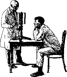
steady and significantly less often jerky. It is caused partly by weakening of the tone of the eye muscles, and partly by weakening of the cortical postural centers. The methods of investigating N. consist of nystagmoscopy (observation of N. by ordinary external examination of the eyes) and nystagmography (recording of N. by a writing instrument), which is a more perfect method of investigation. The presence or absence of N. is determined depending on at what angle of eye deviation it appears. Often it can be noticed already with direct gaze, but more often only after deviation of the eyes to the sides. For observation, a certain point on the surface of the eyeball should be chosen, e.g., a vessel of the conjunctiva or a sharply defined place on the iris. Vestibular N. is especially sharply revealed if the subject wears biconvex glasses (+20D) - Bartels glasses. This achieves almost complete elimination of fixation, which acts as a brake on the course of labyrinthine N. Nystagmography more accurately establishes the characteristics of N. In the basis of some nystagmographs is the principle of transmitting the movement of the eyeball through the eyelid by means of a lever system (fig. 8), in others--through the eyelid by means of compressed air (fig. 9) (Pneumonystagmograph), in thirds--through reflected light: a small mirror is placed on the eyelid of the closed \ eye, onto which a beam of light from an electric lamp falls; the reflected beam is transmitted to a drum covered with light-sensitive paper (Voyacek). The most perfect, but very expensive, is the Dohlman nystagmograph: a concave mirror is fixed on a suction cup made of rubber; the cup is attached to the edge of the cornea by light pressure; thus, the mirror follows all the movements of the eye. The mirror weighs 0.6 g. The light source directed at the mirror reproduces the reflected light point, perceived by the light-sensitive paper. N. can also be recorded cinematographically. The nystagmogram recorded by the Ohm instrument gives a clear idea of the frequency, amplitude, and course of N. On the curve, the slow and fast components of labyrinthine N. are sharply marked, whereas nystagmus caused by eye diseases or caused by a violation of posture appears on the curve in the form of equal height ascents and descents in the form of a wave (fig. 10).
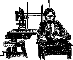
Figure 8. Ohm's nystagmograph.
Etiology of N. The basis of the pathogenesis of spontaneous N. is a violation of the harmonious interaction of the centers and pathways forming the reflex arcs of labyrinthine, vestibular, optokinetic or postural N. This violation is caused by various reasons. Thus, with inflammatory diseases of the middle ear, extending to the labyrinth, sinuses, meninges or to the entire inner ear, as well as with non-inflammatory diseases of the inner ear, labyrinthine N. is observed. When processes extend to the area of the posterior cranial fossa (due to pressure, the presence of otogenic encephalitis in the area of vestibular nuclei, etc.), a large-amplitude vestibular nystagmus is observed. The latter is also observed with organic diseases of the central nervous system along the path of the vestibular system. Among these diseases, necrotic foci in the area of the brain stem, infectious encephalitis (typhus, typhoid, relapsing fevers, erysipelas, etc.), toxic encephalopathies (lead poisoning, botulism), multiple
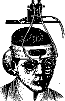
Figure 9. Buys' nystagmograph.
sclerosis (predominantly pendular N., sometimes dissociated N. is also noted), syringobulbaria (rotatory coarse N.). Coarse vestibular nystagmus, observed in cerebellar diseases (tumors), is the result of remote action on the vestibular system located in the medulla oblongata, since experimentally from the side of the cerebellar substance, N. has not yet been obtained. Tumors of the cerebrum can cause vestibular nystagmus, since through remote action they cause increased pressure in the posterior cranial fossa. In addition to the above-mentioned etiological factors, pareses and paralyses of the oculomotor nerves can also be the basis of positional N. Positional nystagmus caused by these causes is fine and very frequent, directed in both directions, but is more often observed on the side of the affected nerve. Since positional nystagmus is caused by a violation of the setting of voluntary movements, it should be expected that N. in such cases can also be caused by diseases of the central mechanisms regulating the voluntary setting of the eyes (gyrus angularis, second frontal convolution, parietal, occipital lobes). Positional spontaneous N., observed with extreme abduction, is the only form that is observed in neurotics in 75% of cases. Diseases and anomalies of the eye, as well as degenerative lesions of the optic pathways and centers, cause optical spontaneous nystagmus. It can occur in the following diseases and developmental anomalies of the eye: microphthalmia, coloboma of the iris and vascular tunic, congenital absence of the iris, central cortical cataract, pronounced anomalies of refraction, complete blindness, as well as color blindness with accompanying phenomena of pigmentary retinitis, then acquired in early childhood diseases of the cornea. N. in the mentioned diseases is pendular, and with extreme eye abduction it becomes jerky and disappears in the dark. In the mentioned diseases, horizontal N. is most common, but rotatory, vertical, and diagonal N. are also observed, as well as dissociated and monocular N. Monocular N. is more often vertical and is often accompanied in children by a nodding movement of the head (spasmus nutans). In the latter, it is caused by staying in poorly lit dwellings. In terms of amplitude, it is usually medium-coarse, while fine N. is very rarely observed. Spontaneous optical N., caused by the above-mentioned causes, is sometimes observed as a familial disease. Latent N. also belongs to the group of optical spontaneous nystagmus, characterized by the fact that when one eye is closed, a medium-coarse N. appears on the open eye, and it is directed towards this same eye. With binocular vision, the mentioned N. disappears. The mechanism of latent N. has not been clarified, just as the mechanism of reflex N., caused by irritation of the trigeminal nerve in conjunctivitis and irritation of the nasal mucosa in operations on the nasal septum and turbinates. The next etiological factor causing the appearance of spontaneous optical N. is many degenerative brain diseases. Spontaneous N. is very rare. Persons who can voluntarily cause N. could often equally cause the contraction of other muscles, usually not allowing isolated voluntary contractions. A special disease is the so-called nystagmus-myoclonia of Lenoble and Obin (nystagmus myoclonica). It is characterized by pendular, less often rhythmic N. with rotatory, diagonal, most often with horizontal direction. This N. is accompanied by trembling of the head muscles (tremor of the head), increased reflexes, intellectual and vascular disorders. Spontaneous nystagmus in tower skull is most likely caused by phenomena accompanying reduced vision. N. of coal miners. N. is very common among miners, thus being their professional disease. Thus, in the USSR in the Anjero-Sudzhensky district of the Tomsk region, Sergeeva found among miners from 12% to 22% nystagmatics. This figure, as well as similar statistical data noted by authors in other countries, justifies the attention paid to the professional hygiene and pathology of nystagmus of coal miners. In Germany, England, Belgium, since 1913, governments have established special commissions consisting of representatives from miners, the coal industry, specialist physicians - otoneurologists and ophthalmologists for the study of nystagmus of miners. In Liege in 1908, a special clinic was founded for the purpose of studying and treating N. In 1927, at a scientific conference of physicians in the city of Stalin, a resolution was passed to organize a commission for the study of nystagmus of coal miners similar to analogous organizations in other countries. In 1931, at a conference of the Stalin Institute of Pathology and Occupational Hygiene, a plan was developed for organizing a collective expedition for the study of N. of coal miners. The expedition began work in May 1931. Not all categories of workers engaged in underground work are equally affected by N. Miners are in first place (75% of all cases of N. of coal miners). Loaders, scrapers, sled runners, tunnelers give a significantly smaller percentage of diseases. The close connection between the occurrence of nystagmus in miners and their professional activity is evident from the table (Table 1), where the percentage of nystagmatics increases with the length of service in underground work. However, the fact that among miners in the USA and South America N. is not observed contradicts the position that this N. is caused exclusively by working conditions in mines. The observation that a Scottish miner suffering from N., after moving to work in mines in the USA, ceased to suffer from the mentioned disease and upon returning to working conditions in Scottish mines fell ill again, indicates that not all causes of N. in mine working conditions have been sufficiently studied. Table 1. (Borrowed from Vigdorchik). Length of underground work Less than 2 years From 2 to 5 years 6 to 10 years 11 to 15 years 16 to 20 years 21 to 25 years Percentage suffering from N. 1.0 1.6 3.9 12.0 24.0 29.0 Length of underground work From 26 to 30 years. 31 to 35 years ! 40 years ! 45 years ! 50 years ! 55 years ! 36 V 41 V 46 V 51 Percentage suffering from N. 39.0 45.0 43.0 65.0 20.0 10.0 Most often, workers aged 25 to 40 years get sick. Cases of illness in adolescence are not uncommon, after three months of underground work. Clinical picture.-The professional disease of workers manifests itself not only with N., but also with trembling of the head, eyelids, extremities and a number of phenomena from other organs (see below). N. in this disease is only the first and more pronounced symptom. N. of coal miners belongs to the category of optical N. However, the clinical picture of N. of coal miners is extremely varied, which complicates differential diagnosis between it and other categories of N. (see below). Similar diversity is also observed with respect to the speed of N. (32-517 per second). N. manifests itself at a certain angle of eye deviation. N. is especially easily caused when looking up. By establishing the limits of eye deviation in all directions in which N. appears, the so-called tremor field is determined. The patient, in order to avoid this field, holds his head tilted accordingly. Nystagmus of coal miners sometimes manifests itself in the dark, and when the patient moves into a lighted room, it disappears. Consequently, dimming has an irritating effect on N., while illumination has a calming effect. In myopic nystagmatics, after applying corrective glasses, N. becomes significantly weaker. These observations lead to the establishment of preventive and therapeutic measures. Movements and changes in head position intensify N. of coal miners. In 2,000 cases (Llewellyn), N. of coal miners in 501 cases was noted with spasm of the eyelids, in 628 cases - trembling of the head. In addition to tremulous phenomena, increased excitability of the sympathetic nervous system, disruption of tendon and skin reflexes, slow gait and speech, a persistent feeling of fear, headaches, heavy sleep, loss of appetite, arrhythmic pulse, depression of the psyche, photophobia are noted. Cessation of work in underground conditions leads to the disappearance of the noted symptoms; after resumption of work in mines, relapses often occur. A predisposition to the disease is noted in certain families and among some peoples; thus, Slavs get sick more often than other races. Of the unfavorable working conditions that can be considered causes of the disease, two moments deserve attention: insufficient lighting in mines and the twisted position assumed by the miner during work with strained upward deviation of gaze. However, the possibility of the pathogenic effect of inhaling gases from certain types of coal is not excluded, which is indicated by the absence of nystagmus of coal miners among workers in the USA working under otherwise equal conditions with Scottish workers. Methods of lighting mines at present are as follows: safety lamps - oil and gasoline, candles and open lamps, electric and acetylene lamps.
Their brightness in units is indicated in Table 2. Table 2. Brightness of various light sources used for illuminating mines (according to Natanson). Light source Brightness in units Oil lamp..... Gasoline lamp . . . . . Candle and open lamp Electric lamp . . . Acetylene lamp . . . 0.5 1.01 0.7 1.75 8-15 From Table 3 it is seen the influence of the type of lighting on the number and character of N. disease. Improvement of lighting conditions has a beneficial effect on reducing diseases and the character of nystagmus. Table 3. (According to Natanson.) Method of lighting Number of severe cases per 1,000 coal miners Safe oil ! lamp......1
35 Safe gaso- ; line lamp . . . .
0 Electric lamp .......... Acetylene lamp Number of pronounced diseases per 1,000 coal miners 57 44 13 All these data with sufficient convinciveness indicate the role of mine lighting in the disease of coal miners with N. A twisted position of the body contributes to irritation of the vestibular organ,-as a result of which vestibular N. appears. But pathological changes in this system in coal miners are rarely observed, although it is quite possible that in persons with increased excitability of the vestibular system due to the twisted position, vestibular N. is added to N. in coal miners. This is indicated by the recording curve of N. in coal miners, containing elements of labyrinthine N. However, the appearance of N. due to insufficient lighting is still difficult to explain, and regarding its mechanism there are several theories: 1) theory of fatigue of eye muscles; 2) connection with poisoning by mine gases; 3) theory of violation of balance centers of eyeballs; 4) theory of counter-rotation of eyes; 5) labyrinthine theory;. 6) lighting theory. Of all theories, the lighting theory best explains the mechanism of N. in coal miners. The similarity of N. in coal miners with non-professional N., for example experimental N. from darkness, N. in children growing in dark rooms (spasmus nutans), and N. in people with weakened vision (p. amblyopicus) speaks in favor of this theory. Differential diagnosis of N. in coal miners. When set at a close distance, N. in coal miners weakens the phenomenon opposite to that observed in labyrinthine N. In terms of direction, amblyopic N. usually proceeds uniformly, most often in the horizontal direction; vertical tremor and wheel-like tremor are very rarely observed, whereas circular and elliptical tremors noted in N. of coal miners are not observed at all in amblyopes. Amblyopic N. more often proceeds concomitantly than N. in miners. Congenital tremor occurs at all angles of eye deviation, whereas N. in miners occurs at a certain angle. Moreover, congenital N. does not depend on a certain position and movement of the body. Professional N. most often proceeds with phenomena of apparent movement of surrounding objects, whereas in persons with congenital N. this is not the case, and most often N. is accompanied by weakened vision. It is very difficult to confuse congenital N. with N. observed at extreme positions of the eyes, proceeding in jerks and in the horizontal direction. Intense convergence prevents the appearance of N. in coal miners. It is necessary to keep in mind that pendular movements of the eyes (the right eye moves up and inward, the left-down and outward) also occur in hemianopia. Preventive measures are as follows: it is necessary to improve the lighting conditions in mines, then before ascending from the mine to wear smoked glasses to avoid a rapid transition from darkness to light. It is necessary to carry out systematic otoneuro-ophthalmological examination to detect the latent form of the disease.-T r e a t m e n t. If the disease has already developed, it is necessary to remove coal miners from underground work. More severe forms are subject to treatment by physical methods in a sanatorium-resort setting.
G. Zimmerman. NITRATES, salts of nitric acid, see Saltpeter.
Related articles
Mentioned in
- Acute Ataxia
- Aniridia
- Aplasia Axialis Extracorticalis Congenita
- Bárány's Method of Investigation
- Bard's Sign
- Belous Symptom
- Deiters' Nucleus
- Dejerine-Sottas Disease
- Duchenne Syndrome
- Dyssynergia
- Galvanic Reaction
- Horner's Syndrome
- Labyrinth
- Methods of Medical Examination
- Multiple Sclerosis
- Occupational Diseases
- Otorhinolaryngological Instruments
- Parinaud Syndrome
- Rossolimo Neuritis
- Spasmus Nutans
Cite this page
“Nystagmus.” Soviet Medical Encyclopedia. English translation of Bolshaya Meditsinskaya Entsiklopediya, 1st ed. (Moscow, 1928–1936), ed. N. A. Semashko. https://sovietmedicalencyclopedia.pages.dev/article/nystagmus/