Otorhinolaryngological Instruments
Historical document, translated for reference. It reflects medical knowledge of the 1920s–30s and is not medical advice.
Summary
This article from the 1st edition of the Great Medical Encyclopedia details the wide range of instruments used in otorhinolaryngology for examination and surgery, including illumination devices, ear instruments, and nasal tools as of the early 1930s.
Encyclopedia article (1928–1936)
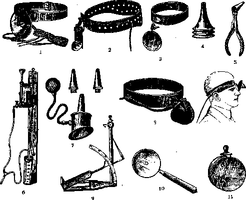
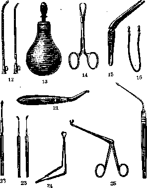
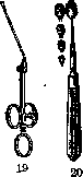
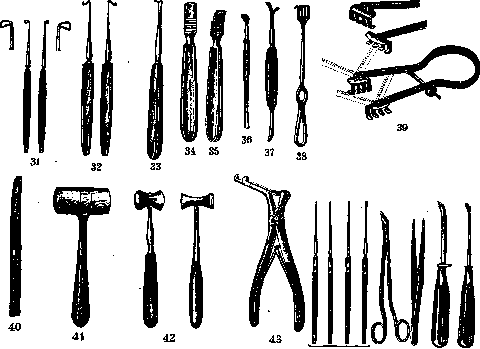
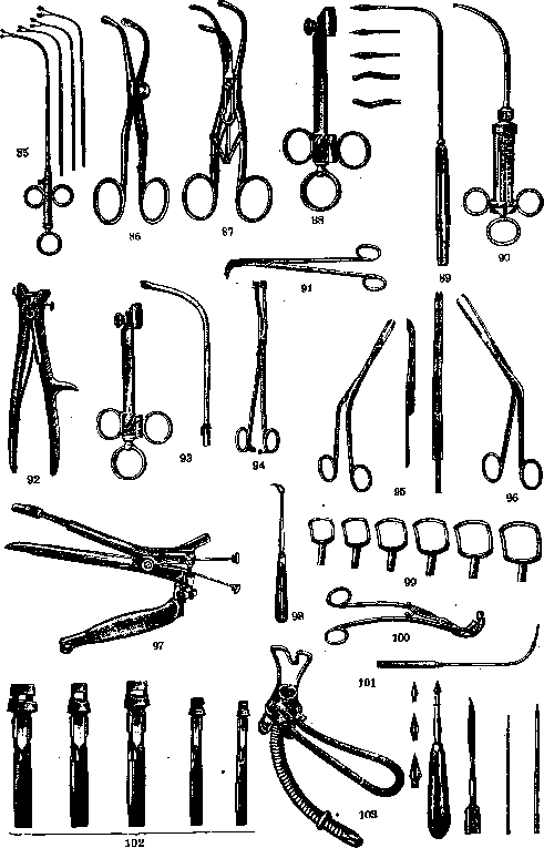
OTORHINOLARYNGOLOGICAL INSTRUMENTS. In otorhinolaryngological practice, a very large number of instruments are used both for examination and for surgical intervention. The most common of these are the following. Illumination devices are divided into illuminators providing direct light and illuminators with reflected light. The first type of illuminator is basically a small spotlight with an electric light bulb inside (with an electric check). By means of a belt, the illuminator is attached to the head on the forehead and directly directs light to the area under examination (Fig. 1 — Clar's lamp, Fig. 2 — Kirstein-Killian's lamp, Fig. 3 — Brünings' lamp). The reflected light illuminator, the so-called reflector, has the appearance of a concave mirror 8–9 cm in diameter and with a focal length of 20–25 cm, with an aperture in the center. The reflector is attached either to a handle (Fig. 10) or by means of a belt on the head to the forehead (Fig. 3). Ear instruments. The ear speculum has the appearance of a funnel. Wilde and von Stein specula are predominantly used. Wilde's speculum (Fig. 4) has a length of 3.5–4 cm, a width at the outer end, which has a slightly thickened edge, of 1.5 cm, and at the rounded inner end of 3–4–5 mm. The von Stein speculum is funnel-shaped and has a width at the outer end of 3–4 cm. Kramer's speculum with a handle (Fig. 5) resembles a nasal speculum. Funnels are made of metal, rubber, glass, and celluloid. It is recommended to use exclusively metal specula, as they are more hygienic and are not spoiled by boiling. To determine the mobility of the tympanic membrane, Siegle's pneumatic funnel is used. It has the appearance of an ordinary funnel, the wider end of which is closed by an obliquely placed glass plate. In the side wall of the funnel there is a short tube connected by means of a rubber tube to a rubber bulb (Fig. 7). The funnel is tightly inserted into the external auditory canal. When the bulb is compressed, the air in the auditory canal is condensed; when the compression of the bulb is reduced, the air is rarefied. Condensation and rarefaction of air in the auditory canal causes movement of the tympanic membrane (see). Various tuning forks (see) have been proposed for hearing examination (König, Lucae, Hartmann, Politzer, etc.). The following tuning forks are most commonly used: C1 — 128 vibrations per second, C2 — 512 vibrations per second, C3 — 1024 vibrations per second, C4 — 2048 vibrations per second, C5 — 4096 vibrations per second. In some cases, Galton's whistle (see Galton's whistle) is also used. To isolate one ear when examining the hearing of the other ear, Bárány proposed a special noise maker (Fig. 11). To examine the vestibular apparatus, a number of devices have been proposed. Bárány's turning chair is used to determine nystagmus depending on rotation. The Brünings otocalorimeter is for determining nystagmus depending on the caloric reaction (Fig. 6). Bárány's nystagmometer consists of a head bandage to which a scale with divisions and a fixator are attached. The nystagmometer is used to measure rotational nystagmus. The Brünings otogoniometer is for the correct positioning of the head and the direction of the eyes during the otocalorimetric test. The von Stein goniometer (Fig. 9) is used for finer diagnostics of balance disorders due to labyrinth damage and brain diseases. Catheters for inflating the ears, proposed by various authors [Troeltsch — Fig. 12 (left), Kramer — Fig. 12 (right), Hartmann, etc.], generally are metal or ebonite tubes of various diameters, 10–15 cm long. The narrow end of the tube is bent in the form of a beak. At the wider end there is a small ring pointing in the same direction as the beak. The position of the beak in the nasopharynx can be judged by the ring. Politzer recommends using a bulb connected by a rubber tube with an olive (Fig. 13) instead of a catheter. The latter is inserted into the nasal cavity. With strong compression of the bulb, air through the nasal cavity enters the nasopharynx and the Eustachian tube. To control the entry of air into the middle ear, an otoscope is used (Fig. 16). To remove foreign bodies from the external auditory canal, various probes, hooks, curettes, and forceps have been proposed. The forceps consist of two handles, a lock, and blades with sharp hooks bent towards each other (Fig. 14). Some authors have proposed ear tweezers with crossing blades (Troeltsch — Fig. 15). Ear probes are used both with threads and buttoned (Stacke's probe — Fig. 17, Ziegler's probe — Fig. 18), as well as with a metal handle. To remove polyps, an ear snare is used. Of the various modifications proposed, Hartmann's model is the most common (Fig. 19). In Herzfeld's forceps, the ends of the blades are bent in the form of branches in different directions: up, down, right, left (Fig. 25). Sharp spoons are used straight and curved (Figs. 20 and 21). For paracentesis of the tympanic membrane, bayonet-shaped or angle-bent spear-shaped needles with handles are used (Politzer's needle — Fig. 26, Lucae's needle — Fig. 22). A number of instruments have been proposed for removing the auditory ossicles. A tenotome is used to detach the malleus from the tendon of the tensor tympani muscle (Schwartze tenotomes — Fig. 23). To remove the malleus, Sexton proposed a corresponding curved forceps (Fig. 24). For the same purpose, Lucae proposed his instrument, consisting of a handle and a rod with one and two bent ends (Figs. 28 and 27). Various instruments are used for the extraction of the incus (Ludewig — Fig. 29, Ferrer — Fig. 30, Zeroni — Fig. 31, Kretschmann — Fig. 32). They are hooks bent at right angles. Instruments for trepanation of the mastoid process. First of all, all instruments commonly used in any major surgical operation must be prepared for this operation, such as: scalpels, tweezers, straight and curved surgical and anatomical scissors, hemostats, needles, needle holders, grooved probe. In addition, it is necessary to have ear tweezers and probes, a raspatory, an elevator, a wound retractor, chisels, a hammer, spoons, polyp and bone forceps. The Luce-Jansen raspatory with a handle (Fig. 33), Farabeuf's raspatory, straight and slightly bent along the plane (Figs. 34 and 35). Stacke's elevator (Fig. 36). Von Stein's raspatory with an elevator (Fig. 37). Wound retractors of various modifications have been proposed (Passow — Fig. 38). Von Stein's self-retaining wound retractor is distinguished by the fact that its hooks are movable in all directions and move in parallel planes depending on the conditions of the wound (Fig. 39). Chisels for trepanation are used straight and curved, smooth and grooved (Fig. 56). Schwartze chisels are smooth and grooved, 3, 5, 7 mm wide. Stacke has proposed long curved chisels 3, 5, 7 mm wide (Fig. 40). Wooden hammers made of beech wood (Fig. 41) and tin hammers (Fig. 42) (according to Brünings) are used during the operation. Von Stein proposed a trephine with three burs and two spotlights for opening the mastoid process. The latter protect against too deep penetration of the bur into the mastoid process (Fig. 48). The advantage of the bur is that it avoids concussion of the skull, which is sometimes observed when working with a chisel. Of the bone forceps proposed by various authors, the Zaufal-Jansen, Luer, Hartmann, and Lucae models are most commonly used (Figs. 43, 49, 50, 92). To open the ear labyrinth, in addition to the instruments indicated above, a dura mater protector (Fig. 51), labyrinth probes (Fig. 44), scissors (Fig. 45), narrow tweezers (Fig. 46), and special narrow chisels are used. The protector has the appearance of a curved raspatory with a blunt end. Nasal instruments. Hartmann's nasal speculum (see Specula — medical specula) consists of two handles, a lock, and two blades, the ends of which are bent at an obtuse angle in the form of branches. The latter are inserted into the nostril opening. When the handles are squeezed, the branches diverge and expand the nostril. Cholewa and Roth specula are of almost the same design. Voltolini and Killian specula differ from the previous ones by longer branches. In Mackenzie's speculum, the branches are covered with ivory plates. Von Stein's speculum is wire-wound; Frankel's speculum is Figure 75. Funnel-shaped specula have been proposed by many authors (Voltolini, Colin); they consist of two trough-shaped blades with a screw; when the latter is rotated, the blades diverge or approach each other. Von Stein proposed a funnel-shaped nasal speculum with a notch for cauterizing the inferior turbinates. The polyp snare. It has the same shape as the ear snare described above and differs from the latter in a slightly larger size of the handle and attachments (Fig. 58). In addition to the snare for removing polyps, various forceps have been proposed (Lublinski, Hartmann — Fig. 53). Scissors and sharp forceps-conchotomes are used for operations on the inferior and middle turbinates (Fig. 54).
Beckmann's scissors are curved on the edge (Fig. 59), Schotz's scissors are curved at an obtuse angle (Fig. 60), Grünwald's scissors have 2 locks (Fig. 61), and Killian's scissors are bent along the plane. Among the conchotomes proposed by various authors, Grünwald's forceps are the most commonly used. Hartmann's conchotome is of a round fenestrated shape (Fig. 63); in Struyeken's forceps (Fig. 64), the upper branch has the shape of a knife, while the lower one has a notch. Instead of separate forceps, a universal handle with attachments of various shapes is used. Instruments for submucous resection of the nasal septum. A narrow scalpel is used for the incision of the mucosa and cartilage (Fig. 65). Killian's knife is a rod whose end has the shape of a semicircular plate with a cutting edge (Fig. 66). Elevators are used straight and curved. Freer's elevator is curved with a handle (Fig. 67). Sturmann proposed a double elevator (Figs. 68, 69, and 70). Ballenger's knife, both straight and curved, is used primarily to remove cartilage. This knife consists of a handle and two branches, between which a rotating blade is placed at the end. Straight and curved chisels, both smooth and grooved (Killian - Fig. 57, Jansen), have been proposed for removing the bony part of the septum. In addition to chisels, one uses
I! 11 1
104 Ю5
Юб
also bone forceps. The most convenient among the various modifications are the Jansen-Halle fenestrated and spoon-shaped forceps. For removing crests, many surgeons use saws (Fig. 76). Instruments for nasal plastic surgery. For Joseph's operation, he proposed the following instruments: a knife (Fig. 71), a saw (Fig. 72), a raspatory for separating the skin from the dorsum of the nose, a raspatory curved at a right angle (Fig. 74), and a correction apparatus. For paraffinoplasty, Brünings' metal syringes (Fig. 78) and Onodi's syringes are predominantly used. Instruments for operations on the accessory nasal sinuses. For puncturing the maxillary sinus, trocars of various types are used [Halle, Krause (Figs. 47, 73, and 104)]; Hartmann proposed a drill, and von Stein proposed a curved needle for a test puncture. It is mainly used for puncturing the maxillary sinus through the middle nasal meatus. For the endonasal opening of the ethmoid labyrinth and the sphenoid sinus, Hayek proposed a set of instruments consisting of knives, hooks, a saw, spoons, and a handle for them (Fig. 84). For the endonasal opening of the anterior cells of the ethmoid labyrinth and the frontal sinus, Halle proposed burrs of various sizes (Fig. 77) and a curved spoon (Fig. 83). For the endonasal opening of the lacrimal duct according to West, the instruments proposed by him are used, consisting of knives, dilators, smooth and grooved, straight and curved chisels, and forceps (Figs. 79-82, 95, 96, 105, and 106). Instruments for the pharynx. Mouth gags of various shapes have been proposed. Doyen-Jessen's model has curved branches covered with rubber guards. In the Doyen-Jessen mouth gag (see), the branches are bent at a right angle. Whitehead's mouth gags are used mainly in operations on the soft palate and nasopharynx (cleft palate, removal of tumors). Tonsillotomes of various shapes have been proposed for tonsillotomy. For tonsillectomy (besides the scalpel and raspatory), it is necessary to have forceps for pulling out the tonsils, curved scissors, and a snare. Jansen's forceps (Fig. 94) with hooks are the most commonly used. Among the proposed various shapes of scissors, Grünwald's curved scissors (Fig. 91) are predominantly used. Instruments for the nasopharynx. Adenotomes of various designs have been proposed for removing adenoids. Beckmann's knife is predominantly used. It consists of a handle and a rod, the end of which has the appearance of a quadrangular window with rounded corners. The upper side of this window has an internal cutting edge (Figs. 98 and 99). Some authors (Doyen, von Stein) have proposed curved nasopharyngeal forceps. Laryngeal instruments. Laryngeal mirrors - see Laryngoscopy. Among the laryngeal knives proposed by various authors, Tobold's curved knife (Tobold - Fig. 89) is the most commonly used. Snares, forceps, and curettes are proposed for removing polyps and infiltrates from the larynx. Krause's snare (Fig. 93) consists of a handle and a curved extension with a snare guide. Brünings' curved, spoon-shaped polyp forceps are the most commonly used (Fig. 85). Among curettes, Cordes' model is predominantly used (Figs. 88 and 102). A guillotine was proposed by Mathieu for removing laryngeal polyps. Curved cotton applicator probes (Fig. 101) are used for applying medication to the larynx. Laryngeal syringes are used for instilling medications into the larynx. They have the shape of an ordinary syringe with a special long curved cannula for the larynx (Fig. 90). Trousseau's, Laborde's, and other forceps (Figs. 86, 87, and 100) are used to widen the tracheotomy opening when inserting a tracheotomy tube into the trachea. The intubation tube (Fig. 103) - see Intubation. Seiffert's laryngoscope is used for direct laryngoscopy and esophagoscopy (Fig. 97). Basically, it consists of two movable spatulas connected to each other by a screw, by means of which they are brought together and moved apart. In addition, the spatulas can also move along the length of the instrument. The upper spatula is flat and provided with a guard at the end to protect the pharyngeal mucosa from injury. The lower spatula has a grooved shape and serves to push the epiglottis forward. - Instruments for bronchoesophagoscopy - see Bronchoscopy.
Related articles
Mentioned in
Cite this page
“Otorhinolaryngological Instruments.” Soviet Medical Encyclopedia. English translation of Bolshaya Meditsinskaya Entsiklopediya, 1st ed. (Moscow, 1928–1936), ed. N. A. Semashko. https://sovietmedicalencyclopedia.pages.dev/article/otorhinolaryngological-instruments/