Otoscopy
Historical document, translated for reference. It reflects medical knowledge of the 1920s–30s and is not medical advice.
Summary
Otoscopy is a crucial method for examining the ear, allowing direct visualization of the external auditory canal, tympanic membrane, and when the latter is destroyed, the middle ear. This article describes the necessary instruments, techniques, and normal otoscopic findings.
Encyclopedia article (1928–1936)
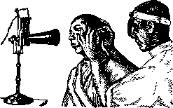
OTOSCOPY, one of the most important methods for examining the ear, in which the external auditory canal, tympanic membrane, and when the latter is destroyed, the middle ear are directly visualized. For otoscopy, it is necessary to have an illuminating instrument. These are divided into instruments that provide direct light (Clar's lamp) and instruments with reflected light. Otologists almost exclusively use the latter instruments. Among them, the frontal reflector is most convenient (see Otorhinolaryngological Instrumentarium). With the help of a headband, the reflector is attached to the head and directed to the right or left eye in such a way that its central opening coincides with the pupil of the eye. One can also use a reflector attached to a handle, which is held in the hand or in the teeth. For prolonged examinations or during surgical procedures, these latter reflectors are not suitable. The source of light for otoscopy can be natural or artificial light. In the first case, one must use diffused light from white clouds, since direct sunlight provides too bright a light and generates significant heat. Natural light has the advantage that it shows a more natural color of the tympanic membrane. For more detailed examination, it is better to use artificial light - kerosene or electric lamps. To give the light source a more uniform surface, frosted light bulbs and special projectors equipped with a system of lenses have been proposed (see Otorhinolaryngological Instrumentarium). The light source is placed to the right and somewhat behind the patient, while the doctor is in front of the patient opposite the light. The light source, the patient's head, and the examiner's head should be at the same height (see figure). Otoscopy should be performed in a darkened room. During otoscopy, the patient's head is turned to the side opposite the ear being examined, approximately 90°, so that the longitudinal axis of the auditory canal coincides with the direction of the light rays from the reflector. With two fingers of one hand, the auricle is pulled upward and backward to straighten the external auditory canal. If the tragus is moved with a finger of the other hand, it is often possible without any special instruments to examine the external auditory canal and even the tympanic membrane. For narrow auditory canals or for more detailed examination, an ear mirror is necessary (mirrors proposed by various authors - see Otorhinolaryngological Instrumentarium). For studying the details of the tympanic membrane, ear loupes and ear microscopes (Voltolini, Trautmann, Veber-Liel, Luscher) and photography of the membrane (Hegener's method) have been proposed. A very valuable instrument in otoscopy is Siegel's pneumatic mirror, with which the degree of mobility of individual parts of the tympanic membrane, scar changes on it, and thickenings can be determined. The ear mirror is inserted into the auditory canal with rotational movements of two fingers of the right hand, inward, approximately 1-11/2 cm. Inserting the funnel into the bony part of the auditory canal is painful and unnecessary, as obstacles are more often encountered in the cartilaginous part of the auditory canal. Obstacles to otoscopy in the external auditory canal include foreign bodies (bugs, flies, cockroaches, sunflower seeds, peas, bones), congenital and acquired pathological changes (exostoses, atresias, cerumen and epidermal plugs, furuncles, diffuse inflammatory processes, polyps, and new growths). In cases of exostoses and incomplete atresias, however, examination of the tympanic membrane is possible with very narrow ear mirrors. Foreign bodies, cerumen, and epidermal plugs must first be removed by washing or with appropriate instruments. Inflammatory processes in many cases require treatment first, and only then is it possible to insert the mirror and examine the tympanic membrane.
Otoscopic picture. 1. The external auditory canal in a normal adult appears as an oval canal about 31/2 cm long, closed at the inner end by the tympanic membrane. In its cartilaginous part, the canal curves forward and downward. At the point where the cartilaginous part transitions into the bony part, a narrowing of the auditory canal (isthmus) is noted. Near the tympanic membrane, another narrowing of the auditory canal is visible due to a bony prominence on the lower wall. The auditory canal is lined with skin, which in the cartilaginous part is equipped with hair follicles, ceruminous glands, and sebaceous glands. Pathological processes change the color of the skin and the shape of the auditory canal. Hyperemia of the skin is often encountered. Narrowing of the auditory canal can be concentric or slit-like (when the upper-posterior wall collapses). 2. Otoscopy of the tympanic membrane - see Tympanic membrane, methods of examination.
a. gelfon.
Related articles
Mentioned in
Cite this page
“Otoscopy.” Soviet Medical Encyclopedia. English translation of Bolshaya Meditsinskaya Entsiklopediya, 1st ed. (Moscow, 1928–1936), ed. N. A. Semashko. https://sovietmedicalencyclopedia.pages.dev/article/otoscopy/