Otolith Apparatus
Historical document, translated for reference. It reflects medical knowledge of the 1920s–30s and is not medical advice.
Summary
The otolith apparatus consists of elevations or spots (maculae staticae) on the walls of the utricle and sacculus in the vestibule of the inner ear. These structures function in detecting linear acceleration and head position in space.
Encyclopedia article (1928–1936)
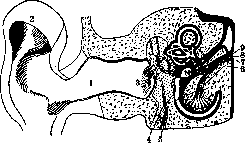
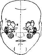
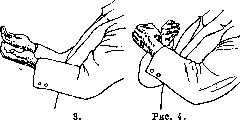
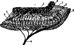
OTOLITH APPARATUS, elevations or spots (maculae staticae) on the walls of the sacs (utriculus and sacculus) of the vestibule of the inner ear (see Internal ear and fig. 1). In humans there are two of them in each ear (O. a. of utriculus and sacculus). The O. a. of utriculus is also called lapillus (stone), the O. a. of sacculus-sagitta (arrow). Their structure is the same, but their shape and position in the skull are different (fig. 2). One can easily imagine the topographical position of utriculus and sacculus in the human cranial cavity by using a mnemonic rule based on a special hand position (fig. 3 and 4). From these figures it is seen that the lapilli (fig. 3) are located horizontally and in the sagittal plane, while the sagittae (fig. 4) are almost vertical and in the frontal plane. These topographical relationships are of cardinal importance for understanding the function and mechanism of examination of the O. a. (see below). Histological structure of the O. a. (fig. 5). In the area of the spot on the membranous wall of sacculus and utriculus, which is lined with bridge-like epithelium, there are supporting cells, hair cells (neuroepithelium connected with endings of the vestibular nerve), gelatinous mass, otolithic plate, and crystals (otolith). The otolithic plate together with the crystals covers the entire surface of the spot and thus determines its shape. The length of the crystals (aragonite - calcium phosphate-carbonate) in humans reaches from 1 to 11 μ, width from 2 to 5 μ. The conglomerate of crystals is held by the otolithic plate, forming a continuous mass. The innervation of the O. a. (fig. 6) is closely connected with the innervation not only of the semicircular canals, but also partly of the cochlea. This connection indicates functional relationships between the semicircular canals and the otolithic system.
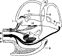
Figure 1. Diagram of the outer, middle, and inner ear (cochlea and labyrinth): 1-poms acusticus ext.; 2-conclia; 3-membrana tympani; 4-tuba Eustachii; 5-m. tensor tympani; 6-aqueductus cochleae; 7-n. cochlearis; 8-porus acusticus int.; 9-n. vestibularis; g-sacculus; u-utriculus.
In humans there are two of them in each ear (O. a. of utriculus and sacculus). The O. a. of utriculus is also called lapillus (stone), the O. a. of sacculus-sagitta (arrow). Their structure is the same, but their shape and position in the skull are different (fig. 2). One can easily imagine the topographical position of utriculus and sacculus in the human cranial cavity by using a mnemonic rule based on a special hand position (fig. 3 and 4). From these figures it is seen that the lapilli (fig. 3) are located horizontally and in the sagittal plane, while the sagittae (fig. 4) are almost vertical and in the frontal plane. These topographical relationships are of cardinal importance for understanding the function and mechanism of examination of the O. a. (see below). Histological structure of the O. a. (fig. 5). In the area of the spot on the membranous wall of sacculus and utriculus, which is lined with bridge-like epithelium, there are supporting cells, hair cells (neuroepithelium connected with endings of the vestibular nerve), gelatinous mass, otolithic plate, and crystals (otolith). The otolithic plate together with the crystals covers the entire surface of the spot and thus determines its shape. The length of the crystals (aragonite - calcium phosphate-carbonate) in humans reaches from 1 to 11 μ, width from 2 to 5 μ. The conglomerate of crystals is held by the otolithic plate, forming a continuous mass. The innervation of the O. a. (fig. 6) is closely connected with the innervation not only of the semicircular canals, but also partly of the cochlea. This connection indicates functional relationships between the semicircular canals and the otolithic system.
The conception of the function of the O. a. is based on an analysis of its anat.-hist. structure, its topographical position in the skull, and the results of experiments performed on them. Seväll (1883) was the first to perform otolithotomy in fish and established a disturbance of equilibrium. Thus, simple opening of the utriculus in rays does not cause functional changes, but after removal of the otoliths a tendency to describe circles toward the operated side and to sink to the bottom is observed; after bilateral removal of the otoliths the animal sinks to the bottom, sometimes somersaulting in the vertical plane. The phenomena are even more markedly expressed after damage to the bottom of the vestibule with the tip of a scalpel. That the above-mentioned phenomena are caused exclusively by damage to the O. a., and not to the semicircular canals, is evident from the fact that isolated damage to the latter is not accompanied in rays by a disturbance of equilibrium. Similar results were noted in experiments with sharks. The circumstance that in animals which by nature lack the possibility of free movement there are no otoliths leads one to ascribe to the otoliths a statico-kinetic function. A coherent theory of the static function of the O. a. was first published by Breuer (Breuer, 1891); with minor corrections made to it by the Utrecht school (Magnus, de Kleijn, Quix), it remains to this day the only one that has gained general recognition.
Figure 2. Position of otoliths in the skull: 1-canalis ant.; 2-canalis post.; 3-utriculus (lapillus); 4-sacculus (sagitta).
Figure.
Figure 5. Diagram of the otolith organ: 1-otolith crystals; 2-ciliary cells; 3-supporting cells; 4-vestibular nerve; 5-membranous wall; 6-filaments; 7-gelatinous mass; 8-otolithic plate.
According to Breuer, the otoliths are organs serving for the perception of movement in a straight line and of the position of the head in space during rest and movement. Breuer was led to this concept by the following line of thought: the otoliths, floating in a liquid specifically lighter than themselves, constantly tend to sink, thereby maintaining in the terminal nerve apparatus continuous irritation transmitted to the brain. Pressing on the cell hairs by inertia during movement in a straight line or else shifting due to the development of centrifugal force during rotational movements or pressing on the hair cells by virtue of their weight at rest, the otoliths in us cause the sensation of movement and the sensation of the resting position of the head and by pressure downward make it possible to determine the vertical direction. Breuer's assumption about the movement of the otoliths was confirmed by the experiments of Kubo (Kubo, 1906-1908) by placing fish in various positions, such as: head up, down, belly down, up. The same author also established deviation of the eyes in case of damage to the otoliths. The disturbance of the sensation of verticality during rotation, which according to Breuer's thought is caused by the displacement of the otoliths, was established in humans by Kreidl (Kreidl, 1893). A more thorough investigation of animals after destruction or damage to the O. a. established (Frohlich, 1904) hypotonia, increased reflexes, weakening of respiration, and disappearance of compensatory wheel-like eye movements (see below). These disturbances for the most part constitute the elements of which the complex phenomenon-disturbance of statics-is composed. The mechanism of work of these individual elements and their manifestation in the form of reflexes in mammals has been studied by Magnus and de Kleijn on decerebrated animals (see Decerebration, decerebrational rigidity). In such a decerebrated animal a state of 'rigidity' is observed, expressing itself in tonic contraction of those muscles whose function consists in maintaining + 90° + 135° -
Figure 6. Innervation of the labyrinth and cochlea: 1-ampulla ant.; 2-n. vestibularis; 3-anastomosis; 4-n. cochlearis; 5-ductus cochlearis; 6-sacculus; 7-macula sacculi; 8-ampulla post.; 9-crus communis; 10-sacculus endolymphaticus; 11-ampulla horizontalis; 12-macula utriculi.
q
gland.
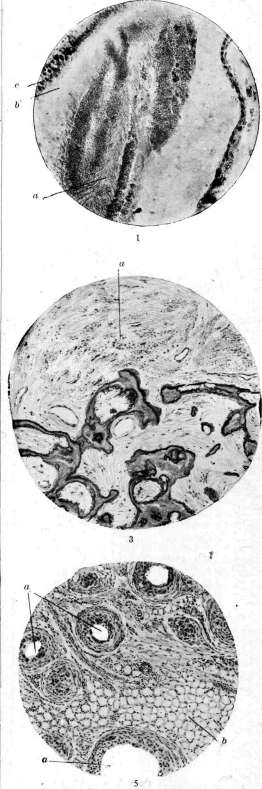
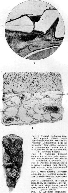
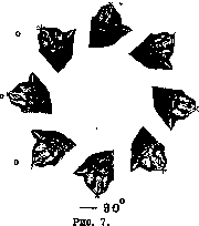
o - ^5 to counteract the force of gravity (extensors of the limbs, raising the neck, extensors of the trunk, raising the tail in animals, and closers of the lower jaw). It turns out that the tone of the extensors is influenced by various positions of the animal's head. However, not only the position of the head in space is important, but also the position of the head relative to the trunk. Therefore, it is necessary to distinguish between otolith (labyrinthine) and neck reflexes (see Magnus-Klein reflexes). By causing decerebration rigidity in a rabbit or cat and then placing the head vertex upward (180°) and rotating the animal around the bitemporal axis along a circular line, it can be established that the limbs assume maximum extensor tone when the position of the utricle is between 0° and +45° [the otoliths of the utricle (lapilli) hang down], and minimum tone between 180° and 135° (in this case the otoliths of the utricle press on the underlying macula statica) (Fig. 7). This observation led Magnus and de Klein to recognize that the irritating factor in the mechanism of excitation of the O. a. under physiological conditions is not pressure, as Breuer once assumed and Quix currently asserts (see below), but hanging. Quix, based on the above-described histological structure and topographical location of the O. a., conceives of the function of the O. a. as follows: if the otoliths approach the spot, i.e., they press on the gelatinous mass, then this pressure is transmitted to the hairs of the neuroepithelium (see histological structure); they are irritated, the process is transmitted to the sensory cells and the nerve fibers connected with them of the n. vestibularis, and then proceeds to the centers of the brain stem. If the otoliths move away from the spot, i.e., hang down, they do not cause physiological irritation. Physiological excitation is caused only by such displacement of the otoliths in which a change in pressure occurs. The above-mentioned Magnus-Klein reflex, elicited at a certain position of the head, was named by Magnus as the position reflex (Lagereflex). That it is actually elicited from the O. a. of the utricle is indicated by the fact that when the animal is centrifuged, in which the otolith of the utricle is torn away (see separate table, Fig. 2), the said reflex on the limbs disappears. All position reflexes originate from the O. apparatus and affect not only the limbs but also the eyes and the neck. The peculiarity of otolith reflexes is that they are tonic, i.e., they last as long as a particular position of the labyrinth lasts, whereas reflexes elicited from the semicircular canals appear to be short-term, rapidly passing. That from the utricle tonic reflexes on the neck are also elicited follows from the analysis of phenomena in rabbits after unilateral removal of the labyrinth; after this, a turning of the head toward the damaged labyrinth is observed. This turning is caused by the fact that the muscles turning the neck toward the intact labyrinth are deprived of their usual tone (due to the loss of labyrinth function), while the muscles of the other side of the neck, which have retained normal tone and are deprived of antagonists, contract maximally. Consequently each utricle is functionally connected with the muscles of the opposite side of the neck. The tonic labyrinthine reflexes (mainly on the forelimbs), as well as their dependence on pressure and hanging of the otoliths, are clearly visible in the following experiment (Fig. 8). When the head is bent forward
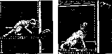
Figure 8.
by 30°, the otoliths produce maximum pressure, resulting in flexion mainly of the forelimbs, while when the head is raised and thrown back by 45°, the otoliths hang down and as a result - extension of the limbs. Similar phenomena are observed in the so-called elevator reaction, which consists in that when the elevator descends, due to the lag of the otoliths, the animal rises (extension of the limbs), while when ascending, the otoliths press and the animal lowers (flexion of the limbs). In humans, this reaction manifests as the sensation of downward movement after stopping an elevator that was moving upward, and the sensation of upward movement after descending in an elevator. This phenomenon is based primarily on the mechanism of pressure and lag of the otoliths. Deaf-mutes, deprived of functioning O. a., do not experience these sensations. However, the participation of the endolymph flow mechanism in the semicircular canals in this reaction is not excluded, which should be assumed from the corresponding experiment (on models of semicircular canals) by Magnus and de Klein. Thus, the semicircular canals and the O. a. react to angular and linear acceleration, but the degree of participation of these organs in the corresponding irritations is such that the semicircular canals are more sensitive to angular acceleration, while the O. a. is more sensitive to linear acceleration. From the analysis of the elevator reaction, it follows that under physiological conditions the irritating factor is pressure and hanging. However, in animals and humans in the position of the head that is normal for them (for example, vertical in humans), the otoliths are located in such a way that they press on the underlying macula. Since in this position animals and humans maintain the correct sensation of the position of the head in space, it is natural to consider this mechanism of irritation specific, and not the one (hanging) that acts in unusual positions of the head. This argumentation is disputed by Quix (see above) against the theory of Magnus and de Klein (see above). Utricular tonic reflexes on the neck in humans are observed only in pathological cases, while on the limbs in a healthy person, 'according to
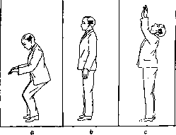
Figure 9. Utricular reflexes on the limbs and eyes: a-bending the head forward causes maximum pressure of the lapilli on the macula, and as a result, flexors are activated in the limbs and trunk, and in the eyes-the superior rectus muscle; b-normal position of the utricle in the vertical position of the head; c-bending the head backward causes maximum hanging of the lapilli on the macula, and as a result, extensors are activated in the limbs and trunk, and in the eyes-inferior rectus muscle.
Quix, can be reproduced (as seen in Fig. 9), if a change in the position of the otoliths of the utricle (lapilli) is caused. This change occurs as the head begins to move from the median position in the sagittal plane (see above-topographical location of lapilli), i.e., forward and backward. The change in the position of the otoliths of the sacculus (sagittae) occurs as the head begins to move from the median position in the frontal plane (see above-topographical location of sagittae), i.e., when tilting to the side. Only then does the function of the sagittae manifest itself. Saccular reflexes in humans, according to Quix, are shown in Fig. 10. In animals, from the side of the otoliths of the sacculus, tonic reflexes can be easily elicited, named by Magnus as restorative, or postural (Stellreflexe). The purpose of these reflexes is to return the animal to the normal position after it has been removed from it. These reflexes include reflexes on the head and
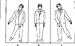
Figure 10. Saccular reflexes on the limbs and eyes: a-right sacculus when the head is tilted to the left shoulder (maximum pressure) causes abduction of the right limbs, adduction of the left ones, and counter-rotation of the eyes to the right; b-normal position of the sacculus in the vertical position of the head; c-left sacculus when the head is tilted to the right shoulder causes abduction of the left limbs, adduction of the right ones, and counter-rotation of the eyes to the left.
eyes; their investigation in animals is carried out as follows: if the animal is held by the pelvis with its head upward, then regardless of the lateral position the animal is in, its head will always assume a vertical position. In the lateral position, the sacculus is irritated, which contributes to the turning of the head to the normal position (in this position, one sacculus, located below, hangs down, while the other, located above, presses on the neuroepithelium). Damage to these otoliths causes the disappearance of the head reflex (see separate table, fig. 1). It has been established that each position of the head in space corresponds to a certain state of contraction of the eye muscles, i.e. a certain position of the eyes, and the position of the eyes remains constant as long as the head maintains its position. The biological significance of this reflex consists in the fact that the turning of the eyes compensates for the deviation of the field of vision caused by the turning of the head, which is why this deviation of the eyes is called compensatory. Since the deviation of the eyes is always directed in the opposite direction to the deviation of the head, the entire phenomenon is called compensatory counter-rotation of the eyes. Compensatory deviation of the eyes can be easily observed in humans and animals; it is especially easily manifested in animals with lateral eye placement (rabbit, guinea pig). A distinction is made between wheel-shaped and vertical counter-rotation. The first is observed when the head is rotated around the bitemporal axis in the sagittal plane and is caused by irritation of the otoliths of the utriculi, which act on the mm. obliq. super, et obliq. infer (fig. 11). The second is detected when the head is rotated around the length of the body in the frontal plane and is caused by irritation of the otoliths of the sacculus, which act on the mm. rectus super, et rectus infer (fig. 12). By the method of decerebration, it has been established that the centers of these reflex arcs are located in the brain stem, starting from the upper cervical segments to the anterior part of the quadrigeminal. Within this area, there are centers located in three groups: 1) for otolith reflexes on the limbs and neck - in the area posterior to the point of entry of the vestibular nerve; 2) for the otolith reflex on the eyes - between the area of entry of the acoustic nerve and the nuclei of the oculomotor nerves; 3) centers for positional reflexes on the head - in the midbrain area. The physiological significance of the latter lies in obtaining an idea of the true position of the head in space; centers of reflexes on the eyes - to maintain optical space; centers of reflexes on muscles - to maintain balance. In light of these experiments, it is easy to understand a number of facts. For example, the assessment of the degree of head tilt relative to the vertical line becomes less accurate as the head is tilted back more and more. A tilt of 75° is taken as the horizontal position, i.e. 90°. In this case, there is increased irritation of the otoliths of the utriculi. A similar mechanism of irritation or damage to the otolith apparatus explains the disturbance of vertical perception by pilots during flights, as well as the appearance of a number of reflexes that together constitute the symptom-complex of the disease of pilots. It is known that when the head is in a certain position (for example, when the head moves along the bitemporal axis), the sense of orientation in space will be completely absent, and the otoliths in relation to the maculae in this case are in a state of hanging; This point in space is called the blind spot of the static organ (Quix). Opposite this blind spot in space there is an area where, when the head is in this position, the otoliths exert pressure and spatial orientation is maintained - the yellow spot of the static organ. Altered excitability of the O. apparatus in the sense of increase or decrease causes a deceptive sensation of the relationship between the individual and surrounding space, and as a result - dizziness, sometimes accompanied by pallor of the face, vomiting, nausea, sweating as a result of reflex irritation of the vagus and sympathetic nervous systems. The mechanism of the appearance of this (otolith) dizziness is analogous to the mechanism of the vestibular syndrome observed in sea sickness. The dependence of the appearance of this disease on abnormal irritations of the otolith apparatus is established by analyzing the movement of the ship during sea rolling (pitching - movement of the ship from top to bottom and bottom to top - action of the otoliths of the utriculus, rolling - action of the otoliths of the sacculus), as well as by the fact that deaf-mutes, devoid of functioning labyrinths, do not suffer from sea sickness. The data obtained experimentally on animals and noted in observations form the basis of the clinic of diseases of the O. a. However, these data obtained on animals cannot be unconditionally transferred to humans. Thus, in all cases of isolated disease of the otolith apparatus in humans described in the literature, there is mention not of compensatory eye movements caused by a certain position of the head, but of nystagmus, i.e. what we usually consider to be caused by the semicircular canals, and only the circumstance that this reaction is tonic and prolonged makes us consider it otolithic. This is explained by the fact that in lesions of the otolith apparatus in humans, it is necessary to assume the appearance not only of compensatory eye movements but also of nystagmus, for which there are prerequisites in the innervation of the labyrinth (see above), which indicate a 'functional interaction between the semicircular canals and the otolith apparatus. The nature of this interaction has not been finally clarified. It is assumed that unusual irritations of the otolith apparatus are sometimes reflexively transmitted to the semicircular canals, as a result of which the so-called 'otolithic nystagmus' (Khilov) is obtained. Symptoms of disease of the otolith apparatus: 1) dizziness in a certain position of the head (Schwindellage) with or without the appearance of nystagmus (Lagenystagmus); 2) overshooting when the head is in a prone, lateral position; 3) disturbance of counter-rotation of the eyes. - Research methodology. 1. After establishing from the anamnesis complaints of dizziness, the patient is laid in turn on all four sides (on the back, on the abdomen, on the right and left side).
Figure 12. Vertical deviation of the eyes when lying on the side.
Figure 11. Wheel-shaped counter-rotation of the eyes in a rabbit when rotating around the bitemporal axis.
centers: 1) for otolith reflexes on the limbs and neck - in the area posterior to the point of entry of the vestibular nerve; 2) for the otolith reflex on the eyes - between the area of entry of the acoustic nerve and the nuclei of the oculomotor nerves; 3) centers for positional reflexes on the head - in the midbrain area. The physiological significance of the latter lies in obtaining an idea of the true position of the head in space; centers of reflexes on the eyes - to maintain optical space; centers of reflexes on muscles - to maintain balance. In light of these experiments, it is easy to understand a number of facts. For example, the assessment of the degree of head tilt relative to the vertical line becomes less accurate as the head is tilted back more and more. A tilt of 75° is taken as the horizontal position, i.e. 90°. In this case, there is increased irritation of the otoliths of the utriculi. A similar mechanism of irritation or damage to the otolith apparatus explains the disturbance of vertical perception by pilots during flights, as well as the appearance of a number of reflexes that together constitute the symptom-complex of the disease of pilots. It is known that when the head is in a certain position (for example, when the head moves along the bitemporal axis), the sense of orientation in space will be completely absent, and the otoliths in relation to the maculae in this case are in a state of hanging; This point in space is called the blind spot of the static organ (Quix). Opposite this blind spot in space there is an area where, when the head is in this position, the otoliths exert pressure and spatial orientation is maintained - the yellow spot of the static organ. Altered excitability of the O. apparatus in the sense of increase or decrease causes a deceptive sensation of the relationship between the individual and surrounding space, and as a result - dizziness, sometimes accompanied by pallor of the face, vomiting, nausea, sweating as a result of reflex irritation of the vagus and sympathetic nervous systems. The mechanism of the appearance of this (otolith) dizziness is analogous to the mechanism of the vestibular syndrome observed in sea sickness. The dependence of the appearance of this disease on abnormal irritations of the otolith apparatus is established by analyzing the movement of the ship during sea rolling (pitching - movement of the ship from top to bottom and bottom to top - action of the otoliths of the utriculus, rolling - action of the otoliths of the sacculus), as well as by the fact that deaf-mutes, devoid of functioning labyrinths, do not suffer from sea sickness. The data obtained experimentally on animals and noted in observations form the basis of the clinic of diseases of the O. a. However, these data obtained on animals cannot be unconditionally transferred to humans. Thus, in all cases of isolated disease of the otolith apparatus in humans described in the literature, there is mention not of compensatory eye movements caused by a certain position of the head, but of nystagmus, i.e. what we usually consider to be caused by the semicircular canals, and only the circumstance that this reaction is tonic and prolonged makes us consider it otolithic. This is explained by the fact that in lesions of the otolith apparatus in humans, it is necessary to assume the appearance not only of compensatory eye movements but also of nystagmus, for which there are prerequisites in the innervation of the labyrinth (see above), which indicate a 'functional interaction between the semicircular canals and the otolith apparatus. The nature of this interaction has not been finally clarified. It is assumed that unusual irritations of the otolith apparatus are sometimes reflexively transmitted to the semicircular canals, as a result of which the so-called 'otolithic nystagmus' (Khilov) is obtained. Symptoms of disease of the otolith apparatus: 1) dizziness in a certain position of the head (Schwindellage) with or without the appearance of nystagmus (Lagenystagmus); 2) overshooting when the head is in a prone, lateral position; 3) disturbance of counter-rotation of the eyes. - Research methodology. 1. After establishing from the anamnesis complaints of dizziness, the patient is laid in turn on all four sides (on the back, on the abdomen, on the right and left side).
Figure 1. Investigation of nystagmus and dizziness in a certain position on the vestibular table of Grahe.
pensatory eye movements, but also nystagma, for which there are prerequisites in the innervation of the labyrinth (see above), which indicate 'functional interaction between the semicircular canals and the otolith apparatus. The nature of this interaction has not been finally clarified. It is assumed that unusual irritations of the otolith apparatus are sometimes reflexively transmitted to the semicircular canals, as a result of which the so-called 'otolithic nystagmus' (Khilov) is obtained. Symptoms of disease of the otolith apparatus: 1) dizziness in a certain position of the head (Schwindellage) with or without the appearance of nystagmus (Lagenystagmus); 2) overshooting when the head is in a prone, lateral position; 3) disturbance of counter-rotation of the eyes. - Research methodology. 1. After establishing from the anamnesis complaints of dizziness, the patient is laid in turn on all four sides (on the back, on the abdomen, on the right and left side).
Figure 14. Figure 15.

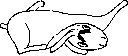
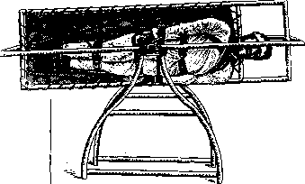
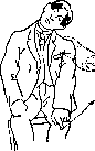
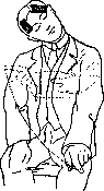
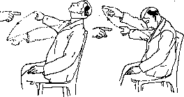
Figure 16. Figure 17. side) and when lying in each of these positions, they wait 10-15 seconds, during which the patient is examined for spontaneous nystagmus, by asking him to look straight ahead, to the sides, up and down (rotary nystagmus is more often noted), and at the same time the patient is asked whether he is experiencing dizziness (figure 13). 2. Pathology of otolith reflexes in the limbs in lesions of the O. a. is revealed in the form of overshooting when performing the pointing test. - The pointing test: the patient sits opposite the seated doctor. The doctor asks him to extend his arm at the shoulder joint in the medial direction and with closed eyes to make vertical movements with the index finger from his knee to meet the doctor's outstretched index finger. Then this test is repeated when the patient tilts his head to the right and left shoulder (fig. 14 and 15). Next, a similar test is performed, but the finger moves in the horizontal plane from outside to inside, both with the head in a straight position and when tilting the head forward and backward (fig. 16 and 17). A normal person with closed eyes always hits the examiner's finger. Tilting the head forward achieves maximum pressure on the otoliths (lapilli) of the utricle, causing flexion of the arms, and tilting backward achieves maximum hanging, causing extension (thus the movements of the arms should occur in the sagittal plane). Impairment of the function of lapilli when they are affected is manifested in the pointing test performed in the horizontal direction with the head in the noted position, by the fact that the subject's finger will miss in the vertical direction and in the sagittal plane (the plane of location of lapilli), i.e. upward or downward. From the analysis of clinical material (Quix) it follows that tilting the head to the shoulder causes irritation of the otolith of the sacculus (sagitta) of the opposite side, causing abduction of the limb on the side of the irritated otolith and adduction of the opposite side (thus the movements of the arms should occur in the frontal plane). Impairment of the function of sagittae when they are affected is manifested in the pointing test performed in the vertical direction with the head in lateral positions, by the fact that the subject's finger will miss in the horizontal direction and in the frontal plane (the plane of location of sagittae), i.e. outward and inward (fig. 14 and 15). The diagnostic value of this test is significantly inferior to the more objective method - examination for counter-rotation of the eyes. 3) Examination for counter-rotation of the eyes is performed with the help of a special apparatus (fig. 18 and 19) as follows. A telescope (1) is set on the iris, magnifying it 8 times. The movement of the eyes is measured by the displacement of the iris when tilting the head. For this purpose, it is necessary that the subject's pupil is at rest, which is achieved by instilling into the corresponding eye 20 minutes before the examination two drops of a 1% solution of pilocarpine. As a result, the pupil is maximally constricted, does not react to light, and the pattern of the iris appears more clearly. A more clearly expressed stripe is selected on the iris, serving as a landmark for measurement. In the telescope, there is a movable thread which is set parallel to the said landmark stripe on the iris, and its position is marked on a board with divisions connected to the eyepiece thread. To set the telescope on the iris, it is necessary to fix the patient's head and eye in relation to the telescope, which is achieved by fixing with the teeth a board with a stent (2), attached to the rod on which the telescope rests. The following authors (according to Kompaneyets) at different tilts of the head obtained different angles of counter-rotation in normal people. Authors Tilt of the head by 30° 35° 45 ° 60° Houben u. Struycken . 6° 2°-5° 10° 8° In pathological cases with reduced function, significantly smaller figures are obtained. This is especially manifested in the difference between tilting the head to both sides, e.g. in a disease of the left O. a. (sacculus) in the sense of reduced function, when tilting the head to the right, counter-rotation to the left is 10°, when tilting the head to the left, counter-rotation to the right is 2°. This method of examination is exceptionally valuable in cases with dizziness phenomena with normal reactions from the semicircular canals. To determine the degree of excitability of the vestibular system in general and the otolith system in particular, for use in pre-flight selection of pilots (a method little suitable for the clinic of diseases of the otolith system), the following method of examination is used (Voyachek-Khilov, fig. 20): the subject sits in a Barany chair, closes his eyes, bends his head and trunk downward, approximately at an angle of 90°. In this position of the subject, he is first rotated to the right at a speed of one revolution in 2 seconds for 10 seconds, i.e. 5 revolutions are made; then the chair is stopped and, without changing the subject's position, they wait 5 seconds, after which the subject is asked to straighten up. At the moment of changing position, a reaction occurs, expressed in tilting of the body and head to one side or another (weak, moderate or strong). After a few minutes, the test is repeated, rotating the subject in the opposite direction. The reaction of overshooting and falling is conditionally divided into 3 degrees: from 0° to 10° the reaction is considered weak, from 10° to 20° - moderate, and from 20° to 45° and more - strong.
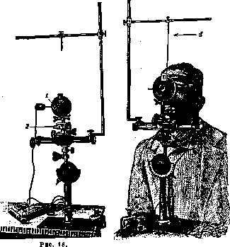
Fig. 19
corresponding eye 20 minutes before the examination two drops of a 1% solution of pilocarpine. As a result, the pupil is maximally constricted, does not react to light, and the pattern of the iris appears more clearly. A more clearly expressed stripe is selected on the iris, serving as a landmark for measurement. In the telescope, there is a movable thread which is set parallel to the said landmark stripe on the iris, and its position is marked on a board with divisions connected to the eyepiece thread. To set the telescope on the iris, it is necessary to fix the patient's head and eye in relation to the telescope, which is achieved by fixing with the teeth a board with a stent (2), attached to the rod on which the telescope rests. The following authors (according to Kompaneyets) at different tilts of the head obtained different angles of counter-rotation in normal people. Authors Tilt of the head by 30° 35° 45 ° 60° Houben u. Struycken . 6° 2°-5° 10° 8° In pathological cases with reduced function, significantly smaller figures are obtained. This is especially manifested in the difference between tilting the head to both sides, e.g. in a disease of the left O. a. (sacculus) in the sense of reduced function, when tilting the head to the right, counter-rotation to the left is 10°, when tilting the head to the left, counter-rotation to the right is 2°. This method of examination is exceptionally valuable in cases with dizziness phenomena with normal reactions from the semicircular canals. To determine the degree of excitability of the vestibular system in general and the otolith system in particular, for use in pre-flight selection of pilots (a method little suitable for the clinic of diseases of the otolith system), the following method of examination is used (Voyachek-Khilov, fig. 20): the subject sits in a Barany chair, closes his eyes, bends his head and trunk downward, approximately at an angle of 90°. In this position of the subject, he is first rotated to the right at a speed of one revolution in 2 seconds for 10 seconds, i.e. 5 revolutions are made; then the chair is stopped and, without changing the subject's position, they wait 5 seconds, after which the subject is asked to straighten up. At the moment of changing position, a reaction occurs, expressed in tilting of the body and head to one side or another (weak, moderate or strong). After a few minutes, the test is repeated, rotating the subject in the opposite direction. The reaction of overshooting and falling is conditionally divided into 3 degrees: from 0° to 10° the reaction is considered weak, from 10° to 20° - moderate, and from 20° to 45° and more - strong. Etiology. The causes of disease of the O. a. appear to be quite diverse: acute, chronic inflammatory and non-inflammatory diseases of the middle and inner ear, infectious, traumatic and intoxication neuritis of the otolith nerves, as well as vascular, infectious, tumor-like diseases of the brain in the area of location of the otolith pathways (medulla oblongata and pons).- Treatment should be carried out in two directions: 1) depending on the clarified etiology, therefore causal, and 2) symptomatic, i.e. along the line of action on the vagosympathetic component of the vestibular syndrome. At the moment of an attack: subcutaneous atropine (Sol. Atropini sulfurici 1 : 1,000.0 pro dosi 1.0) (if there is no therapeutic effect, subcutaneous Sol. adrenalini 1 : 1,000 from 0.3 to 0.5 every two days), hot water bottle
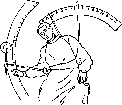
Figure 20.
to the legs, cold compresses (ice) on the head, large doses of bromine internally (Sol. Natr. bromati 12.0 : 200.0 - 1 tablespoon three times a day). As the intensity of the attacks decreases, the dose of bromine is reduced to 1.0 per day and in the post-attack period for 5-6 days, bromine therapy is continued in combination with taking baths at 35° every other day.
Related articles
Mentioned in
Cite this page
“Otolith Apparatus.” Soviet Medical Encyclopedia. English translation of Bolshaya Meditsinskaya Entsiklopediya, 1st ed. (Moscow, 1928–1936), ed. N. A. Semashko. https://sovietmedicalencyclopedia.pages.dev/article/otolith-apparatus/