Hair
Historical document, translated for reference. It reflects medical knowledge of the 1920s–30s and is not medical advice.
Summary
This article from the 1928–1936 Great Medical Encyclopedia details the anatomical structure, microscopic composition, growth, and distribution of human hair, covering its various types, follicle layers, and associated physiological structures.
Encyclopedia article (1928–1936)
HAIR represents a part of the glandular epithelium of the skin, occurring over its entire surface except for the skin of the palms and soles, the palmar and plantar surfaces of the fingers and toes, the dorsal surfaces of the third phalanges of the fingers and toes, the nipple, the glans penis, the inner leaflet of the prepuce, the labia minora, the inner surface of the labia majora in women, and the red border of the lips. In hair, one distinguishes a shaft protruding above the skin and a root immersed in it, ending in a thickening called the bulb. Hairs also occur in the form of thin (from 5 µ) light vellus hairs (lanugo). Hairs sit obliquely to the skin surface, and hairs with the same inclination are arranged along specific lines, generally coinciding with the lines of skin cleavage (Langer's lines). Diverging, these lines form in places so-called "whirls," from which hairs diverge in spirally curved rays in various directions. Hairs in humans are mostly found in groups of 3 together, and in the distribution of hairs of each group, as well as in the interrelation of these groups with each other, a regular arrangement can also be noticed (on the trunk, generally transverse). By appearance, one can distinguish (aside from vellus hairs) longer and thicker (50-200 µ) hairs: mostly curly sexual hairs, bristle-like eyebrows and eyelashes, and the longest hairs of the head. The number of hairs per 1 sq. cm ranges from 320 (crown) to 18 (back of the hands). The total number of hairs on the scalp ranges from 80,000 (in redheads) to 140,000 (in blonds). Depending on race and partly individuality, the properties of hair in shape, color, grouping, etc., vary significantly. The change in color of grey hairs is caused mainly by the appearance of air bubbles in the cuticle and medulla of the hair. Under the microscope, one can distinguish in the hair shaft: 1) cortical substance, 2) medullary substance, and 3) cuticle (see Figure 1). The medullary substance is absent in vellus and generally thinner hairs. The cortical substance consists of elongated horny cells with pigment granules and air bubbles (especially in grey hairs). The medullary substance is formed by irregularly rounded cells, partly with small teeth and processes on the surface. The hair is covered by a cuticle consisting of keratinized flat cells, layered shingle-like from top to bottom, i.e., with the free edge pointing upward. The hair sits in a hair follicle, which consists of a connective tissue sheath and two epithelial sheaths—outer and inner. The connective tissue sheath is formed by a dense layer of fine fibrous connective tissue with fibers arranged transversely (on the inside) and longitudinally (on the outside), and by a glassy membrane (see Fig. 2). Structural diagram of a hair follicle: a—follicle funnel; b—follicle neck; c—hair bulb; d—outer epithelial sheath of the hair; e—inner epithelial sheath; f—follicle cuticle; g—hair cuticle; h—cortical substance; i—medullary substance; k—bulb epithelium; m—fatty tissue; n—musculus arrector pili; o—hair papilla; p—sebaceous gland (after Ormsby).
The glassy membrane of hyaline-like structure is smooth on the outside, while on the inner side it is covered with transverse ridges. Gradually thinning, it extends deep down to the bottom of the pouch, where the hair papilla sits—a cone-shaped or mushroom-
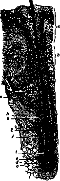
shaped formation consisting of a dense plexus of fine connective tissue and elastic fibers, and rich in blood vessels and nerves. It occupies a notch in the lower part of the hair bulb and merges with the dermis at the base. The first outer epithelial sheath represents the direct continuation of the Malpighian layer of the epidermis and has a structure analogous to it. In the inner epithelial sheath, three layers are distinguished: 1) Henle's layer, 2) Huxley's layer, and 3) the hair cuticle. Henle's layer consists of a single row of keratinized cells of irregular shape. Huxley's layer is formed by 2-3 rows of also keratinized polygonal cells with processes penetrating outward in places between the cells of Henle's layer. The sheath cuticle has the same structure as the hair cuticle, but its plates are layered (shingle-like) with the free edge pointing downward. At the site where the sebaceous glands flow in, the inner epithelial sheath and hair cuticle thin out and disappear. Hair growth (on average 1 cm per month) and the growth of the inner epithelial sheath occur due to the multiplication of epithelial cells covering the hair papilla (matrix), with the cells of the cortical substance keratinizing, apparently without forming keratohyalin granules beforehand. In the inner epithelial sheath, the cells of Henle's layer keratinize earliest, where the grains of so-called trichohyalin appear below the level of the equator of the papilla, in Huxley's layer—higher, and in the cuticle even higher, at the level where the cells of Huxley's layer already keratinize. The new formation of pigment is apparently connected with the life activity of pigmented stellate cells (melanoblasts) found among the epithelium of the bulb. Each papilla is accompanied by several sebaceous glands and bundles of smooth muscle tissue in the form of the arrector pili muscle. These muscles usually have the appearance of a lanceolate plate, which is attached by its narrowed base to the connect
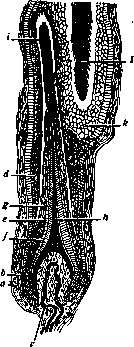
Figure 2. Hair cycle; a—connective tissue sheath; b—glassy membrane; c—hair papilla; d—outer epithelial sheath of the hair; e—inner epithelial sheath; f—sheath cuticle; g—hair cuticle; h, i—young hair; k—remnants of the outer sheath of the falling hair; l—Henle's club (after Ebner).
ive tissue sheath.
to the connective-tissue sheath below the point of entry of the sebaceous glands, and above, narrowing somewhat again, weaves into the tissue of the papillary layer. This muscle, by contracting, facilitates the emptying of the sebaceous glands, places the hair more vertically and protrudes it upwards, simultaneously drawing the skin inward at the place of its attachment (hence cutis anserina). Blood vessels supplying the hair papilla branch off from the deep vascular network (a twig into each papilla), while the walls of the hair follicle are nourished from the superficial arterial network. Hairs are very rich in nerves, which on the surface of the glassy membrane break down into terminal twigs. During the shedding of hair, the reproduction of cells covering the papilla ceases; the cells of the bulb keratinize, and it becomes solid, with brush-like protruding, keratinized cells of the cortical layer—Henle's bulb (see Figure 2). This bulb, having separated from the papilla due to the pressure of the elastic walls of the hair follicle, gradually rises upwards, lingers for a more or less long time near the neck of the follicle, and then falls out. Along with the hair, the inner epithelial sheath also rises upwards, while the papilla remains connected to it by a thin cellular strand. Further, the entire follicle shortens, and at the same time the hair papilla rises somewhat, entailing the connective-tissue sheath, which is as it were turned inward. The very process of hair regeneration begins with the proliferation of cells of the thickened part of the epithelial sheath at the site of attachment of m. arrector pili (the so-called Wulst). These cells, clothing the papilla, form a new matrix and bulb. As the new hair grows, the papilla returns downwards to its former place. Usually, among the hairs on the head, 25% (and more) dead hairs are found, which indicates a relatively frequent shedding of them. Hairs on the head remain on average for 2-4 years, while long ones (1 m and more) must have an age of up to 10 and more years. The lifespan of vellus hair is considered to be, on average, 200-300 days. Hair germs appear for the first time at the beginning of the 4th month of intra-uterine life. The grown hairs usually fall out even during intra-uterine life and are later replaced by new ones. Hair growth is under the influence of the endocrine-vegetative apparatus (see Hairiness), N. Chernogubov. Hair in forensic medical relations. Hairs found in the hands and on the surface of a corpse, on the weapons or clothing of a suspect, can serve as very strong evidence in exposing a criminal, which is why they have long attracted the attention of forensic physicians and are, in appropriate cases, the subject of detailed research. Regarding the order of forensic medical research of hair, it must first be noted that the collection of hair for research should be carried out with great caution, since sometimes hairs can scatter even with a slight movement of air. First, the hairs are examined with the unaided eye, determining their color, length, softness, fineness, curliness, and cleanliness; if it concerns exhumed corpses, then those changes that occurred from the corpse's stay in the ground are noted. After this, the hairs are examined under a microscope. During microscopic examination, the following questions are resolved: 1. Whether the hair belongs to a human or an animal. This is comparatively easily recognized by its structure consisting of three parts—the cuticle (cuticula), the cortical substance, and the medulla (see Figure 3). The cuticle consists of very thin, non-nucleated keratinized scales arranged in a tile-like manner, with their uncovered teeth directed toward the free end of the hair; by this feature, it is easy to distinguish the peripheral end from the central one. In humans, the cuticle consists of thin scales closely adjacent to each other, due to which the hair has a smooth surface or one with fine, barely noticeable serration; in contrast to this, in animals the cuticle is formed by larger cells, clearly protruding, with significantly lagging scale tips, which gives the hair a serrated or saw-like appearance, as in the badger, bat, and others. The cortical layer of human hair consists of longitudinally elongated, tightly connected keratinized cells that give the hair longitudinal striation, and constitutes its main mass and often the sole one; conversely, in animals this layer is very thin, often in the form of a small strip. Upon treatment with sulfuric acid or caustic alkali, the cortical layer can be split into very long ribbon-like "hair fibers," which upon further treatment break down into cortical fibrils and rod-like nuclei. In the cortical layer of colored hairs, pigment is always present—either in a dissolved state, uniformly coloring the cells, or in the form of grains of various colors; these grains are distributed either uniformly or accumulate in the peripheral part or in the central one. Upon treatment with chlorine water or hydrogen peroxide, the pigment is discolored, first acquiring a golden-fair color. The medulla of human hair occupies about V" of its diameter, more often less, and is far from constant: in vellus hairs it is always absent, in thin ones frequently; it contains air, which is why in transmitted light it appears black, in reflected light—in the form of a white strip of uneven thickness, repeatedly interrupted in places. Upon treatment of human hair with caustic alkali or concentrated nitric acid, the medulla can be split into small flattened longitudinal cells containing pigment granules. In animal hairs, the medulla occupies the greater part of the thickness and consists of sharply expressed cells, either round-oval or polygonal; some contain air, which gives the medulla a variegated

Figure 3. 1—bat hairs; 2—hair from a human head with a developed medulla; 3—thin rat hair; 4—hare hair.
appearance; the medulla cells in certain animals exhibit a very beautiful arrangement in rows, often in a spiral direction, which makes it possible to determine the animal origin of the hair and the class of the animal. However, individual animal hairs may, as an exception, bear a resemblance to human ones, especially in dogs, bears, and others; nevertheless, when examining several hairs, this similarity of single hairs cannot make it difficult to determine whether they belong to a human or an animal. 2. The question of whether a hair was pulled out or fell out is resolved, according to the opinion of the majority of authors, by the state of its bulb: a pulled-out healthy hair usually has a downward-opening moist bulbous root and remnants of the hair follicle; a fallen-out one has a smooth, dry atrophic root, closed at the bottom. According to Minakov's research, it is far from always possible to correctly resolve this question by the shape of the bulbs. According to his observations, on pulled-out viable hairs, the vaginal membranes (cuticle of the inner sheath, layers of Huxley, Henle, and the outer sheath) are present on the root part of the shaft; on those same hairs pulled out without sheaths, the cuticular scales of the root are always turned downwards and crumpled. On fallen-out hairs, there are never any vaginal membranes; the root part of the shaft and the bulb are completely smooth, and the scales of the root cuticle are neither turned down nor compressed. Dying-off hairs are constantly pulled out with a keratinized inner sheath enveloping the atrophic bulb, and not the root part of its shaft, as in growing hairs. 3. The question of from which place hairs were pulled out is resolved by the length, thickness, shape of the cross-section of the hair, and the property of the free end; usually one has to deal with hair from the head, beard, pubic region, less often from other parts of the human body. According to Minakov, the thickest hairs in adults are those of the mustache, beard, sideburns (0.143-0.166 mm), then the pubic region (0.126-0.153 mm), eyelashes, eyebrows, and nostrils (0.110-0.125 mm), armpits (0.101-0.119 mm), back of the hand and lower leg (0.094-0.104 mm), head (0.064-0.096 mm); the thinnest are vellus hairs (0.020 mm). In cross-section, head hair gives a circle or oval; the hair of eyebrows and eyelashes in the middle of its length has the shape of an elongated oval, towards the ends—rounder; on the hairs of the beard, mustache, sideburns, nostrils, the cross-sectional shape is angular, most often triangular; on the pubic and armpit regions—an elongated oval, less often a semicircle and kidney-shaped; curly hairs can give the shape of a narrow oval, a kidney, less often an angular one. It is necessary to examine both ends of the hair; the length of an entire hair can already give an indication of its origin; the ends of long hairs have the shape of an acute, long, about 2 cm, cone, but this happens rarely, because by virtue of various mechanical influences the free end splits into a tassel (in women, artists, servants of religious cults, etc.) (see Figure 4). Upon recent haircutting, the end of the hair has a rectangular or oblique-angled profile, with well-defined corners with sharp scissors; blunt ones
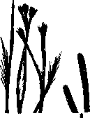
Scissors cause flattening, serration, and small cracks at the ends of the hair. A cut end of the hair becomes polished after two weeks, and the corners along the cut edge are rounded, while after 2–3 months they begin to form a tassel. From blows with a blunt instrument, the hair undergoes flattening and longitudinal splitting. A torn end of the hair appears irregularly stepped. During burning or curling, the hair twists, and its end appears under the microscope as club-shaped and swollen, with small air bubbles. Under the influence of mechanical impacts, the original shape of the hair end undergoes polishing, especially on short hairs of the limbs due to constant friction from clothing, and appears rounded, partly club-shaped; the latter shape is especially pronounced on axillary hairs, around the anus, and the genital organs due to sweat and strong friction; this shape makes it possible to determine the origin of presented hairs. Determining the identity of hair presents a very difficult task requiring great experience and caution. For hair comparison, it is necessary to microscopically examine and measure at high magnification (about 600), using a micrometer
a large number of hairs and take the average of them; the thickness of the hair, the medulla, and the cortical layer are subjected to measurement; the character and location of pigment and other features are noted. One must be extremely cautious with a conclusion regarding the similarity, and especially the identity, of hair; one can only speak with certainty about a greater or lesser similarity, since in the same person individual hairs on the head can be completely dissimilar to each other, while, on the other hand, in two different persons hairs can be almost identical. Occupational coloring of hair occurs in persons working in an atmosphere containing certain dust particles which, settling on the hair, impart a corresponding color to it. Thus, in those working in coal mines, as well as those dealing with charcoal, soot (e.g., chimney sweeps, colliers, stokers, etc.), light hair often becomes black from coal dust and soot; in bakers, millers, starch makers, hair, on the contrary, turns white from dust; in coppersmiths, green coloring of the hair is observed, which can persist for up to 15 years after a change of profession (Oesterlein); Beigel observed an indigo worker with dark blue hair due to indigo lumps scattered in the hair. Figure 4. 1--Split hair ends from a woman's head; 2--polished hair ends from the shin. as far as possible a large (WOLF'S JAW The latter type of hair coloring can depend not only on the deposition of coloring substances on the surface of the hair, but also on penetration into the hair substance itself. Occupational hair coloring can serve as an indication of the trade of the person whose hair is being examined. The normal color of hair sometimes changes significantly in corpses that have spent a long time in the ground: black hair may acquire a red-chestnut color, and light hair a chestnut color; this must be kept in mind when establishing the identity of the deceased, although among hairs that have changed their color, some that have retained their former color are always encountered.
Related articles
Mentioned in
Cite this page
“Hair.” Soviet Medical Encyclopedia. English translation of Bolshaya Meditsinskaya Entsiklopediya, 1st ed. (Moscow, 1928–1936), ed. N. A. Semashko. https://sovietmedicalencyclopedia.pages.dev/article/hair/