Muscles
Historical document, translated for reference. It reflects medical knowledge of the 1920s–30s and is not medical advice.
Summary
This article from the 1930s Soviet encyclopedia describes the histology of muscle tissue, focusing on the morphological and functional characteristics of smooth and striated muscle fibers, including their structure, arrangement, and innervation.
Encyclopedia article (1928–1936)
MUSCLES.
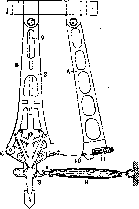
I. Histology. General-morphologically, the tissue of contractile substance is characterized by the presence of differentiation in the protoplasm of its elements into a specific fibrous structure; the latter are spatially oriented in the direction of their contraction and constitute the morphological substrate of the specific contractile function of M. Arranged parallel to each other, the visible elements of this structure, the so-called myofibrils, appear in the form of the thinnest (up to 0.2μ in diameter) threads, distinguished by their characteristic property of true positive uniaxial anisotropy. Depending on whether the myofibrils are optically homogeneous or constructed from alternating fragments of optically heterogeneous substance along their length, two main types of muscle tissue are distinguished: smooth muscles and striated muscle tissue. 1. The morphological and functional unit of smooth muscle tissue is the smooth muscle fiber (cell-fiber)—an elongated (in vertebrates), spindle-shaped, somewhat flattened along its long axis, single-nucleated formation, averaging 100-200μ in length and 4-6μ in width (extreme dimensions 240-500μ in length, 4-13μ in width). Fibers are encountered that are forked at one or both ends. The elongated, so-called "rod-shaped" nucleus is located somewhat eccentrically in the middle of the cell length. The protoplasm of the cell-fiber appears delicately striated due to the presence of the thinnest homogeneous myofibrils arranged parallel to the long cellular axis. The nucleus is surrounded by a certain amount of sarcoplasm (protoplasm not differentiated into fibrils), accumulating at its poles and forming the thinnest layers between the bundles of fibrils; in it are found the "cell center" located near the nucleus, and chondriosomes scattered throughout the cellular body. In the composition of the tissue complex, the elements of smooth muscle are connected to each other by fibers, the so-called myoglia, having the characteristic of "reticular" fibers of connective tissue, sometimes taking on a collagenous type and forming net-like sheaths on the surface of each muscle cell. Architectonically, the smooth muscles of man appear either in the form of thin, individual bundles embedded in connective tissue or in the form of continuous contractile membranes, and in the latter case, the fibers in the composition of the muscular plate are either arranged parallel to each other or form net-like interweavings of muscle bundles. Functionally, smooth muscle tissue in man constitutes the so-called involuntary musculature of the internal organs, vascular system, and skin covering and is always innervated directly by the peripheral neuron of the autonomic nervous system. 2. Striated muscle tissue is optically heterogeneous due to the structure of the myofibrils that make it up; it is represented in the vertebrate organism either by morphologically individualized entities (skeletal musculature) or by a syncytially connected system of muscular crossbars (type of cardiac muscle). The structural unit of "skeletal" musculature is the so-called striated muscle fiber, a multi-nucleated, strongly elongated cylindrical formation with pointed or rounded ends. The average size of the fiber in man is from 4 to 8 cm in length with a diameter of 40 to 50μ (the smallest observed length - 2 mm, the largest - about 12.7 cm, the smallest diameter - 9μ, the largest - 150μ). The myofibrils in the composition of the fiber are arranged in the form of columns separated by larger or smaller amounts of sarcoplasm, which on its cross-sections give the picture of the so-called fields of Köigheim. The numerous nuclei are distributed along the entire length of the muscular element; they are located in the peripheral, sub-sheath layer of the fiber in mammals with poor sarcoplasm. The entire fiber is clothed in the sarcolemma, a shell formerly homologized with cell membranes but now recognized as a connective tissue formation; it is intimately connected on one side with the substance of the muscle fiber through the fibers that make it up and on the other side with the tissue of the perimysium (perimysium). The characteristic transverse striation observed in microscopic examination of the striated muscle fiber is due on one hand to the heterogeneity of the optical structure of its myofibrils and on the other to the presence of transverse, crossing the entire fiber, thinnest membranes (intercalated discs). Located along the length of the muscle fiber at strictly the same levels, the isotropic and anisotropic areas of neighboring myofibrils give the optical phenomenon of alternating, parallel to each other light and dark stripes, as if cutting across the entire fiber in the transverse direction (the so-called discs); this picture is further complicated by the lines of optical sections of the intercalated discs, which also cross the muscular element transversely at certain intervals of its length. Alternating at a certain area of the muscle fiber, the discs form the so-called muscle segments, characterized by the same sequence of discs making up each of them. Scheme of transverse striation within one segment. The boundary between two adjacent segments is considered to be the intercalated disc crossing the fiber between two isotropic discs, connected at its periphery with the sarcolemma (disc Z of modern nomenclature, telophragma of Heidenhain, thin dark disc of French authors, stripe of Amici-Krause of old nomenclature); adjacent to it is an isotropic disc (I of modern nomenclature, light disc of French authors); followed by a wide anisotropic disc (A of modern nomenclature, thick dark disc of French authors). In ordinary transmitted light in a stretched fiber, the middle part of A appears lighter, forming a light stripe (h of modern nomenclature, stripe of Hensen), crossed in the middle by the thinnest intercalated disc M (mesophragma of Heidenhain, stripe of Merkel); then comes another isotropic disc I and finally the segment is closed by the next intercalated disc Z. Thus, in letter expression the scheme has the form: z + i + (h + A + M) + h + i + z. Appearing as a solid disc in polarized light, A possesses true positive anisotropy, while the anisotropy of Z is explained by the phenomenon of so-called depolarization. The fibrous discs A and I, besides their metamicroscopic structure, differ in their ability to absorb light, density and probably chemical composition. The properties of the mesophragma M are almost unknown: this disc is little accessible to observation and is clearly visible only on fixed preparations of maximally contracted fiber. The average height of the entire segment in vertebrates is about 2.5μ. The sarcoplasm of the striated muscle fiber is characterized by the presence in it of granular formations of different morphological value: along with true sarcosomes, representing the chondrioma of the fiber and properly arranged between the fibrils at the levels of disc I (in A - grains), here are also encountered droplets of lipoids and lumps of glycogen. Interesting are the argentophilic transverse networks of fibers discovered by Veratti, located at the level of discs I on both sides of Z and possibly identical with the trophospongium of Holmgren. In the composition of the muscular complex, the "muscle," the striated muscle fibers are connected by means of the fibrous connective tissue of the perimysium, the fibers of which pass into the fibrous framework of the sarcolemma. The question of the connection of fibers with the corresponding tendon is currently under discussion: while according to the opinion prevailing in modern literature this connection is entirely carried out through the fibers of the perimysium, a number of researchers insist on the presence of a direct transition of myofibrils in the area of the tendon end of the muscle fiber into the collagenous fibers of the tendon, bundles. Unlike skeletal musculature, striated cardiac muscle is characterized by a) the syncytial arrangement of the muscular crossbars making it up, b) the axial arrangement of nuclei in connection with the accumulation of sarcoplasm in the axial region of these crossbars and c) the presence of special stripes crossing these at indefinite distances, the so-called "insertion regions"; the morphological and functional significance of the latter is still controversial. Previously homologized with cell boundaries, these formations are now considered either as growth points of the cardiac muscle syncytium or are given the significance of microscopic intermediate tendons, serving as a support point during the contraction of the latter. Literary data on the morphology of contraction of striated M. up to the present time are extremely incomplete and contradictory: it can be considered generally accepted that the morphological substrate of contraction is precisely the myofibrils, but even the question of which of the myofibrillar discs the shortening of each myofibril predominantly occurs from is a subject of discussion. Also not finally clarified is the morphological significance and the so-called
«striations of contraction» (CS in modern nomenclature), observed on fixed preparations of maximally contracted striated muscle fiber instead of its normal striation.-Due to the above, modern theories of muscle contraction have long been constructed without a close connection with the morphological data of the finest microstructure of muscle tissue. All these theories, starting from Engelmann's, recognize the direct cause of the shortening of the fibrils as the property of the colloidal protein gel (in this case the substance of the muscle fibrils) to change its external form under the influence of energy changes in the surrounding environment (resp. sarcoplasm). These changes, according to the generally accepted view, are in turn due to chemical processes occurring in the muscle fiber at the moment of its excitation. The mechanism of this change in shape of the contractile fibrils is explained either 1) by an increase in the osmotic tension of their substance depending on the appearance at the moment of excitation of new products of decomposition (the so-called osmotic theories of McDougall and Tsunz), or 2) by a change in surface tension at the surfaces separating the fibrils and sarcoplasm (the theory of d'Arsonval-Bernstein), or finally 3) by the swelling of the anisotropic substance of the muscle fibrils when hydration of the proteins composing it occurs at the moment of contraction (the theory of Engelmann, Pauly, Fürth). This swelling should occur either at the expense of water coming from the isotropic fibrillar sections (Engelmann), or from the surrounding sarcoplasm, or at the expense of the water of the anisotropic sections themselves; the latter in this case must be composed of ultramicroscopic ellipsoidal granules suspended in a liquid medium; their long diameter is parallel to the direction of contraction. Engelmann explained the changes in the hydration capacity of the protein gel by the influence of the significant increase in temperature of the muscle fiber observed during contraction, which is the result of the oxidative processes occurring at the moment of excitation. - In contrast to Engelmann's «thermodynamic» theory, modern «chemodynamic» theories of muscle contraction accept this hydration as the result of the change in the reaction of the substance of the muscle element occurring at the moment of excitation. At the same time, according to Fürth and Pauly (see also the data of Meyerhof and Embden), the moment that resolves contraction is the appearance of acidic products of decomposition in the muscle; Embden, however, believes that here, on the contrary, alkaline products play a role.-Carefully conducted Hill's observations on the course of heat production in striated M. in various stages of its activity refute the classical theory of Engelmann; in connection with the latest research of Meyerhof and Embden, Hill's experiments provide the basis for «chemodynamic» theories. Functionally, the striated musculature of vertebrates is as a rule the musculature of voluntary movement and is innervated by the motor neuron of the cerebrospinal nervous system. The automatically working cardiac muscle is innervated according to the type of smooth muscle tissue.
A. Mislavsky. II. Physiology. Functions of M. The main function of M. is to bring about movement and mutual displacement of individual links and parts of the body. The basis of these movements lies in the processes of M. contraction, with which it is most convenient to begin the study of the physiology of muscular activity. M. can be made to contract by acting on it with various agents: mechanical influences, chemical reagents, heat, and finally electric current. In experiments with muscle, it is desirable to take as a stimulus a factor that 1) would not damage the M., 2) would act immediately on the M., 3) could be dosed, and 4) the action of which could be quickly started and stopped. Mechanical action is usually accompanied by damage to the superficial fibers. This, however, can be avoided if the M. is stimulated through its nerve (indirect stimulation). In this case, a small device called a tetanomotor is used, consisting of an electromagnet and a movable armature that is set into vibrational trembling, transmitted in the form of light blows to the motor nerve of the muscle. Thermal action on the muscle can be carried out by quickly changing the t° of the M.; this is usually accompanied by some damage to the M. Chemical stimulation (acids, citrates and oxalates of sodium, alcohol, chloroform) primarily affects (and this is understandable) the superficial fibers. The most convenient and satisfying all the above requirements is stimulation with the help of induction current. The source of the latter is usually the so-called Dubois-Reymond induction apparatus (see Dubois-Reymond induction apparatus), in which it is possible to change the voltage of the induction current by shifting and separating the primary and secondary coils. This type of current, in contrast to direct current, does not cause polarization. To study muscle contractions, the M. is placed in a so-called myograph. The frog's gastrocnemius muscle (m. gastrocnemius) is fixed immovably in its upper part. When the M. contracts, its lower end (Achilles tendon) raises a lever, which is equipped with a feather that writes on a smoked kymograph tape. Since the muscular pull acts on the short arm of a second-class lever, while the recording is made with the long arm, the myograms are obtained as many times larger than the actual contractions as the long arm is longer than the short one. A very weak electric shock is unable to cause muscle contraction. The moment when the muscle first contracts is called the threshold of excitation. With further strengthening of the stimulating current, the single contractions of the muscle also increase, but this increase occurs only up to a certain maximum limit, upon reaching which the contractions of the M. no longer increase. Between the moment the M. receives a single stimulus and the moment the contraction begins, a certain period of time passes, which for the frog's M. averages 0.01 seconds and is called by Helmholtz the period of latent excitation, or the latent period. The phases of contraction and relaxation usually last 0.05 seconds each. In the case where the M. is fatigued, the total time of a single muscle contraction increases mainly due to the lengthening of the relaxation phase. A stimulus that falls on the muscle before the latter has had time to return to a state of relaxation gives rise to so-called summation of muscle contractions. With sufficiently frequent stimulation, the M. does not have time to relax and comes into a state of prolonged, so-called tetanic contraction, or tetanus (Helmholtz). The longer a single contraction of the M. lasts in a given species of animal, the lower the frequency of stimulation required to produce tetanus; with short single contractions of the M., even very frequent stimulation may only cause single muscle contractions, but not produce tetanus. Thus, the frog's gastrocnemius muscle comes into a state of tetanic contraction already with 20 shocks of induction current per second (the duration of its single contraction is 0.1 seconds), while the wing muscles of insects do not produce tetanus even with 200 stimulations per second (the duration of single contraction is 0.003-0.040 seconds). The Russian scientist N. E. Vvedensky proposed his own theory to explain deviations in the magnitude of tetanus, calculated as the result of simple superposition (summation) of identical muscle contractions. According to Vvedensky, there exist optimal rates (frequencies) and strengths of stimulation that give maximum tetanic contraction. In these cases, the stimulation not only causes a single contraction but also increases the excitability of the muscle tissue to subsequent stimuli, as a result of which the tetanus turns out to be greater than in the case where all contractions would be completely identical. Excessively strong or excessively frequent stimulation—the pessimum (see)—gives a smaller effect than stimulation that is weaker or less frequent—optimal. The cardiac M., as is known, does not produce tetanic contraction but can only contract rhythmically. The latter is due to the presence in it of the so-called refractory phase, when the cardiac muscle becomes inexcitable, unresponsive to stimuli falling on it. Questions related to the reaction of the muscle to one and two subminimal, i.e., below the threshold of excitation, and supramaximal stimulations have been studied in detail by Vvedensky and his students (see Parabiosis, Pessimum). A subthreshold stimulus to the M. is unable to cause its contraction, but as we saw above, it can increase the excitability of the muscle and with a certain frequency of such stimuli cause maximum contraction. Two successive maximal stimuli give completely different effects depending on the duration of the interval between them. With a large or small interval, the amplitude of contraction is very large; with a very small interval (e.g., one-thousandth of a second), two maximal stimuli act as one supramaximal stimulus, and the contraction obtained is not larger but, on the contrary, smaller (stage of parabiosis). Wave of contraction. When a muscle that has been acted upon by curare poison is stimulated, its contraction is not observed immediately along its entire length, but first at one point—the point of application of the stimulus (with indirect stimulation through the motor nerve—at the location of the nerve's end plates). The contraction of the M. from this point occurs immediately in both directions, towards both its ends. The speed of this wave of contraction (Kontraktionswelle) in different animals, according to the research of Bernstein and Hermann, proved to be different: in the frog about 3 m/sec., in the rabbit 5-10 m/sec., in humans 10-15 m/sec. For smooth M., the speed of propagation of the excitation wave is less than for striated muscles. Law of average load and pace. Mechanical work is expressed, as is known, by the product of force and distance. In the case of lifting a load, instead of force we can put the weight of the load. Naturally, the question arises about the dependence that exists between the weight of the load being lifted and the mechanical work done in this process. This dependence can be easily found if one takes a nerve-muscle preparation or the M. of a frog without a nerve and records the heights of load lifting with different loads. Such experiments have been performed many times and have become classical; they showed that with an increase in load, mechanical work increases, but only up to a certain limit; upon reaching it, it begins to decrease and at a certain maximum load falls to zero. Mechanical work is expressed in gram-centimeters (g/cm) or kilogram-meters (kg/m). This dependence is usually called the law of average load. A similar dependence can also be found with respect to the pace (speed) of work. At a certain average pace, mechanical work is greatest. These two dependencies cannot be applied indiscriminately and uncritically to the whole organism, where the connection of the M. with the circulatory system ensures the supply of oxygen to the muscle tissue and the removal of certain substances formed as a result of muscular work. The participation of the nervous system in muscle contractions also changes the process of muscular work so much compared to the contractions of a nerve-muscle preparation that the dependencies observed on the latter are significantly complicated. This is especially true with respect to the law of average speed (pace). Fatigue of M. Under the influence of prolonged work, a series of changes usually designated as fatigue occurs in the M. in a state of functioning. This term is one of the most indefinite and imprecise in physiological terminology, and under it different authors understand far from the same thing (see Fatigue). In its simplest form, the phenomena of fatigue are manifested in the work of an isolated muscle, periodically stimulated by induction electric current (stimuli of maximum strength). At the beginning, the contractions of the M. hardly change in height, and then the decrease becomes noticeable and progressively increases until finally the muscle completely stops contracting. The more the M. is loaded and the more frequent the stimulations, the sooner its fatigue sets in. The total duration of contraction during fatigue increases mainly due to the lengthening in time of the relaxation phase.
A very fatigued M. may not even give complete relaxation (residual contraction or contracture). Along with slowing down the entire process of contraction, the speed of the contraction wave also decreases. During fatigue, the contraction wave may even stop at the site of irritation, and a so-called idiomydullary bulge forms here. Finally, during fatigue, the strength of the M. decreases and the threshold of irritation increases. A muscle that has reached a state of complete fatigue and does not react to any irritation can, even outside the organism, again return to a working state, albeit to a very incomplete degree. This phenomenon still has no scientific explanation. An M. connected to the circulatory system of the organism, and even better, an M. not removed from its place and retaining all its nerve and vascular connections, resists fatigue much longer than an isolated M. At present, physiology is dominated by a very primitive concept of fatigue, reducing almost all phenomena to the accumulation in the tissues of the working muscle of some 'fatigue substances.' When the connection with the vascular system is preserved, these substances are supposedly quickly washed out of the fatigued M., and it again acquires working capacity. In an isolated M., there is no washing out, and the hypothetical substances are oxidized by oxygen diffusing into the M. from the air. Of course, the phenomena of muscle fatigue are much more complex than many modern scientists imagine. There are many reasons to believe that fatigue is associated with colloid-chemical processes and occurs with the direct participation of the autonomic and cerebral nervous systems. If even the phenomena of fatigue in a single M. have not yet been scientifically explained, it is clear that the much more complex process of fatigue of the entire organism is far from its scientific interpretation. The fatigue of human M. is usually studied with the help of an ergograph (see Ergography). The weight lifted and lowered due to flexions and extensions of the finger or hand can vary in weight. The tempo can also vary. By selecting different values for both, one can find on the work of the finger the same laws of average tempo and load that were considered for the isolated M., but from this one by no means can make broad generalizations concerning the entire organism. Ergograms are individual in nature: in some, the decrease in amplitude occurs slowly, in others, on the contrary, almost immediately after the start of work. One should not think that ergograms reflect only the fatigue of the working finger muscle; despite fixing the forearm, the work of lifting the load is not limited to the participation of only finger flexors, but as fatigue sets in, more and more new muscles are involved in the work. The ergograph is often used to assess a person's work capacity, and attempts have even been made to apply it in production. Such attempts must be approached with great caution. The magnitude of work capacity in professional labor is determined by a number of factors, including psychological factors (interest in work, competition, shock work). That is precisely what labor differs from work, as it is a bio-social phenomenon. When assigning a worker work on an ergograph, the researcher excludes this moment and studies the work capacity of the subject not in relation to the professional labor performed by the subject, but in relation to the uninteresting and meaningless work of lifting a load with a finger or hand. The total amount of energy released by the M. strictly corresponds to the work performed by it. This has recently been proven by precise experiments of Hill's students {Hill}-Azuma and Fenn. Meyerhof, Embden, and their students established that most of the broken down hexosephosphoric acid is restored again in the relaxation stage of the M. In a fatigued state, resynthesis processes occur half as energetically as in a fresh state, and the M. loses much valuable energy-source substance. It has been proven that the smaller the part of the original substances is finally burned in the M., the higher its efficiency. Hill compares the M. with a self-charging battery after each partial discharge. Elsewhere, he draws an analogy between the M., in which hexosephosphoric acid breaks down into lactic and phosphoric acids and then resynthesizes again, and a self-charging spring. M. as a receptor. Usually, M. is considered as an organ of movement and the other role of M.-the role of a receptor apparatus-is pushed into the background. Inside muscle tissue, and especially in muscle tendons, there is a huge number of nerve endings signaling the central nervous system about the degree of tension of the M. and consequently about the position of the body's links at various moments of movement, about the direction and speed of the latter. M. thus is an organ with a dual role-receptive and effector. Since the impulses going centripetally from the endings in the M. give information about the internal state of the organism and its parts, the English physiologist C. Sherrington called these endings proprioceptive. However, objections are raised against this term, since in a number of cases, receptor endings give an idea of the properties of objects of the external world. For example, a person holding a stone in an outstretched hand and thus evaluating its weight undoubtedly uses for this data on the degree of contraction of his muscles, data obtained from proprioceptive elements. Long before Sherrington, the famous Russian physiologist I. M. Sechenov gave another name to the sensations we receive from our muscles. He called these sensations 'dark muscle feeling,' thereby emphasizing the difference between them and sensations received from the sense organs. M. and its relationship to the nervous system. As is known, M. is part of the reflex arc. Numerous experimental studies have shown that M. of some animals can sometimes function without the participation of the nervous system and that apparently the early link of the reflex arc was precisely the M., and then in the order of phylogenetic development, the M. caused the appearance and improvement of sense organs and the central nervous system (Samoylov). M. gave rise to the central nervous system, this most complex formation that we know, and then in the process of evolution this system began to perform a number of new independent functions; M., however, moved to the role of an executive organ. According to the views of I. M. Sechenov, M. was precisely that formation that essentially influenced the function of some sense organs. If the human ear can evaluate the duration of different sounds, it is completely unable to evaluate the duration of pauses-silent intervals of time. The latter could be accomplished, according to Sechenov's thought, only if the pauses were filled with muscle feeling with its characteristic tenseness. This circumstance actually occurs in walking. 'In view of the fact that each placement of the foot on the ground is accompanied by a sound, walking at various speeds is for consciousness a periodic series of short sounds, the intervals of which are filled with tensile elements of muscle feeling. Here therefore is the school in which hearing could learn to evaluate the different duration of intervals within the limits of acceleration or deceleration of the step in walking' (Sechenov, 'Elements of Thought'). On the other hand, the evaluation of distances with the help of the eye is also accomplished thanks to muscle feeling. The complex of impulses coming from the endings embedded in the six muscles of the eye to the brain gives us a much more complete idea of the shape of objects, their distance from us and from each other, of the direction, character, and speed of their movements than in the case of a stationary eye. As a result of a series of ingenious examples and constructions, I. M. Sechenov comes to the conclusion that muscle feeling was and is an analyzer of time and space. Muscular work. There is no professional work in which one muscle or an isolated group of muscles participates. As a rule, many muscles participate in work, and the longer the work is performed and fatigue progresses, the greater the number of muscles is involved in it. Part of the musculature performs the function of fixing various joints or parts of the body in a given position, another part directly participates in the work. Tension of the M., not accompanied by further approximation of its ends and the body parts to which it is attached by means of tendons, we conventionally call static work. This term, rooted in physiology, is incorrect from a physical point of view, since mechanical work presupposes the presence of some displacement caused by the action of a force applied to the body. The term 'static,' however, indicates precisely the absence of movement. However, since this name for the isometric tension of the M. when holding a load, fixing body parts, etc. has not been replaced by another more rational one, it has to be preserved and used.
The activity of muscles, the ends of which move the links of our body and objects connected with them (tools, loads), relates to dynamic work. The organism as a whole does not remain indifferent to the activity of its own musculature. The main factor in this regard is the increasing need of active muscles for nutrients and, first of all, for oxygen. Usually any more or less significant muscular work is accompanied by a general increase in blood pressure in the cardiovascular system, a redistribution of blood in the body in the sense of a greater supply to its active musculature, and an expansion and increase in the number of capillaries in the working muscles. According to the data of A. Krogh, the number of open capillaries in 1 mm3 of the cross-section of a working muscle increases 10-100 times compared to the resting state, and the total surface area of the capillary network in 1 liter of muscle tissue increases 12-130 times. There is an assumption that the local expansion of blood vessels in working muscles is caused by an increase in the active reaction in the muscles. Atzler and Lehmann found that the pH in a working muscle decreases by 0.2 and that even such a weak acid as carbonic acid can cause vasodilation. The latter leads immediately to two consequences: an increase in the inflow of blood and an increase in the outflow of venous blood and lymph. As for the redistribution of blood in the body, it occurs by narrowing the lumen of the vessels of internal organs and a corresponding expansion of the vessels of active muscles. Weber, and after him Lehmann, showed that the blood filling of muscle groups increases even before the start of work, already at the mere thought of possible work. Static work, in contrast to dynamic work, is characterized precisely by the fact that the vessels of isometrically tense muscles are compressed by the muscles themselves; the blood supply to muscle tissue during static work is much worse than during dynamic work, in which the contractions of the muscles are rhythmically replaced by their relaxations. Possibly, this is the reason for the greater fatigue of muscles during static work. Poor blood supply, difficult washing out of metabolic products from tense muscles, and with fixation of the chest cavity, disruption of respiration and blood circulation in the whole organism—this is what characterizes static work as a rule. The increase in pressure in the arterial system during muscular work is due, on the one hand, to the difficulty from time to time in the outflow of blood through active muscles, and on the other hand, to the increased frequency of heart contractions and the increased capacity of individual systoles. This pressure (maximum) can reach very high values (up to 200 mm and higher). Therefore, in developed arteriosclerosis, muscular work is contraindicated. The following table gives an idea of the dependence between the power (intensity) of work and the volume of blood ejected by the heart per minute: Dependence between power of work and minute volume. State of organism Power of work (in kgm/min) Minute volume of blood (in liters) Rest ...... Work I ...... » II ...... » III...... » IV...... » V...... 0 180 500 700 900 1100 3.0 8.5 12.5 14.0 16.0 21.6 Venous circulation also does not remain unchanged during muscular work. During dynamic work, the increased excursions of the chest cavity promote the inflow of venous blood from the hollow veins into the right atrium, as intrathoracic pressure decreases at this time. Due to the more vigorous activity of the heart, blood passes more forcefully through the capillary network and enters the venous system under greater pressure. Finally, the contractions of the leg muscles squeeze blood from the veins toward the heart (venous valves do not let it back), and thanks to all these factors, the blood filling of the right heart during diastole increases significantly. This circumstance, according to E. Starling's well-known 'law of the heart', leads to an increase in the volume of systole and leads to an increased supply of blood and oxygen to active muscles. A different situation occurs during static work. A fixed chest cavity (when lifting or carrying heavy loads, when moving handcarts, wheelbarrows, carts, etc.) prevents the inflow of blood from the venous system into the right heart due to the high air pressure created in it. If, in addition, the huge muscle masses—the legs—are in a state of tension, then the conditions for blood movement through the veins of the legs upward to the heart become very difficult. All this leads to stagnation of blood in the legs (profuse dilation of the cutaneous veins) and (during the static work itself) in the veins of the face and head. The harmful effect of static work on the body has been emphasized many times by many researchers, and there is no doubt that the most important factor here was the disorders in blood circulation. The effect of muscular work on the respiratory activity of a person is enormous. Even with slight muscle work, breathing becomes more frequent and uneven. Without touching the question of what exactly in this case is the excitant of the respiratory center—carbonic, lactic, or other acids formed during muscle work—it is necessary to note that there is a certain and (within certain limits) linear dependence between the power of the work performed and the magnitude of pulmonary ventilation, as shown in the following table (compiled according to Boothby's data). Dependence of pulmonary ventilation on power of work. State of organism Power of work (in %) ventilation Rest ...... » II...... » III...... » IV...... » V...... 182 256 320 347 521 100 178 283 288 306 438 As we see, the dependence has a linear character in this case. The research of Hill and other physiologists and biochemists has established a number of extremely interesting facts relating to the role of lactic acid in the process of respiration. As is known, the organism after more or less difficult work does not immediately return to its normal resting state. Pulmonary ventilation and the amount of O2 absorbed are increased, and the ratio of CO2: O2 in the exhaled air, or the so-called respiratory coefficient (RQ), also remains increased for some time, indicating the predominant combustion of carbohydrates. Chemical analysis of the blood during this period (the recovery, or reparative period) shows a greater content of lactic acid than at rest. By the end of the recovery period, pulmonary ventilation and the volume of oxygen absorbed return to normal. The same happens with RQ and the content of lactic acid. Barry-Himwich established that in the recovery period in the tissues of non-working muscles, the lactic acid released into the blood is oxidized. Having compared a number of experimentally established facts, Hill and his colleagues came to the conclusion that the excess oxygen absorbed in the recovery period goes to the oxidation of part (approximately 1/4-1/3) of the lactic acid; the other, larger part, as mentioned above, is resynthesized into hexosephosphoric acid. The CO2 formed as a result of the oxidation of lactic acid is not completely released with the exhaled air, but combines with the alkali of the blood that has just been freed from lactic acid. Consequently, during work, the lactic acid entering the blood in excess binds part of the alkali and thereby forces a larger amount of CO2 to be released with the exhaled air than is formed at each given moment. Conversely, during the recovery period, the disappearance of lactic acid from the blood due to its oxidation and resynthesis leads to less release of CO2 into the air ('retained' carbonic acid). All this, of course, affects the fluctuations of the respiratory coefficient. The release of lactic acid into the blood during heavy muscular work is of enormous importance, and without it a person could not perform any significant muscular work. For each organism there is a certain limit for oxygen absorption; if the work is light, then the 'oxygen requirement' (or Oxygen requirement in Hill's terminology) is below this limit and the organism easily copes with the work. Approximately the same thing happens if the need is very close to the limit. In the third case, however, when the oxygen requirement exceeds this limit, 'oxygen intake' turns out to be insufficient, and a certain 'oxygen debt' is formed, which is repaid with the help of the release of lactic acid into the blood only during the recovery period. After heavy work, as is known, oxygen absorption remains elevated for a long time. The changes occurring in the organism during muscular work have been studied very little, but even what is known now allows us to think of the significant participation of the endocrine system and both components of the autonomic nervous system. Experiments indicate an increased, compared to normal, entry of adrenaline into the blood during muscular work and an increase in the percentage of sugar content in the blood.
It was not possible to establish a direct relationship between the power of work and the amount of sugar in the blood. By examining the urine, an increased excretion of various nitrogen- and phosphorus-containing chemical substances was found, but certainly all of this is clearly insufficient to clearly understand the course of intermediate metabolism with respect to at least the main substances (proteins, carbohydrates and fats) during and after muscular work. Physiologists are accustomed to the amazing coordination that is manifested in the human body at rest; during work, however, especially in a trained* person, the coordination of the activities of various body systems reaches a very high degree of perfection due to the activity of a number of coordinating mechanisms. Coordination is carried out by means of the cerebral and autonomic nervous systems and the accessory apparatus of the latter (endocrine system). One can with full right say that the organism as a whole appears with special distinctness precisely when performing complex and difficult muscular work.
K. Kekcheev. Basic mechanical phenomena in M., methods of their registration and representation. The basic mechanical property observed in M. is the variability of its length, which constitutes the basis of all its motor properties. Upon closer examination of this phenomenon, it turns out that the length of M. changes as a result of a very large number and variety of causes, both purely physical and physiological. These causes and their effects have not yet been brought into any systematic theory, and the mechanics of M. continues to remain the subject of numerous experimental investigations. Changes in the length of M. are studied mostly on the muscles of frogs (usually m. sartorius or m. gastrocnemius), which depending on the experimental setup are either completely isolated, or are prepared along with the n. ischiadicus, or finally are kept in situ, with only the distal end being prepared and connected to the recording instrument. The recording of changes in muscle length is usually carried out with the help of a myograph, which is a stand with two hooks or clamps to which the ends of the M. being studied are attached. One of the hooks is immovable, while the other is connected in one way or another
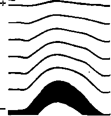
Figure 1. Photomyogram of a single muscle twitch of a frog muscle (Bethe and Happel). The signs + and - indicate the points of application of the electrodes. The upper broken line is the moment of irritation. In the photograph, it is clearly visible that the excitation of the muscle (increase in elastic properties) begins at the lower end of the M., near the cathode: the lower segments begin to contract first and pull the upper segments toward themselves, temporarily stretching them.
with a pen that writes on a smoked drum (kymograph). In some varieties of myograph (Schenck, Blix, Kries), both ends of the M. are connected to movable writing instruments. Bethe (A. Bethe) used an extremely delicate method of optical recording to analyze changes in the length of M., interesting in that with its help not only the total length of the entire M. is recorded, but also changes in the lengths of individual consecutive segments of the muscle. Bethe places the M., divided along its length into a series of segments to which black silk threads are attached, in the focal plane of a projection apparatus and casts its translucent image onto a slit photocymograph, in which the displacements of each of the silk threads appear as curves (fig. 1). Along with changes in length, M. is also capable of experiencing changes in tension, i.e., force tending to bring its ends together. Since there is no action without reaction, the tension of an M. not loaded by any stretching force is always zero, whatever its physiological state. Tension arises in the muscle only in the presence of a stretching force (whereas a change in length may not require any external force) and in magnitude is always determined by the magnitude of this external force, although the cause of the tension may have nothing to do with this force. The tension of a muscle is measured by the most diverse dynamometers (see Dynamometry). As a dynamometer for the frog M., modifications of the myograph are used. In those cases where it is desired to record changes in the tension of M. at constant length (isometric processes in M., see below), it is fixed between two almost immovable clamps, one of which is connected to a stiff dynamometric spring and to a writing pen. If it is desired to record simultaneously both changes in length and changes in tension of M., it is loaded on the movable end either 1) with a weight or a lever with a large moment of inertia, the tension being calculated from the acceleration imparted to M. by this weight, or 2) with a compliant spring (auxotonic process, see below), the resistance of which is taken into account by the degree of stretching it undergoes, or finally 3) with a significant weight which M. can lift only relatively slowly, so that the weight acts on it mainly by its weight and not by its inertia (isotonic process, see below). In addition to changes in length and tension, M. also experiences changes in cross-sectional area and transverse hardness. The cross-sectional area of M. increases when it shortens and decreases when it lengthens, since the volume of M. does not change during these processes. Transverse hardness changes in parallel with the change in the longitudinal tension of M. Indeed, if M. overcomes an external stretching force, this means that it with an equal and opposite force tends to shorten. Meanwhile, simultaneous transverse compression by another external force decreases its cross-sectional area, i.e., tends to lengthen M.; therefore such compression encounters resistance in the tension of M. Both of these phenomena are thus so-called derivative, secondary manifestations of both basic processes and therefore represent
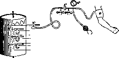
Figure 2. Diagram of the myographic installation of Bruzhes: a1-air cuff on the human arm; a2-manometer; a3-chamber with an elastic partition for equalizing pressures; a4-Marey capsule; A - writing pen; B, C, D - electromagnetic markers.
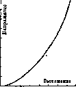
little theoretical interest; however, since M. in the intact organism is much more accessible to transverse influences than to longitudinal ones, both of the mentioned phenomena are widely used to evaluate the states of M. in humans. The registration of transverse expansions of M., which should testify to its contractions, was carried out on humans by Bruzhes and Johnen and applied by them to the study of labor processes; the diagram of Bruzhes' installation is given in fig. 2. The registration of transverse hardness (sclerometry) has found wide application for the approximate evaluation of the longitudinal tension of M. in humans (Noyons, Uexkull, Exner Tandler, Mangold) and consists in measuring the depth to which a weight placed on it (more precisely on the skin over it) sinks into M. This static sclerometry has in the present time given way to a more perfect dynamic (ballistic) one, which measures the elastic recoil exerted by M. on a hammer striking it (Noyons, Gildemeister, Bethe, Steinhausen, Richter). The hammer is suspended like a pendulum and strikes with a completely definite force against a plate applied to the skin over M., and the duration of its contact with the plate is measured. The two main variables of muscle mechanics - length and tension - are connected by a close internal relationship. If one studies both of these variables in parallel on an unexcited (resting) M., it turns out that the tension of M. increases with an increase in its length (stretching) and decreases with the shortening of M. The relationship between both variables can be represented graphically in the form of the so-called length and tension diagram (Langen-Spannungsdiagramm, D.-N.-diagram) (see e.g. fig. 3). A more careful study of the D.-N.-diagram of passive stretching of a resting M. shows that even in this particular case the relationship between both quantities is not constant: the D.-N.-diagram changes depending on the speed of stretching and on the biochemical state (e.g. degree of fatigue-Fig. 3. Length and tension diagram (D.-N.-diagram) of an isolated muscleЖdea" niiili, on the contrary, with gradual shortening of M. The point of intersection of the D.-N.-diagram curve with the abscissa axis obviously represents the state of M. in which its tension is zero, i.e., the state to which M. comes when no external forces act on it. The length of M. in this state is designated as the resting length (Ruhelange), or the natural length of M. Both these names are not quite appropriate, since the rest of M. is usually contrasted with its excitation, and the opposition of the same word to tension can lead to errors; as for the natural length, we cannot speak of it when we cannot recognize other lengths of M. as unnatural. The most correct thing would be to call this length the unstressed, or free length of the muscle. The relationship between length and tension is even more complex and peculiar if one turns to the study of it in M. excited to one degree or another. Here there is such a chaotic variety of experimental material that it is difficult to sort out
in the
it is not yet possible with sufficient certainty at the present time. (The main theoretical premises for the analysis of these facts are given below.) The measurement of the relationship between length and tension of M. can be performed both on an isolated M. and on a living subject (human). In the frog's M., this measurement can be carried out in two ways. The first method, introduced by Weber and used by Marey, Haycraft, Wertheim, Christen (Marey, Haycraft, Wertheim, Christen), and others, consists in gradually loading or unloading a resting or tetanized M. with weights, while the tip of the M., connected to a writing pen, itself records on a kymograph the changes in its length caused by different loads (i.e., tensions). The curves obtained by these methods have often been subjected to theoretical analysis, but these analytical attempts have not yet led to consistent results. In any case, it can be considered a fact that the L.-T. diagram of passive stretching is undoubtedly not a straight line, i.e., that there is no proportionality between the increase in length and the increase in tension, which is characteristic over wide ranges of inelastic bodies and known as Hooke's law (Hooke). As can be seen from Figure 3, the L.-T. diagram is a curved line (Wertheim incorrectly took it for a hyperbola), which, as the length of the M. increases, goes increasingly steeply upward *. This means that the more a muscle is stretched, the more strongly it resists further stretching. If to lengthen the M. by 1 mm it is sufficient to increase the load by 1 g, then to lengthen it by another 1 mm will require a further increase in load not by 1, but by 1.2 or 1.5 g. True, these experiments on an isolated M. are carried out in much wider ranges of stretching than is possible in a living organism, and it is undoubtedly the case that in the latter case the deviation of the muscle from Hooke's law during passive stretching is small. Experiments making it possible to obtain the L.-T. diagram of M. are also possible on a living human. Spiegel balanced the leg of a raised leg of a person lying on their back with the help of a lever with weights, and then by hanging weights to the leg itself, he caused passive flexion of the knee and thus stretching of the m. quadriceps femoris, the tension of which was estimated by the weight of the hung weights. By this method, the tension of the studied M. could be brought up to 8-9 kg. Vereschagin applied a similar setup, but much more successfully chose as the experimental M. the group of posterior thigh muscles (mm. biceps fem., semitendinosus and semimembranosus), taking advantage of the fact that these muscles, passing over two joints at once, can be passively stretched to incomparably greater lengths when the hip joint is bent: up to 115% of the original length (taken as 100%) and up to 70-80 kg of tension. This setup is shown in Figure 4a. The setups of Spiegel and Vereschagin were used by the authors to study muscle tone (see Tone)-a phenomenon understood very differently by various researchers, but recently increasingly identified with the elastic state of an unexcited M., i.e., with a syndrome characterized precisely by the L.-T. diagram of longitudinal stretchability of the muscle. It must be emphasized that the similarity between the L.-T. diagram of a living toned M. (Fig. 46) and the same diagram of an isolated M. (Fig. 6) should not be understood in
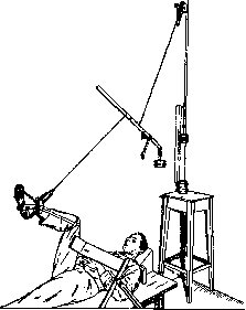
Figure 4a. Vereschagin's setup for measuring muscle tone.
the sense of complete identity. An isolated M. does not experience any nerve influences, whereas in the whole organism the M. is continuously under the influence of the central and autonomic nervous system, and its L.-T. diagram reflects in itself the synthesis of local (purely muscular) and central phenomena. Among such central, tonic phenomena is, for example, the hump of initial inhibition (Bremsung) in Fig. 46, which is absent in an isolated muscle. The methods of both authors have found wide application in neurological clinics, allowing for reliable quantitative assessment of pathological tone of M. The second method for quantitative determination of the elastic properties of M., applied so far only to the frog's M., was developed by Bethe, Steinhausen, and Richter (Steinhausen, Richter) and is based on the mentioned ballistic principle. In Richter's ballistic elastometer (Fig. 5), the M. is attached at one end to a fixed clamp, at the other to the hook of pendulum B, against which hammer A strikes. The blow of the hammer deflects the pendulum and thus suddenly stretches the M.; after this, the hammer flies back, and the pendulum, attracted by the stretched M., makes one or two swings and then calms down. As Steinhausen showed, the duration of the first swing stands in a very simple relationship to the measure of elasticity (the so-called modulus of elasticity) of the M.; the difference, however, between the duration of the first deflection of the pendulum and the duration of its return is just as closely related to the internal friction of the M., or muscle viscosity (see below). The advantage of the ballistic method over the gradual loading method is that in sudden, shock-like stretching of the M., the influence of the previous prolonged stretched state, which very noticeably changes the character of the L.-T. diagram, is excluded. Theoretical premises for the mechanics of M. Any body that gives an L.-T. diagram in which (as can be seen in Fig. 3) an increase in length is accompanied by an increase in tension is classified by mechanics as a longitudinally elastic body. M. is a longitudinally elastic body, although, as indicated above, it is not subject to Hooke's law of proportionality. The degree of elasticity of a body is characterized by the modulus of elasticity, or Hooke's modulus. The modulus of elasticity is the ratio of the increase in tension per unit of cross-sectional area (in kg per cm2 of the body's cross-section) to the relative increase in length accompanying this tension. If an M., stretched to 1/10 of its original (unstressed) length, shows a longitudinal tension of 0.8 kg per cm2, then the modulus of elasticity (more precisely-the average value of the modulus) of this M. is 0.8 : 1/10=8 kg/cm2. As mentioned above, this ratio, which determines the modulus of elasticity, is not the same for different stretches of the M.; the more the M. is stretched, the higher the value of the modulus. Therefore, the modulus of elasticity of M. cannot be expressed by a single number, as is possible, for example, for steel or rubber. At best, one can speak of the average value of the modulus, but even for this value, due to many contributing factors, various authors have obtained very poorly agreeing figures. Wundt (Wundt) gives for the M. of an adult human values from 35 to 86 kg/cm2, for the m. sterno-cleido-mast. of a dog-142 kg/cm2, for the M. of a frog-9.4 kg/cm2. Richter by the ballistic method found for the m. sartorius of a frog values from 5.2 to 9.4 kg/cm2. Measurements of tone of human M., made by Vereschagin, give figures approaching Richter's-10-12 kg/cm2. Stretching of M. requires expenditure of work not only to overcome the rapidly increasing tension, but also to overcome internal friction in the muscular substance. The work expended to overcome tension is returned back to the M. when it shortens (as the spring of a mechanism returns the work spent on its
winding) and is therefore called the reversible part of the work, whereas the expenditure to overcome friction irreversibly turns into heat. The internal friction of M., or muscle viscosity, depends on the speed of stretching or shortening of the M.: the faster the length of the M. changes, the greater the friction. Experiments to determine the coefficient of muscle viscosity (Steinhausen, Richter), i.e., the relationship between the speed of stretching and the force of resistance to friction, have not yet led to reliable results.
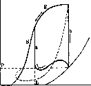
Figure 5. Richter's ballistic elastometer: A-hammer; B-pendulum; M-muscle; 5-writing pen. The hammer strikes plate 6 against plate 7. Muscle viscosity significantly affects the D.-N.-diagram of M. Let line I (figure 6) be the D.-N.-diagram of very slow stretching of M., in which viscous resistance is negligible. Then when the same M. is stretched at a noticeable speed, the D.-N.-diagram will follow line II, and during elastic shortening of M., it will return to zero along line III. Indeed, if static stretching of M. to magnitude A corresponds to static stress Aa, then when M. is stretched to the same magnitude at a noticeable speed, we will experience 1) the same static resistance Aa and 2) friction resistance ab, or in total resistance Ab, greater than Aa. During shortening, internal friction will resist the shortening of M. and thereby weaken its strength; therefore, when passing through length A during shortening, M. will exhibit stress which will be less than the static stress Aa by the magnitude ac, i.e. will be equal to Ac. From the nature of the D.-N.-diagram it follows that the area of the figure bounded by curves II and III represents in scale the irreversible work expended to overcome internal friction during the forward and reverse stroke of M.; all work expended on stretching M. will be represented by the area between curve II, the x-axis, and the extreme ordinate Bd, while the reversible part of work (returned by the muscle during shortening)-the area between curve III, the x-axis, and the ordinate Bd. The work of M., like that of any elastic body, occurs in two successive phases. In the 1st phase, accumulation of potential energy by the muscle occurs, in the second-its transformation into mechanical work. The sources of energy for the first phase may be different: elastic potential energy can accumulate in M. both due to previous passive stretching and due to an internal chemodynamic process occurring during excitation of M. and releasing a certain portion of mechanical energy. On the contrary, the source of energy for the second phase in all cases is the same-elastic potential energy of M. In other words, under identical external conditions, the course of muscle contraction will be completely identical, regardless of the sources that charged M. with energy in the first phase; the observed enormous diversity of forms of this course is determined exclusively by the diversity of external conditions accompanying the activity of M. (see below). Between an unexcited M., charged with energy due to previous passive stretching, and an excited M., having received an energy charge through a chemodynamic pathway, there is, however, a significant quantitative difference. Both unexcited and excited M. are elastic bodies, quite similar in their mechanical properties; however, the quantitative characteristics of the elastic properties of both these states of M. are different: 1) the free, or unstressed length of M. decreases during excitation. If an unloaded frog M. is placed in a Ringer-Locke solution to free it from the influence of its own weight and then subjected to tetanization through a nerve, it will shorten. 2) The modulus of elasticity increases during excitation. Like that of unexcited Mm, it varies at different degrees of stretching of M., so that in this case too the D.-N.-diagram has the form of a curve, but unlike the resting M.-this curve bends upward to a certain limit, after which it begins to go more and more gradually. D.-N.-diagrams of excited (I) and resting (II) M. are shown semi-schematically in fig. 7. The free length of excited M. is represented by point A, of resting M.-by point a. Up to point B, the steepness of curve 1 (i.e. the modulus of elasticity of excited muscle) increases, then begins to decrease. At point C it already compares with the steepness (c) of curve II, and at point D (the existence of this latter cannot be considered strictly proven) it becomes zero, i.e. the excited M., when stretched to length D, loses its elastic properties. Figure 6.
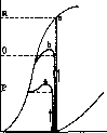
Figure 7. Figure 6. D.-N.-diagram of passive stretching and shortening of an isolated muscle. Figure 7. D.-N.-diagram of excited (I) and unexcited (II) muscle: A-free length of excited; a-of unexcited muscle. To the present day, between supporters and opponents of the "all or nothing" law (see) a heated discussion is being conducted as to whether the transition of elastic properties of M. from state II to state I occurs suddenly (by a jump) or gradually, in other words, whether intermediate states of the type of curve III (fig. 7) occur, even if only for very short times each time. This dispute has not yet received experimental resolution, although indirectly many facts speak in favor of a jump-like change. It should be noted that only experiments on a microscopically small section of one fiber can be decisive in this dispute. If one observes an entire multi-fiber M. over its entire length, then even if the change of states of each fiber occurs suddenly, it will inevitably be masked by the different timing of entry of individual fibers, as well as by the fact that both the excitation wave and the elastic wave propagate along M. not instantaneously. During tetanization of M., each new excitation shock transfers M. from state II to state I, after which together __ with the fall of the action current wave of M. it is obviously immediately again returned to state II. T.O. at a tetanization frequency of 50 impulses per second, M. that many times per second changes elastic state II to I and back. The fact that during this the length of the muscle does not manage to change that many times per second (at least noticeably to the eye) is explained exclusively by the internal friction of M., its viscosity, which does not allow the length of M. to change with the required speed and thus is an extremely useful property for the feasibility of tetanus. In fig. 8 it is very clearly seen how tetanization of the same frequency acts on a muscle with high viscosity (top) and with low viscosity (bottom). The curves of length and tension changes during tetanus (D.-N.-diagrams) for both cases in fig. 8 are given schematically in fig. 9 next to the main passive stretching curves I and II; a-tetanus of a viscous muscle; the area of the oval-work of one impulse; b-trembling of a less viscous muscle; the area of the oval-work of lifting and lowering a load. The influence of external conditions on muscle contraction. Relying on the basic D.-N.-diagram fig. 7, one can with great clarity trace the peculiarities of the course of muscle contraction under various conditions. The graphical representation of all these processes in the form of D.-N.-diagrams provides the best way to understand, at least in the most general terms, the chaotic abundance of experiments conducted on muscle contraction.-Figure 10 shows the isometric tension of M. (the phrase "isometric contraction" obviously makes no sense). In the unexcited state, the tension is zero, in the excited state it reaches magnitude P. Various cases of auxotonic contractions, i.e. those in which tension increases as the muscle shortens (for example when stretching a spring with a muscle), are shown in fig. 11. Line a represents a strictly auxotonic (ideal) case; line b-case of auxotonic contraction of a muscle with high viscosity. Since the increase in static tension of the spring in this case goes along line a, the excess b over a is "obviously quite analogous to the excess A over I (fig. 6), and the area between b and a represents the work of overcoming internal friction of the muscle.--Cases of isotonic contractions are shown in fig. 12. Curve a-slow (almost strictly isotonic) lifting of a large load P; b-faster lifting of a medium load Q. From the figure it is clear that this lifting is "isotonic" only in name. At the beginning of the lift, the muscle has to overcome 1) the weight of the load Q=lm, 2) its inertia st, proportional to acceleration, and 3) its own internal friction rs, depending on the speed of contraction. Additions (2) and (3) raise the initial tension
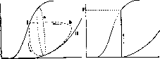
Figure 11.
Figure 9.
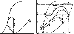
Figure 10. Figure 9. D.-N.-diagrams of incomplete tetanus of a viscous muscle (a) and a less viscous one (b). Cf. with fig. 8. Figure 10. D.-N.-diagram of isometric tension of a muscle.
Figure 12. Figure 11. D.-N.-diagrams of auxotonic muscle tensions. Figure 12. D.-N.-diagrams of so-called isotonic muscle contractions. The displacement of the muscle by the amount rt over the tension sufficient for static support of the load Q. By the end of the ascent, the upward movement of the load slows down, and it stretches the muscle with a force that is less than its own weight by the amount uv. In connection with this, despite friction and the overall tension of the muscle b decreases. The useful work expended on lifting is measured by the rectangle mlkn or by the area mlswvkn, equal to it; the expenditure on internal friction is determined by the area Irkvwsl. The curve c depicts the changes in the inertial resistance of the load Q. The curve d of Fig. 12 corresponds to the case of throwing the load R, which during rapid contraction of the muscle flies over the equilibrium position and then performs one or two damped oscillations around this position. The useful work of lifting is the rectangle ochur, the work of friction is the area between the solid curve d and the dashed curve e. - Figure 13 characterizes contractions in which a load initially supported can be lifted only when the excited muscle has already developed tension equal to its weight. These contractions obviously begin strictly isometrically, and then follow the type of curves in Fig. 12. The line a is the lifting of the load P, the viscosity of the muscle is great; b is the lifting of a heavier load Q, the viscosity is small (throwing); c is an impossible load L (isometric tension).-On Fig. 14
Author Henke and Knorz Hermann Rosenthal Franke. . Beck . . . Rosenthal Camarero Figure 13.
Figure 14. Figure 13. D.-N.-diagrams of muscle contractions with an initially supported load. Figure 14. D.-N.-diagrams: a-contraction from impact; b-contraction with initial delay. a is presented-contraction from impact (An-«chlagszuckung), in which a lightly loaded muscle first contracts to a certain length L, at which it hits an obstacle, forcing it to further tense isometrically (Schenck, B Их); b-contraction with initial delay (Aniangshemmung): the muscle is held until full tension is developed, and then released into flight with the load P. The area between part b' of this curve and the dashed curve is the expenditure on friction. Experiments of this type were conducted by A. Fick. The muscle as an organ and as an element of the bone o-m y i n e ch-nogo skeleton. The individual working elements of skeletal muscle are of the same type and obey the same laws in any muscle, but the whole muscle is not a simple bundle of fibrils: it is an organ in which the arrangement, connection, and combined effect of the action of fibrils can be very diverse depending on the function that this muscle has to perform. Since the modulus of elasticity determines the relationship between stresses and relative elongations of the muscle, it is obvious that with given elastic properties the absolute excursion of the end of the muscle will be the greater, the longer its fibers. On the other hand, again with given elastic properties, the overall tension of the muscle must change in proportion to the cross-sectional area (the number of parallel working fibers). Thus, the excursions of the end of the muscle are proportional to its length, the tensions are proportional to the cross-sectional area. Consequently, the work of the muscle, measured by the product of tension and distance traveled, is proportional to the product of the cross-sectional area of the muscle by its length, i.e., the volume of the muscle or the amount of contractile substance contained in it. - Figure 15 schematically depicts two completely equivalent muscles of different shape. Both these muscles have the same volume, therefore- the same power, and in addition the moment of their action on the joint is the same, since the three times stronger muscle is applied to a three times shorter lever arm.- As an attempt to evaluate the force of the muscle per unit cross-sectional area, mention should be made of experiments on the so-called absolute force of the muscle. According to Franke, the absolute force of the muscle is the greatest stress per 1 cm2 of cross-sectional area that the muscle can develop through tetanus under the most favorable conditions (i.e., at length B, Fig. 7). The imperfection of methods and some vagueness of the definition have led to not too consistent results among different authors (the main data-see in the table). However, the same total work (determined, as stated, by the volume, not the shape of the muscle) can be produced
Muscle Triceps surae Flexores brachii Triceps surae Mm. masticatorii Biceps brachii Brachialis int. Triceps brachii Object of study Abs. force in kg/cm2 Human 5.9 » 8Д » 6.2 » 10.0 » 11.4 » 12.1 » 16.8 Frog 1.8 » 2.8 Crustacea (crustaceans) 1.8-3.2 Coleoptera (beetles) 3.4-6.9 Lamellibranch. (bivalves) 12.4
Figure 15. Diagram of two equal-power muscles with the same moments, of different shape and location.
in different ways: either as overcoming a small force over a long path or, conversely, as movement against a greater force over a short path. To the first type of work are adapted muscles with long fibers, usually spindle-shaped (e.g. m. biceps br.) or fan-shaped (e.g. m. temporalis); to the second type - short-fibered muscles with a large cross-section: parallel-fibered (e.g. m. rhomboideus) or pennate. (e.g. m. rectus femoris).-Figure 16 shows very clearly that the work performed by a pennate muscle (lifting a large load to a small height) and the equivalent spindle-shaped one (lifting a small load to a greater height) is the same. Indeed, if the weight of the pennate muscle consisted of two parts A and B, then the work of lifting the entire weight to the dashed position would not differ from the work of lifting only piece B and placing it on the upper side of the remaining in its original position-part A, and this is exactly what the1second muscle does. The various basic forms of 'cut' of the muscle are found in the body in: infinitely diverse combinations and variations (see Muscular system). Here we should turn to the general typical conditions of muscle work in the skeletal system. In the vast majority of cases, skeletal muscles have to perform the rotation of one skeletal part relative to another. Therefore, as in the mechanics of any rotation in general, the action of the muscle has to be characterized not by its tension itself, but by the so-called moment of tension relative to the axis of the joint it serves. The moment of tension is measured as the product of the longitudinal tension F (Fig. 17) by the arm of its action, i.e., by the length of the perpendicular R, dropped from the center of the joint to the longitudinal axis of the muscle. With a complex shape of the muscle and diverse directions of its fibers, as the tension of the muscle one has to take the resultant of the tensions of all its parts, and as the longitudinal axis of the muscle-the line of action of this resultant.-From Fig. 17 it is easy to see that with different positions Figure 16. Diagram illustrating the equality of work performed in different ways by a pennate and spindle-shaped muscle. and links the moment M. may turn out to be different even with the same longitudinal tension F, since the length of the action arm R changes from the displacement of the links. Usually the greatest value of the arm R corresponds approximately to a right angle between the links, and in this position the muscle can develop the greatest moment (and not the 'greatest force', as is usually said).
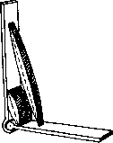
*%> The law of equality of action and reaction also applies to moments. According to this law, the moment of action of the M. on link B (Fig. 17) is equal and opposite to the moment of its action on link A. Therefore, the tension of one M. cannot yet determine which of the links will begin to move when it is tensed: for A to move, it is necessary to fix B, and vice versa (by an external force or some other M.); otherwise both links move towards each other. It is very important to understand this for the case of a muscle overcoming external forces. Let a load P be placed on the hand with the forearm horizontal (Fig. 18), which must be held. Two external forces act on the elbow joint: the moment of the load + PR and the moment of the weight of the forearm with the hand + pr. For equilibrium, the moment of the M. (elbow flexor) must be equal and opposite to their sum, +(PR+pr). But since, as stated above, the moment of the M. is bilateral, it will not only support the loaded forearm with the moment -(PR+pr), which is directly purposeful, but will also pull the shoulder forward in the direction of flexion with the same moment -(PR+pr). This is not only unnecessary but also harmful, because for maintaining equilibrium the shoulder must be immobile, but this M. is powerless to help in any way. To hold the shoulder, it is necessary to apply to it from the outside the same moment -(PR+pr); this has to be done by the muscular system of the scapula and shoulder girdle. But the action of these latter M. is again bilateral and in turn needs external support; moreover, in addition to the moment PR+pr coming from the forearm and hand with the load, a weight moment of the shoulder p'g' also acts on them, and for equilibrium of the entire system, a moment -(PR + pr + p'g') must act on these M. from the outside. Reasoning in this way, it is easy to see that when holding a load on the hand, not only the M. closest to it are tensed, but also many M. throughout the body-from the point of application of the external force of the load and up to the nearest external point of support (e.g., the support of the feet on the ground). The same thing is observed in the same order during movement, when M. overcome not the static force of gravity, but dynamic forces (friction, inertia, etc.). In all cases, the moments of M. increase from the periphery to the center due to the addition of new additional external moments p'g', p"g", etc. We give some figures representing the moments of muscles of different joints in some simplest processes, taking the moment of the M. of the wrist as a unit (Bernshtein, Popova). Moment Processes in the wrist in the elbow in the shoulder joint 22.0 16.7 7.6 9.3 Holding the horizontally extended hand . . . Holding the hand, semi- Piano strike . . . Postal stamping . . 1 1 1 6.8 6.7 4.6 5.4 From the table it is seen that in wrist movements (piano strike, stamping), the relative role of the M. of the wrist increases, but still far lags behind the load from distant M., which have no direct relation to this movement. The application of the law of action and reaction described above is also used to measure moments of M. on a person (Bernshtein). For this, the sum of the moments of all external forces is measured on one side or the other from the joint being studied. As stated, the resultant moment of all M. of this joint must be equal and opposite to the measured sum of external moments.
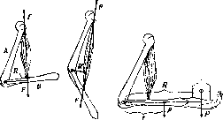
Figure 17.
Figure 18. Figure 17. Moment of a muscle in various positions of the joint. Figure 18. Moments of external forces (weight of the forearm with the hand and load) acting on the elbow flexors.
N. Bernstein. Electrical phenomena in M. The processes taking place in M. under conditions of its active state are accompanied by electrical phenomena, as was firmly established by Matteucci and Dubois-Reymond (see Bioelectric currents, Animal electricity). The electric currents arising in M. during its contraction are called action currents, in contrast to the resting current, which appears in a resting M. when its damaged part is connected through a galvanometer to its undamaged points (see Animal electricity). As is known, the action currents arising during tetanic contraction are so strong that with their help a second muscle can be irritated if its motor nerve is placed on the first. This phenomenon is called secondary contraction. When the capillary electrometer and string galvanometer were applied to the study of electrical phenomena in M., it was possible to show that in a single contraction there is a single electrical oscillation corresponding to the occurrence of a single wave of excitation, for example, under conditions of irritation of the nerve of a given muscle by a single make or break induction shock. The photographic recordings obtained at this time, or so-called electromyograms, make it possible to approach more closely the study of single electrical oscillations in M. The electrical process in a single impulse has a wave-like course. The descending part of the electrical oscillation is somewhat more stretched compared to the ascending part. The length of the electrical wave is less than the contractile wave and changes under the influence of temperature. As for the latent period of the electrical effect after the application of irritation, it passes very quickly. Some authors consider it equal to zero. The latent period is easier to detect when muscles are cooled. In the isometric regime of muscles, a steeper drop of the electrical curve is observed than in the isotonic one. According to Samoilov, contraction in the isotonic regime has an inhibitory effect on the magnitude of the electrical oscillation. Furthermore, it was found that the height of the electrical oscillation increases up to certain limits with the loading of M. by a weight. In tetanus, although the mechanical effect of contraction is continuous, the telephone and string galvanometer can easily detect the intermittent nature of this contraction. Namely, corresponding to the frequency of impulses entering the muscle and causing tetanic contraction, in the telephone, connected to the muscle by special electrodes, we have a corresponding number of membrane oscillations. In connection with this, Vvedensky established the concept of lability, by which he meant the ability of M. to give a certain number of electrical oscillations (action currents) per unit time at a known rhythm of irritation. When exciting M. from a nerve with an intermittent current with a period of 100 per second, Vvedensky established that the muscle gives 100 electrical oscillations only for a short time, and then it begins to transform the impulses entering it into a smaller number. Accordingly, instead of a given tone from 100 irritations per second (in the tuning fork-current interrupter circuit), we begin to hear a lower tone in the telephone. At a relatively higher frequency of irritations, a musical tone is not immediately obtained in the telephone, but a noise, which is replaced by a rumble, which depends on the muscle canceling out a series of impulses coming from the nerve by transformation (in the physiological sense). Partial blocking of excitation waves occurs, according to Vvedensky, in the myoneural transmission, at the endings of the motor nerve. Undoubtedly, the amplitude and frequency of electrical oscillations in the muscle change together with a change in the lability of the muscle, or - what is the same - together with an increase or decrease in the speed of processes occurring in M. Since as fatigue sets in the lability of the muscle decreases, the ability of the muscle to reproduce a given frequency of irritation in the form of electrical oscillations also decreases. The data obtained by Vvedensky with the help of the telephone later received brilliant confirmation in works with the string galvanometer, which makes it possible to photograph action currents. The ability of the muscle to transform indicates the manifestation of the muscle's own rhythm. The latter is easily detected when the muscle is irritated with a constant current. The electromyogram shows a series of electrical oscillations in the muscle in response to the passage of current through it. In the muscle of a turtle, the own rhythm of excitation is slower than in a frog. In a turtle, so-called absolute refractoriness, falling on the ascending part of the electrical curve, and a relative refractory stage at its descending part, can be easily observed at this rhythm. As expected, in warm-blooded animals the own rhythm is faster compared to cold-blooded ones. The study of action currents of skeletal muscle during voluntary movement has become very important. It sheds light on the rhythm of the central nervous system and in any case makes it possible to penetrate more deeply into the activity of the centers. The main hindering moments here are the above-mentioned transformation processes in the transmission of impulses from one physiological instrument to another and the difference in lability. From Vvedensky's data it is known that the nerve has the highest lability, or the ability to reproduce the rhythm of irritation; it is followed by the muscle. As for the centers and endings of the motor nerve, they show a certain similarity in their lability and stand below the nerve and muscle. This means that the phenomena of transformation, remodeling of the rhythm for passing impulses, are most likely to occur in the centers and in myoneural transmissions. The law of relative lability, established by Vvedensky in his work with the telephone, can have a guiding value in the study of the rhythm of the most diverse physiological instruments, up to and including the rhythm of the centers. Furthermore, it should be borne in mind that proprioceptive nerves must also have an influence on the electromyogram. When they are excluded, muscles usually show a lower frequency of electrical oscillations and greater regularity in their succession. The study of action currents of the muscle during voluntary contraction showed that voluntary contraction gives intermittent action currents of different periodicity, which is explained by the fact that 'in voluntary contraction, muscle fibers receive impulses not simultaneously, but some earlier, others later, as if the nerve centers are firing at random'. This is Brücke's characteristic comparison. When conducting action currents from the human forearm to a string galvanometer during voluntary contraction, Piper found the main rhythm to be 50 per second, with additional oscillations (Nebenzacken) added to the basic ones. In fatigue, the number of main waves decreases to 25-35 per second. Cobb and Forbes studied fatigue with an ergograph and observed a decrease in the number of main waves to 9-20 per second with an increase in their amplitude. The study of electrical phenomena in pathological states of the musculature showed the following. In Wilson's disease, a muscle in a state of rigor shows very small action currents, which apparently stand in no relation to the intensity of contraction. In Thomson's disease, after the cessation of voluntary contraction, the muscle remains in a state of tension for some time, with oscillating action currents being observed, as if voluntary innervation were still continuing. When studying ataxia in humans, Weizsäcker found a certain decrease in the frequency of electrical oscillations during voluntary movement compared to normal. As for tonic contractions, in contrast to tetanus, they apparently are not accompanied by intermittent action currents. Thus, Wachholder was unable to observe action currents during tone when studying the development of narcosis in cats. Whether there is a long negative oscillation during tone remains unclear for now. Under conditions of maintaining tension in the human muscle, significantly smaller action currents are observed than with the slightest movement. With small tensions, the action currents are remarkably small. For the theory of electrical phenomena in the muscle, the question of the relationship between the electrical phenomenon and the mechanical effect of contraction is of deep interest. It should be noted that this question has not yet been resolved. There are only the following data: when the action current of the muscle captured by the string galvanometer is recorded photographically together with the curve of a single muscle contraction, in the rough conditions of the experiment it is observed that the electrical effect precedes the muscle contraction and falls on its latent period. One might think that the electrical process is the result of preparatory chemical processes, which then cause the muscle contraction itself. However, by registering the mechanical effect of contraction by an improved method, Einthoven found that the latter almost coincides with the beginning of the electrical one. On this basis, Einthoven believes that the action current is connected with the contractile process itself.
After Bishop and Gilson, using cathode rays and an oscilloscope, found that only the descending part of the electrical monophasic curve coincides with the mechanical effect of a single contraction. The authors consider that only this part of the curve is due to the contractile process itself, while its ascending part is due to the excitation wave. Thus, since the magnitude of the action current does not always change in parallel with contraction, Beritov believes that the electrical effect is due to the formation of other chemical substances, and not those on which contraction directly depends. In connection with the question raised, the electrical phenomena in muscle poisoning by veratrine are of particular interest. As is known, under the action of this poison, after the usual contraction, a prolonged shortening of the muscle occurs. From Garten's experiments, it can be seen that this prolonged contraction is accompanied by a prolonged action current. Under conditions of severe poisoning, no oscillatory currents are observed in the muscle. If the above data of Samoylov are compared with the data of Bishop and Gilson, it can be assumed that the contractile process is a kind of compensatory process aimed at restoring the disturbed equilibrium in the muscle cell. And therefore, under normal conditions, the electrical and contractile processes are in the closest connection with each other, n. Rezyvyakov. III. Chemical statics and dynamics of M. I. Chemical composition of M. Striated and smooth M. differ in composition, and these differences are more quantitative than qualitative. The composition of striated M. is better studied, while less attention has been paid to the chemistry of smooth muscle. Composition of striated M. The composition of M. includes proteins, fats, carbohydrates, a number of extractive nitrogenous and non-nitrogenous substances, and inorganic substances. - Protein substances. Striated M. contains several protein substances, to which various authors gave different names. These proteins participate in the process of clotting of muscle plasma, which various authors also explained differently. The proteins of M. and their participation in clotting were most fully studied by Fürth, whose nomenclature is now the most widespread. Muscle plasma can be squeezed out with a press from fresh, just-cut-out animal M. in the form of a yellowish liquid. This muscle plasma, like blood plasma, clots, and the plasma of cold-blooded animals clots quickly, while the plasma of warm-blooded animals clots slowly; after clotting of the plasma, muscle serum remains, which has an acidic reaction to litmus. The clotting of muscle plasma depends on the transformation of two proteins contained in the plasma, named by Fürth myosin and myogen; they turn into insoluble protein substances myosin-fibrin and myogen-fibrin, of which the clot formed during the clotting of muscle plasma consists. According to Fürth, during the clotting of myogen, soluble myogen-fibrin is first formed, which then turns into insoluble myogen-fibrin; therefore, clotting can be represented by the following scheme: Myogen Myosin
♦ ^ 4.
Soluble myogen-fibrin Myosin-fibrin
I. Insoluble myogen-fibrin. When juice is pressed from dead muscles, muscle serum is immediately obtained. Myogen (myosinogen, myoprotein) is not precipitated when its solution is dialyzed in a medium salt solution. Myogen, especially in the presence of salts and at elevated temperatures, converts into soluble myogen-fibrin, and then into insoluble myogen-fibrin. Myogen is the main protein of muscle plasma (70-80% of all proteins). In the plasma of frog and fish muscles, there is much soluble myogen-fibrin. Myosin is a typical globulin, dissolving in neutral salt solutions and precipitating when its solutions are diluted with water. The usual method of obtaining myosin from dead muscles is to extract it with a 5-10% solution of ammonium chloride and precipitate it from the solution by diluting with water. Muscle stroma. After removing all proteins soluble in water and in ammonium chloride solution, a residue remains—the muscle stroma, consisting mainly of insoluble proteins, so-called myostromin. Fat is always present in muscles, indeed in small quantities (about 1%). The fat content in some animals can reach 30% (eel). It also increases significantly in fatty degeneration of muscles. Of phosphatides, lecithin is mainly contained in muscles. In heart muscles, there are more phosphatides (lecithin and cephalin) than in limb muscles. Cholesterol is always present in muscles (from 0.03% to 0.23%). Phosphatides and cholesterol are part of the sarcoplasm. According to Embden, red muscles are richer in phosphatides and cholesterol, as well as in sarcoplasm, than white muscles, which are capable of faster contractions but at the same time fatigue more quickly. Glycogen is the reserve carbohydrate of muscles. Its reserves vary and depend on a number of conditions, primarily on food and on the physical work performed by animals; with good nutrition and at rest, there is much glycogen in muscles, while during starvation or during heavy muscular work, its content sharply decreases. In humans, muscles contain on average 0.4% glycogen. Lactacidogen (see) is an intermediate product of carbohydrate metabolism in muscles. Striated muscles, different in their function, contain different amounts of lactacidogen: white muscles contain more of it, red muscles less. Inositol (see) is present in muscles in small quantities; its role is not clarified. One of the intermediate products of the transformation of hexose, which is cleaved off from lactacidogen, is lactic acid, which can be detected in muscles in larger or smaller quantities depending on the functional state of the muscle. Extractive nitrogenous substances of muscles. In striated muscles, various non-protein (extractive) nitrogenous substances are contained; namely, according to Fürth and Schwarz, in 100 g of muscles there are from 0.327 to 0.328 g of these extractive substances. Of this amount, ammonia accounts for 4.5-7%, purine substances for 6.1-11.1%, creatine and creatinine for 26.5-37.1%, carnosine fraction for 30.3-36.3%, carnitine, methylguanidine and other similar bases for 8.2-15.3%, urea, polypeptides, amino acids for 8.3-16%. Creatine (see) is contained in different amounts in different muscles; limb muscles are richer in it than heart muscles. Furthermore, white limb muscles are richer in creatine than red muscles, e.g., the m. biceps femoris of a rabbit is richer in creatine than the m. semitendinosus (Table 1). Homonymous muscles of different animals (dogs, rabbits) also differ in creatine content. But if one takes homonymous muscles or groups of homonymous muscles of different individuals of the same species (e.g., different rabbits), then provided that nutrition and all other living conditions are normal, the creatine content in them differs greatly in constancy. Therefore, for example, various researchers, determining the creatine content in the muscles of the hind leg of rabbits, always found the same value—0.522%. The creatine content in muscles can increase under the influence of a number of causes, e.g., during starvation (at its beginning), during avitaminosis, after severe cooling, after poisoning with phosphorus, guanidine, after extirpation of the parathyroid glands, during tetany, during training, etc. The content of creatine phosphate (see Creatine) increases in acute forms of polyneuritis, during training, after poisoning with phosphorus and tetrahydro-β-naphthylamine, etc. Creatinine (see) is contained in muscles in very small quantities: e.g., in human muscles only 0.003-0.007%. Carnosine (see) is contained in muscles of different animals in fairly large quantities, namely—from 0.05% to 0.3%. There are only first preliminary indications regarding changes in carnosine content in muscles under different conditions, e.g., an indication of a decrease in content during starvation and an increase during training. Carnitine (see Meat extract) is contained in muscles in small quantities (from 0% to 0.03%). Adenosine phosphoric acid, or adenylic acid (Embden) during deamination, which takes place, for example, during muscle contraction, is converted into inosinic acid; the latter was isolated from meat extract by Liebig. Table 2. Content of various anions and cations in muscles. Ions % Ions % K...... 2.5-4.0 Fe..... 0.04-0.2 Na..... 0.6-1.6 1.7-2.5 Mg..... 0.2-0.3 Cl..... 0.4-0.8 Ca..... 0.02-0.2 1.9-2.3 Inorganic substances. Of the inorganic substances of muscles, the largest are phosphoric acids (H3PO4) and salts of pyrophosphoric acid (H4P2O7), the presence of which in muscles was recently established by Lohmann. Pyrophosphoric acid is found in muscles in combination with adenylic acid in the form of adenylenepyrophosphoric acid. Among anions, Cl and SO4 are also contained in muscles, and among cations K, Na, Mg, Ca, Fe (Table 2). Table 3 shows data on the content of the above-mentioned substances in muscles of different animals. The chemical composition of smooth muscles has been studied less than that of striated muscles, and the available data mainly concern the muscles of the stomach and bladder. In smooth muscles, protein substances similar to myosin and myogen, as well as nucleoprotein, are found. Furthermore, glycogen, lactacidogen, inositol, creatine, creatine phosphate, creatinine, purine bases, and lactic acid have been found in their composition. There is much less creatine, creatine phosphate, and lactacidogen in smooth muscles than in striated muscles (Table 4). Table 4. Content of creatine phosphate and lactacidogen in smooth muscles* (in percentage of fresh muscle substance). Muscles Uterus of rabbit....... Muscular stomach of rooster Creatine phosphate 0.034 0.047 Hexose monophosphate 0.100 0.064 * According to data of the Ukrainian Biochemical Institute Table 3. Quantitative composition of striated muscles according to Hammarsten (in percentages). Muscles | Composition of substances | mammals birds cold-blooded Solid substances . . . 21.7 -27.8 72.2 -78.3 20.7 -26.3 1.0 - 1.5 16.6 -20.0 3.0 -10.6 0.3 - 0.52 0.07 - 0.286 0.007- 0.01 0.2 -0.3 0.019- 0,'03 0.07- 0.17 0.1- 0.37 0.084-0.235 0.003 Organic substances . . Inorganic substances . Creatine........ Creatine phosphate *.......... Carnitine ....... Purine bases . Lactacidogen* .... Inosit......... * According to research of the Ukrainian Biochemical Institute. 22.5-28.2 71.8-77.5 21.7-26.3 1.0- 1.9 17.4-20.0 3.0-11.0 0.3- 0.5 0.076-0.251 0.07-0.13 0.092-0.235 20.0 80.0 18.0-19.0 1.0- 2.0 14.4-15.2 3.0- 8.7 0.23- 0.7 0.408 0.3 0.053-0.088 Regarding inorganic substances, one can note the greater content of chlorine in smooth muscles compared to striated muscles. P. Rigor mortis of muscles. Rigor mortis of muscles occurs when the blood supply to muscles ceases, either after the death of the animal, or after the muscles are cut out of the animal's body, or as a result of ligation of the corresponding artery. In rigor mortis, lactic acid accumulates in muscles, and their reaction becomes acidic. Under the influence of lactic acid, due to the swelling of the colloidal anisotropic substance of muscle fibers, muscles shorten, become hard and opaque. Subsequently, the rigor mortis muscles become soft again; this can be explained by the fact that a further increase in lactic acid content causes protein coagulation, which in turn reduces the ability of colloids to bind water and release water. Some authors hold different views on the phenomena occurring during rigor mortis. Thermal rigor develops under the influence of high temperature; in warm-blooded animals it occurs at 50°, and in frog muscles at 40°. Chemical rigor occurs when isolated muscles are immersed in distilled water and when acted upon by acids of chloroform, ether, alcohol, many alkaloids, etc.
A characteristic feature of rigor mortis is not only the formation of lactic acid, but also the transformation of pyrophosphoric acid into orthophosphoric acid (Loman). III. Metabolism and energy in M. The beginning of work on the chemistry of muscle contraction was laid by the research of Fletcher and Hopkins, who established that the activity of M. is always accompanied by the formation of lactic acid in them, which, as later shown by Meyerhof, disappears during the relaxation of M. Then Embden showed that this process goes through the stage of formation of lactacidogen. Until recently, in the study of the chemical dynamics of M., almost exclusive attention was paid to the exchange of carbohydrates, attributing to them the main role. Glucose, formed in M. from glycogen, before turning into lactic acid, combines with phosphoric acid, forming lactacidogen (see), which is thus an intermediate product of carbohydrate metabolism in M. From it, in further transformations, lactic acid is formed [lactacidogen breaks down into glucose and phosphoric acid, and glucose, passing through further intermediate stages (see Carbohydrate metabolism), turns into lactic acid]. The formation of lactic acid occurs under anaerobic conditions: muscle contraction is not associated with oxygen consumption; oxygen consumption is associated with the recovery period of M. (rest). During recovery, the lactic acid formed during contraction disappears in such a way that part of it (1/4) is further oxidized to CO2 and H2O, and part (3/4) is again converted into glucose and glycogen. Thus, in working M., the formation and disappearance of lactic acid occurs. [This process of disappearance of lactic acid is also the result of two opposite processes (see below).] As work continues, a struggle takes place between these two opposite processes; during intense work, the second process cannot keep up with the first, and the content of lactic acid in M. increases. M. becomes tired, but its working capacity is still preserved. With the accumulation of a certain amount of lactic acid, complete fatigue sets in, and M. stop working. During the ensuing rest period, lactic acid disappears from M. as a result of two opposite, mutually related processes: due to the oxidation of two molecules of lactic acid, six other molecules are converted into glycogen. After rest, working capacity returns to the muscle, but the rested M. is no longer the same muscle that was at the beginning of its work. If isolated M. work in an oxygen-free environment, they quickly reach a state of complete exhaustion, simultaneously with the accumulation of large amounts of lactic acid in them; recovery of muscle working capacity is only possible in the presence of oxygen, since only under aerobic conditions does the oxidation of lactic acid occur. The ratio between the total amount of lactic acid disappearing during rest and the amount of lactic acid oxidizing during the same period is not a constant value; this coefficient------------------------------------- depends on ^
oxidized lactic acid depends on the state of the M., their fatigue, training, etc., and can vary within limits from 6/1 to 3/g. The carbohydrate metabolism in M. consists, according to Meyerhof, of two phases: the first phase is anaerobic (work phase); during it, glycogen, passing through the stage of lacticidogen formation, breaks down into molecules of lactic acid: 4/n (C6H10O5)n + 4H2O + 4H3PO4 → 4C3H6O3. H3PO4 + 4H2O → 8C3H6O3 + 4H3PO4; the second phase is aerobic (recovery phase); during it all 8 molecules of lactic acid disappear (2 of its molecules are oxidized to CO2 and H2O, and 6 molecules are again converted to lacticidogen and glycogen): 8C3H6O3 + 3H3PO4 + 6O2 → 3C6H11O5.H3PO4 + 6CO2 + 9H2O → 3/n (C6H10O5)n + 3H3PO4 + 6CO2 + 9H2O. Thus, during contraction of M., exothermic processes of carbohydrate metabolism occur, and during rest, endothermic phases. -Energy exchange in connection with these carbohydrate metabolism processes is depicted by Meyerhof and Hill (Hill) as follows: heat production occurs both during contraction and during rest, with 50% of energy falling on the contraction period and 50% on the recovery period. The heat of contraction, released during the work period, is not formed immediately at the beginning of contraction, but in 3 phases: the first phase of heat production coincides with the beginning of contraction, the second phase of less significant heat production - with the period of maintaining the achieved tension (shortening), and the third phase (phase of more significant heat production) - with the period of relaxation. Only after this, in the presence of oxygen, does heat production associated with the recovery of M. begin, stretching over a longer period. Meyerhof believes that lactic acid is formed during contraction of M. Embden thinks that part of the lactic acid formed during muscle activity arises after contraction of M., and therefore looks upon it as a substance causing relaxation of M. The role of creatine phosphoric acid in the dynamics of M. Recent research has shown that in the energy exchange in M. not only carbohydrates participate. Meyerhof previously recognized that the exothermic process of converting carbohydrates into lactic acid gives only a large part of the energy released (in the form of work and heat) during contraction of M., and after the discovery of creatine phosphoric acid, he thought that the remaining part of energy is given by the process of its breakdown. Indeed, according to the latest data, an important role should be attributed to creatine phosphoric acid in the chemistry of muscle activity; during contraction of M. it breaks down into creatine and orthophosphoric acid and is then resynthesized; this synthesis can proceed under anaerobic conditions, i.e., can occur during the first phase of contraction. The breakdown of creatine phosphoric acid is an exothermic reaction, with 110-120 calories being released for each gram of phosphoric acid cleaved off. Rapidly contracting (white) M. contain more creatine phosphoric acid than slowly working (red) ones; during training of M., associated with an increase in their work capacity, the content of creatine phosphoric acid in them increases; these facts indicate a certain role of creatine phosphoric acid in the chemical dynamics of M. If it is made impossible to form lactic acid during contraction of M. (for example, by the action of monoiodoacetic acid on M.), then all energy is formed at the expense of the breakdown of creatine phosphoric acid; it is even assumed that under normal conditions in M. during contraction, the breakdown of creatine phosphoric acid occurs, providing energy for it, while the carbohydrate metabolism processes may participate indirectly (for example, providing energy for the reverse synthesis). In any case, the processes of breakdown of creatine phosphoric acid and the process of converting glucose into lactic acid should be considered important in terms of energy in M. On the contrary, the processes of glycogen breakdown, breakdown of lacticidogen into glucose and phosphoric acid, as well as the processes of transformation of other phosphorus compounds occurring in M. - adenosine phosphoric and pyrophosphoric acids - do not play an energetic role. Transformations of adenylic acid in M. Adenosine phosphoric acid is also a substance, the breakdown of which is associated with the work of M.; during contraction of M. (at the beginning of contraction), ammonia is cleaved off from adenosine phosphoric acid, and as a result of such deamination, it is converted into hypoxanthine phosphoric or inosine acid. During relaxation of M., the reverse synthesis of adenosine phosphoric acid occurs by the reverse attachment of ammonia to inosine acid. In the reverse synthesis of adenylic acid, according to Parnas, not the ammonia that was released during deamination of adenylic acid is attached to inosine acid, but ammonia formed in M. during deamination of amino acids. - Pyrophosphoric acid is present in M. in combination with adenylic acid in the form of adenylophosphoric acid. Pyrophosphoric acid can be enzymatically cleaved into two molecules of orthophosphoric acid: H4P2O7 + H2O → 2H3PO4; such breakdown occurs during prolonged work, during rigor mortis, and during autolysis. The role of pyrophosphoric acid during contraction of M. has not yet been clarified; it is assumed that during contraction, synthesis of pyrophosphoric acid occurs, and during rest - breakdown. For the role of lacticidogen, see Lacticidogen. Thus, in the working M., the following chemical processes occur: breakdown of creatine phosphoric acid into creatine and phosphoric acid, breakdown of carbohydrates with formation of lactic acid passing through the stage of lacticidogen formation and its breakdown into glucose and phosphoric acid; oxidation of lactic acid; deamination of adenylopyrophosphoric acid and reverse amination of inosine acid and probably still other, as yet unknown processes. All these processes do not occur in isolation, but are undoubtedly interconnected with each other; it is quite probable that endothermic processes of synthesis of one substance proceed at the expense of the exothermic breakdown of another substance. The orthophosphoric acid formed in M. during their contraction as a result of the breakdown of creatine phosphoric and hexose phosphoric acids does not all go to the reverse synthesis of these substances; part of it passes from M. into the blood and then into the urine. For muscle activity, as is evident from the above, not only carbohydrates and nitrogenous substances, but also phosphoric acid are necessary; it is therefore understandable that the external delivery of phosphoric acid, especially during heavy physical work, has a favorable effect on the work capacity of M. All the above-described chemical processes cause a number of physicochemical changes in the muscles. First of all, the beginning of shortening of M. is characterized by a change in the concentration of hydrogen ions; namely, the breakdown of lacticidogenu as well as the breakdown of creatine phosphoric acid and deamination of adenylic acid cause a decrease in the concentration of hydrogen ions, i.e., shift the reaction in the direction of alkalinity at the site of these processes (probably in the interfibrillar colloids). This decrease in the concentration of hydrogen ions Embden considers the moment causing contraction, and the subsequent increase in the concentration of hydrogen ions due mainly to the formation of lactic acid - the moment causing relaxation of M. The second characteristic physicochemical process during muscle contraction Embden considers the sudden increase in the permeability of the boundary layer of muscle fibers. Both these processes are closely related to each other. -The influence of training on the chemical composition of M. Training, increasing the work capacity of M., causes certain changes in their chemistry. Trained M. are found to contain more glycogen, creatine, and creatine phosphoric acid. On the content of lacticidogen, on the contrary, training has no effect.
A. Palladius. (Continuation of the article Muscles-see article 735.) Muscles №№ Latin and Russian name. Synonyms. Form and position Origin and insertion Innervation. and relation to segments Abductor digiti minimi longus manus (long abductor of the little finger of the hand). [Rare variation.] A long flat ribbon, which can even reach the humerus, to the lateral epicondyle, lying on the medial surface of the forearm See abductor digiti quinti manus Abductor digiti quinti manus (abductor of the little finger of the hand). Syn.: abductor digiti minimi manus. The largest of all M. hypothenaris, located in the form of a flat ribbon, lies most laterally and superficially under the skin, covered only by m. palmaris brevis Orig.: pisiform bone, tendon of m. flexoris carpi ulnaris, lig. pisohamatum, sometimes lig. carpi transversum. Ins.: base of the proximal phalanx, its ulnar edge N. ulnaris-r. profun-dus. Cvil, VIII and Di. Abductor digiti quinti pedis (abductor of the fifth toe). Syn.: abductor digiti minimi pedis. Represents a relatively thick and long bundle, located immediately under the skin along the lateral and plantar edge of the foot; it is this muscle that mainly forms the relief of the lateral edge of the foot Orig.: calcaneus-processus lateralis tuberis calcanei and its plantar surface, fifth metatarsal bone-tuberositas ossis metatarsalis V, plantar aponeurosis. Ins.: base of the proximal phalanx of the 5th toe N. plantaris medialis. Ly and Sn Abductor hallucis (abductor of the great toe). Syn.: pollicem abducens. The largest muscle of the foot, in the form of a thick prismatic muscle bundle with a strong tendon, lies along the medial edge of the foot under the skin and fascia, covering the tendons, joint and bones of the medial edge and forming a cushion for the great toe of the foot Orig.: calcaneus-processus medialis tuberis calcanei, lig. laciniatum and plantar aponeurosis. Ins.: base of the proximal phalanx of the great toe, medial sesamoid bone and bursa of art. metatarso-phalangeae N. plantaris medialis. Ly and Si Abductor pollicis brevis: caput radiale and caput ulnare (short abductor of the thumb of the hand). Syn.: abductor internus pollicis (Cruveilhier), externus (Soemmering). Consists of two small flat bundles, converging at the point of insertion, lying immediately under the skin in the lateral part of the thenar eminence, which it mainly outlines; the muscle covers m. opponens pollicis, as well as the entire deep head of m. flexoris pollicis brevis Orig.: caput radiale: terminal tendon of m. abductoris pollicis longi, fascia antibrachii volaris; caput ulnare: navicular bone-tuberositas ossis navicularis and lig. carpi transversum, sometimes to the os pisiforme and the place of origin of the muscles of the hypothenar eminence. Ins.: base of the proximal phalanx of the thumb-its basis and radial sesamoid bone N. medianus. Cvi,VII 6 Abductor pollicis intermedius (Frohse and Frankel) (intermediate abductor of the thumb of the hand). [Variation.] See m. abductor pollicis longus Abductor pollicis longus (long abductor of the thumb of the hand). Syn.: abductor pollicis major, s. internus, s. bicornis, s. m. extensor ossis metacarpi pollicis, s. m. abductor carpi. Represents a large flat bipennate muscle belly, passing into a thin long tendon; it is located on the distal half of the forearm on its dorsal surface and partly on its radial edge, in the proximal half it is covered by m. extensor carpi radialis brevis and extensor digitorum communis, in the distal half it lies immediately under fascia antibrachii and skin; its tendon is well visible under the skin, especially sharply protrudes when abducting the thumb and forms the radial border of the "anatomical snuffbox" Orig.: ulna-facies dorsalis of it, membrana interossea; radius-facies dorsalis radii. Ins.: first metacarpal bone-its basis-lateral part of it N. radialis-r. profun-dus. Cvi, VII (vill) Accessorius ad digastricum (accessory to the digastric muscle of the mandible). [Rare variation.] A small bundle from the angle of the mandible to the lateral edge of the anterior belly of the digastric muscle See m. digastricus of man. Blood supply Action Variations A. ulnaris-r. volaris Abduction of the little finger profundus '! ! 1) Rarely absent; 2) often fuses with t. flexor brevis digiti quinti; 3) may have two or three heads; 4) one of the heads may originate higher from os pisiforme, i.e. a) from lig. carpi transversum, b) from fascia antibrachii, c) from m. flexor digitorum sublimis, d) from m. flexor carpi ulnaris, e) from ulna, f) from humerus or from its middle or even from epicondylus medialis humeri-this is m. abductor digiti minimi longus A. plantaris medialis Abduction and flexion of the fifth toe One of the most variable muscles of the human body; due to its various points of attachment to the proximal phalanx, it may be divided into two b. or m. independent layers; Le Double described three cases of additional M.(m. abducteur accessoire du petit orteil), located laterally and above the normal muscle Arcus plantaris, rr. perforantes from a. arcuata and aa. metatarsales Strengthens the medial part of the arch of the foot and thus shortens it; flexes and abducts the great toe 1) The muscle often, if not always, consists of two portions: deep and superficial, as evidenced by their separate innervation by small branches of n. plantaris medialis; 2) often there is a tendon to the second toe and not only to the medial side, but also to the lateral A. radialis-r. volaris superficialis Abduction of the thumb and partly its opposition; Aa. interosseae volaris and dorsalis Abduction not only of the thumb but mainly of the entire hand; in addition its palmar flexion helps m. flexor carpi ulnaris and radialis Variations of this muscle are very common and diverse. 1) The muscle belly may be completely absent due to its complete fusion with mm. extensores pollicis longus or brevis; 2) it may be double throughout its length; 3) more rarely it is triple; 4) as a norm its tendon may divide into two, more rarely into three or four; 5) one of them may fuse with m. abductor pollicis brevis (its caput radiale)-this is m. abductor pollicis intermedius; 6) additional tendons may attach: a) to os metacarpale I, b) to lig. carpi transv., c) to the muscles of the thenar eminence: m. flexor pollicis brevis, m. abductor pollicis brevis, m. opponens; the latter connection sometimes gives the form of a two-bellied muscle; 7) the most interesting connection is with m. trapezius, existing in all monkeys; 8) more complex combinations of the above variations with each other are possible ' ! „ Latin and Russian name. Syn-<NsJN'8 onyms. Form and position ! Origin and insertion Innervation and relation to segments Acromio-clavicularis profundus (Tisne and Rousseau) (deep acromioclavicular muscle). [Once found variation.] A small muscle between the clavicle and scapula, covered by m. deltoideus Orig.: clavicle-at the border of the middle and lateral thirds. Ins.: scapula-processus acromialis Acromio-clavicularis superficialis (Gruber) (superficial acromioclavicular muscle). 1 [Very rare variation.] A small muscle bundle located between the clavicle and scapula under m. deltoideus Orig.: clavicle-at the border of the middle and lateral thirds. See m. infraclavicularis and t. praeclavicu- Ins.: scapula-processus acromialis, laris lateralis. [Very rare variation.] 1 its apex Adductor brevis (short adductor muscle of the thigh). Syn.: adductor parvus, s. profundus parvus femoris, s. femoris brevis, s. subpubiofemoralis, s. caput profundum m. tricipitis. The muscle has a flat belly with parallel fibers; located on the medial surface of the thigh: on the anterior surface of mm. adductoris magni and minimi, covered in front by superficial m. pectineus and t. adductor longus; it is separated from other adductors by the fact that on its anterior surface lies r. anterior n. obturatorii, and on its posterior-r. posterior of it Orig.: pubic bone-facies anterior, covered by the origins of mm. adductor longi and gracilis. Ins.: femur;-linea aspera femoris (upper third of the medial lip) behind mm. adductor longus and pectineus, fusing with the tendon of m. adductoris magni N. obturatorius - r. anterior. Lh_iv ! Adductor hallucis: caput obliquum and caput transversum (adductor of the great toe). Syn.: abductor hallucis (Cruveilhier). Caput transversum =m. transversalis pedis, s. plantae. Caput obliquum=m. interosseus ad indicem pertingens. Represents two independent bundles, which have completely different directions at an angle close to right angle; between them is a large space for all art. branches emerging from arcus volaris profundus; the muscular belly lies in a depression on the middle and lateral toes and only its attachment falls on the first toe;
M. closed by m. flexor digitorum brevis and the tendon of m. flexoris digitorum longus. Origin: caput obliquum: cuboid and third cuneiform bones, lig. calcaneo-cuboideum plantare, lig. plantare longum, second-fourth metatarsal bones - basis ossis metatarsalis II-IV; caput transversum: the capsule of the articulation of the proximal phalanges of the third-fifth toes and lig. transversa capitulorum. Insertion: proximal phalanx of the great toe and the lateral sesamoid bone. N. plantaris lateralis - r. profundus; it also frequently anastomoses with n. plantaris medialis. Si and tg Adductor longus (long adductor of thigh). Syn.: adductor magnus, s. superficialis, s. secundus femoris, s. m. pubio-femoralis, s. m. adductor pectinealis. A flat, irregularly triangular sheet of muscular fibers, which begins and ends with tendons, lies on the medial surface of the thigh between m. pectineus on the lateral side and m. gracilis on the medial, in front of mm. adductor brevis and magnus and is covered in front by m. sartorius. Origin: pubic bone - below tuberculum pubicum and the fibrous cartilage symphysis ossium pubis. Insertion: thigh - linea aspera femoris (middle third of the medial lip), fusing with the tendons of mm. adductoris magni and brevis and with the aponeurosis of m. vasti medialis. N. obturatorius - r. anterior. Ln and sh Adductor magnus: portio flexoria and portio adductoria (great adductor of thigh). Syn.: adductor profundus magnus femoris, s. m. custos virginum, s. m. ischio-femoralis, caput tertium tricipitis. This is one of the largest M. of the human body, having the form of a thick, broad, more or less triangular sheet, situated on the medial surface of the thigh between it and the pelvis; it lies on m. semimembranosus and m. glutaeus maximus, is covered in front by m. vastus medialis, m. sartorius and m. adductor longus, on the medial side it is adjacent to m. gracilis, which also covers it; thus only a small middle portion of the M. on the medial surface of the thigh is not covered by muscles and lies on the superficial fascia and the skin. Origin: ischial bone - tuber ischiadicum. Insertion: portio adductoria: thigh - linea aspera femoris (medial lip) by means of tendinous arches from tuberositas glutaea to the distal third of the thigh, fusing with the tendons of the superficial adductors; portio flexoria: thigh - medial epicondyle by means of a strong tendon and fuses with the aponeurosis of m. vasti medialis. N. obturatorius - r. posterior for portio adductoria and n. ischiadicus - a branch of n. tibialis for portio flexoria. Ln and iv-portio adductoria and Liv and Ly- portio flexoria 473
MUSCLES OF MAN
474 Blood supply j 1 Action Variations i 1 , A. obturatoria and aa. perforantes Adducts the thigh and rotates it outward 1) Very often M. consists of two parts, separated by a slit, in which one a. perforans lies; 2) once division into three bundles was observed Arcus plantaris and a. arcuata-rr. perforantes, and aa. metatar-seae Mainly strengthens the arch of the foot, but in addition caput transver-sum narrows, a caput obliquum shortens the arch; also partly adducts the big toe and plantarly flexes it Caput obliquum: 1) may also send a tendon to the second toe and originate from os metatar-sale I; 2) may attach to the tendon or belly of m. flexoris hallucis brevis; 3) may originate with a bundle only from one of the bones; 4) may have additional bundles along its lateral edge to the first phalanx of the second toe. Caput transversum: 1) may be absent altogether; 2) may originate only from IV and V tarsophalangeal joints or from III and IV or finally from III, IV and V; 3) very often there is an additional muscle bundle in the first interosseous space between m. interosseus dorsalis I and caput obliquum of this M.*, which goes to caput transversum and ends in it by means of a tendon A. femoralis (a. pro-funda femoris and a. pudenda externa) and a. hypogastrica (a. obturatoria) Adducts the thigh and rotates it 1) sometimes may be duplicated; 2) its attachment outward 1 may descend to the knee and to condylus medians; 3) sometimes may fuse more or less completely with ' m. adductor brevis and m. pectineus 1 j j A. obturatoria and aa. perforantes Adducts the thigh-portio adductoria and flexes it in the knee-portio fle-xoria 1) Duplication is often observed with formation of separate portio adductoria and portio fle-xoria; 2) may fuse at tuber ischiadicum with m. semimembranosus; 3) once beginning with two heads near the beginning of m.semimem-branosus was observed; 4) in one case an additional belly of m. semimembranosus went from fascia m. adduc-torismagni, and then in the middle of the thigh it connected with the common mass of M.; 5) caput breve m. biceps femoris may by means of a thin bundle connect with m. adductor magnus №№ Latin and Russian name. Synonyms. Form and position Innervation and relation to segments Beginning and attachment shene k seGment Adductor minimus (small thigh adductor). Syn.: adductor quadratus, s. adductor femoris (cruris) minimus, s. adductor tertius femoris. Represents the uppermost part of m. adductoris magni and has the form of a fairly wide triangular plate, is located between the pelvis and the upper third of the medial surface of the thigh and lies against the lateral edge of m. abductoris magni; fibers of both M. have different directions and in the upper parts cross each other Begin.: ischial bone-ramus inferior (anterior surface) and pubic bone-synostosis ischio-pu-bica. Att.: thigh-linea aspera femoris (proximal third), starting from trochanter minor N. obturatorius - r. anterior. Ьц i iy Adductor pollicis: caput obliquum and caput transversum (big toe adductor of the hand). Syn.: mesotbenar, pars profunda hy- pothenaris pollicis. The largest and deepest of the muscles of the thenar eminence; consists of two heads in the form of muscle ribbons, located at an angle to each other; between them there is a slit, through which arcus volaris profundus and the deep branch of n. ulnaris pass. M. is largely covered by m. flexor pollicis brevis, its superficial head, tendons of mm. fle-xoris digitorum sublimis and profundi and m. lumbricalis I and II and only its final distal part is not covered by muscles; the M. itself covers mm. interossei exter-ni I and II Begin.: caput obliquum: II and III metacarpal bones-their base, capitate bone and lig. radiatum; caput transversum: II and III metacarpal bones, their heads and facies volaris III metacarpal. Att.: base phalanx of the big toe-its base, medial sesamoid bone artic. meta- carpo-phalangeae N. ulnaris-r. profundus and in 10% of cases also a branch of n. te-diani. Cvni (Dj), as well as sometimes Cvi i vii (n. medianus) p Adductor prostatae Fibers of m. levatoris ani, which go to the prostate gland Alaris (M. of the ala of the nose). Syn.: alaris major, dilatator pinnarum proprius, myrtifor-mis, depressor alae nasi, dilatator narium proprius+levator alae narium proprius, leva-tor proprius alae nasi, dilatator nasi. A small fan-shaped muscle bundle on the ala of the nose Begin.: maxilla-margins of ar-eg-turae piriformis nasi. Att.: skin of the ala of the nose N. facialis-r. infra-orb it alis ( 1 t Anconaeus (elbow M.). Syn.: anconaeus quartus, s. posterior, s. parvus, s. m. bra-chialis posterior, s. m. epicondylo-cubitalis. A small M. in the form of a pyramid, apex attaching to the arm - epicondylus lateralis humeri, and base to the olecranon; is located immediately under the skin Begin.: arm-epicondylus lateralis humeri, lig. collaterale laterale, tendinous plate between m. extensor carpi ulnaris and m. extensor digitorum communis. Att.: ulna-margo dorsaiis, its proximal quarter N. radialis. Суп i VII, Anomalus maxillae (Albinus) (anomalous M. of the maxilla). Syn.: rhomboideus + + lateralis narium. A flat muscle ribbon or cylindrical or fusiform bundle on the anterior surface of the maxilla under m. quadratus labii superioris Begin.: maxilla-its proces- sus naso-frontalis. Att.: maxilla-fossa canin a Antitragicus (antitragic muscle). A small muscle plate on the antitragus Begin.: auricle-antitragus and to cauda helicis. Att.: skin of the auricle-incisura antitragica N. facialis Apicis nasi (M. of the tip of the nose). Syn.: levator nasi proprius, compressor narium minor. [Inconstant M.] A small bundle on the lower half of cruris lateralis cartilaginis alaris majoris Begin.: ala of the nose-cartilago alaris N. facialis nasi major, its crus laterale. Att.: skin of the tip of the nose } i I Arrectores pilorum (M. raising hairs). Syn.: pili, s. expressores sebi Smooth muscle fibers of the upper Sympathetic nerve layer of the true skin, directed system to the hair follicles and sebaceous glands Blood supply Action Variations A. obturatoria Adducts the thigh and rotates it outward According to some anatomists this is only a variation of m. adductoris magni, with which it may fuse to different degrees, up to complete loss of its independence Arcus volaris рго-fundus Caput olbiquum-flexion of the big toe and caput transversum-its adduction Possibly more complete separation of both heads into separate M., then caput transversum is called m. transversus man us (Halette) A. angularis, a. ma- xillaris externa and a. ethmoidalis ant. from a. ophthalmic a Dilates the ala of the nose i) Development of M. varies greatly; 2) sometimes absent altogether; 3) the anterior bundle may originate from the upper edge of the ala cartilage and go to the tip of the nose-this is m. levator alae pro-prius (Arnold), or t. dilatator anterior (Theile); 4) at the anterior edge there may be a small bundle-this is m. alaris minor, it departs from the anterior edge of the ala cartilage i i A. interossea recur- rens from a. interossea dorsalis Extends the arm in the elbow joint and tightens the joint capsule A very constant M., rare variations of which consist in closer connection with neighboring M.: with m. triceps brachii (its caput laterale), with t. extensor carpi ulnaris and sometimes to its duplication or division into two by means of connective tissue layer Attaching to parts of immovably connected bones, apparently devoid of function 1) May be absent altogether; 2) may vary in size from 5 to 20 mm; 3) may consist of several portions; 4) may send bundles to m. nasalis, m. caninus, caput infraorbitale and caput zygomaticum m. quadrati labii superioris Pulls the antitragus 1) May divide into two crossing bundles; 2) individual fibers may reach the tip of cauda helicis-these are mm. caudae helicis Lowers the tip of the nose i Often may be absent or variably developed Raises the hairs and makes the skin 'goose-flesh' | Latin and Russian name. Synonyms. Form and position Beginning and attachment Innervation and relation to segments Articularis genu (more correctly genus) (M. of the knee joint). Syn.: subcruralis exter- nus and internus. A flat muscle plate on the anterior surface of the thigh, lying on the bone itself under m. vastus intermedius Begin.: thigh-facies anterior. Att.: joint capsule of the knee joint, its anterior and lateral surface N. cruralis Articularis humeri novus (Gfantzer) (new M. of the shoulder joint). [Variation.] See m. tensor semivaginae articula-tionis humero-scapularis Ary-epiflrlotticus (aryepiglottic M.). Syn.: epiglottideo-arytaenoideus, depressor epiglottidis, Hiltoni See m. arytaenoideus obliquus Arytaenoideus obliquus (oblique arytenoid M.).
Syn.: ary-corniculatus, s. ary-taenoideo-corniculatus, s. interarytaenoideus obliquus. A small muscle bundle on the dorsal surface of the arytenoid cartilages. Origin: arytenoid cartilage-processus muscularis. Insertion: the opposite cartilage, and its continuation along the lateral wall of vestibuli laryngis to epiglottis-this is m. ary-epiglotticus. N. vagus-ramus laryngeus inferior, its ramus posterior. Arytaenoideus transversus (transverse arytenoid muscle). Syn.: interarytaenoideus proprius. A small muscular plate located on the dorsal surface of the arytenoid cartilages. An unpaired muscle lying on the posterior surface of both arytenoid cartilages. N. vagus-ramus laryngeus inferior, its ramus posterior. Atlantico-basilaris internus (Gruber) (atlanto-basilar muscle). [Rare variation (3-4%).] A spindle-shaped, triangular, or prismatic bundle located on mem-brana atlanto-occipitalis anterior, medial to m. longus capitis. Origin: atlas-tuberculum anterius, more rarely slightly below-lig. lon-gitudinale anterius and axis. Insertion: occipital bone-basis ossis occipitalis. Atlantico-mastoideus (Gruber). [Rare variation (1 in 33 or 1 in 5).] A spindle-shaped or triangular bundle between atlas and occipital bone laterally. Origin: atlas-processus transversus. Nn. cervicales I and II. Insertion: temporal bone-processus mas-toideus. Auricularis anterior (anterior muscle of the auricle). Syn.: auricularis anticus, s. attrahens auriculae, s. epicranius superficialis, s. temporalis superficialis, s. protrahens auriculae, s. novus conchae proprius. A very small bundle of diverging short muscle fibers located in front of the auricle slightly above the zygomatic arch in the superficial fascia of the temple. Origin: galea aponeurotica and fascia temporalis superficialis together with fibers of m. auriculo-frontalis. Insertion: auricle-spina helicis and spina et eminentia conchae. N. facialis-ramus temporalis. Auricularis anterior profundus (Cruveilhier) (deep anterior muscle of the auricle). Part of fibers of m. auricularis anterior. Auricularis anticus profundus (Cruveilhier). Fibers extending from arcus zygomaticus to tragus. Auricularis inferior (inferior muscle of the auricle). [Very rare variation.] Represents several muscle fibers lying in the subcutaneous fat layer above fascia parotidea. Origin: fascia parotideo-masseterica. N. facialis-n. auricu-laris posterior. Insertion: auricle-eminentia conchae. Auricularis posterior (posterior muscle of the auricle). Syn.: auricularis posticus, retra-hens auriculae, epicranius auricularis posterior. Represents a rather significant (3-4 cm) muscle bundle located across the mastoid process on the bone immediately under the skin. Origin: temporal bone-processus mas-toideus (with two teeth) and occipital bone-linea nuchae superior. Insertion: auricle-posterior surface, eminentia conchae. N. facialis-n. auricularis posterior. Blood supply. Action. In variations. Tenses the knee bursa. R. laryngeus sup. and narrows the entrance to the larynx. Crico-thyreoideus from a. thyreoidea superior. R. laryngeus superior from a. crico-arytaenoidea from a. thyreoidea superior. Narrows the posterior part of the vocal fissure. A. temporalis super-ficialis. Pulls the auricle forward. 1) Sometimes absent; 2) variations consist of elongation of the muscle forward, so that the lower bundles may be parallel to arcus zygomaticus. A. auricularis posterior. Pulls the auricle backward. 1) May be absent; 2) may consist of separate portions (up to 5-6); 3) their origin: protuberantia occipitalis externa, fascia colli superficialis, aponeurosis of m. trapezii; with m. transversus nuchae may form a biceps muscle; 4) often its lower bundles reach fascia parotideo-masseterica; upper fibers of platysma may extend backward to it and adhere to it.
B. M. E. t. XIX. №№ Latin and Russian name. Synonyms. Form and position. Origin and attachment. Innervation and relation to segments. 36 Auricularis superior (superior muscle of the auricle). Syn.: attollens auriculae, attentio-nis, epicranius auricularis superior. Represents a thin layer of rather long separate muscle fibers, having different shape and size; lies immediately under the skin on galea aponeurotica, which separates it from fascia temporalis. Origin: galea aponeurotica and fascia temporalis superficialis. Insertion: auricle-posterior part) and p. auricula-posterior surface-eminentia scaphae et triangularis fossae and spina helicis. 37 Auriculo-frontalis (Gegenbaur) (auriculo-frontal muscle). Syn.: epicranius temporalis, orbi-to-auricularis. [Variation.] In well-developed state represents a quadrangular plate of thin muscle fibers, located in the temple area between the lateral edge of m. fron-talis and medial edge of m. auricularis superioris, on galea aponeurotica, immediately under the skin. Fibers of m. epicranii, which in full development represent a quadrangular plate between m. frontalis on one side and mm. auricularis superior and anterior on the other. N. facialis-ramus temporalis. Auriculo-iniacus (Hamy). [Rare variation.] Muscle fibers of fused m. transversi nuchae and m. A small biceps muscle located on the occipital bone along linea nuchae immediately under the skin. Auriculo-styloideus, (Duverney) (auriculo-styloid muscle). Syn.: stilo-auricularis, auriculo-glossus, depressor auriculae. [Rare variation.] A small muscle bundle in the space between the auricle and styloid process. Origin: external auditory passage-cartilago meatus externi. Insertion: temporal bone-processus sty-loideus. Azygos pharyngis(HenapHaH muscle of the pharynx). A small muscle bundle on the posterior wall of the pharynx. Origin: occipital bone-tuber-culum pharyngeum. Insertion: pharynx-raphe pharyngis. Biceps brachii: caput longum and caput breve (biceps muscle of the arm). Syn.: flexor anti-brachii radialis, flexor biceps internus, quadri-
sh \
gemimis brachii.' M. has two heads, beginning with long tendons in the form of bands; they pass into spindle-shaped muscle bellies and fuse more or less completely into one large muscle mass, which passes into a strong tendon in the form of a cord; from it a fibrous plate (lacertus fibrosus) extends medially. M. is located on the anterior surface of the arm under the superficial brachial fascia and under the skin; it borders laterally with m. triceps brachii-caput laterale, and medially with its caput mediale, the tendon of t. latis-simi dorsi, t. coraco-brachialis and t. brachialis, being separated from them by means of septi intermuscularis lateralis and medialis. Origin: caput longum: scapula-tuberositas supraglenoidalis scapulae,-caput breve: scapula-processus coracoideus scapulae, its apex. Insertion: radius-tuberositas radii, fascia antibrachii-lacertus fibrosus musculo-cutaneus, cv and vi. Blood supply. Action. Variations. A. temporalis superficialis, a. auricularis posterior and a. occipitalis. Pulls the auricle upward. 1) It may be absent; 2) it may split into separate bundles; 3) its size may vary; 4) the entire M. may attach to fascia temporalis and not reach the auricle; 5) it may consist of two layers; 6) it may have inscriptio tendinea; 7) it may fuse with m. biceps major. Various forms of muscle fibers from single to a complete quadrangular plate; approximately the same thickness as m. frontalis. 1) This small M. has various forms: it may consist of one belly or be two-bellied, with an intermediate tendon; 2) sometimes it is divided into two parallel bellies (biceps) either along its entire length or only at its end; 3) it may attach: a) to processus styloideus; b) to m. stylo-glossus; c) to the cervical aponeurosis. It has been observed several times, but Le D oцЫe believes that it should occur much more frequently than is generally thought. A. axillaris-rr. mus- Flexes the arm at the elbow joint culares and a. brachia- and supinates the forearm; in the plevs-rr. musculares joint it participates |in abduction of the arm (abductio)-the long head, and in bringing the arm into a flexed position (anteversio)-both heads. M. is subject to the most frequent variations. 1) Cases of absence of one or the other head are very rare, and cases of absence of both heads are extremely rare; 2) cases of additional initial heads of M. are common, which may originate: laterally from processus coracoideus, from the shoulder joint capsule, from the tendon of m. pectoralis majoris, from the upper and even middle part of the humerus, directly above t. brachialis (caput access., s. tertium-10%), from septum intermusculare med.; 3) cases of increased attachment areas are common, which may extend to ulna (m. coraco-ulnaris), to adjacent muscles: m. pronator teres, flexor carpi radialis, t. brachio-radialis; 4) both heads may be completely separated up to the tendon, in which case one portion is called m. coraco-radialis, and the other t. gleno-radialis; 5) in case of underdevelopment of the first of them M. reduces to m. gleno-radialis; 6) in case of underdevelopment of t. gleno-radialis M. reduces to a form with one belly-m. coraco-radialis; 7) M. may be three-headed in the following combinations: a) to the normal heads an additional one from the humerus, from its middle or lower third is added; b) to the normal heads a third one from the tendon of m. pector. majoris, or from the upper part of the humerus, or from the joint capsule is added; c) to the normal m. gleno-coracoideus a doubled head of m. coraco-radialis is added; 8) M. may have four heads in the following forms: a) to the two normal ones a third from the middle third of the humerus and a fourth from tuberculum majus humeri are added; b) to the two normal heads two portions from the humerus, originating at the level of the upper tendon of m. brachialis, are added; c) in absence of capitis longi there may be double caput coraco-radiale, a third head from the medial surface of the humerus and a fourth from tuberculum majus; d) with normal heads a third head originates from the medial surface of the arm, and a fourth from the lateral lip of sulci intertubercularis; e) similar to the previous one, but with a fourth head from the tendon of m. pectoralis; f) caput longum, double caput breve and fourth from the tendon of m. pectoralis; g) caput longum '16 №№ Latin and Russian names. Synonyms. Form and position. Origin and insertion. Innervation and relation to segments. To the sh o l a i Biceps femoris: caput longum and caput breve (two-headed M. of thigh). Syn.: biceps :-cruris, flexor cruris externus, flexor cruris fibularis, flexor cruris tibialis. M. has two heads-a long one, which originates with a tendon from the pelvis, and a short one from the thigh; each of them has a flat, wide, plate-like belly, which fuse together and pass into a narrow, short tendon; M. lies superficially under the skin on the lateral part of the posterior surface of the thigh and borders laterally with m. tensor fasciae latae and tractus ilio-tibialis, and medially with t. semitendinosus. Origin: caput longum: ischial bone-tuber ischiadicum, together with m. semitendinosus; caput breve: femur-linea aspera femoris, middle third of the lateral lip, septum intermusculare. Insertion: fibula-capitulum fibulae, tibia-condylus lateralis tibiae and fascia cruris. N. peronaeus communis-caput breve and n. tibialis - caput longum. Liv-V and Si-caput breve; Si and L-caput longum. Brachialis (brachial M.). Syn.: brachialis anterior, s. internus, brachiaeus internus, flexor antibrachii ulnaris, flexor cubitalis internus. A large fleshy M., covering the entire anterior surface of the humerus in its lower half and itself being covered by the two-headed M. almost throughout its entire length, except for the lower part of the medial half. Origin: humerus-facies anterior from tuberositas deltoidea to the capsule of the elbow joint and septa intermuscularia laterale and mediale. Insertion: ulna-tuberositas ulnae and fuses with the capsule of the elbow joint. N. radialis (in 75% of cases), n. musculo-cutaneus, very often also n. medianus. Cv and Cyi. Brachio-radialis (brachio-radial M.). Syn.: regulator radii, supinator longus, s. primus, satelles a. radialis, pauperum, mendicantium. M. represents a long, spindle-shaped, somewhat flattened band, lying along the lateral edge of the forearm in its upper half; in the lower half it passes into a long, tapering tendon; it is located immediately under the superficial fascia of the forearm and the skin; its belly borders dorsally with mm. extensor carpi radialis longus and brevis, and volarly with m. pronator teres and m. flexor carpi radialis. Origin: humerus-margo lateralis humeri above epicondylus lateralis, septum intermusculare laterale. Insertion: radius-facies lateralis proximally from processus styloideus radii. N. radialis. Su and VI. Broncho-oesophageus (broncho-esophageal M.) [Frequent muscle.] A thin, triangular, muscular plate, which extends from the posterior wall of the left bronchus to the longitudinal muscles of the esophagus. Buccinator (buccal M. or M. of trumpeters). Syn.: buccae, buccinatorius, contrahens communis buccarum labiorumque. It is a thin plate, woven from muscle bundles, the greater part of which cross at an acute angle, either from the upper jaw to the lower lip or from the lower jaw to the upper lip, and only the marginal fibers go without crossing from the upper jaw to the upper lip, and from the lower jaw to the lower lip; M. lies in the depth of the cheek under a layer of fatty accumulation of the cheek. Origin: maxilla-processus alveolaris maxillae, tendinous arch between processus pyramidalis ossis palatini and hamulus pterygoideus, raphe pterygomandibularis, crista buccinatoria and mandible-processus alveolaris mandibulae.
Ex.: skin of the angle of the mouth and m. orbicularis oris J 1 1 N. facialis-rr. bucca-les, plexus buccinatorius Blood supply Action Variations doubled, caput breve and fourth from the humerus; 9) M. may have five heads in the following forms: a) besides the normal third from the humerus, fourth from m. deltoideus, fifth from the lip of bicipital sulcus; b) besides the normal M., a second biceps with its own tendon and with three heads: two from the humerus, and the last from the short head of the biceps M.; c) besides the normal third head from the humerus, fourth from processus coracoideus, and fifth from capsula articularis A. circumflexa femo- ris medialis, aa. per- iorantes and branches of a. poplitea Caput breve-flexes the leg at the knee joint and with the knee bent rotates the leg outward; caput longum-additionally rotates the thigh outward, flexes the leg backward at the hip joint (retroversio) and strengthens it when standing M. is characterized by frequent and diverse variations. 1) Caput breve may originate: from fascia lata, linea aspera medialis, epicondylus latera-Iis, m. adductor magnus, vastus lateralis and even lig. sacro-tuberosuni; 2) caput breve may connect with the flexor portion (longitudinalis) of m. adduc-toris magni; 3) caput longum may originate: from the sacrum, coccyx, lig. sacro-tuberosum and m. glutaeus maximus; 4) caput longum may connect with m. semitendinosus by an intermediate muscle bundle; 5) caput longum may connect with m. adductor magnus; 6) M. may give a bundle to fascia cruris-this is m. tensor fasciae cruris; 7) both heads may be completely independent; 8) caput breve may be completely absent; 9) on the other hand caput breve may be divided into several bundles; 10) there may be additional bundles from the thigh or pelvic bones A. brachialis, aa. collaterals ulnaris superior and inferior and a. radialis - a. recurrens Flexes the arm at the elbow joint] Variations of this M. are comparatively few. 1) Fairly frequent cases of division of M. into 1 two heads on both sides of tuberositas deltoi-dea; 2) there may be an additional bundle from septum intermusculare mediale; 3) it may have close relationships with mm. deltoideus, coraco-brachialis, pro-nator teres, supinator longus, biceps; 4) additional bundles may attach to radius, to chorda obliqua, to fascia antibrachii, to the elbow joint, to the tendon of m. bicipitis; 5) there may be muscle bundles to m. flexor pollicis longus; 6) once absence of M. was observed with underdevelopment of radius Aa. collaterales et recurrens radiales 1 Flexes the arm at the elbow joint, weakly supinates (up to 20° and only in cases when supination begins from the position of extreme pronation) and significantly stronger (up to 100°) pronates 1) Several cases of absence of M. on one side, and once on both sides were observed; 2) the origin may extend to tuberositas deltoidea and connect with m. deltoideus and t. brachialis; 3) the attachment site may shift, on one side it may only reach the upper edge of t. рго-natoris quadrati, and on the other descend below processus styloideus to os lunatum and to the base of ossis metacarpalis III; 4) cases of detachment of a thin bundle from it, which attached near m. pronator teres to the radius-were also observed-this is m. supinator verus; 5) often the tendon splits into two, and very rarely the entire M. may divide into two bundles Aa. labiales superior et inferior from a. ma-xillaris externa, a. transversa faciei from a. temporalis superfi-cialis and aa. buccina-toria, alveolaris superior posterior, men-talis and infraorbitalis from a. maxillaris in-terna Compresses the cheeks when blowing or spitting, protects the mucosa from being bitten during chewing 1) Division of M. into two layers; 2) a defect or thinning in the middle part is observed; 3) the origin of M. on both upper and lower jaws may be reduced; 4) there may be an additional bundle from the tendinous arch from fascia b.uccalis; 5) a small bundle from the lower portion may attach to the lower jaw near tuber men tale or together with t. теп-talis to the skin of the chin; 6) a separate bundle may pass to ductus parotideus; 7) M. may have close relationships with m. masseter, with t. constrictor pharyngis superior (m. bucco-pharyngeus), with t. zygomaticus №№ Latin and Russian names. Synonyms. Form and position Origin and attachment Innervation and relationship to segments Bucco-labialis (bucco-labial M.). Fibers of m. buccinatoris for the lips N. glosso-pharyngeus Buceo-pharyngeus (bucco-pharyngeal M.). See constrictor pharyngis superior and m. buccinator Bundle of t. constrictoris pharyngis superioris, which originates from raphe pterygo-mandibularis i ; Bulbo-cavernosus (bulbo-cavernous M.). Syn.: accelerator urinae, compressor bulbi ; its posterior part (centrum perinaeale), (muliebris), s. bulbi urethrae, constrictor sphincter ani externus, in men cunni, s. pudendi (muliebris), ejaculatorius lower surface of bulbi urethrae. seminis, levator penis, pubo-cavernosus, Ex.: in men: bulbus urethrae, cor-sphincter vaginae (muliebris); bulbo-ureth- ralis common name for M. of both ralis sides in men clitoridis, bulbus vestibuli, its dorsal surface and mucous membrane of vestibuli vaginae N. pudendus Caninus (canine M., dog M.). Syn.: leva- ! Ex.: upper jaw--fossa cani-tor angularis (anguli) oris superior, levator; papod ioramen infraorbitale. labiorum communis, triangularis superior, ! Ex.: skin of the angle of the mouth and inter- ; by tendon fibers into m. tri-angularis and orbicularis oris a small muscular ribbon on the upper- angularis and orbicularis oris most upper jaw, covered by a collection of '' of the lip fat, m. quadratus labii superioris and t. 1 zygomaticus ] N. facialis-r. zygomaticus Cephalo-pharyngeus (occipito-pharyngeal M.). Syn.: occipito-pharyngeus. A small ribbon or cord between the base bone and pharynx; located on its posterior surface Ex.: occipital bone-pars basi-laris in front of condylus occipitalis. Ex.: pharynx, its posterior wall N. vagus-rr. pharyn-gei Cerato-arytaenoideus (Gruber) (crico-arytaenoideus M.) [Rare variation.] Muscle fibers on the posterior surface of the laryngeal cartilages between the lateral edge of m.crico-arythaenoideiposterioris and cartilagi- nis thyreoideae Ex.: arytenoid cartilage-pro- cessus muscularis. Ex.: thyroid cartilage-cornu infe- rius Cerato-cricoideus(Tourtual S.) (crico-cricoideus M.). Syn.: kerato-cricoideus, cri-co-cerniculatus, crico-thyreoideus posticus. [Variation.] A small muscle bundle located on the posterior surface of the laryngeal cartilages, having fibers opposite in direction to t. crico-thyreoideus and different innervation Ex.: cricoid cartilage-lamina cartilaginis cricoideae and m. crico- arytaenoideus posterior. Ex.: thyroid cartilage-cornu in-i'erius N. laryngeus inferior - its small branch Cerato-glossus (crico-glossus M.). Posterior bundles of t. hyo-glossi Ex.: hyoid bone-cornu majus. Ex.: body of tongue N. hypoglossus Cerato-pharyngeus (crico-pharyngeal M.). Syn.: hyo-cerato-pharyngeus. Part of the fibers of m. constrictoris pharyngis medii. Between the hyoid bone and pharynx Ex.: hyoid bone-cornu majus ossis hyoidei. Ex.: lateral wall of pharynx N. vagus-rr. pharyn-gei 5v Cervico-costo-humeralis (Gruber) (cervico-costal-humeral M.). [A variation observed once.] A muscle belly (11 cm) originating with two tendons lay dorsally to the clavicle and m. subclavius, ventrally to vena subclavia and plexus brachialis, directed dorsally to m. pectoralis minor and ventrally to the attachment of m. subscapu-laris and attached tendinously to the humerus Ex.: VII cervical vertebra-pro- cessus transversus and I rib.
Ex.: humerus-crista tuberculi minoris near the attachment of m. latissimi dorsi Unknown 404: Blood Supply Action Variations i l Compresses the urethra in men and vestibulum vaginae in women i 1 1 A. infraorbitalis and a. 1 Pulls the angle of the mouth upward and medially buccinatoria from a.! maxillaris interna and! branches of a. maxillaris1 extemae i 1) Its medial origin may reach m. nasa-! lis and in this case M. appears to be adjacent to m. incisivus superior; 2) gives bundles to pars trans-versa in. nasalis; 3) connects with m. anomalus maxillae, with caput infraorbitale and zygomaticum m. quadrati labii superioris, with m. orbicularis oris and incisivus superior; 4) divides into two portions- lateral and medial or superficial and deep, and even into three completely equal bundles R. pharyngeus from a. pharyngea ascendens (a. carotis externa) Elevates the pharynx t) Is absent very rarely; 2) seg-mentation of this M. was once "observed; 3) its origin may shift: a) to the inferior surface of the temporal pyramid-m. petro-pharyngeus,-ends in m. pte-rygo-pharyngeus (part of m. constrictoris pnaryngis superioris); b) to the inferior surface of the auditory tube-m. salpingo-pbaryngeus (see t. constrictor pharyngis superior); c) to the sphenoid bone from its small tubercle or ligamentum Civirftni- m. spheno-pharyngeus; 4) may receive an additional small bundle from the aponeurosis of the soft palate According to Furbringer it occurs much more frequently than indicated and represents part of the fibers of m. crico-arytenoidei postici, which attach to the thyroid cartilage Is often absent altogether, found on 33 corpses-7 times, on 26 corpses-5 times, on 28 corpses-5 times; may be present only on one side Depresses the root of the tongue Compresses the pharynx '1) Close to it is m. cervico-hume alls (Ranson); it begins from the VI cervical vertebra-- processus transversus and attaches to tubercu- lum minus humeri 49ti №№ Latin and Russian name. Synonyms. Form and position tt Innervation and rela- Origin and attachment shenie k segments Cervico-byoideus (Le Double) (cervical-hyoid M.). [Rare variation, see sh. omo-hyoideus. ] Orig.: VI and "VII cervical vertebrae-processus transversalis Insert.: os nyoideum Chondro-coracoideus. [Variation.] See m. pectoralis major Chondro-epitrochlearis. [Variation.] See m. pectoralis major Chondro-glossus (hyo-glossic M.). [Variation.] Middle fibers of m. hyo-glossi Orig.: hyoid bone-cornu minus ossis liyoidei. Insert.: root of the tongue-merges into m. longitudinalis linguae superior N. hypoglossus Chondro-pharyngeus (middle pharyngeal constrictor). Represents the isolated upper bundle of the middle pharyngeal constrictor Orig.: hyoid bone-cornu minus ossis hyoidei. Insert.: lateral wall of pharynx N. vagus-rr. pharyn-gei Ciliaris: librae meridionales, radiales and cir-culares (ciliary M.). Syn.: lig. ciliare, tensor choriodeae-fibrae meridionales and radiales (m. Bruckii),- m. Mulleri-fibrae circulares, compressor lentis Smooth muscle fibers located on the outer surface of orbiculi ciliaris and coronae ciliaris; consists of three groups of fibers: meridional fibers (fibrae meridionales, s. Bruckii) go from the border between the sclera and cornea to orbiculus ciliaris, radial fibers (f. radiales) go more inward and circular fibers (f. circulares, s. Miilleri) surround the ciliary edge of the iris N. oculomotorius - through ganglion cilia-re-nervi ciliares Ciliaris Riolani (muscle of the eyelashes). Syn.: sub-tarsalis A thin bundle of muscle fibers located at the edge of the eyelids, around the ducts of the tarsal glands 64 ] Clavicularis novus (Gantzer). [Atypical muscle.] See t. inri'aclavicularis j i Cleido-cerYicalis (clavicular-cervical M.). j See m. omo-nyoideus 1 [Variation.] ! i : 66 i Cleido-hyoideus (Gruveilhier) (clavicular-hyoid M.). [Variation.] See m. omo-hyoideus Cleido-mastoideo-occipitalis (clavicular-mammary-occipital M.). [Variation.] See m. sterno-cleido-mastoideus Cleido-mastoideus (clavicular-mammary M.) [Variation.] See m. sterno-cleido-mastoideus Cleido-fascialis (Macalister) (clavicular-fascial M.). [Variation.] See m. omo-hyoideus Cleido-occipitalis (clavicular-occipital M.) [Variation.] See m. sterno-cleido-mastoideus Coangustator labii inferioris (Merkel) Part of m. orbicularis oris 72 1 Coccygeus (coccygeal M.). Syn.: abductor coccygis (ventralis), s. caudae anterior, leva-tor coccygis, Шо-coccygeus, ischio-coccy-geus, ischio-caudalis, triangularis coccygis, spino-caudalis Orig.: ischial bone-spina iscluadica. Insert.: IV and V sacral vertebrae and coccyx, their lateral edges and ligamentum sacro-spinosum Plexus pudendus.S(n)i III, iv (V) Blood Supply Action Variations i Occurs as a separate part of m. hyo-i glossi, on 3 corpses-1 time (according to Bochdalek'y), on 6 corpses-1 time (according to Macalister*y), on 68 corpses - 1 time (according to Le Double) R. pharyngeus from a. pharyngea ascendens (a. carotis externa) Compresses the pharynx Aa. ciliares poste- riores longae and aa. ciliares anteriores Eye accommodation | Branch of a. glutaea inferior, a. haemor- rhoidalis inferior and a. sacralis lateralis 1) The M. is absent very often; 2) The M. consists of muscle and tendon layers, which^vary in various relative quantities, with the latter predominating in most corpses, but sometimes it can be the opposite; 3) cases of doubling and tripling of the M. are possible, 4) its connection with m. levator ani is possible №№ Latin and Russian name. Synonyms. Form and position tt Innervation and rela- Origin and attachment shenie to segments Complexus major (greater complexus muscle). The French mean by this name the entire triangular mass of muscle bundles between the transverse processes of kr-6-thoracic vertebrae and the four lower cervical ones, as well as often the spinous processes of the VII cervical and I thoracic vertebrae on one side and the occipital bone on the other (=m. semispinalis capitis); some German anatomists call only the lateral part of this M. by this name. See m. semispinales Orig.: 1) 5-7 upper thoracic vertebrae and VII cervical vertebrae- their processus transversus, 2) 4 lower cervical vertebrae- their processus articulares and basis processus transversus, 3) VII cervical and I thoracic vertebrae- their processus spinosus. Insert.: occipital bone between linea nuchae superior й inferior The first three cervical nerves-rami poste- riores Complexus minor (of French authors) (lesser complexus muscle). Syn.: complexus раг-vus, trachelo-mastoideus, mastoideus late- ralis. A flat, not wide, long muscular ribbon on the lateral surface of the neck in front of m. complexus major Orig.: lower cervical and upper thoracic vertebrae-processus transversus. Insert.: temporal bone-processus mastoideus, its posterior edge and apex N. occipitalis major, as well as III and IV cervical nerves- their rami posteriores Complexus profundus(Testut) (deep com-j Orig.: II thoracic vertebra-рго- plexus M.). cessus transversus. A rarely occurring thin muscle bundle located under m. complexus major. See m. semispinales Compressor bulbi proprius (proper compressor of the bulb of the urethra.) The posterior and deep layer of m. bulbo-cavernosi (virilis) 1 77 Compressor narium minor (Arnold) (minor compressor of the nostrils) See m. apicis nasi i 78 Compressor prostatae (compressor of the prostate gland) A bundle of m. levatoris ani, which goes to the prostate gland Compressor sacci lacrimalis (compressor of the lacrimal sac) Part of bundles of m. orbicularis oculi Compressor urethrae (Guthrie) (compressor of the urethra). Syn.: trans- verso-urethralis Circular fibers of the urethra in women i 81 1 Compressor vesiculae et ductus seminis (Vi-j ner Ellis) (compressor of the seminal vesicle ; and duct) Muscle fibers between both | seminal vesicles | Constrictor cunni profundus, s. vestibuli The most anterior bundles of m. transver-si perinaei profundi in women Constrictor isthmi faucium superior, s. posterior (compressor of the isthmus of the faucae) Part of fibers of m. pharyngo-palatini Constrictor pharyngis inferior: m. thyreo-pharyngeus-t-m. crico-pharyngeus (inferior pharyngeal constrictor). Syn.: laryngo-pharyn- geus. A thin muscular layer in the lower part of the pharynx Orig.: with two bundles: a) thyreo-pharyngeus - thyroid cartilage; b) crico-pharyngeus - cricoid cartilage. Insert.: posterior wall of pharynx N. vagus-rr. pharyn-gei Constrictor pharyngis medius: m. chondro-pharyngeus+m. cerato-pharyngeus (middle pharyngeal constrictor). Syn.: hyo-pharyngeus. A thin muscular layer in the middle part of the pharynx Orig.: with two bundles: a) m. chondro-pharyngeus-hyoid bone- cornu minus and lig. stylo-hyoideum; b) m. cerato-pharyngeus-hyoid bone-cornu majus.
Ex.: posterior wall of the pharynx N. vagus-rr. pharyngei Blood supply Action Variations Raises the head and slightly bends it to its side 1) Most often consists of two or three portions: medial-m. biventer cervicis, middle-splenius major in the narrow sense and m. trachelo-occipitalis; 2) m. biventer may have three bellies; 3) origin may descend to the VIII thoracic and ascend to the I cervical vertebrae-their transverse processes; 4) similarly, origin from the spinous processes may be entirely absent or descend to the V thoracic vertebra; 5) may fuse with m. longissimus dorsi and m. splenius minor Raises the head and slightly bends it to its side 1) May be entirely absent; 2) may originate only from the VI and VII cervical vertebrae; 3) may consist of two separate bundles: one from the five lower cervical, and another from the five upper thoracic vertebrae; 4) may be a two-bellied bundle from the fusion of m. complexus major and minoris; 5) attachment may extend to the occipital bone-linea nuchae superior | ! : i [ 1 1 1 1 | | 1 1 1 s R. pharyngeus from a. pharyngea ascendens (a. carotis externa) Compresses the pharynx 1) Separate bundles may pass into the wall of the esophagus-m. oesophageus (Meckel and Winslow) and into the thyroid gland (m. thyreo-adenoideus); 2) may connect with additional small bundles to mm. sterno-hyoideus, thyreo-hyoideus, crico-thyreoideus, crico-arytaenoideus posterior and with the trachea R. pharyngeus from a. pharyngea ascendens (a. carotis externa) Compresses the pharynx 1) Both bundles may be independent; 2) m. cerato-pharyngeus may be entirely absent; 3) there may be an additional bundle from cartilago triticea; 4) may be inseparable from m. constrictor pharyngis inferior, exchange fibers with m. stylo-glossus and m. thyreo-hyoideus №№ j Latin and Russian names. Synonyms. Form and position Origin and attachment Innervation and relation to segments Constrictor pharyngis superior: mm. salpingo-pharyngeus, pterygo-pharyngeus, bucco-pharyngeus, mylo-pharyngeus and glosso-pharyngeus (superior pharyngeal constrictor). Syn.: cephalo-pharyngeus, gnatho-pharyngeus. A thin layer of muscle bundles in the upper part of the pharynx, originating from different points. Orig.: salpingo-pharyngeus-Eustachian tube; pterygo-pharyngeus-sphenoid bone-processus pterygoideus; bucco-pharyngeus-buccopharyngeal fascia and raphe pterygo-mandibularis; mylo-pharyngeus-mandible-m. mylo-hyoideus; glosso-pharyngeus-tongue-m. transversus linguae. Ex.: posterior wall of the pharynx N. vagus-rr. pharyn-gei i j Coraco-brachialis (coraco-brachial muscle). Syn.: coracoideus, perforatus, levator numeri internus. Represents a larger or smaller prismatic muscle bundle between the coracoid process of the scapula and the medial surface of the humerus; in front it is largely covered by m. biceps. Orig.: scapula-processus coracoideus, its apex, together with the tendon of m. pectoralis minor and m. bicipitis brachii-caput coracoideum. Ex.: humerus-facies medialis humeri along the line of cristae tuberculi minoris, septum intermusculare mediate N. musculo-cutaneus. CVI and VII 88 Coraco-brachialis inferior (inferior coraco-brachial muscle). [Variation.] See m. coraco-brachialis i 89 ; Coraco-brachialis longus (long coraco-brachial muscle). [Variation.] See m. coraco-brachialis Coraco-brachialis minor (short coraco-brachial muscle). Syn.: coraco-brachialis superior. [Variation.] See m. coraco-brachialis Coraco-cervicalis (coraco-cervical muscle). ; See m. omo-hyoideus , [Variation.] j j 92 Coraco-clavicularis singularis anterior (Gru-; Ьег). [Variation.] See m. pectoralis minimus Coraco-radialis (Krause) (coraco-radial muscle). [Variation.] Orig.: scapula-processus coracoideus. Ex.: radius See m. biceps brachii j Coraco-ulnaris (Krause) (coraco-ulnar muscle). [Variation.] See; m. biceps brachii Orig.: scapula-processus coracoideus. Ex.: ulna Corrugator labii inferioris (Santorini) (muscle, wrinkling the lower lip). Syn.: protractor labii inferioris Fibers that go from the skin of the chin or fascia of m. quadrati labii inferioris to the medial edge of m. triangularis 96 Corrugator supercilii (muscle, wrinkling the eyebrow). Syn.: corrugator glabellae, frontalis verus, corrugator Coiteri. A small muscle bundle in the medial 1/2 of the eyebrows, located immediately under the skin. Orig.: frontal bone-sutura fronto-maxillaris, glabella and arcus superciliaris. Ex.: skin of the eyebrow (caput supercilii) and galea aponeurotica N. facialis-r. temporalis Costo-deltoideus (costo-deltoid muscle). Syn.: acromio-clavicularis lateralis, costo-acromio-clavicularis, tensor fasciae deltoideae a margine axillare scapulae. [Variation.] Orig.: scapula-margo axillaris. Ex.: m. deltoideus or its aponeurosis soe Blood supply Action Variations R. pharyngeus from a. pharyngea ascendens (a. carotis externa) Compresses the pharynx 1) Individual bundles may be more or less independent; 2) bundles of mm. mylo-pharyngei and bucco-pharyngei may be rudimentary; 3) points of origin of individual bundles may shift: a) pterygo-pharyngeus may originate only from the upper or lower edge of lamina interna processus pterygoidei or even only from liamulus pterygoideus; this last one may be independent (m. pterygo-pharyngien extrinseque-Cruveilhier), mylo-pharyngeus may represent the outermost fibers of m. mylo-hyoideus; glosso-pharyngeus-continuation of m. hyo-glossi, m. genio-glossi and m. transversi linguae Aa. circumflexae hu-jMoves the arm forward, upward and meri anterior and poste-j
of the middle line rior from a. axillaris \ 1) Very rarely absent altogether; 2) attachment may begin from the shoulder joint; 3) the upper part of the M. may be independent - this is m. coraco-brachialis minor or superior; its form, size, and attachment may vary; it may attach: a) to collum chirurgicum; b) to crista tuberculi minoris; c) to septum intermusculare mediale; d) to the tendon of m. latissimus dorsi (m. levator tendinis m. latissimi dorsi-Gruber); e) to the tendon of m. subscapularis; f) to the capsule of the shoulder joint; 4) the middle part of the M. may be independent - this is m. coraco-brachialis medius; it may very rarely be completely absent, be pierced by n. musculo-cutaneus or be divided into two portions, may fuse with m. brachialis, its medial part; 5) the lower part of the M. may be independent - this is m. coraco-brachialis inferior or longus; it may reach to epicondylus medialis humeri A. angularis from a. maxillaris, r. frontalis from a. temporalis superficialis and a. supraorbitalis from a. ophthalmica Wrinkles the eyebrow 1) Not very rarely absent; 2) often not sharply delimited from m. orbicularis oculi; 3) origin may vary along as far as fovea trochlearis and to foramen supraorbitale; 4) a tendinous connection of both antimeres has been observed Described by several anatomists, this M. varies in its form (round or flat), in its composition (many or few muscle fibers), in its isolation, in its origin from margo axillaris and in its attachment (sometimes to the scapula and to the outer edge of the clavicle) №№ Latin and Russian name. Synonyms. Form and position Origin and attachment Innervation and relation to segments Costo-coracoideus (costo-coracoid M.). [Variation.] See m. latissimus dorsi Costo-fascialis (Quain) (costo-fascial M.). [Variation.] A bundle which goes from m. sterno-hyoideus to fascia colli Costo-fascialis cervicalis (Macalister) (cervical costo-fascial M.). [Variation.] Orig.: first rib. Insert.: fascia endothoracica Cremaster (M., raising the testicle). Syn.: cremaster externus, suspensorium testis, tunica carnea testis, s. cremasterica, s. erythroides, s. vasculosa, s. Tirilis testis musculus, suspensorius Orig.: separate bundles from m. transversus abdominis and m. obliquus abdominis internus, from the upper edge of lig. inguinalis and from the anterior wall of vaginae m. recti abdominis. Insert.: around the testicle N. genito-femoralis- p. spermaticus externus n LI and II Cremaster internus (M. internal, raising the testicle) 1 Name for smooth muscle fibers of tunicae vaginalis communis Crico-arytaenoideus lateralis (lateral crico-arytenoid M.). A small b. or m. triangular bundle on the lateral wall of the larynx Orig.: cricoid cartilage-arcus cartilaginis cricoideae and lig. crico-thyreoideum medium. Insert.: arytenoid cartilage-processus muscularis cartilaginis arytaenoideae N. vagus- n. laryngeus inferior (r. anterior) Crico-arytaenoideus posterior (posterior crico-arytenoid M.). Syn.: crico-arytaenoideus posticus. A small fan-shaped M. on the posterior surface of the cricoid cartilage Orig.: cricoid cartilage-lamina cartilaginis cricoideae. Insert.: arytenoid cartilage-processus muscularis cartilaginis arytaenoideae N. vagus - n. laryngeus interior (r. posterior) Crico-hyoideus (crico-hyoid M.). [Variation.] ! See m. thyreo-hyoideus i Crico-oesophageus anterior, medius and lateralis (crico-esophageal M.) Muscle fibers to the esophagus Crico-thyreoideus: pars recta and pars obliqua (crico-thyroid M.). Syn.: crico-thyreoideus anticus. A comparatively large M., consisting of two bundles and located on the lateral surface of the thyroid cartilage Orig.: cricoid cartilage-arcus cartilaginis cricoideae. Insert.: thyroid cartilage-lamina thyreoidea in two portions N. vagus-n. laryngeus superior (r. externus) Crico-thyreoideus internus (internal crico-thyroid M.). Syn.: crico-thyreoideus superior A small muscle bundle, located internally from m. crico-thyreoideus Orig.: cricoid cartilage-facies externa of its arcus. Insert.: thyroid cartilage-its margo inferior N. vagus-n. laryngeus superior (r. externus) ! Deltoideus: portio clavicularis, portio acromialis, portio spinata (deltoid M.). Syn.: deltoides, elevator, s. extensor, s. attollens humeri. The M. has a triangular outline, the base of the triangle begins from the girdle of the upper extremity (clavicle and scapula), and the apex of the triangle attaches to the humerus; the entire mass of the M. represents a semi-cone consisting of interlacing muscle bundles; it covers the shoulder joint and lies on mm. supraspinatus, infraspinatus, teres major, biceps brachii and coraco-brachialis, partially covering them, and is covered from the outside only by the superficial fascia of the arm and skin. Usually consists of three b. or m. independently beginning portions, which then merge into one M. Orig.: portio clavicularis: clavicle-extremitas acromialis, its anterior edge; portio acromialis: scapula-processus acromialis; portio spinata: scapula-spina scapulae, its lower edge and sometimes fascia infraspinata. Insert.: humerus-tuberositas deltoidea N. axillaris. C(IV). V and VI Blood supply Action Variations A. spermatica externa from a. epigastrica inferior interna and a branch of a. pudendae externae from a. femoralis Raises the testicle Mass and length of muscle fibers are very variable, as well as origin and connection with M. giving it their fibers Aa. laryngeae Narrows the vocal fissure Aa. laryngeae Widens the vocal fissure 1) May consist of separate bundles; 2) there may be muscle connections with m. constrictor pharyngis inferior and m. arytaenoideus transversus A. thyreoidea inferior Pulls the thyroid cartilage forward and downward and tenses the vocal cords 1) Division into two independent M. many anatomists consider normal; 2) it often sends bundles to adjacent M.: m. constrictor pharyngis inferior, m. sterno-thyreoideus, m. cerato-cricoideus and more rarely to the cartilages of the trachea Some anatomists suppose that it is more characteristic of Negroes, but Le Double saw it also in whites; it is present in chimpanzees and gibbons A. circumflexa humeri posterior and a. thoraco-acromialis from a. axillaris, r. deltoideus a. profundae brachii from a. brachialis All three portions together abduct the arm to the horizontal position (abductio); portio clavicularis abducts forward (abductio) and slightly rotates inward (pronatio); portio acromialis abducts outward (abductio); portio spinata abducts backward, but only 45° (according to Duchenne (retroversio) and rotates outward (supinatio) 1) Portio clavicularis and portio acromialis may be partially or completely absent: Gruber once saw instead of portio acromialis a slit four fingers long and two fingers wide; 2) Very often portio clavicularis is independent-m. delto-clavicularis; more rarely portio spinata is isolated-m. delto-spinalis, s. deltoideus posterior; cases of division of the M. into a greater number of bundles have been observed; 3) sometimes there is an additional bundle along the posterior edge: from fascia infraspinata-m. infraspinatus deltoideus, from margo vertebralis scapulae-m. basio-deltoideus or from margo axillaris-m. costo-deltoideus, in one case such a bundle went over portio spinata to fascia deltoidea superficialis; 4) connections with adjacent M. are observed; usually - close connection of its terminal tendon with the tendon of m. pectoralis majoris and m. brachialis, more rarely with m. trapezius; occasionally m. brachio-radialis may reach to tuberositas deltoidea and connect with m. deltoideus by means of muscular or tendinous fibers; 5) in isolated cases have been observed: an additional bundle from spina scapulae to the middle third of fasciae brachii; a bundle from musculus
HUMAN MUSCLES
612 Latin and Russian names. Synonyms. Form and position 1 tt | Innervation and relation to segments i юэ a B ! Ш ! o n Depressor auriculae (Lauth) (M., depressing the auricle). Syn.: stylo-auricularis, auriculo-glossus. A small muscular bundle, described several times. Origin: cartilaginous part of the external auditory canal. Insertion: temporal bone - processus styloideus or m. stylo-glossus or cervical aponeurosis | Depressor septi nasi (M., depressing the nasal septum). Syn.: depressor septi mobilis, s. apicis naris, m. nasalis labii superioris, naso-labialis. | Depressor supercilii (Lesshaft) (M., depressing the eyebrow). Syn.: depressor palpebrarum, s. capitis supercilii, dilatator superior sacci lacrimalis, m. superciliaris medialis. Medial fibers of the palpebral part superioris, which ascend upward to the root of the eyebrow. N. facialis 113 Depressor supercilii lateralis (lateral M., depressing the eyebrow). [Variation.] Separate fibers of m. orbicularis oculi, departing from the lateral side of the palpebral part 114 Depressor urethrae muliebris (Santorini) ! Part of the fibers of m. transversus perinaei profundus 115 Dermo-scrotalis (Sebileau) Part of the muscular fibers of the scrotum 116 Detrusor urinae (sphincter of the urinary bladder). Outer layer of tunicae muscularis -Plexus sacralis vesicae urinariae Diaphragma: pars sternalis, pars costalis, pars lumbalis (diaphragm or thoraco-abdominal M.). Syn.: diaphragmaticus, phrenicus, septum transversum, interseptum. It represents a thin muscular plate, in the center of which is a tendinous plate; the muscle fibers are divided according to their origin into sternal, costal, and lumbar parts; the lumbar part consists on each side of three crura: medial, intermediate, and lateral; the diaphragma has the shape of an irregular dome, with a somewhat depressed tendinous center, located between the thoracic and abdominal cavities, with its convexity facing the thoracic cavity. Origin: pars sternalis - sternum - processus xiphoideus, its posterior surface; pars costalis: from VII to XII ribs - their inner surface; pars lumbalis: crus mediale - XII thoracic and lumbar vertebrae (to III on the right and to IV on the left) - their bodies and lig. longitudinale anterius; crus intermedium - II lumbar vertebra, its body; crus laterale - from Gallus' tendinous arches. Insertion: tendinous center - centrum tendineum diaphragmatis. Nn. phrenici and nn. intercostales (VII-XII). CTV v IV and V 1 i ! 118 Diaphragmatico-hepaticus (diaphragm-liver M.). [Rare variation.] 119 1 Diaphragmatico-oesophageus (diaphragm-esophageal M.). [Very rare variation.] i | See m. diaphragma [ Diaphragmatico-retromediastinalis (diaphragm-posterior mediastinal M.).[Very rare variation.] See m. diaphragma | Blood supply Action Variations infraspinatus (margo inferior scapulae) to the attachment point of m. deltoidei; additional bundles to m. teres major and minor; a small portion of m. subscapularis at margo axillaris scapulae, which was innervated by n. axillaris; 6) possible: a) cases of doubling of the M., i.e., its splitting into deep and superficial layers (m. scapulo-humeralis digastricus Gruber); b) a small bundle under m. deltoideus from processus acromialis to the shoulder joint capsule - m. acromio-capsularis; c) separate muscle bundles above m. deltoideus - remnants of panniculus carnosus Depresses the tip of the nose Depresses the eyebrow ! Aa. vesicales superiores et inferiores Contracts the urinary bladder A. pericardiaco-phrenica and a. musculo-phrenica from a. mammaria interna, a. phrenica superior from aorta thoracalis and a. phrenica inferior from aorta abdominalis Depresses the tendinous center of the thoraco-abdominal barrier 1) Complete absence of the diaphragma was observed only in severe malformations, most often in acephalia; partial developmental defects in the form of non-union (asyntaxia) between pars lumbalis and costalis are occasionally observed; 2) the lumbar bundles may descend lower and pass into the fascia of mm. psoatis and quadrati lumborum, reach crista iliaca, the aponeurosis of m. transversi abdominis; 3) aberrant bundles are very often observed: to the duodenum - m. suspensorius duodeni (Treitz), to the stomach - m. phreno-gastricus, to flexura duodeno-jejunalis, to the peritoneum (faisceaux peritoneo-diaphragmatiques), to the tissue around the aorta, spleen, to the liver (m. diaphragmatico-hepaticus), to the esophagus (m. diaphragmatico-oesophageus), to the posterior mediastinum (m. diaphragmatico-retromediastinalis); 4) atypical muscle bundles in the tendinous center of the diaphragma, going in different directions, are often observed. B. M. E. t. XIX. №№ Latin and Russian names. Synonyms. Form and position Origin and insertion Innervation and relation to segments Digastricus mandibulae: venter anterior and venter posterior (digastric M. of the mandible). Syn.: digastricus maxillae inferioris, s. ossis hyoidei, digastrico-myloideus, biventer mandibulae, s. ossis hyoidei, depri-mens biventer. The M. consists of two large or small cylindrical bellies, connected between themselves at an angle by means of an intermediate tendon, which is connected to the hyoid bone. The M. is located between the mandible, hyoid bone, and mastoid process of the temporal bone; the posterior belly touches m. stylo-hyoideus, m. stylo-glossus, nn. glossopharyngeus, vagus and hypoglossus, a. carotis and v. jugularis interna; The M. lies on m. mylo-hyoideus and is covered by m. platysma, fascia superficialis and skin. Origin: venter anterior: mandible - fossa digastrica; venter posterior: temporal bone - incisura mastoidea. Insertion: hyoid bone - corpus and cornu majus ossis hyoidei. Venter anterior: -n. trigeminus, - n. mandibularis (its r. mylo-hyoideus); venter posterior: n. facialis, its r. digastricus. Si Dilatator ani (M., dilating the anal opening) Lower medial bundles of the lower end of the longitudinal muscular layer of the rectum Dilatator ani internus (internal M., dilating the anal opening) Lower end of laminae mucosae of the rectum Dilatator canaliculorum lacrimalium (dilator of the lacrimal canals) Muscle fibers of the lacrimal part of m. orbicularis oculi, which end in the lacrimal canals Dilatator pupillae (dilator of the pupil). Syn.: membrana pigmenti iridis, lamina pigmenti iridis Radially arranged smooth muscle fibers of the iris Plexus ciliaris sympathicus Dilatator pylori (dilator of the gastric outlet) Muscle fibers of the longitudinal muscles of tunicae muscularis ventriculi Dorsi-axillaris (Macalister) (dorsal-axillary M.) Muscle fibers in the axillary fossa, see m. latissimus Dorso-epitrochlearis(dorsal-epitrochlear M.). Syn.: latissimo-condyloideus, anconaeus quintus. [Rare M.] See m. latissimus Dorso-fascialis (dorsal-fascial M.). [Rare variation.] See m. trapezius Epicranius (cutaneous M. of the head). Syn.: cranii cutaneus, occipitofrontalis, capitis. Consists of a tendinous plate - 'galea aponeurotica, into which muscle bundles are woven: m. frontalis, proceri, occipitalis, auricularis superioris and auriculo-frontalis (see these muscles) N. facialis Epistropheo-basilaris (epistropheo-occipital M.). [Variation.] See m. longus capitis Epitrochleo-anconaeus (Gruber) (shoulder-elbow M.). [Not rare variation (1:4)]. Syn.: epitrochleo-cubitalis, epitrochleo-olecranius, olecrano-epitrochlearis, anconaeus trochlearis, s. quintus, s. sextus, s. minimus, s. parvus. Insignificant muscle fibers, on the medial half of the posterior surface of the elbow joint under the skin, under fat accumulation, and under the superficial aponeurosis. Origin: humerus - epicondylus medialis humeri.
Ex.:ulna-olecranon ulnae N. ulnaris-its branch Blood supply j Action Variations of A. lingualis and a. sub-mentalis (venter anterior), a. occipitalis and a. auricularis posterior (venter posterior) Raises the hyoid bone M. is subject to very frequent and diverse variations. 1) Venter anterior may be absent; 2) between both anterior bellies there may be various additional bundles: mm. mento-hyoideus, digastrico-myloideus, interdigastricus; 3) from the angle of the mandible an additional bundle may go to the lateral edge of ventris anterioris or to fascia suprahyalis or to the intermediate tendon-oto m. accessoriusaddigastri-cum; 4) venter anterior may consist of two layers: superficial and deep; this is-m. tri-gastricus; it may end in fossa digastrica of its own or opposite side, in the aponeurosis of m. mylo-hyoidei, on os hyoideum; 5) the anterior bellies may fuse with each other; 6) venter posterior may begin from processus sty-loideus, linea nuchae superior, from m. splenius, m. trapezius; 7) it may not cross m. stylo-hyoi-deus; 8) on M. there may be inscriptio tendinea; 9) it may attach to the angle of the jaw-m. ma-stoideo-maxillaris; 10) there are cases of doubling; 11) from M. a bundle may go to m. serpa lo-pharyn-geus; 12) if only tendon fibers remain from venter posterior, then this is-m. mento-hyoideus transversus Circulus arteriosus iri-dis-anastomosis of branches of aa. ciliares poste-riores longae and ante-riores Dilates the pupil [ j Blood supply, see separate muscles Moves the hairy part of the scalp (see separate muscles) i i Brings the olecranon to the medial epicondyle of the humerus M. is variable in shape (ribbon-like, or cylindrical, or spindle-shaped, or triangular, or even two-bellied), in its dimensions (up to 55 mm), but in the ratios between muscular and tendon elements, but it is always well separated from neighboring muscles №Л Latin and Russian name. Synonyms. Shape and position Origin and insertion Innervation and relation to segments Extensor carpi radialis brevis (short radial extensor of the wrist). Syn.: extensor carpi rectus, radialis externus brevis, s. secundus. Prismatic belly of M. on the dorsal surface of the forearm along its lateral edge partly immediately under the skin, partly covered by mm. extensor carpi radialis longus; its tendon on the forearm is covered by m. abductor pollicis longus and extensor pollicis longus Origin: humerus-epicondylus lateralis humeri, lig. collaterale la-terale and lig. annulare radii. Insertion:III metacarpal bonev its base, also sometimes and II N. radialis-r. pro-fundus. C<v). VI and VII Extensor carpi radialis longus (long radial extensor of the wrist). Syn.: extensor adductoris, radialis externus longus, s. primus. M. has a fairly large flat spindle-shaped belly and a very long tendon; the belly is located immediately under the skin along the lateral edge of the elbow on the dorsal side, adjacent to t. brachio-radialis and partly covered by t. extensor carpi radialis brevis; the tendon on the forearm is covered by mm. brachio-radialis, abductor pollicis longus and extensor pollicis longus Origin: humerus-margo lateralis humeri, epicondylus lateralis and septum intermusculare late-rale. Insertion: II metacarpal bone, its base N. radialis, r. pro-fundus. C(V), VI and VII Extensor carpi ulnaris (ulnar extensor of the wrist). Syn.: extensor adductorius, s. cubitalis posterior, s. epicondylicus, s. ulnaris externus. Long spindle-shaped belly of M. lies along the medial edge of the dorsal surface of the forearm immediately under the skin and is adjacent on the lateral side to m. extensor digiti minimi proprius, partly covering the origin of both it and m. extensoris indicis proprii; its short tendon is visible under the skin along the medial edge of the dorsal side of the hand Origin: humerus-epicondylus lateralis humeri, lig. collaterale, ulna-margo dorsalis ulnae and fascia antibrachii. Insertion: V metacarpal bone-tuberositas ossis metacarpalis V N. radialis-r. pro- fundus. C(vi), VII and CviII Extensor digiti minimi proprius (proper extensor of the little finger of the hand). Syn.: extensor digiti . quinti proprius. Elongated spindle-shaped belly lies on the dorsal surface of the forearm in its lower half, between m. extensor carpi ulnaris and m. extensor digitorum communis, immediately under the skin; its tendon lies along the medial edge of the dorsal surface of the hand Origin: humerus-epicondylus lateralis humeri, lig. collaterale la-terale, lig. annulare radii and fascia antibrachii. Insertion: dorsal aponeurosis of the fifth finger N. radialis-r. pro-fundus. Cv, VI and VII Extensor digitorum brevis manus (Gruber) (short extensor of the fingers of the hand). Syn.: maniaeus, extensor brevis digiti indicis, vel medii, indicator anomalus brevis et extensor brevis anomalus medii digiti, pe-diaeus manus, extensor brevis digitorum, extensor brevis proprius s. lateralis medii digiti. Not a very rare muscle (1 in 10 or 1 in 15), of various shapes Origin: radius. Insertion: most often to the index finger, as well as to the middle and fourth fingers N. radialis-r. pro-fundus Extensor digitorum communis (manus) (common extensor of the fingers of the hand). Syn.: extensor digitorum radialis. Large spindle-shaped bipennate belly of this M. lies in the middle part of the dorsal surface of the forearm between m. extensor carpi ulnaris and extensor digiti minimi proprius on the medial side and mm. exten-sores carpi radiales longus and brevis on the lateral side, immediately under fascia antibrachii and skin; its tendon in the lower part is covered by m. abductor pollicis longus and m. extensor pollicis brevis Origin: humerus-epicondylus lateralis humeri, lig. collaterale la-terale, lig. annulare radii and fascia antibrachii. Insertion: II-V fingers, their dorsal aponeurosis . radialis-r. pro-fundus. Cyi, vil and VIII Extensor digitorum longus (pedis) (long extensor of the toes of the foot). Syn.: ex- condylus lateralis tibiae and small i tensor digitorum communis longus pedis, ber-cova bone-capitulum and crista Я. ischiadicus-p. re- onaeus profundus Liv и v and Si Blood supply Action Variations A. collateralis radia-lis from a. profunda brachii and a. recurrens from a. radialis Dorsal flexion of the wrist 1) From the tendon of the muscle an additional tendon may go to m. extensor carpi radialis longus, i.e. the muscle attaches also to the base of the II metacarpal bone; 2) there may be cases of intermediate fibers to m. extensor carpi radialis longus-this is m. extensor carpi radialis intermedius (Wood); 3) usually M. begins together with m. extensor carpi radialis longus, but cases of fusion of both have been observed on a greater or lesser extent up to complete fusion into one M.: the described cases of absence of this M. are explained by their complete fusion; 4) cases of doubling of A. collateralis radialis from a. profunda brachii and a. recurrens from a. radialis have been observed Extends the wrist and abducts it in the radial direction, and also flexes the arm in the elbow joint 1) Frequent additional tendons: to mm. abductor pollicis brevis, interosseus dorsalis and volaris I, to the base of the first metacarpal bone, to os multan-gulum majus; 2) there may be cases when there are five heads of m. extensoris radialis; 3) a case of formation of an intermediate tendon connection with m. abductor pollicis longus is known.
Other variations, see m. extensor carpi radialis brevis A. interossea dorsalis Slight dorsal flexion (extension) of the hand, but mainly abduction toward the ulnar side, and also flexion of the arm in the elbow joint 1) Partial fusion with m. triceps brachii, anconaeus, extensor digiti quinti and abductor digiti quinti is observed; 2) there may be duplication of the muscular belly and attachment to the IV and V metacarpals; 3) often (1:8) there is a dorsal accessory tendon, which attaches to the articulatio metacarpo-phalangea V-m. ulnaris digiti quinti (Macalister) A. interossea dorsalis Extension of the little finger 1) Complete absence is apparently possible; 2) an additional origin from the dorsal surface of the ulna has been observed; 3) rarely is there division of the tendon into two, three, or four heads; even more rarely is there duplication of the muscular belly throughout its length; 4) fusion with the tendon to this finger from m. extensor digitorum communis is observed; 5) attachment to the radius is observed (1 time in 10 corpses) The form of M. is very diverse: 1) once 4 tendons to the II, III and IV fingers and to the dorsal surface of the fifth metacarpal were observed; 2) cases with three tendons have been described; 3) two tendons to the II and III fingers were more often observed; 4) finally, a single tendon may go to the II, or III, or IV fingers A. interossea dorsalis Dorsal flexion (extension) of the fingers and hand 1) The number of tendons is very variable; usually 4, but can rise to 12 and drop to 3 due to the absence of a tendon for the index or little finger; 2) there may also be an additional tendon for the thumb; 3) the muscular belly for the index finger may have greater independence; 4) the belly for the little finger may be absent; 5) it is possible for the muscular belly to divide into two, three, and even four independent bundles; 6) variations of connecting tendons are very common A. tibialis anterior Dorsal flexion (extension) rather of the entire foot than of individual fingers 1) The number of tendons and their attachment points on the dorsal surface can significantly increase and even double; in places 523
MUSCLES OF MAN
524 Latin and Russian names. Synonyms. Form and position. Innervation and relation to segments. Origin and attachment. shenie to segments <щ S O) к o s. pedis sublimis, s. digitorum tibialis. The long muscular belly lies on the lateral-anterior surface of the leg in the groove between m. tibialis anterior on the medial side and m. peronaeus longus on the lateral side, covered in front by fascia cruris and skin; the tendon beginning on the anterior surface of M. divides on the foot into four terminal tendons anterior fibulae, membrana interos- 1 sea, its upper quarter, septum intermusculare anterius and fascia cruris. Act.: II-V toes, their dorsal-j nal aponeurosis and os metatarsale V and i IV-in the form of m. peronaeus tertius Extensor digitorum pedis brevis (short extensor of the toes). Syn.: pediaeus, pedilus. The small common belly lies on the lateral part of the dorsal surface of the foot immediately under the skin, crossed only by the tendons of m. extensoris digitorum lon-gi; its tendons are also covered by the tendons of the long extensor of the toes. In the closest connection with it is m. extensor hallucis brevis Orig.: calcaneus-corpus cal-canei, its dorsal surface, in front of the entrance to sinus tarsi and lig. cruciatum, its lateral part. Act.: by three tendons to the dorsal aponeuroses of II-IV toes N. peronaeus profundus. Liv and V and Si Extensor hallucis brevis (short extensor of the great toe). Syn.: extensor primi internodii hallucis. Represents a b. or m. independent medial bundle of m. extensor digitorum brevis Orig.: calcaneus-corpus cal-canei, its dorsal surface, in front of the entrance to sinus tarsi, and lig. cruciatum, its lateral part. Act.: great toe-proximal phalanx N. ischiadicus-n. peronaeus profundus. L]V and V И Si Extensor hallucis intermedius (intermediate extensor of the great toe). [Variation.] Orig.: fibula, membrana interossea, distal to the origin of m. extensoris hallucis longi. Act.: tendinous to t.. extensor hallucis brevis at the level of articul. cuneo-metatarsalis I Extensor hallucis longus (long extensor of the great toe). The small muscular belly lies in the middle and lower thirds of the leg between m. tibialis anterior and m. extensor digitorum longus; its tendon is well visible on the dorsum of the foot under the skin along the medial border of the foot Orig.: fibula-facies medialis, its two middle quarters and membrana interossea. Act.: first toe-nail phalanx N. ischiadicus-p. peronaeus profundus. Liv, V (Si) Extensor indicis proprius (proper extensor of the index finger). Syn.: extensor digitorum manus profundus, indicator, indicatorius, abductor indicis. A narrow long spindle-shaped belly located on the forearm under m. extensor digitorum communis, next to t. extensor pollicis longus, and the long tendon passes transversely over the dorsum of the hand Orig.: ulna-facies dor-salis ulnae, its distal third. Act.: index finger, its dorsal aponeurosis N. radialis-r. profundus. Cyi, vil and VIII Extensor pollicis brevis (short extensor of the thumb). Syn.: extensor pollicis minor, s. primi internodii pollicis. A small muscular ribbon located in the lower part of the forearm along the lateral edge of its dorsal surface, partly covered by superficial extensors and partly directly under the skin; its tendon forms the radial border of the "anatomical snuffbox" together with m. abductor pollicis longus Orig.: ulna-crista ulnae and membrana interossea, radius-facies dorsalis radii. Act.: first finger-proximal phalanx N. radialis - r. profundus. C(vi), VII and C VIII Extensor pollicis longus (long extensor of the thumb). Syn.: extensor pollicis major, s. tertius, s. secundi internodii pollicis Orig.: ulna-crista ulnae, membrana interossea and radius-facies dorsalis radii. Act.: thumb-distal phalanx by means of the dorsal aponeurosis N. radialis - r. profundus. C(vi), VII andVIII Blood supply Action Variations of attachment may be: ossa metatarsalia and articulationes metatarso-phalangeae; 2) division of M. was observed: a) into two parts: medial-for II and III toes and lateral for IV and V toes; b) into two parts: one for II, III and IV toes, and the other merged with m.peronaeus j tertius; c) into four independent bundles; 3) m. peronaeus tertius represents an independent part of this M., attaching to os metatar-sale IV and V; 4) tendons from t. extensor digitorum to t. extensor haliucis longus were observed; 5) possible fusions with t. extensor haliucis longus; intermediate bundles to m. tibialis anterior, to t. extensor haliucis brevis, to t. extensor digitorum brevis pedis; 6) aponeurotic bridges between the tendons of M. A. tarsea lateralis from a. dorsalis pedis and r. perlorans from a. rego-paea Dorsal flexion (extension) of II-IV toes in a. metatarso-phalangea 1) Very rarely it may be absent; 2) it may rarely have five tendons; 3) more often there are four tendons; 4) rarely cases with two and one tendon were observed; 5) closer relations with the tendon m. extensoris digitorum communis and with tt. interrossei are possible; 6) displacements are often observed: a) origin on one of ossa cuneiformia, cuboideum or proximal ends of ossium metatarsalium; b) attachment not to aponeuroses but to phalanges; c) both origin and attachment simultaneously-various cases of underdevelopment of M. A. tarsea lateralis from a. dorsalis pedis and r. periorans from a. rego-paea Dorsal flexion of the great toe (extension) Occasionally it may be absent altogether A. tibialis anterior Dorsal flexion (extension) of the great toe, but also extension of the entire foot and its pronation Sometimes sends a bundle to os metatarsale I A. interossea dorsalis Extends the index finger 1) Occasionally may be absent; 2) the muscular belly may be doubled; 3) the tendon may be double and triple; 4) the tendon may go to III and IV fingers; 5) tendon connections with the thumb or its extensor are possible Aa. interosseae vola-ris et dorsalis i Extends, and to some extent abducts, the thumb 1) May be absent altogether; 2) may originate only from the radius; 3) may fuse with m. abductor pollicis longus; 4) the tendon may be double and go to the nail phalanx Aa. interosseae vola-ris et dorsalis Extends, and to some extent abducts, the thumb 1) The tendon of M. may receive an additional tendon from m. extensor digitorum communis, which may be considered more or less normal-m. ypsiloformis; 2) the tendon may send an additional tendon to the index finger, Latin and Russian names. Synonyms. Form and position Origin and attachment Innervation and relation to segments й п o H o O. A long muscular ribbon located in the lower part of the forearm, in depth between m. extensor indicis proprius on the medial side and m. abductor pollicis longus on the lateral, under m. extensor digitorum longus; its tendon forms the ulnar border of the "anatomical snuffbox" i Fixator baseos stapedis (M., supporting the base of the stapes). A microscopic M. reinforcing lig. annulare baseos stapedis Orig.: temporal bone-cavum tympa-ni of its pyramid, fenestra vestibuli. Act.: stapes-eras posterius stapedis Flexor carpi radialis (radial flexor of the wrist). Syn.: radialis internus, anterior, anticus,flexor manus radialis, palmaris major. A bipennate long Flat muscular belly lies in the middle of the palmar surface of the forearm, covered only at the proximal end by lacertus fibrosus and m. palmaris longus, and further only by fascia anti-bracliii and skin; the long tendon in the distal third of the forearm is located most radially of all flexors Orig.: humerus-epicondylus medialis humeri, septa intermuscu-laria and fasciasuperficialis antibracnii. Act.: second metacarpal bone, palmar surface of its base N. medianus. Cvi, VII and C(pi) Flexor carpi radialis brevis (Wood) (short radial flexor of the wrist). Syn.: radialis internus brevis, profundus, minor. [Not rare variation (1 in 20). ] Form, size and position are very variable Orig.: radius-facies late- ralis or anterior in the middle third. Act.: bones of metacarpus or carpus or ligamentum carpi transversum Flexor carpi ulnaris; caput humerale and caput ulnare (ulnar flexor of the wrist). Syn.: ulnaris internus, cubitalis internus. The M. starting with two heads represents a long muscular belly with a strong terminal tendon; occupies the ulnar border of the forearm, lying under fascia antibrachii and skin; borders with t. flexor digitorum sublimis on the lateral side and covers m. flexor digitorum profundus Orig.: caput humerale: humerus-epicondylus medialis humeri, septum intermusculare; caput ulnare: ulna-olecranon, facies dorsalis ulnae, its upper two thirds, and fascia antibrachii.
E.g.: pisiform bone and with the help of lig. piso-hamatum and piso-metacarpeum to the hook-shaped and V metacarpal bone N. ulnaris, as well as sometimes n. medianus. C(vn), VIII and Di Flexor digastricus indicis (two-bellied flexor of the index finger). [Variation.] See flexor digitorum sublimis and profundus Flexor digiti quinti brevis accessorius pedis (accessory short flexor of the fifth toe) See m. flexor digiti quinti brevis Flexor digiti quinti brevis manus (short flexor of the fifth finger of the hand). Syn.: flexor digiti minimi manus, s. proprius. A small flat muscular ribbon on the palmar surface of the hand next to m. abductor digiti minimi on the medial side and m. opponens digiti quinti on the lateral, and immediately under m. palmaris brevis and skin. Origin: hamulus ossis hamati and lig. carpi transversum. Insertion: fifth finger, its proximal phalanx on the palmar surface N. ulnaris. C(vil), VIII and Di Flexor digiti quinti brevis pedis (short flexor of the fifth toe). Syn.: flexor digiti minimi brevis. A small spindle-shaped bundle on the sole next to m. abductor digiti V near the lateral edge of the foot. Origin: fifth metatarsal bone, its base, lig. plantare longum and tendon sheath of m. peronaei longi. Insertion: V finger, first phalanx, its base N. plantaris lateralis. Si and II Flexor digitorum brevis pedis (short flexor of the toes). Syn.: flexor digitorum perforatus, s. sublimis, s. pediaeus, s. pedilus internus, pterno-dactyleus. Origin: calcaneus-tuber calcaneum, its lower surface and plantar aponeurosis, its proximal end N. plantaris medialis. Lv and Si Blood supply Action Variations to the proximal phalanx of the big toe; once four tendons were observed: two to the distal phalanx of the first toe and two to lig. annulare; 3) closer relationships are possible: with m. extensor indicis proprius-this is m. extensor pollicis indicisque, m. extensor pollicis brevis, abductor pollicis longus; 4) duplications of M. were observed Branches of a. radialis Flexes the hand and turns it inward (pronates) 1) The most common variations are in the attachment site: a) lig. carpi transversum; b) os naviculare; c) os multangulum majus; d) metacarpale II, III and IV; e) combinations of the previous ones; 2) the origin of M. may be on radius and ulna; 3) additional bundles from mm. biceps and bracbialis may be present; 4) it may be fused for a longer or shorter distance with m. palmaris longus, pronator teres, flexor digitorum superficialis; 5) once a M. with three origins was observed: from m. biceps, epicondylus medialis and processus coronoideus Flexes the hand 1) The origin of M. occupies different places on the radius, but in addition individual fibers may originate: a) from m. pronator teres; b) from fascia antibrachii; c) from m. flexor pollicis longus; d) from ulna; e) from epicondylus medialis; 2) attachment may be: a) to os carpi-m. radio-carpeus (Gruber); b) to os metacarpi-m. radio-metacarpeus (Gruber); c) to ossa carpi and metacarpi-m. radio-carpo-metacarpeus (Gruber); d) to wrist ligaments or palmar aponeurosis-m.carpo-palmaris (LeDouble) Aa. collaterals from a. brachialis and a. ulnaris Palmar and ulnar flexion 1) Part of the fibers may be innervated by n. medianus and form an additional belly, but the entire mass of M. may also have double innervation; 2) M. may attach to lig. carpi volare, lig. carpi transversum, os metacarpale IV or III or to articulatio metacarpo-phalangea digiti V, to m. abductor digiti V; 3) absence of capitis ulnaris on both hands was observed once; 4) closer relationships with m. palmaris longus and with caput mediale m. tricipitis brachii are observed R. volaris profundus from a. ulnaris Flexes the proximal phalanx of the little finger 1) It may be completely absent; 2) it may fuse with m. abductor and opponens digiti quinti; 3) it may have an additional tendon to the head of metacarpalis V; 4) under the name m. flexor brevis digiti minimi accessorius, a small bundle located above M. and originating from the antebrachial aponeurosis has been described several times A. plantaris lateralis Flexes the little finger in the plantar direction and strengthens the longitudinal arch of the foot 1) It may often be absent; 2) an additional belly from lig. talo-naviculare-m. flexor digiti quinti accessorius was observed; 3) according to Le Double, incomplete or even complete fusion with m. opponens digiti V is possible A. tibialis posterior and a. plantaris lateralis and medialis Plantar flexion of the toes 1) Occasionally M. may be absent; 2) closer relationships with neighboring M. are observed: a) with m. adductor hallucis-pars obliqua; b) with m. opponens hallucis; 3) additional ' Latin and Russian names. Synonyms. Shape and position Innervation and relationship to segments Origin and insertion No A fairly thick muscular plate located on the arch of the foot directly above aponeurosis plantaris; three or four terminal tendons split, passing through the tendons of m. flexor digitorum longi. Insertion: II, III and IV (V) fingers, their middle phalanges Flexor digitorum longus pedis (long flexor of the toes). Syn.: flexor digitorum profundus, s. perforans pedis, flexor tibialis. A semi-pennate long muscular belly lies on the entire medial surface of the leg on the posterior surface of the tibia, with m. tibialis posterior on the lateral side, and behind and on the medial side m. soleus. The tendon of M., which began in the lower third of the leg, crosses on the foot the tendon of m. flexoris hallucis longi and t. tibialis posterioris, lies on m. flexor digitorum brevis and divides into its 4 terminal tendons. Origin: tibia-facies posterior, from linea poplitea to the epiphysis of fibula, and the fibrous arch between both bones. Insertion: II-V fingers-distal phalanges N. tibialis. Ly and Si Flexor digitorum profundus (manus) (deep flexor of the fingers of the hand). Syn.: flexor digitorum perforans, s. communis profundus. A large, wide, but flat muscular belly lies on the entire palmar surface of the ulna and interosseous membrane, and below on m. pronator quadratus, soon passes into a wide tendon, dividing on the palm into 4 terminal tendons; on the lateral side M. is adjacent to m. flexor pollicis longus, and from above is completely covered by m. flexor digitorum sublimis. Origin: ulna-facies volaris ulnae, proximal two-thirds of it, and membrana interossea, it surrounds tuberositas ulnae and margo dorsalis ulnae, reaches the origin of m. flexoris carpi ulnaris. Insertion: II-V fingers-distal phalanges, their bases N. medianus and n. ulnaris. Cvi I and VIII and Di Flexor digitorum sublimis: caput humerale and caput radiale (superficial flexor of the fingers of the hand). Syn.: flexor digitorum superficialis, s. perforatus, flexor digitorum secundi internodii. The unique shape of this M. is due to the fact that first it is formed from two heads-the long, humeral, for III-V fingers and the shorter but wider radial, for the index finger, that secondly individual muscle bundles lie in two layers: outer for III and IV and deep for II and V, and that thirdly M. has lodges: one for m. palmaris longus and another for m. flexor carpi radialis: both these M. almost completely cover it. The two tendons that began on the forearm divide on the hand each into two (bifurcation); the terminal tendons pass through the tendons of m. flexoris digitorum profundi. Origin: caput humerale: humerus-epicondylus medialis humeri and ulna-processus coronoideus ulnae; caput radiale: radius along an oblique line medially and proximally from the attachment of m. pronator teres. Insertion: II-V fingers, middle phalanges, their lateral edges N. medianus. Cvi, CviII and Di Flexor hallucis brevis: caput laterale and caput mediale (short flexor of the big toe). Syn.: flexor brevis pollicis pedis. Two small muscular ribbons on os metatarsale I, adjacent on the lateral side to M. abductor hallucis and forming a bed for the tendon of m. flexoris hallucis longi. Origin: I (II and III) cuneiform bone, lig. calcaneo-cuboideum plantare, often also the tendon of m. tibialis posterioris and plantar aponeurosis. Insertion: caput laterale: big toe, its proximal phalanx, lateral sesamoid bone, tendon of m. abductoris hallucis; caput mediale: big toe, its proximal phalanx, medial sesamoid bone, tendon of m. abductoris hallucis Caput laterale - n. plantaris lateralis;Si and n. Caput mediale - n. plantaris medialis;
Ly and Si Blood supply Action ch [ Variations of bundles: a) tendon bundle to os cuneiforme primum; b) to the first phalanx of the second toe A. tibialis posterior Flexes the toes, but in addition flexes, supinates and adducts the entire foot This M. is subject to numerous variations, depending on its common origin with m. flexor hallucis longus and reducing to the following groups: 1) both M. may receive additional bundles: a) from m. flexor tibialis posticus; b) from fibula; c) from tibia; d) from fascia cruris; e) from condylus lateralis femoris; f) from ligaments of the knee joint-m. accessorius longus; g) from os calcaneum-m. accessorius brevis; 2) various degrees of connection between these M.-not only their muscle bellies, but especially their terminal tendons; 3) isolation of certain bundles-m. flexor digiti secundi proprius for the second toe and m. flexor digiti V proprius for the fifth toe R. muscularis from a. ulnaris H a. interossea volaris Flexes the terminal phalanges of II-V toes 1) Separate bundles for individual toes may be isolated, most often for the index finger (see m. flexor digastricus indicis), the latter bundle may be independent to its origin at tuberositas ulnae: 2) separate bundles for III-V toes are often connected to each other by thin long tendons, sometimes they serve as the origin for mm. lumbricalium; 3) there may be closer relationships with mm. palmaris longus, flexor pollicis longus, flexor digitorum sublimis, lumbricales; 4) there may be an additional bundle: a) from processus coronoideus-m. accessorius ad flexorem profundum digitorum (Gantzer); b) from the radius below the attachment of m. pronatoris teretis Branches of aa. radialis and ulnaris Flexes the middle phalanges of II-V toes 1) Absence of one or another head is observed, especially often for the little finger; 2) tendons may not split, but attach only to the radial edge of the middle phalanx; 3) caput radiale may have different degrees of development, it may even be completely absent or present only for I-II toes; 4) it may fuse with the more superficial layer; fuse or replace m. palmaris longus, i.e., reach the aponeurosis palmaris, or with fascia antibrachii; or connect with mm. pronator teres, flexor carpi radialis, brachioradialis and very often with M. of the deeper layer: with m. flexor digitorum profundus or with t. flexor pollicis longus or with tt. lumbricales; 5) very often there is an intermediate tendon to the head of m. flexoris digitorum profundi for the II toe-this is m. flexor digastricus indicis; 6) M. rarely may divide into 4 separate bundles; 7) the tendon to the V toe may be absent and then a separate tendon for it continues into the muscle belly, originating from different places: ulna, m. flexor digitorum profundus, lig. carpi transversum, aponeurosis palmaris and others; 8) the tendon to the IV toe may be absent; 9) it may divide into 4 independent bundles along the entire length of M.; 10) a separate bundle for the II or III or IV toe may be isolated A. plantaris medialis and arcus plantaris Plantar flexion of the big toe 1) Often M. originates from the tendon sheath of m. tibialis posterioris №№ Latin and Russian names. Synonyms. Shape and position Origin and attachment Innervation and relation to segments Flexor hallucis intermedius (Frohse and Frankel) {intermediate flexor of the big toe of the foot}. [Variation.] Orig.: leg at the level on one side of lig. laciniatum, on the other-fascia profunda m. abductoris hallucis, calcaneus bone (with two separate bellies). Att.: sesamoid bones of the big toe Flexor hallucis longus (long flexor of the big toe of the foot). Syn. flexor digitorum fibularis, flexor digitorum pedis peronaeus, caput fibulare m. flexoris hallucis communis. Strong, fairly large and long muscle belly lies on the posterior surface of the leg between m. tibialis posterior and mm. peronaei, in depth covered by t. soleus; its tendon on the sole lies in the deepest layer under m. flexor digitorum brevis and between two layers of t. flexoris hallucis brevis Orig.: fibula-facies posterior, its lower two thirds, membrana interossea and septum intermusculare posterius. Att.: big toe, terminal phalanx, its base N. tibialis. Ly and Si and Sn Flexor pollicis brevis: caput superficiale and caput profundum (short flexor of the thumb of the hand). Syn.: caput superficiale-abductor internus; caput profundum-caput superficiale (Henle). M. consists of two small flat muscle ribbons lying one above the other, separated from each other by the tendon of t. flexoris pollicis longi and forming the distal part of the thenar eminence (eminentia thenar) Orig.: caput superficiale: lig. carpi transversum; caput profundum: trapezium and trapezoid bones and capitate bone and the first metacarpal bone, its base. Att.: articulatio metacarpo-phalangea pollicis and the radial sesamoid bone Caput superficiale-n. medianus; caput profundum - n. ulnaris. CVI and VII Flexor pollicis longus: caput radiale and caput humerale (long flexor of the thumb of the hand). Syn.: flexor pollicis proprius longus, longissimus pollicis. Long, unipennate flat M.-caput radiale, with a narrow additional ribbon-caput humerale, lies along the radial edge of the forearm in one layer with m. flexor digitorum profundus, covered above by superficial flexors; its tendon, which begins on the forearm, passes on the palm between two layers of m. flexoris pollicis brevis Orig.: caput radiale: radius-facies volaris radii from tuberositas to the beginning of m. pronator quadratus and membrana interossea; caput humerale: humerus-epicondylus medialis humeri. Att.: thumb, terminal phalanx, its base N. medianus-p. interosseus volaris. Cvi, Frontalis (frontal M.). Syn.: epicranius frontalis, frontis Orig.: skin of the eyebrow and with the help of neighboring muscles os frontale, below the attachment of m. corrugator supercilii. Att.: galea aponeurotica N. facialis-rr. temporales Galea aponeurotica (aponeurotic aponeurosis of the head). Syn.: galea tendinea aponeurotica, pericranium. Tendinous plate under the hairy part of the scalp, with which mm. frontalis, occipitalis, auricularis superior, auriculo-frontalis are connected Gastrocnemius: caput mediale and caput laterale (gastrocnemius M.). Syn.: gemelli surae, biceps surae. Two large meaty heads-medial and lateral, ovoid in shape, lie on the posterior surface of the leg, in its upper half, directly under the skin and fascia cruris and cover mm. plantaris, popliteus, soleus, and under the latter mm. flexor digitorum longus, flexor hallucis longus and tibialis posterior; long, thick, wide above and narrowing downward tendon lies in the lower half of the leg immediately under the skin and fascia cruris Orig.: caput mediale: thigh-planum popliteum femoris, condylus medialis and knee joint capsule; caput laterale: thigh-planum popliteum femoris, condylus lateralis and knee joint capsule. Att.: calcaneus-tuber calcaneus-tendo calcanei Achilli, together with m. soleus and m. plantaris N. tibialis. L(iy),v and Si and и Blood supply Action Variations A. regonae Flexes the big toe, in addition flexes the entire foot in the plantar direction, supinates and adducts it 1) From the tendon of M., tendon branches often depart on the foot at the point of its crossing with the tendon of m. flex. dig. longi (chiasma plantare), directed along the course of the tendons of m. flexoris digitorum longi to the terminal phalanges of II, III and (IV) toes, which is even considered normal, and the above description-variation; 2) in case of absence of a bundle from m. flexor digitorum longus to the second toe, m. flexor hallucis longus gives a significant tendon to this toe; 3) rarely there is m. tensor vaginae mucosae m. flexoris hallucis longi, which originates from sustentaculum tali R. volaris superficialis and r. princeps pollicis from a. radialis and arcus volaris profundus Flexes the big toe 1) There may be cases of underdevelopment of one or another head; 2) caput profundum may fuse with m. adductor pollicis, abductor brevis and opponens Branches of a. radialis, ulnaris and interossea volaris Flexes the terminal phalanx of the big toe 1) It may fuse: a) with m. flexor digitorum profundus; b) with t. sublimis; c) with both at once; d) with pronator teres; e) with t. brachialis; 2) often under it there is a special M.-m. radialis internus (Gruber) or t. flexor carpi radialis brevis (Frohse and Frankel), which attaches to os carpale I or os metacarpale I; 3) there may be an additional bundle from processus coronoideus-m. accessorius ad pollicem (Gantzer), which is closely connected with m. pronator teres; this additional bundle may vary in its development and its relationships with neighboring M.
A. supraorbitalis and a. lacrimalis from a. ophthalmica, r. frontalis from a. temporalis superficialis and a. angularis from a. maxillaris externa. Raises the skin above the eyebrows and the eyebrows themselves upward 1) Extremely rare complete underdevelopment of M.; 2) different degrees of development of M.; 3) various forms of fusion with mm. corrugator supercilii, depressor supercilii and procerus are observed; 4) between m. frontalis and m. auricularis superior there may be muscle fibers - m. auriculo-frontalis; 5) once a muscular connection with m. occipitalis was observed (possibly through t. auriculo-frontalis); 6) a case of inscriptio tendinea in the muscle was observed Aa. surales from a. poplitea Flexes the foot in the plantar direction and supinates it; in addition, it flexes the leg at the knee joint; with the foot fixed, it flexes the thigh backward (e.g., when lowering onto a chair) 1) The lateral head is occasionally absent or rudimentary; 2) both heads may be separated from each other throughout their entire length; 3) they may have a separate tendon from m. soleus even throughout its entire length; 4) os sesamoidum in caput mediale is observed in isolated cases and in caput laterale somewhat more frequently (1:6); 5) sometimes an additional bundle of m. gastrocnemius tertius is observed, see below; 6) the medial head may give off a bundle to fascia cruris - m. tensor fasciae cruris Latin and Russian name. Synonyms. Shape and position Origin and insertion Innervation and relation to segments Gastrocnemius tertius (third gastrocnemius M.). [Variation.] A small muscle bundle on the posterior surface of the thigh and under m. gastrocnemius on the leg Origin: in various places - thigh - planum popliteum femoris or labium internum lineae asperae femoris or sheath of vessels popliteorum and n. ischiadici. Insertion: calcaneus - tuber calcaneum - tendo calcaneus Gemelli: superior and inferior (M. twins). Syn.: secundus and tertius quadrigemini, marsupium carneum, marsupiales externi. Two tiny spindle-shaped bundles above and below m. obturator internus Origin: ischial bone - tuber ischiadicum. Insertion: thigh - fossa trochanterica Branches from plexus sacralis. L4 and L5 and S1 and II Genio-epiglotticus (genio-epiglottic M.). Syn.: glosso-epiglotticus, elevator epiglottidis Fibers of m. genio-glossi that go to the base of the epiglottis N. hypoglossus Genio-glossus (genio-glossic M.). Syn.: expulsor linguae Origin: mandible - spina mentalis mandibulae. Insertion: root of the tongue N. hypoglossus Genio-hyoideus (genio-hyoideus M.). Syn.: geniohyalis. A small, spindle-shaped, but somewhat flattened bundle, located next to its pair on the neck under the chin, covered by skin and m. mylo-hyoideus and covering t. genio-glossus Origin: mandible - spina mentalis mandibulae. Insertion: hyoid bone - corpus ossis hyoidei N. hypoglossus Genio-hyoideus superior (Ferrein) (superior genio-hyoideus M.) The most posterior bundles of m. transversi linguae Genio-pharyngeus (Winslow) (genio-pharyngeus M.) Part of the fibers of m. constrictoris pharyngis superioris Glosso-palatinus (palato-glossic M.). Syn.: palato-glossus, glosso-staphylinus, constrictor isthmi palatini Origin: lateral edges of the base of the tongue. Insertion: fuses with its antimer at the base of the uvula N. vagus - rr. pharyngei Glutaeus maximus (gluteus maximus M.). Syn.: glutaeus magnus, major. Represents one of the most massive muscles of the body, surpassed only by a few M. (m. quadriceps cruris); consists of coarse b. or m. parallel bundles, separated by connective tissue layers and has the shape of a wide, thick but flattened rhombus: The M. also passes onto the postero-lateral surface of the gluteal region into a wide aponeurotic plate, continuing into the wide ligament of the thigh; The M. lies in the gluteal region under the skin with fatty layer and fascia coxae, s. glutaea, covering all M. of this region except part of m. glutaei medii Origin: ilium - below linea glutaea superior, sacrum - its lateral edge, coccyx, fascia lumbodorsalis and lig. sacrotuberosum. Insertion: upper bundles: fascia lata, pass into tractus iliotibialis; lower bundles: thigh - tuberositas glutaea femoris N. glutaeus inferior. L4 and L5 and S1 Blood supply Action Variations 1) M. may end in different places: a) in the medial, b) in the lateral head, c) in both heads of m. gastrocnemii, d) in its tendon; 2) it may originate from m. biceps femoris, m. semitendinosus, t. adductor magnus A. glutaea inferior Rotates the thigh outward 1) Often one or the other, more rarely both together are absent; 2) gemellus inferior may sometimes be duplicated; 3) gemellus superior may attach to articulatio coxae; 4) the tendon of m. gemelli superior may fuse with the tendon of m. piriformis and m. obturator internus Pulls the epiglottis upward and forward Pulls the tongue forward May send fibers: 1) to the epiglottis - m. genio-epiglotticus; 2) to the pharynx - in the space between os hyoideum and m. stylo-glossus; 3) to lig. stylo-hyoideum; 4) m. genio-glossus accessorius (Luschka) - a bundle from the very lowest part of spinae mentalis into the thickness of the muscle A. thyreoidea superior and a. hyoidea from a. lingualis Pulls the hyoid bone upward and forward 1) Both antimers may be fused; 2) very often there is an accessory bundle to cornu majus; 3) fusion may occur: a) with venter anterior m. digastricus; b) with m. mylo-hyoideus; c) with m. genio-glossus and m. hyo-glossus j A. glutaea inferior from a. hypogastrica, a. circumflexa femoris medialis from a. femoralis and a. perforans prima from a. profunda femoris Tenses the wide ligament of the thigh, abducts and extends the leg at the hip joint, - when standing, straightens the forward-bent torso No significant variations were observed. 1) Once a division into two layers throughout the entire length and once in its lower part was observed; 2) the anterior edge of M. occasionally fuses with m. tensor fasciae latae; 3) posteriorly M. sometimes also originates from the spinous processes of the lower lumbar vertebrae; 4) posteriorly M. may not have an origin on lig. sacrotuberosum or os sacrum or os coccygis; 5) the number of bundles constituting M. is individually very variable A. glutaea superior from a. hypogastrica and a. circumflexa femoris lateralis from a. femoralis When standing, it tilts the body to its side. Abducts the free leg, but in addition, the anterior bundles rotate the thigh inward, and the posterior ones - outward 1) It may fuse anteriorly: a) with m. glutaeus minimus; b) with m. piriformis, either in the form of intermediate bundles or as complete fusion throughout the entire length; 2) division into two layers was observed several times №JST Latin and Russian name. Synonyms. Shape and position Origin and insertion Innervation and relation to segments ; 177 Glutaeus minimus (gluteus minimus M.). Syn.: glutaeus minor, s. profundus, s. tertius. Represents a comparatively thick muscle plate approximately in the shape of a triangle, with the base upward toward the iliac crest, and the apex downward toward the greater trochanter; lies on the ilium on the lateral part of its wing, adjoining posteriorly to m. piriformis, partially covered by m. glutaeus medius and completely by m. glutaeus maximus and tensor fasciae latae Origin: ilium - :lateral surface between linea glutaea anterior and inferior. Insertion: thigh - trochanter major, anterior and superior surface N. glutaeus superior L4, L5 and S1 Glutaeus quartus (fourth gluteus M.). Syn.: invector femoris, scansorius. A broad bundle near the anterior edge of m. glutaei minimi Origin: ilium, near spina iliaca anterior superior. Insertion: thigh - trochanter major, anterior edge Gracillimus oculi (Bochdalek) (gracilis M. of the eye). Syn.: gracillimus orbitae, orbitalis, obliquus accessorius oculi A thin small bundle that sometimes accompanies the belly of m. obliqui superioris oculi and attaches in various places ; 180 Gracilis (gracilis M.). Syn.: gracilis, internus, adductor gracilis, femoris internus. Represents a long, thin, not wide muscular ribbon, located on the medial surface of the thigh immediately under the skin and fascia lata; covers all adductor M. and lies anteriorly to m. sartorius and posteriorly to m. semimembranosus Origin: pubic bone - ramus descendens, its anterior surface, near symphysis ossium pubis.
Ex.: tibia-tuberositas tibiae and fascia cruris N. obturatorius-branch anterior. L(n)in and iy 1 181 Harlingi (muscle of Harling). [Variation.] A bundle from m. semimembranosus, which goes to m. adductor magnus Helicis major (large muscle of the helix of the ear). Long but narrow muscle bundle under the skin on the helix. Origin: auricle-helix-spina helicis. Insertion: auricle-eminentia fossae triangularis N. facialis Helicis minor (small muscle of the helix of the ear). Very small muscle bundle under the skin on the helix. Origin: auricle-helix, its rima helicis. Insertion: auricle-helix, its spina helicis N. facialis Hyo-fascialis (Gruber) (subhyoid-fascial muscle). [Variation.] See m. omo-hyoideus Hyo-glossus: m. basio-glossus, cerato-glossus and chondro-glossus (subhyoid-lingual muscle). Three small bundles (the widest-basio-glossus), located between the hyoid bone and the root of the tongue on the sides of the neck, in depth under venter posterior m. digastrici, m. mylo-hyoideus and on m. constrictor pharyngis superior. Origin: a) basio-hyoideus-hyoid bone-basis; b) cerato-hyoideus-cornu majus and c) chondro-glossus-cornu minus. Insertion: substance of the tongue N. hypoglossus Hyo-trachealis (subhyoid-tracheal muscle). [Variation.] See m. thyreo-hyoideus Iliacus: caput internum and externum (iliacus muscle). Syn.: iliacus internus. Broad muscular plate over the entire inner surface of the iliac wings (caput internum) and from the anterior superior and inferior iliac spines to the medial surface of the upper thigh. Here it is partly covered on the lateral side by m. sartorius, partly only by skin and fascia cruris; on the medial side it lies adjacent to m. pectineus. Origin: a) caput internum: iliac bone-fossa iliaca to linea terminalis; caput externum-spina iliaca anterior superior and inferior. Insertion: thigh-trochanter minor femoris and adjacent parts of the thigh-linea aspera Branches of plexus lum-balis. Lin and iy Ilio-appendicularis (Jonnesco and Juvara) Smooth muscle fibers of plica ileo-caecalis Blood supply Action Variations A. glutaea superior from a. hypogastrica and a. circumflexa femo-ris lateralis from a. fe-moralis Abducts the thigh. When standing, it tilts the pelvis, and consequently the body to its side 1) Between m. glutaeus medius and minimus there may be a portion which is called m. invector femoris; 2) The muscle is very rarely divided into two bundles: anterior and posterior; 3) often the muscle fuses with m. piri-formis and m. glutaeus medius; 4) very rarely additional bundles to mm. tensor fascia lata and vastus externus were observed; 5) several anatomists observed an independent muscle under m. glutaeus minimus, attached to the capsule of the hip joint-m. glutaeus quintus (Macalister) The muscle is often found, but is variable in its relation to m. glutaeus minimus, in its size and in its attachment: a) to trochanter major; b) to capsula articularis; c) to trochanter minor A. pudenda externa and a. profunda femoris from a. femoralis and a. ohturatoria from a. hypogastrica Adducts the thigh; additionally it flexes the leg in the knee joint, and the flexed leg turns outward 1) It may sometimes in the distal third of the thigh send a tendon to fascia lata; 2) it may in the lower part be completely inseparable from mm. sarto-rius and vastus medialis; 3) in its origin it may be divided into several bundles; 4) the length and width of the muscle may vary individually 1) It may sometimes be absent; 2) it may fuse: a) with m. helicis minor; b) with m. tragi It may fuse with m. helicis major A. pharyngea ascen-dens and branches of a. lingualis from a. carotis externa Pulls the tongue downward (depresses the root) 1) All three of its main bundles may be independent; 2) to them may be joined a fourth from cartilago triticea-m. triticeo-glossus; 3) there is often an additional bundle to m. sterno-thyreoideus A. ilio-lumbalis from a. hypogastrica and a. circumflexa ilium profunda from a. iliaca externa Flexes the thigh in the hip joint and turns it outward; with fixed thigh it flexes the trunk forward 1) It may have a connecting bundle with m. psoas major in the place where n. femoralis passes between them; 2) occasionally a separate superficial bundle was observed; 3) caput externum may also sometimes represent an independent muscle-m. iliacus minor or ilio-capsularis or ilio-capsulo-trochantericus B. M. E. vol. XIX. Nos. Latin and Russian name. Synonyms. Form and position Origin and insertion Innervation and relation to segments Ilio-costalis: pars lumbalis, pars thoracalis (dorsi) and pars cervicalis (iliocostal muscle). Syn.: sacro-lumbus, sacro-lumbalis; ilio-costalis lumborurn, lumbo-co-stalis; ilio-costalis dorsi-accessorius ad m. ilio-costalem, costaljs dorsi; ilio-costalis cervicis-cervicalis descendens et ascendens. The most lateral of the long back muscles; it forms a massive muscular mass which begins from the sacrum and iliac bone and continues, becoming thinner, to the lower cervical vertebrae; the muscle consists of bundles which attach to the ribs near their angles; medially from the same places begin accessory bundles; along the entire length of the back these bundles are divided into three parts: lumbar, thoracic and cervical. Origin: pars lumbalis: sacrum-crista sacralis lateralis and fascia lumbo-dorsalis, iliac bone-crista iliaca-dorsal part; pars thoracalis (dorsi): c. XII to VII ribs-angulus costae, medially from the attachment of the previous portion; pars cervicalis: from VI to III ribs-angulus costae, medially from the attachment of the previous portion. Insertion: pars lumbalis: lumbar vertebrae-processus laterales, deep layer of fasciae lumbo-dorsalis and lower VI-IX ribs-angulus co-starum; pars thoracalis: 6 or 5 upper ribs-angulus costae; pars cervicalis: from VI to IV (III) cervical vertebra-tuberculum posterius processus costo-transversarii Spinal nerves- their posterior branches: from the first lumbar, all thoracic and cervical to Csh. Li to Csh Ilio-costalis cervicis See m. ilio-costalis Ilio-costalis dorsi See m. ilio-costalis Ilio-costalis lumborurn See m. ilio-costalis Ilio-lumbalis (iliolumbar muscle) Fibers of m. quadrati lumborurn, which go from the iliac bone-crista iliaca to the lumbar vertebrae Iio-psoas See m. iliacus and m. psoas Incisivi labii superioris et inferioris (incisive muscles upper and lower). Syn.: incisivi inferiores et superiores, accessorii buccinatoris, s. orbiculares, adductores, ad-ductores anguli oris, protractores anguli oris, protrusores. Small but rather long ribbons, which, starting from the bones of the upper and lower jaw near the midline, go obliquely to the outer edges of the lips: the upper muscle to the upper lip, the lower muscle to the lower lip; they lie under the mucous membrane and are interwoven in the fibers of m. orbicularis oris. Origin: upper and lower jaw-juga alveolaria dentium caninorum superiora et inferiora. Insertion: mucous membrane of the lips at the corners of the mouth in the mass of m. orbicularis oris N. facialis-rr. buc-cales Incisurae helicis (muscle of the incisure of the helix of the auricle). [Rare muscle.] Syn.: incisurae auriculae, s. majoris auriculae, carti-laginis meatus acustici externi, dilatator concbae auriculae, intertragicus. Origin: cartilage of the external auditory meatus cartilago meatus acustici externi. Insertion: tragus of the auricle-facies anterior tragi Infraclavicularis (Bardeleben) (infraclavicular muscle). Rare atypical muscle, it represents a flat muscular bundle in the area of m. pectoralis majoris and deltoidei in fascia superficialis. Origin: above pars clavicularis m. pectoralis majoris.
Ex.: disperses in the fascia over m. pectoralis major and deltoideus. Branch of n. thoracalis anterior. Infrascapularis minor (Gruber). Lower bundle of m. subscapularis. Blood supply. Action. Variations. Unilateral contraction helps to bend the trunk and neck to its side; bilateral straightens the bent forward trunk and neck. 1) Variations consist of reduction or increase in the number of points of origin and attachment of the bundles; 2) pars lumbalis with one deep bundle may attach to lig. lumbo-costale; 3) there the first bundle of partis thoracalis may sometimes also originate; 4) the sequence of bundles may be disrupted due to the absence of some of them; 5) the attachment of partis dorsalis is often limited only to the second rib; 6) pars cervicalis may attach only to vertebrae V-VII; 7) closer relationships with neighboring muscles are possible. Aa. labiales superior and inferior from a. axillaris externa, from a. infraorbitalis, and also branches from a. ophthalmica - upper; a. mentalis and a. submentalis - lower. They pull the lips forward during sucking, whistling, kissing. Incisivus superior: 1) it may reduce to several bundles or even to complete absence; 2) on the other hand, it may increase and occupy the space up to fossa canina and to the beginning of m. caninus or rise along m. nasalis, or reach the skin of the wings of the nose, or connect with the superficial bundles of partis transversae m. nasalis, or with the lower end of m. anomali maxillae; 3) the attachment may also vary: it may end fan-shaped in the mucous membrane of the cheek up to the very beginning of m. buccinatoris or adjoin the medial edge of m. caninus or some fibers may pass through m. caninus into the nasolabial fold. Incisivus inferior: 1) it may also reduce to disappearance; 2) the bundles may not lie close to each other but diverge in the lateral direction, with the lower ones even attaching to the lower jaw above foramen ovale; 3) the attachment of the bundles often merges with m. mentalis; 4) sometimes the bundles may lie close to m. platysma, quadratus labii inferioris and m. triangularis. Rare variations also belong here: 1) m. claviculae novus (Gantzer), which originates tendinously from the sternal edge of the clavicle and over m. deltoideus passes into the fascia of the arm; 2) m. supraclavicularis (Luschka) - originates from articulatio sterno-clavicularis and passes into the fascia of m. deltoidei; 3) m. praclavicularis lateralis (Gruber) or m. acromio-clavicularis (superficialis) - originates from the clavicle at the border of its lateral and middle thirds, crosses the beginning of m. deltoidei and attaches to the apex of acromion. 18 №№ Latin and Russian name. Synonyms. Form and position. Origin and attachment. Innervation and relation to segments. Infraspinatus: portio superior, portio intermedia and portio inferior (infraspinatus M.). Syn.: rotator humeri posticus. A flat triangular mass of muscle fibers occupying the entire infraspinous fossa; it is divided according to its origin and direction of fibers into three portions: upper with parallel fibers, middle with converging fibers and lower with parallel fibers; M. is covered: above in the lateral part by m. deltoideus, above in the medial part by m. trapezius and below by m. latissimus dorsi and teres major; only the middle part of M. is covered only by superficial fascia and skin. Orig.: portio superior: scapula - spina scapulae; portio intermedia: scapula - surface of the entire fossa infraspinatae; portio inferior: scapula - margo axillaris directly near m. teres minor. Insert.: humerus - tuberculum majus humeri, its middle facet. N. suprascapularis. Cv and VI [ 200 Intercartilagineus (intercartilaginous M.). Fibers from mm. intercostales interni, located between the cartilaginous ribs. Interclavicularis anticus digastricus (Gruber) (anterior interclavicular digastric M.). [Very rare variation.] See m. sterno-clavicularis anterior. A case when a bilaterally developed M. originates with one common median tendon above manubrium sterni. Interclavicularis superior (Hyrtl) (interclavicular M.). [Rare variation.] Fibers directed between the sternal ends of the clavicles ventrally from lig. interclaviculare. Intercostales externi (external intercostal M.). Syn.: inspiratorii. They are layers of muscle fibers that fill all eleven intercostal spaces; the fibers go from above and behind forward and downward, originating from crista costae laterally from sulcus inferior costae of the overlying rib to the nearest underlying rib; M. leave unfilled: a) a small triangular space behind and at the dorsal edge of the rib, where mm. levatores costarum adjoin them, and b) the anterior parts where they are replaced by shiny tendon fibers - ligamenta coruscantia, or 'fascia' intercostalis externa. On adjacent ribs from tuberculum costae to the beginning of the costal cartilages, and further to the very sternum, the muscle fibers are more often replaced by tendon ones. Spinal nerves from Di to Dxi, their anterior branches. Di-Dxi Intercostales intermedii (Eisler) (intermediate intercostal M.). The muscle bundles described by Eisler, which are located between the external and internal intercostal M.; they differ from the first in direction (from above downward and from the midline laterally) and are separated by a long thin nerve, and from the second they are separated by the trunk of the intercostal nerve and intercostal vessels; M. is on the space up to angulus costae. Intercostales interni (internal intercostal M.). Syn.: expiratorii. They are a layer of muscle fibers filling all eleven intercostal spaces; their fibers go from below and behind upward and forward, originating from the upper edge of the ribs and attaching to the lower edge of the overlying rib medially from sulcus inferior costae; the muscle fibers are thickest in the anterior parts and become thinner toward the dorsal edge, being replaced there by tendon fibers - 'fascia' intercostalis interna. On adjacent ribs from the sternum to angulus costae, and closer to the sternum the muscle fibers may be replaced by tendon ones. Spinal nerves from Di to Dxi, their anterior branches. Di-Dxi Interdigastricus (Bianchi) (interdigastric M.). [Variation.] See m. digastricus mandibulae. 55$ Blood supply. Action. Variations. A. transversa scapulae from a. subclavia and a. circumflexa scapulae from a. axillaris. Rotates the humerus outward, and the raised arm backward. 1) Close relationships with m. teres minor are common; 2) rare cases of connection of the lower portion with m. deltoideus; 3) possible isolation of the upper bundles into a separate M. - m. infraspinatus minor (Knott); 4) isolated superficial bundles have been observed - m. infraspinatus superficial (Thomas). \ i , Aa. intercostales posteriores from a. subclavia and aorta thoracalis and aa. intercostales anteriores from a. mammaria interna and a. musculo-phrenica. Participates (according to different authors differently) in the process of respiration. 1) Some bundles may be absent, but all may be absent and replaced entirely by tendon fibers; 2) starting from the eighth intercostal space, the bundles may end at the place of emergence of rami lateralis of intercostal nerves; on IX-XI intercostal spaces, the bundles may be interrupted at the place of origin from ribs of m. serrati posterioris and m. obliqui abdominis externi; 3) one or more bundles may extend ventrally to the sternum; 4) the number of M. may increase in the presence of a cervical or thirteenth thoracic rib; 5) sometimes exchanges of fibers with m. serratus anterior, mm. intercostales interni and m. obliquus abdominis externus are observed. Aa. intercostales posteriores from a. subclavia and aorta thoracica, aa. intercostales anteriores from a. mammaria interna and a. musculo-phrenica. Participates (according to different authors differently) in the process of respiration. 1) The three upper M. sometimes extend to the very spine, often merging with mm. subcostales; 2) near the sternum, portions from mm. intercostales intermedii are sometimes woven into this M.; 3) in the ventral area, tendon continuations of X, XI and XII ribs into m. obliquus abdominis externus (inscriptiones tendineae) and correspondingly muscle fibers continue (mm. intercostales intermedii); 4) ventral bundles often have a direction parallel to the ribs; 5) mm. intercartilaginei - layers of muscle fibers, often sharply separated from mm. intercostales interni or even representing a separate M.; 6) in different places, defects of muscle fibers are sometimes observed. №№ Latin and Russian name. Synonyms. Form and position. Origin and attachment. Innervation and relation to segments. Interfoveolaris (Hofmann). [Variation.] Syn.: pubo-transversalis, pubio-peritonaealis, tensor laminae posterioris vaginae m. recti abdominis, pubo-rectalis. See m. transversus abdominis. Intermetacarpalis I (Frobse and Prankel) (first intermetacarpal M.) [Rare variation.] Muscle bundle between ossa metacarpalia I and II. Interossei dorsales manus (dorsal interossei M. of the hand). Syn.: interossei externi manus, s. posteriores, extensores interossei, bicipitis manus, metacarpales externi; m. interosseus dorsalis I = abductor indicis.
They represent four elongated spindle-shaped pinnate muscle bundles with two heads, located in the spaces between the metacarpal bones and extending with their tendons to the base of the first phalanges. Origin: I-IV metacarpal bones-dorsal surfaces of two adjacent fingers. Insertion: II, III and IV fingers-their first phalanges, basis, articulationes metacarpo-phalangeae, dorsal aponeurosis of them and tendons m. extensoris digitorum; int. I origin: from Mtc. I and Mtc. II and insertion: to II finger; int. II origin: Mtc. II (dorsal head) and Mtc. III (dorsoulnar head) and insertion: to III finger; int. III origin: Mtc. III (dorsoulnar head) and Mtc. IV (dorsal head) and insertion: to III finger; int. IV origin: Mtc. IV (dorsoulnar head) and Mtc. V (dorsal head) and insertion: to IV finger N. ulnaris-ramus profundus. CviII and Di Interossei dorsales pedis (dorsal interosseous muscles of the foot). Syn.: interossei externi pedis, s. superiores, bicipitis pedis, metatarsales externi, s. superiores. They are four muscle bundles, which in cross-section have a prismatic and triangular shape, and if viewed in their natural position-flat bipinnate M., filling all the space between the metacarpal bones from the dorsal side; in this space they form an aponeurotic plate, which passes into a cylindrical tendon. Origin: with two heads from all the tarsal bones, from their sides facing each other and lig. plantare longum. Insertion: II-IV fingers-their first phalanges (basis), their lateral edges, as well as lig. accessoria plantaria. M. int. I-origin: Mtt. I-lateral surface only at the base (caput med.) and Mtt. II-the entire medial surface (caput lat.) and a small tendon from os cuneiforme I and insertion: to the second finger (medial surface of the base of the first phalanx); int. II-origin: Mtt. II-the entire lateral surface (caput med.) and Mtt. III-the entire medial surface (caput lat.) and insertion: to II finger (lateral surface of the base of the first phalanx); int. III-origin: Mtt. III-the entire lateral surface (caput med.) and Mtt. IV-the entire medial surface (caput lat.) and insertion: to III finger (lateral surface of the base of the first phalanx); int. IV-origin: Mtt. IV-the entire lateral surface (caput med.) and Mtt. V-the entire medial surface (caput lat.) and insertion: IV finger (lateral surface of the base of the first phalanx) N. plantaris lateralis. Si and II Interossei plantares (plantar interosseous M.). Syn.: interossei pedis inferiores, metatarsales interni. Three small muscle bundles in the second, third and fourth intermetatarsal spaces, on the medial surfaces of the third, fourth and fifth metatarsal bones; cylindrical tendons attach from the medial side. Origin: III-V metatarsal bones-their base and plantar surface (upper third) and lig. plantare longum. Insertion: to their own fingers (III-V) first phalanges, medial surface of their bases. M. int. I origin: Mtt. III-plantar surface of the base and proximal third and insertion: III finger; int. II origin: Mtt. IV, as the previous and insertion: IV finger; int. III origin: Mtt. V, as the previous and insertion: V finger N. plantaris lateralis. Si and and Interossei volares (palmar interosseous M.). Syn.: interossei interni manus, s. simplices, flexores breves profundi manus, digitorum manus profundi, metacarpales interni, palmares. They are four spindle-shaped muscle bundles located in the interosseous spaces on four Origin: II metacarpal-ulnar edge and IV and V metacarpal bones-radial edge. Insertion: I, II, IV and V fingers-first phalanges, articulationes metacarpo-phalangeae and dorsal aponeurosis. M. int. I-origin: Mtc. I-ulnar N. ulnaris-r. profundus. Sush and Di Blood supply Action Variations Arcus volaris profundus They abduct II and IV fingers from the axis, i.e., from III finger (adductors), flex the fingers in the metacarpophalangeal joint (first phalanx) and extend in articulationes interphalangeae (middle and nail phalanges) 1) Some of the M. may be absent; 2) they may be double; 3) m. interosseus II may attach to the index finger, which becomes the axis of the hand instead of the third finger; 4) m. intermetacarpalis I (see No. 208) Arcus plantaris and aa. metatarseae plantares They flex the fingers in the metatarsophalangeal joints (flex the first phalanx) and extend the middle and nail phalanges, in addition they abduct the fingers from the axis of the foot, which passes through the second (and not the third, as in the hand) toe (abduction in adults is negligible). They participate in strengthening the arch of the foot Ruge noted the displacement of muscles from the plantar side to the dorsal side as the child develops, similarly he pointed out that in the human embryo M. have only one head and are not bipinnate Arcus plantaris and aa. metatarseae plantares They flex III-V fingers only in articulatio metatarso-phalangea, bring them closer to II finger and slightly participate in the formation of the arch of the foot Arcus volaris profundus It adducts II, IV and V fingers to the axis of the hand, which passes through III finger (mm. adductors), in addition it flexes the fingers in the metacarpophalangeal joint and extends in the interphalangeal joints The bundles may be double-all or only some of them, repeating the shape of the main M. №№ Latin and Russian name. Synonyms. Shape and position Origin and insertion Innervation and relation to segments of the metacarpal bones, except the third, on their sides facing this third finger-the axis of the hand. The first interosseous represents an easily detachable bundle-m. adductor pollicis edge and Mtc. II-base of the radial edge and insertion: I finger; int. II origin: Mtc. II-ulnar edge and insertion: II finger; int. III origin: Mtc. IV-radial edge and insertion: IV finger; int. IV origin: Mtc. V-radial edge and insertion: V finger Interosseus prior indicis (Albinus) Part of m. interossei dorsalis I manus Interspinales (inter spinous M.). Small muscle bundles between the spinous processes Between the spinous processes of all vertebrae except IV-X thoracic Spinal nerves, their posterior branches Intertransversarii anteriores longi (Testut) (anterior long intertransverse M.). [Variation.] See m. intertransversarii ventrales Intertransversarii dorsales: cervicales, thoracales and lumbales (dorsal intertransverse M.). Syn.: intertransversarii posteriores; intertransversarii lumbales = intertransversarii posteriores mediales, interaccessorii, interobliqui, interarticulares lumborum. Very tiny muscle bundles, deeply lying on the dorsal surface between adjacent transverse processes and having retained the original segmental arrangement; from ventral intertransverse they are separated not only topographically, but also by their innervation from the posterior branches of spinal nerves Cervicales: origin: all cervical vertebrae-processus transversus-their facies caudalis and insertion: cervical vertebrae from II to VII-processus transversus (tuberculum posterius), first rib-tuberculum and collum costae I and first thoracic vertebra-processus transversus. Thoracales: 1) origin: first thoracic vertebra-processus transversus and insertion: second thoracic vertebra there; 2) origin: X vertebra there and insertion: XI vertebra there; 3) origin: XI vertebra and insertion: XII-processus mammillaris accessorius. Lumbales: origin: all lumbar vertebrae-processus mammillaris and insertion: all lumbar vertebrae-processus mammillaris and accessorius Spinal nerves of the corresponding segments, their posterior branches Intertransversarii laterales: cervicales and lumbales (lateral cervical and lumbar intertransverse M.) Lateral parts from mm. intertransversarii dorsales, which are separated from the latter M. with good muscle development Spinal nerves of the corresponding segments, their posterior branches Intertransversarii longi (long intertransverse M.). [Variation.] See mm. intertransversarii dorsales Intertransversarii ventrales: cervicales and lumbales (ventral intertransverse M.). Syn.: intertransversarii anteriores. Small muscle bundles between transverse processes, located on the ventral side in the cervical and lumbar regions Cervicales: all cervical vertebrae-processus costo-transversus-their tuberculum anterius; lumbales: all lumbar vertebrae-processus lateralis Spinal nerves of the corresponding segments, their anterior branches Intertransversarius lateralis longus See mm. intertransversarii ventrales (cervicales) and scalenus medius Ischio-bulbosus (Macalister) (ischio-bulbar M.).
Syn.: constrictor, s. depressor urethrae et vaginae, dilatator, s. retractor urethrae, erector accessorius Anterior bundles of m. transversi peri-naei profundi in women Ischio-calcaneus (Hinterstoisser) (ischio-calcaneus M.). [Variation.] Bundle of muscle fibers from m. biceps femoris or m. semitendinosus Blood supply Action Variations Extend the forward-bent spine Bend the spine to their own side 1) Bundles passing to adjacent muscles are observed very frequently: ilio-costalis, longissimus and splenius cervicis; 2) mm. inter-transversarii longi are often formed, passing over one or more vertebrae; 3) some bundles may be absent i Bend the spine to their own side! 1) The last bundle may often be absent; 1 2) between IV and V lumbar vertebrae the M. may also be reduced to a small oblique strip attaching to lig. ilio-lumbale Bend the spine to their own side 1) Sometimes some of them are very poorly developed or may even be absent altogether; 2) the bundle between atlas and axis may be very small, and may also be absent altogether; 3) sometimes directly lateral to them there are bundles which pass over the nearest processes and, becoming longer, attach to the vertebrae above-mm. intertransversarii anteriores longi (Testut), in the cervical region-this is m. transversalis cervicis anticus (Luschka) or m. singularis in collo (Albinus), or mm. transversarii cervicis anteriores (Retzius), or m. intertransversarius lateralis longus, s. transversarius cervicis medius (Tornblom) №№ Latin and Russian name. Synonyms. Form and position Origin and attachment Innervation and relation to segments 223 Ischio-cavernosus (ischio-cavernous M.). Syn.: depressor, s. erector clitoridis, sustentator, s. director, s. erector penis Origin: ischial bone-tuber ischiadicum and ramus ascendens ossis ischii. Insertion: tunica albuginea at the root of corpus cavernosum penis or clitoridis N. perinaeus 224 Ischio-coccygeus (Browning) (ischio-coccygeus M.). Syn.: obturator-coccygeus (Savage) Posterior bundles of m. levatoris ani, which go from the ischial bone-spina ischiadica to the coccyx 225 Labii proprius (proper M. of the lip). Syn.: labii externus, s. rectus, compressor labii, suctionis, cutaneo-mucosus. See m. orbicularis oris Muscle fibers which are in each lip separately in the form of round or flat bundles, located at the edges of the lips under the mucous membrane; macroscopically they are not distinguishable Latissimo-condyloideus. Syn.: anconaeus quintus. [Variation.] See m. latissimus dorsi 227 Latissimus dorsi: pars scapularis, vertebralis, iliaca and costalis (broad M. of the back). Syn.: dorsalismaximus, aniscalptor, anitensor. The largest M. of the body; has the form of a wide, flat triangular plate, with its base along the midline of the body on the back and its apex directed toward the humerus; lies immediately under the superficial fascia and skin, and only the upper angle of pars vertebralis is covered by m. trapezius; the M. in turn covers m. serratus posterior inferior, superficial back M., m. rhomboideus, m. obliquus abdominis externus, m. serratus anterior (partially); the upper edge contacts m. rhomboideus, and its tendon adjoins m. teres major Origin: pars scapularis: scapula-angulus inferior; pars vertebralis: VII-XII thoracic vertebrae-processus spinosus, fascia lumbodorsalis; pars iliaca: iliac bone-crista iliaca, its inner third; pars costalis: (IX) X-XII ribs. Insertion: humerus-crista tuberculi minoris humeri N. subscapularis-r. thoracodorsalis. (C7) C8 and T1 228 Levator alae narium major (great M. elevating the ala of the nose). Syn.: levator posterior. [Variation.] Medial position of capitis angularis m. quadrati superioris 229 Levator ani: pars anterior and posterior (M., elevating the anal opening). Syn.: levator intestini recti, sedem attollens, diaphragma pelvis; pars anterior-m. pubo-coccygeus; pars posterior-m. iliaco-coccygeus. A broad flat plate of funnel shape Origin: pars anterior: pubic bone-ramus horizontalis, its posterior surface; pars posterior: fascia obturatoria and arcus tendineus fasciae pelvis. Insertion: ischial bone-spina ischiadica N. pudendus 56 5 Blood supply Action Variations Compresses the cavernous body of penis and clitoris and thereby compresses v. dorsalis 1) Sometimes may give bundles to mm. bulbo-cavernosus, sphincter ani and transversus perinaei; 2) Houston described a small bundle going from both antimers to the dorsal surface of penis to vena dorsalis penis-m. compressor venae dorsalis penis A. thoracodorsalis from a. subscapularis and branches of a. circumflexa humeri anterior and posterior Rotates the shoulder inward, lowers the raised arm and throws it onto the back with the palm outward 1) 1 time complete absence of pars vertebralis, iliacae and scapularis was observed; the M. attached only to VIII-XI ribs; 2) the origin may extend down to I lumbar vertebra; 3) may extend up to V-IV thoracic vertebrae; 4) the origin on crista iliaca may either extend far ventrally, so that trigonum lumbale is absent, or be reduced to the absence of this part of the M.; 5) the rib origin may be limited to X and XI ribs; 6) very often an additional bundle arises from the fascia of teres major near angulus inferior scapulae; 7) the terminal tendon may fuse with the tendon of m. teres major throughout or partially; 8) 1 time in the absence of m. teres major the tendon of m. latissimus was split into two layers; 9) from the costal part larger or smaller bundles pass to fascia axillaris; sometimes these bundles may separate from m. latissimus to a greater degree-this is m. costo-coracoideus; the tendon of such a bundle may reach processus coracoideus or the shoulder joint capsule through the space between the lateral and medial trunks of plexus brachialis; it can also be called the 'muscular subclavian arch'; 10) associated with the tendon of m. latissimus may be: a) the 'muscular subclavian arch', which is connected with m. pectoralis major, b) occasionally near the tendon there may be a bundle going to the long head of m. triceps or remaining independent-this is m. dorso-epitrochlearis, s. latissimo-condyloideus, s. anconaeus quintus; it goes to septum intermusculare mediale humeri; c) occasionally bundles go from processus coracoideus to the dorsal and ventral surfaces of the tendon of m. latissimus-this is m. coraco-brachialis minor, s. secundus, s. brevis; if this M. goes medial to the subclavian nerve and vascular bundle, it is called m. levator tendinis latissimi; 11) very rarely there is a bundle from processus spinosus of VI thoracic vertebra, adjoining the cranial edge of m. latissimus dorsi and reaching angulus inferior of scapula; Hofmann calls it rhomboideus minimus (but this is incorrect, as it has common innervation with m. latissimus) A. pudenda Elevates the perineum and anal opening and narrows it Part of the fibers pass by the prostate, tightly fusing with it-m. compressor prostatae; part attaches to the urinary bladder; in women this muscle passes near the vagina and connects with its muscle fibers №№ Latin and Russian name. Synonyms, Form and position Origin and attachment Innervation and relation to segments Levator oesophagi (M., elevating the esophagus) Longitudinal muscle fibers of the esophagus, which go from the cricoid cartilage Levator palpebrae superioris (M., elevating the upper eyelid). Syn.: orbito-palpebralis, tarsalis superior. A broad triangular muscle bundle located under the upper wall of the orbit Origin: sphenoid bone, around foramen opticum and the sheath of n. optici. Insertion: upper eyelid-tarsus palpebrae superioris N. oculomotorius Levator prostatae See m. pubo-vesicalis Levator scapulae (M., elevating the scapula). Syn.: levator, s. elevator anguli scapulae, rhomboideus anterior, patientiae, adjutor splenii. Represents a large muscular plate, located on the posterior and lateral surface of the neck in the same plane with mm. scaleni between the transverse processes of the lower cervical vertebrae and the dorsal angle of the scapula; it adjoins in front to m. scalenus and behind to m. splenius; almost the entire M. is covered by m. trapezius, only the origin of the M. lies directly under the skin Origin: IV-VII cervical vertebrae-processus costo-transversarius-their tuberculum posterius. Insertion: scapula-angulus medialis to spina scapulae N. dorsalis scapulae and branches from plexus cervicalis. C7 and C8 Levator tendinis latissimi (Gruber) (M., elevating the tendon of the broad M. of the back). [Not too rare variation.] See m. latissimus dorsi Levator urethrae (M., elevating the urethra) Anterior, medial bundle of m. levatoris ani Levator vaginae (Luschka) (M., elevating the vagina) Outer, longitudinal layer of tunicae muscularis vaginae Levator veli palatini (M., elevating the soft palate). Syn.: levator palati mollis, petro-salpingo-staphylinus, compressor tubae Eustachii.
A small round bundle between the soft palate and the temporal bone, located laterally from the choanae and medially and posteriorly from the tensor veli palatini. Origin: temporal bone-facies inferior partis petrosae ossis temporalis and the cartilaginous part of the auditory tube. Insertion: converges with its antimere on the midline of the soft palate. N. facialis. Longissimus: pars thoracis, pars lumborum, pars cervicalis and pars capitis (long muscle of the back). Syn.: semispinatus, transversalis; longissimus thoracis=l. dorsi; longissimus cervicis= transversalis, s. posterior major; longissimus capitis = complexus minor, trachelo- mastoideus, transversalis capitis. Forms the middle part of m. sacro-spina-lis, the lateral part of which is m. ilio-costalis, and the medial part is m. spinalis; initially presents a wide and thick muscular mass, which gradually decreases in volume and can be compared to a long pyramid, with the apex directed toward the skull; a feature of the M. is that it receives auxiliary bundles, varying in number and in their places of origin. Pars lumborum: lateral bundles: lumbar vertebrae--processus lateralis and fascia lumbo-dorsalis, deep lamina;-medial bundles: lumbar vertebrae-tuberculum accessorium. Origin pars thoracis: superficial aponeurosis, upper sacral, lumbar and lower thoracic vertebrae-processus spinosus, as well as tuberculum accessorium and tuberculum mamillare; insertion pars thoracis: lateral fibers: from XII to II Spinal nerves of all cervical, thoracic and lumbar segments, their posterior branches. Si and Sn Blood supply 1 Action Variations A. ophthalmica Raises the upper eyelid i A. cervicalis ascen- dens, a. vertebralis and a. transversa colli to a. subclavia Raises the scapula and brings it closer to the midline; with a fixed scapula, it tilts the neck to its side 1) Complete absence is very rare; 2) greater or lesser independence of individual bundles, especially the bundle from the atlas; 3) reduction or increase of initial bundles or their aberration is very common; the two upper bundles show the greatest constancy; bundles may originate from all cervical and even from the first thoracic vertebrae; 4) additional bundles may originate from m. trapezius, m. splenius capitis, m. longissimus cervicis, from the fascia of m. splenii, from the tendinous arch above nervi et vasa occipitalia in the area of origin of m. trapezii and m. sterno-cleido-mastoidei from the dorsal edge of the latter, from linea nuchae superior (from the temporal bone scale in monsters), from processus mastoideus, from the aponeurosis of m. serrati posterioris superioris, from the inferior surface of m. rhomboidei, from I or II or from both ribs; 5) variations in the area of attachment are insignificant: the bundle from the atlas may attach to I or II ribs, to the cranial edge of the first bundle of m. serrati anterioris; may be independent; 6) this aberrant bundle dorsally may attach to the tendon of m. rhomboidei minoris, to the fascia or tendon of m. serrati superioris posterioris or to the spinous process of the VII cervical vertebra; it is called: rhombo-atloides, adjutor splenii, singu-laris splenii accessorius, splenius accessorius Raises the soft palate, dilates the fauces and constricts the auditory tube With unilateral contraction it turns to its side, with bilateral contraction it straightens the bent forward spine 1) Often there is a reduction of lateral attachment points of m. longissimi dorsi to the ribs: upper bundles may be absent up to the V rib; medial ones may reach the VII cervical vertebra; 2) m. long, cervicis on one side may reach X-XI thoracic vertebrae, on the other-attach only to three; its superficial bundles may extend beyond the atlas to the posterior edge of proc. mastoideus; when strongly developed it is m. trachelo-mastoideus minor, s. accessorius (Luschka); 3) m. longissimus capitis may be completely absent; on the other hand it may caudally extend to the VIII thoracic vertebra; in length the M. may divide into two bellies: one on the five lower cervical vertebrae, the other on the five upper thoracic ones №№ Latin and Russian names. Synonyms. Form and position Origin and attachment Innervation and relation to segments 0> Я ч o н o a N of origin and flowing into the mass of M. from its inner surface. M. lies immediately under m. latissimus, m. trapezius, m. rhomboideus and mtt.. scaleni posteriores superior and inferior and covers part of the short back muscles rib-angulus costae; medial fibers: all thoracic vertebrae-processus transversi. Origin pars cervicalis: upper thoracic (IV-VI) and lower' cervical vertebrae-processus transversi; insertion pars cervica'lis: cervical vertebrae (V-II)-processus costo-transversarius, its posterior tubercle. Origin pars capitis: lower cervical vertebrae (VII-III)-processus costo-transversarius; insertion pars capitis: temporal bone-processus mastoideus Longitudinalis inferior linguae (inferior longitudinal M. of the tongue). Syn.: lingualis inferior, s. profundus, stratum musculare linguae - longitudinale Muscle fibers that run in the sagittal direction on the lower part of the tongue on both sides of the midline N. hypoglossus Longitudinalis superior linguae (superior longitudinal M. of the tongue). Syn.: lingualis longitudinalis superior, noto-glossus Muscle fibers that run in the sagittal direction on the upper part of the tongue from its root to the tip N. hypoglossus Longus capitis (long M. of the head). Syn.: rectus capitis internus major, anticus major. A small mass of muscle bundles located on the bodies of the anterior cervical vertebrae and their processus costo-transversarius; they partly cover the upper bundles of m. longi colli Origin: III-VI cervical vertebrae- processus costo-transversarius, its tuberculum anterius. Insertion: occipital bone-pars basi-laris ossis occipitalis on the sides of tuberculum pbaryngeum Spinal nerves- their anterior branches. Ci-iv» and according to others Ci-IV or Csh-V Longus colli: pars medialis verticalis, pars superior obliqua and pars inferior obliqua (long M. of the neck). Syn.: longus colli+ m. longus atlantis (Henle), rectus colli-f-obliqui colli superior and inferior (Luschka). An elongated triangular mass of muscle bundles; the wide base of the triangle is located on the spine, and the apex on the transverse process of the fifth cervical vertebra-its tuberculum anterius; only partly covered by m. longus capitis, and mainly by the pharynx, esophagus and vessels of the neck Origin: pars medialis: III-I thoracic vertebrae and VII-V cervical-corpus vertebrae, their ventral surface; pars superior obliqua: V-II cervical vertebrae-processus costo-transversarius, its tuberculum anterius; pars inferior obliqua: upper thoracic vertebrae-their bodies. Insertion: pars medialis: (IV) JII-I cervical vertebrae-corpus vertebrae, their ventral surface; pars superior obliqua: atlas-tuberculus anterius and epistropheus-corpus; pars inferior obliqua: VII and VI (V) cervical vertebrae-tuberculum anterius Spinal nerves from II to VI, their anterior branches. Csh-vill Lumbo-styloideus (Broca) Part of the fibers of m. longissimus dorsi on the lumbar vertebrae Lumbricales manus (worm-like M. of the hand). Syn.: fidicinales, fidicini, flectantes primum internodium. Four small spindle-shaped muscle bundles extending from the tendons of m.flexor digitorum profundus in the area of the metacarpal bones to the radial edge of the bases of the first phalanges of II-"V fingers Origin: tendons of m. flexoris digitorum profundi. Insertion: II-V fingers-dorsal aponeurosis, its radial surface N. medianus-IhIIM. and p. ulnaris-III and IV M. CviII and Di Lumbricales pedis (worm-like M. of the foot). Four small spindle-shaped muscles Origin: tendons of m. flexoris digi-i torum longi. N. plantaris medialis for I and (II) M., p. plan- Blood supply Action Variations A. profunda linguae Shortens the tongue and makes it thicker A. dorsalis linguae Shortens the tongue and curves its surface in the sagittal direction A. cervlcalis ascen-dens and a. vertebralis from a. subclavia and a. pharyngea ascendens from a. carotis externa With bilateral contraction it tilts the head forward 1) The number of initial bundles may decrease to three due to the absence of the bundle from the VI vertebra and increase to seven due to origin from epistropheus and atlas; 2) cases of division into two layers or even two independent M. have been described; 3) transition of bundles from one side to the other at their attachment point has been observed once, more often there is a connection with m. longus colli; 4) in 4% there is m. atlanto-basilaris internus (Gruber), which is a spindle-shaped M., originating from tuberculum atlantis anterius and attaching near m. longus capitis; 5) the origin of this M. may shift to epistropheus or to Hg. longitudinale anterius- this is m. epistropheo-basilaris (Gruber), s. m. rectus / capitis anticus medius (Walsham); 6) in one case the tendon of this M. fused with the tendon of a detached bundle of m. longi colli >
A. vertebralis, a. cervicalis ascendens and profunda from a. subclavia, as well as the upper aa. intercostales posteriores turn to their side, and with bilateral contraction bend the neck (and head) forward 1) Extension of the muscle to the IV vertebra; 2) attachment to the heads of I, II, and III ribs; 3) often a tendonous ligament with m. longus capitis; 4) often M. may be independent M.; 5) pars obliqua superior may fuse with m. scalenus anterior and m. longus capitis; 6) pars obliqua inferior may fuse with mm. intertransversarii ventrales; 7) pars medialis may fuse with its antimere Arcus volaris sublimis Flexes the fingers at the metacarpophalangeal joint Variations are more common than the normal form (45%, according to Froment, and 61%, according to Kopsch, however according to other authors much less frequently): 1) they may all be absent; 2) they may originate: m. lumbricalis I additionally from the tendon of m. flexoris pollicis longi, flexoris digitorum sublimis; m. lumbricalis III from Hg. carpi transversum; m. lumbricalis III and IV by means of special tendons from t. flexor digitorum profundus; 3) often (35%) m. lumbricalis I, II and IV attach to the radial surface of the corresponding fingers, i.e. II, III and V, and m. lumbricalis III splits and attaches with one tendon to the ulnar edge of III, and the other to the radial edge of IV; 4) they may double Aa. plantaris lateralis and medialis Flexes the fingers in the plantar direction 1) The muscular and tendonous parts may double; 2) m. lumbricalis IV may depart №№ Latin and Russian name. Synonyms. Form and position Origin and attachment Innervation and relation to segments 245 - (Continued) muscle bundles going from the tendons of m. flexor digitorum longus in the area of the metatarsal bones to the medial edge of the bases of the first phalanges of II-V fingers Pr.: II-V fingers-first phalanx, its medial edge, and from here to the dorsal aponeurosis taris lateralis for (II) IIIhIVM.Lvh Si (and) Masseter: portio superficialis and portio pro-funda (masticatory M.). Syn.: masseterus, mansorius, masticatorius, mandicatorius, massiter, mandibularis externus, jugo-ma-xillaris. Short but powerful quadrangular muscular plate consisting of two layers-superficial and deep, more independent in the anterior part and more fused in the posterior, but distinguishable from each other by the slightly different direction of the fibers; M. is located on the space between the zygomatic arch and the angle of the lower jaw, covered above by skin, fascia ra-rotideo-masseterica, partly by the parotid gland, m. platysma and m. risorius and in turn covers m. buccinator, tendon of m. temporalis and m. pterygoideus externus Or.: portio superficialis: zygomatic arch-its lower edge; portio pro-funda: zygomatic bone-its inner surface and fascia temporalis. Pr.: lower jaw-tuberosi-tas masseterica N. mandibularis-r. masseteric us. I-mandibular arch '. 247 Masto-carotideus fasciculus (mastoid-carotid M.). [Rarest M.] Or.: temporal bone-processus ma-stoideus. Pr.: sheath of a. carotis at the level of the thyroid cartilage Mastoideo-triticeus (Westarini Kresi). [Once encountered variation.] Or.: medial surface of m. sterno-cleido-mastoidei. Pr.: corpusculum triticeum in lig. thyreo-hyoideum laterale Mentalis (mental M.). Syn.: levator menti, s. labii inferioris, elevator labii inf. proprius, incisivus, s. incisorius inferior, su-sperbus, corrugator menti, conscribens. A small conical-shaped muscle bundle with its apex on the lower jaw and base in the skin of the chin Or.: lower jaw-juga alve-olaria of the incisors and canines. Pr.: skin of the chin N. facialis-r. margi-nalis mandibulae Mento-hyoideus (Macalister) (mental-hyoid M.). [Variation.] See m. diga-stricus mandibulae An additional bundle of ventris an- terioris of m. digastricus, which goes from os hyoideum to the chin, not fusing with m. digastricus Mento-hyoideus (Gruber) (mental-hyoid M.). [Variation of m. digastri-cus mandibulae.] A variation when venter anterior does not fuse with venter posterior, but attaches to os hyoideum Mento-hyoideus transversus (Schwegel, H. Virchow) (transverse mental-hyoid M.). [Variation of m. digastricus.] A transverse bundle located medially from venter anterior and passing in a transverse direction to the other side Multifidus: longus and brevis (multifidus M.). Syn.: multifidi, multifidus spinae; for the cervical part-transversalis colli; for the thoracic-semispinatus, transversalis dorsi interiores; for the lumbar-sacer, transversalis lumborum. Represents separate muscle bundles, located throughout the vertebral column between the transverse processes or their derivatives, deep under m. semispina-lis and long back M.; in this case the bundles of mult, longus pass three vertebrae, while breves only two i Or.: sacrum-facies posterior to IV foramen sacrale posterius, lig. sacro-iliacum posterius, iliac bone-crista iliaca, fascia lumbo-dorsalis, lumbar vertebrae-processus mamillaris, thoracic-processus transversus, VII-IV cervical-processus articularis. Pr.: all vertebrae to IV cervical-processus spinosus. Spinal nerves to III cervical-their posterior branches Mylo-hyoideus (mandibular-hyoid M.). Syn.: diaphragma oris, transversus mandibulae, mylo-glossus, mylo-hyalis. Represents a not very thick, consisting of two symmetrical halves muscular plate, stretched between Or.: lower jaw-linea mylo-hyoidea. Pr.: hyoid bone-corpus os-sis hyoidei and raphe between spina mentalis and os hyoideum N. mandibularis-p. mylo-hyoideus. Mandibular arch Blood supply Action Variations from m. interosseus dorsalis IV; 3) t. lumbricalis I may depart from the tendon of m. tibialis postici or m. flexoris hallucis; 4) rarely one or another or even all may be absent f A. masseterica from a. maxillaris interna, a branch of a. maxillaris externae, transversa faciei, a. buccinatoria and a. alveolaris superior posterior Raises the lower jaw 1) Complete absence was observed once (pho-comelia); 2) possible partial or almost complete separation of both portions; 3) fusion of the deep portion with m. temporalis and m. buccinator; 4) additional bundles: from lig. temporo-mandibulare, behind m. zygomaticus; from maxilla to the anterior edge of mas-seteris; between both portions to the posterior edge of arcus zygomatici near tuberculum articulare; 5) superficial bundles were observed from fascia collisuperficialis near anguius mandibulae: 6) once both portions attached above the angle of the jaw to a special ridge that ran arch-wise from processus condyloideus through the outer surface of rami mandibulae to the anterior edge of processus coronoidei (Fleissig) A. Iabialis inferior and a. submentalis from a. maxillaris externa and a. mental is from a. maxillaris interna Raises the skin of the chin 1) Different degrees of its development and accordingly different sizes of its origin area; 2) it can very rarely split into two parallel portions , Unilateral contraction rotates, bilateral straightens the spine Possible absence of individual muscle bundles R. mylo-hyoideus from a. maxillaris interna, a. submentalis from a. maxillaris externa and a. sublingualis Raises the hyoid bone 1) Complete absence is possible; 2) fusion of both antimers into one; 3) attachment to os hyoideum may be extremely insignificant; 4) it may divide into several bundles; 5) it may split into two bundles, between which m. stylQ-hyoideus may pass; 6) it may fuse with B. M. E. t. XIX. 1Э 579 . №№ Latin and Russian name. Synonyms. Form and position Origin and attachment Innervation and relation to segments Й o and p. e both halves of the lower jaw on the space between the hyoid bone and the chin; M. is covered by skin, fascia superficialis with m. platysma and on the sides venter anterior of m. digastricus, and itself in turn covers mm. genio-hyoideus and genio-glossus Mylo-pharyngeus (mandibular-pharyngeal M.). Syn.: mylo-glossus (Douglas) The lower portion of m. constrictor is pharyngis superioris, which goes from the posterior part of lineae mylo-hyoideae and under the last posterior root teeth Nasalis: pars alaris (inferior) and pars trans-versa (superior) (M. of the nose). Syn.: pars alaris: myrtiformis, depressor alae nasi, s. labii superioris proprius, s. labii superioris et pinna-rum nasi, s. alae nasi et septi mobilis, depressor labii superioris et constrictor alae nasi; pars transversa: elevator alae nasi, transversus and dilatator pinnae proprius, compressor naris, s. nasi major, s. naris, triangu-larisnasi, attrahens, compressor naris. A delicate flat triangular plate located on the nose under the skin directly above the upper border of the wings of the nose; the base of the triangle of both sides meet on the bridge of the nose Or.: upper jaw-fossa ca-nina to jugum alveolare of the lateral incisor and canine. Pr.: pars alaris-skin of the wings of the nose and nasal septum: pars trans-versa: through the cartilaginous nasal septum to the common aponeurosis between the M. of both sides N. facialis Naso-labialis (nasolabial M.).
Syn.: nasalis labii superioris, depressor narium, s. septi mobilis narium, s. apicis nasi. The highest bundles of the outer zone of m. orbicularis oris, which are directed to the skin of the nostrils and nasal septum. Obliquus auriculae (oblique muscle of the auricle). A small group of separate short muscle fibers on the posterior surface of the auricle. Origin: auricle-eminentia fossae triangularis. Insertion: auricle-eminentia conchae. N. facialis. Obliquus capitis inferior (inferior oblique muscle of the head). Syn.: obliquus capitis major. A fusiform muscle bundle, located obliquely between the first and second vertebrae on the arches in depth beneath the more superficial neck muscles: m. semispinalis capitis and m. splenius. Origin: second cervical vertebra-processus spinosus epistrophei. Insertion: first cervical vertebra-processus transversus atlantis. N. suboccipitalis-r. posterior and a branch of n. occipitalis majoris. Obliquus capitis superior (superior oblique muscle of the head). Syn.: obliquus capitis minor. A flat muscle ribbon, located obliquely on the lateral and posterior surface of the neck between the occipital bone and the atlas: it lies adjacent to m. rectus capitis major and is covered by mm. complexus, splenius and partly by m. sternocleidomastoideus. Origin: first cervical vertebra-processus transversus atlantis. Insertion: occipital bone-linea nuchae inferior, its lateral part. N. suboccipitalis-r. posterior. Obliquus externus abdominis (external oblique muscle of the abdomen). Syn.: obliquus superficialis, s. descendens, s. major. The largest and most superficial muscle of the abdominal wall, consisting of muscular and tendinous parts. The muscular plate has the form of a large or small irregular rectangle and is formed from 7-8 muscle bundles beginning with teeth on the 7-8 lower ribs between the teeth of m. serratus anterioris; the base of the triangle touches the crest of the ilium, the other leg goes approximately along linea mamillaris, the hypotenuse lies on the ribs. The tendinous plate lines the entire anterior abdominal wall from arcus costarum above to spina iliaca anterior superior and symphysis ossium pubis below. Origin: V-XII (bony) ribs, their outer surface. Insertion: ilium-labium externum cristae iliacae, lig. inguinale, pubic bone-tuberculum pubicum, linea alba. Nn. intercostales V-XII thoracic and first lumbar, nn. iliohypogastricus and n. ilioinguinalis. Blood supply. Action. Variations. Adjacent muscles: m. digastricus, m. sternohyoideus, m. omo-hyoideus, m. stylo-hyoideus, m. genio-hyoideus. A. angularis-r. palatine and a. labialis superior from a. maxillaris externa and a. ethmoidalis anterior from a. ophthalmica. Pars alaris brings the wings of the nose together and narrows the nasal opening; pars transversa pulls the soft parts of the nose backward (Braus). 1) Pars alaris may be attached only to septum mobile nasi; 2) pars transversa may vary greatly in its dimensions; 3) pars transversa may send a bundle to the inner surface of m. procerus on its side and to the outer surface of the opposite side; 4) additional bundles may be present; 5) it may be absent; 6) it may be inseparable from m. orbicularis oris. A. occipitalis and a. vertebralis. Rotates the atlas and head to its side, and with bilateral action bends the head backward. 1) It may occasionally be duplicated; 2) along the lateral edge a bundle may go to processus mastoideus or to incisura mastoidea; 3) once a bundle was observed that went from processus spinosus epistrophei to its transverse process, bent over it like a pulley, directed caudally and attached to the transverse process of the third cervical vertebra. A. occipitalis and a. vertebralis. Rotates the head to the opposite side, bends the head backward with bilateral action. Only one variation is known - division of the muscle into two layers (duplication). A. circumflexa ilium interna and a. epigastrica inferior from a. femoris externa, a. musculophrenica and a. epigastrica superior from a. mammaria interna, aa. intercostales and a. thoraco-lateralis from a. axillaris. With bilateral action it lowers the thoracic cage, brings it closer to the pelvis and bends the trunk forward. It participates in the formation of the abdominal press. Unilateral contraction bends and rotates (torsio) the trunk to the opposite side. 1) Complete absence has not been observed, but bundles from VIII and IX ribs may be partially absent; once the muscle began from VII-XII ribs between angulus costae and cartilaginous ribs and from VIII cartilaginous rib and, becoming tendinous at the level of the navel, attached only to the pelvis; 2) the number of bundles may vary: often a bundle from V and XII ribs is absent, in the latter case the muscle dorsally begins from lig. lumbo-costale; increase in the number of bundles either due to origin from IV rib or duplication of their number (according to different authors varying: on VII-VIII, VIII-IX, VIII-X and X-XI); 3) the length of the attachment to crista iliaca may vary, so that trigonum lumbale (Petiti) may be absent; 4) a tendinous connection with m. serratus ant. (fairly often), m. latissimus dorsi, m. serratus. Latin and Russian names. Synonyms. Form and position. Origin and insertion. Innervation and relation to segments. The muscle lies immediately under the skin and superficial fascia, contacts mm. pectoralis major and serratus anterior above, m. latissimus dorsi behind (except trigonum Petiti) and covers m. obliquus abdominis internus and mm. intercostales externi. The tendinous plate participates in the formation of laminae externae vaginae m. recti, and its lower edge on the space between spina iliaca anterior superior on one side and tuberculum pubicum and symphysis ossium pubis on the other forms the Poupart's ligament-lig. inguinale. Obliquus externus abdominis secundus (second external oblique muscle of the abdomen). Syn.: obliquus ext. accessorius, s. profundus, s. minor. [Variation.] A case of complete duplication of m. obliqui externi abdominis, see m. obliquus externus abdominis. Obliquus inferior oculi (inferior oblique muscle of the eye). A long narrow muscle ribbon is located in the lower part of the orbit between its floor and m. rectus inferior in the mass of corpus adiposi orbitae, arches around the eyeball from medial to lateral side and attaches tendinously to the eyeball. Origin: maxilla-margo infraorbitalis, laterally from the lower edge of the lacrimal crest of the lacrimal bone. Insertion: eyeball-its lateral surface behind the equator. N. oculomotorius. Obliquus internus abdominis (internal oblique muscle of the abdomen). Syn.: obliquus ascendens, s. alter, s. interior, s. profundus. A broad, flat plate consisting of muscular and tendinous parts. The muscular part consists of fan-shaped diverging muscle fibers and is located on the sides of the abdomen in the space between the costal arch and the iliac crest, as in a bony frame; the tendinous part occupies the anterior abdominal wall from linea mamillaris to the middle line of the abdomen. The muscle is covered by m. obliquus externus abdominis, and itself covers m. transversus abdominis. Origin: fascia lumbodorsalis-outer lamella, ilium-linea intermedia cristae iliacae, lig. inguinale. Insertion: XII-X(IX) ribs-their lower edge and linea alba. Nn. intercostales (VIII, IX, X), XI, XII and n. iliohypogastricus and n. ilioinguinalis. Obliquus superior oculi (superior oblique muscle of the eyeball, or trochlear muscle). Syn.: obliquus longus, patheticus, trochlearis. The long muscular belly lies in the upper medial angle of the orbit medial and superior to m. rectus oculi medialis, has a long narrow round tendon, which bends over the trochlea, changes its sagittal direction to lateral and dorsal at an acute angle and with an expanded tendon attaches to the eyeball. Origin: sphenoid bone-around foramen opticum, from the common tendon ring around n. opticus-annulus tendineus communis (Zinni). Insertion: eyeball, its upper surface behind the equator. N. oculomotorius. Obturator externus (external obturator muscle). Syn.: flexor cruris fibularis. The most hidden and inaccessible muscle of the entire body, located in the depth of the adductor muscles of the thigh, under m. ilio-psoas, m. pectineus and m. adductor minimus and occupies the space between the pelvis and thigh; the muscle has the appearance of a flat triangular mass and consists of three bundles: upper-from ramus horizontalis ossis pubis, middle-from ramus descendens of it, and lower-from corpus and ramus ascendens of the ischium. Origin: hip bone-facies externa, edges of foraminis obturati and membrana obturatoria. Insertion: thigh-fossa trochanterica and capsule of the hip joint. N. obturatorius-r. posterior. Obturator internus (internal obturator muscle). Syn.: marsupialis, bursalis. A fan-shaped muscle of muscle fibers converging into a flat long tendon; at a right or acute angle it bends over incisura ischiadica minor; the muscle fibers lie in the pelvis, and the tendon is outside between the pelvis and the greater trochanter.
In the pelvis, the M. contacts m. levator ani, and the tendon is surrounded by mm. gemelli and covered by m. glutaeus maximus. Origin: iliac bone-facies pelvina, around foramen obturatum and membrana obturatoria. Insertion: thigh-fossa trochanterica. Branch from plexus sacralis directly or through n. pudendus. Lry, L5, and S1, S2 (ill) Blood supply Action Variations posterior inferior, mm. intercostales externi; 5) under the name of duplication-m. obliquus externus secundus, s. accessorius, s. profundus, s. minor- descriptions of bundles lying between m. obliquus externus and internus are common; this also includes m. rectus lateralis abdominis (Kelch), which extended from the middle of the caudal edge of the X rib to the middle of labii externi cristae iliacae; separately stands m. protractor arcus cruris (Gruber), which extended between the aponeurosis of mm. obliqui externi and interni and attached to ramus superior ossis pubis dorsally from the attachment point of m. recti; 6) fairly common additional bundles from V-VII ribs, which run in a transverse direction to the ventral surface of the sheath of the straight M. of the abdomen; 7) tendonous bridges are very rarely found in the M. A. ophthalmica - r. muscularis Rotates the eyeball upward and to the side A. circumflexa ilium interna and a. epigastrica inferior from a. iliaca externa, a. musculo-phrenica and a. epigastrica superior from a. mammaria interna, as well as the lower aa. intercostales Participates in the formation of the abdominal press (see m. obi. ext.); with unilateral contraction, it flexes and rotates the trunk in the lumbar region to its side (torsio) 1) Once, absence of portionis inguinalis, defects in the cranial and ventral parts of the M. were observed; 2) The M. occasionally attaches to the IX and even VIII ribs, but in exceptional cases it may rise to V-IV and even to III ribs; 3) sometimes tendonous bridges are described, more often one, but their number can reach 3-6; 4) fusion with m. transversus abdominis has been described by several authors A. ophthalmica - r. muscularis Rotates the eyeball downward and to the side Fibers that attach to the trochlea are called m. tensor trochleae A. obturatoria and a. circumflexa femoris medialis Rotates the thigh outward 1) It may fuse with m. pectineus; 2) the tendon of the M. may fuse with the tendon of m. piriformis; 3) the upper bundle may sometimes be separated from the other bundles by n. obturatorius; 4) a case of an additional bundle of m. adductor brevis was observed; A. glutaea inferior, a. obturatoria and a. pudenda interna Rotates the thigh outward 1) The M. was once reduced to a flat, translucent plate; 2) the number of aponeurotic layers in the muscle mass can vary from 3 to 6; 3) there may be a separate part of the M. consisting of fibers originating from the outer surface of the pelvis; 4) The M. may fuse with m. piriformis; 5) The M. may receive additional bundles from different points: a) eminentia ilio-pectinea; b) tendon of m. psoas minoris; c) fascia pelvis; d) lig. sacro-tuberosum; e) spina ischiadica; f) III sacral vertebra; g) os pubis №№ Latin and Russian name. Synonyms. Form and position Origin and attachment Innervation and relation to segments 268 Occipitalis (occipital M.). Syn.: occipiti, epicranius occipitalis. A flat, thin sheet of muscle fibers on the occipital bone immediately under the skin and fascia superficialis. Origin: occipital bone-linea nuchae suprema. Insertion: galea aponeurotica. N. facialis-r. auricularis posterior 269 Occipito-hyoideus (occipito-hyoid M.). Syn.: occipito-pharyngeus. [Rare muscle.] Origin: occipital fascia near protuberantia occipitalis. Insertion: hyoid bone-cornua ossis hyoidei, sometimes also m. pharyngeus. Occipito-parotideus (occipito-parotid M.). [Rare variation.] See m. transversus nuchae. Omo-cervicalis (scapulo-occipital M.). Syn.: levator claviculae, s. cinguli, s. scapulae anterior, s. major, omo-cleido-transversarius, acromio-basilaris, atlanto-acromialis, atlanto-scapularis, basio-humeralis, cervico-humeralis, trachelo-clavicularis, trachelo-scapularis, cleido-omo-atlanticus, cleido-epistrophicus, trachelo-clavicularisimus, omo-atlanticus. [Rare M. (2%).] Origin: atlas-tuberculum posterius or anterius, or atlas and epistropheus, or III vertebra-its transverse process, or fascia of t. levatoris scapulae, or tendon of t. longi capitis. Insertion: clavicle-extremitas acromialis claviculae. Third cervical nerve-its posterior branch. Cm. Omo-hyoideus: venter superior and venter inferior (scapulo-hyoid M.). Syn.: omo-hyalis, coraco-hyoideus, costo-hyoideus, scapulo-hyoideus. A long, thin muscular ribbon with a tendonous part in the middle-a two-bellied M. Located in an arc along the lateral side of the neck and covered differently at various points of its extent: near the midline only by skin, fascia superficialis and m. platysma; further laterally m. sterno-cleido-mastoideus also joins here; in the middle third lateral to this M. (triangle subclaviculare) again only skin, fascia superficialis and m. platysma; in the posterior third-skin, fascia superficialis colli and m. trapezius. In turn, the M. covers: in the anterior part m. thyreo-hyoideus and sterno-thyreoideus; further-the larynx, thyroid gland, and vessels and nerves of the neck; in the posterior part mm. scaleni and the upper edge of m. serratus anterioris. Origin: scapula-margo superior scapulae around incisura and lig. transversum scapulae superius. Insertion: hyoid bone-corpus ossis hyoidei, its lower edge. Ansahypoglossi. Opponens digiti quinti manus (M., opposing the little finger of the hand). Syn.: opponens digiti minimi, adductor digiti minimi, s. ossis metacarpi quinti manus. A broad, flat triangular M., covered by m. abductor and flexor digiti minimi brevis and in turn covers the origin of m. interossei volaris IV. Origin: hamate bone-hamulus ossis hamati, lig. carpi transversum. Insertion: V metacarpal bone, its ulnar edge. N. ulnaris-r. profundus. CvIII and Di Blood supply Action Variations A. auricularis posterior and a. occipitalis Pulls the hairy part of the head backward 1) It may be absent altogether; 2) its size may increase to fuse behind at the midline with its contralateral, and laterally to processus mastoideus and to m. auricularis posterior; 3) it may fuse with m. auricularis posterior and with m. auricularis superior; 4) it may divide into portions; 5) it may attach to the auricle. A. transversa coll. A. thyreoidea superior. r. hyoideus from a. lingualis, a. cervicalis superficialis, a. transversa coll. and a. transversa scapulae. Lowers the hyoid bone 1) It may be absent on one or both sides; 2) venter inferior may either be completely reduced or replaced by a tendon; 3) venter superior may also be absent and then from venter inferior a tendon extends to os hyoideum or to fascia supraclavicularis, while venter superior may pass and disperse in the fascia of the neck-this is m. hyo-fascialis (Gruber); 4) venter superior may divide into 2 or 3 bundles, which attach fan-wise to os hyoideum, or to it and to the thyroid cartilage, or to cornu majus, or to m. hyoglossus, or to the sheath of the neck vessels, or to m. geniohyoideus, or fuse with m. sterno-thyreoides; 5) venter inferior may duplicate or a bundle may detach from it to m. sterno-hyoideus or m. sterno-cleido-mastoideus or partially extend to the transverse process of the VI cervical vertebra, partly into m. scalenus anterior; 6) the entire M. may duplicate-m. omo-hyoideus alter; 7) the tendonous bridge may vary in length and shape; 8) the origin at the scapula varies in extent from lig. transversum scapulae superius to almost the entire upper edge, may extend to processus coracoideus, lig. coraco-claviculare, to the acromial edge of the clavicle, processus acromialis, or, losing connection with the scapula, attach to the first rib or the dorsal surface of the clavicle-m. cleido-hyoideus; 9) there may also exist an additional portion from the clavicle-m. cleido-omo-hyoideus in various forms and relationships with neighboring M., tendons, and bones; 10) The M. may pierce m. sterno-thyreoideus; 11) other atypical forms may also be observed, such as m. sterno-scapularis-from the cranial edge of the scapula, fascia supraspinata-into the fascia and tendon of m. sterno-thyreoidei; m. coraco-cervicalis-a rudimentary form when the tendon is lost in fossa supraclavicularis; m. cleido-cervicalis or cleido-fascialis: from the middle of the dorsal surface of the clavicle or from its medial third it passes into the fascia of m. sterno-hyoidei. volaris profundus from a. ulnaris. Opposes the little finger to the other fingers 1) It may be absent altogether; 2) it may have an additional head from the aponeurosis of the forearm; 3) it may divide into two bundles; 4) it may be closely connected with m. flexor brevis and abductor pollicis. №№ Latin and Russian name. Synonyms. Form and position Origin and attachment Innervation and relation to segments Opponens digiti quinti pedis (M., opposing the little finger of the foot). Syn.: opponens digiti minimi.
Short triangular M., covered by m. abductor digiti quinti and covers m. flexor brevis. Origin: lig. plantare longum and tendon sheath of m. peronaei longi. Insertion: V tarsal bone N. plantaris lateralis. Opponens pollicis (M. opposing the thumb of the hand). Thin (two-layered) muscular plate of large or triangular shape, covered by m. abductor pollicis brevis and in turn covers m. adductor pollicis and deep head of m. flexoris pollicis brevis, as well as partially the tendon of m. abductoris pollicis longi. Origin: trapezium bone-tuberositas ossis multanguli majoris and lig. carpi transversum. Insertion: I metacarpal bone N. medianus. Orbicularis oculi: pars orbitalis, pars palpebralis, pars lacrimalis and pars superciliaris (circular M. of the eye). Syn.: palpebrarum, palpebrarum primus, orbicularis palpebrarum, m. supercilii, orbicularis Iatus + ciliaris, palpebras claudentes, semicircularis, sphincter oculi; pars superciliaris-depressor supercilii, s. capitis supercilii, superciliaris medians; pars lacrimalis-sacci lacrimalis Horneri, tensor tarsi, lacrimalis posterior. Flat, thin elliptical mass of muscle fibers located along the edge of the orbit and upper part of the cheek and zygomatic bone (pars orbitalis) and along the edge of the eyelids (pars palpebralis); pars orbitalis is closely fused with fascia superficialis and with the skin and its fatty layer by means of tendon plates and covers the origin of m. zygomatici and m. quadrati labii superioris; pars palpebralis is separated by loose tissue from the skin and from cartilago tarsi. Origin: pars orbitalis: maxilla-processus nasalis and crista lacrimalis, frontal bone-pars nasalis ossis frontalis and lig. palpebrale mediale; pars palpebralis: lig. palpebrale mediale and saccus lacrimalis; pars lacrimalis: lacrimal bone-crista lacrimalis; pars superciliaris: lacrimal bone-facies orbitalis. Insertion: pars orbitalis: lateral angle of the orbit (Poirier denies this attachment), where the upper and lower M. pass into each other; pars palpebralis: lig. palpebrale laterale; pars lacrimalis: continues along the edges of both eyelids into m. ciliaris (Riolani); pars superciliaris: skin of the medial part of the eyebrow N. facialis-rr. temporales and zygomatici. Orbicularis oris (circular M. of the mouth). Syn.: sphincter oris, s. labiorum, constrictor labiorum, s. prolabiorum, constringens, osculatorius. Consists of fibers, mostly originating from mm. buccinator, caninus, triangularis, zygomaticus, risorius and incisivi superior and inferior-these are fibrae radiees Poirier. But in addition there are also circular fibers-fibrae circulaires (Poirier), representing a single unpaired M. in his opinion, and according to Brans' consisting of two semicircular M.-upper and lower lip, since in the latter's opinion there exists under the mucous membrane a tendon layer, something like inscriptio tendinea, to which the fibers of m. orbicularis oris and part of the fibers of m. buccinatoris are attached; these fibers represent a kind of constrictor or sphincter oris. Finally there are anteroposterior fibers-compressor oris N. facialis-rr. buccales i. Orbicularis malaris (Henle) (circular cheek M.). [Variation.] See m. orbicularis oculi. Orbitalis (Mulleri). [Mullerian (orbital) M.] Smooth muscle fibers located in the depth of the orbit around fissura orbitalis inferior in the so-called membrana orbitalis, which closes this fissure N. sympathicus. Palmaris brevis (short palmar M.). Syn.: carpicus, cutaneus manus, carpo quaedam quadrata. Flat trapezoidal plate consisting of individual muscle fibers running parallel immediately under the skin of the ulnar part of the hand in its proximal half. Origin: palmar aponeurosis-its ulnar edge, lig. carpi transversum. Insertion: skin of the hypothenar eminence N. ulnaris-r. superficialis. Blood supply Action Variations A. plantaris lateralis Plantar flexion of V tarsal bone and strengthening of the arch of the foot Very often absent (in about half of the cases according to Le Double) R. volaris superficialis and a. princeps pollicis from a. radialis, arcus volaris profundus Opposes the thumb to the entire hand 1) May be completely absent; 2) may fuse with m. abductor or flexor pollicis brevis; 3) once consisted of two portions throughout its length A. angularis from a. maxillaris externa, a. frontalis and a. transversa faciei from a. temporalis superficialis, a. infraorbitalis from a. maxillaris interna and a. supraorbitalis from a. ophthalmica Pars orbitalis-squints and closes the eye; pars palpebralis-closes the eyelids; pars lacrimalis-dilates the lacrimal sac; pars superciliaris-lowers the medial end of the eyelids 1) Diversity is observed in the extent of the M.; thus, the lateral parts may adjoin bundles coming from fascia temporalis, on the other hand, in the peripheral part of partis orbitalis inferioris there may be a gap between the outer and inner segments of the M.; 2) close relationships may exist between pars orbitalis inferior and m. depressor supercilii; sometimes a bundle of varying thickness is observed, which originates at the medial angle of the eye and goes to os zygomaticum or margo infraorbitalis near caput infraorbitale m. quadrati labii sup.; a bundle to m. levator palpebrae superioris (Knott) was even observed; 3) cases of variations in the medial and lateral connections of the upper and lower bundles of partis palpebralis are very frequent; among them can be noted: the transition of the medial ends through the midline and the connection of antimers in the area of glabellae-m. transversus glabellae (Ruge); Eisler observed m. transversus radicis nasi, when fibers from both sides went from a straight line between crista lacrimalis and arcus superciliaris and, passing to the opposite side, were woven into the skin of the beginning of the eyebrows and the side of the nose; 4) sometimes some fibers of partis orbitalis go to the skin of the cheek-this is m. orbicularis malaris Aa. labiales superior and inferior from a. maxillaris externa, as well as branches from a. infraorbitalis (upper lip) from a. maxillaris interna and a. submentalis from a. max. ext. and a. mentalis from a. max. int. (lower lip) Compresses the lips and closes the mouth In each lip there are macroscopically indistinguishable fibers, which receive different names: m. suctionis, compressor labii, rectus labii, labii proprius, cutaneomucosus A. ulnaris Tenses the skin of the hypothenar eminence 1) The absence of the M. is apparently very rare; 2) sometimes originates from os hamatum and os triquetrum. Latin and Russian names. Synonyms. Shape and position Origin and insertion Innervation and relation to segments 281 Palmaris longus (long palmar M.). Syn.: flexor manus medius. Spindle-shaped muscle belly of very variable size passes into a very long cord-like tendon, which on the palm passes into the wide palmar aponeurosis; the M. lies on the forearm superficially, immediately under the skin and fascia superficialis, between m. flexor carpi radialis and flexor digitorum sublimis, which it partially covers. Origin: humerus-epicondylus medialis humeri, septum and fascia antibrachii. Insertion: aponeurosis palmaris N. medianus. Papillae valvulae bi et tricuspidalis (papillary M. of the cardiac valves). Syn.: contractors chordae vaginae. Origin: inner surface of the wall of the ventricles of the heart. Insertion: by means of chordae tendineae-to the atrioventricular valves. Pectinati (pectinate M.) Muscle fibers of the auricle. 284 Pectineus (pectineal M.). Syn.: pectinalis, adductor superficialis, primus femoris, lividus. Small flat muscular plate of quadrangular shape between os pubis and the most proximal part of the medial surface of the thigh; part of the M. lies directly on the skin and fascia cruris (triangle of Scarpa), and itself covers m. obturator externus and the upper part of m. adductoris minimi. Origin: pubic bone-pecten ossis pubis, tuberculum pubicum, lig. pubicum superius. Insertion: femur-linea aspera femoris below trochanter minor (linea pectinea) N. obturatorius-r. anterior (often absent) and n. femoralis. Pectoralis intermedius (intermediate pectoral M.). [Rare M.] Origin: III and IV ribs at the border of the cartilaginous and bony parts and the third intercostal space. Insertion: fascia of the arm above mm. biceps and coraco-brachialis. Pectoralis major: pars clavicularis, pars sterno-costalis (superior and inferior) and pars abdominalis (major pectoral M.). Syn.: portio clavicularis-m. cleido-brachialis. The M. represents a large mass of muscle bundles located on the anterior-upper part of the chest, between the clavicle, sternum and upper six ribs on one side and the humerus on the other. These bundles are divided according to their place of origin into three or four large or small independent parts: upper, or clavicular, middle-sternocostal (upper and lower) and lower-abdominal. All these bundles converge in a radiate manner, become tendinous, and the tendon of the clavicular portion becomes superficial, while the tendons of the other portions approach under it; thus the tendon has the shape of a very narrow bag, open to the shoulder joint and filled with adipose tissue.
The shape of the M. changes depending on the position of the arm. The M. is covered in the upper part by skin, fascia superficialis and platysma, in the middle by skin, fascia superficialis and the mammary gland, and in the lower part only by skin and fascia superficialis; in turn, the M. covers mm. pectoralis minor, subclavius, intercostales; finally, the M. contacts its upper edge with m. deltoideus, and its lower edge with mm. obliquus abdominis externus and serratus anterior. Origin: pars clavicularis: clavicle - its medial half or even two-thirds; pars sterno-costalis: sternum - membrana sterni and II-VI (VII) ribs - their cartilaginous parts; pars abdominalis: vagina m. recti abdominis - its anterior lamella. Insertion: humerus - crista tuberculi majoris humeri. Nn. thoracales anteriores I and II. C(v), VI, VII and D. Blood supply Action Variations Branches a. ulnaris. Palmar flexion of the hand. Variations are so frequent that it is difficult to determine the typical form of the M. Complete absence is often (22-27%, according to other authors less frequently, Le Double accepts an average of 11.2%); 2) the entire M. may be fleshy or only the middle part, or conversely only the upper or lower part may be bipennate, it may be entirely tendinous; 3) it may originate from fascia antibrachii, lacertus fibrosus, m. flexor carpi ulnaris or radialis, humerus, radius and ulna; 4) it may attach already to fascia antibrachii, thenar, carpus, m. abductor pollicis longus; 5) the M. may be double or only the tendon or the whole thing, both parts may attach to different points or originate from different places, e.g. one on ulna, the other on radius. pudenda externa and circumflexa femoris medialis from a. femoralis and a. obturatoria from a. hypogastrica. Raises the thigh, rotates it outward and adducts it. 1) It may rarely be absent altogether; 2) it may originate from articulatio coxae; 3) it may attach to trochanter minor; 4) it may fuse with m. obturator externus or with m. adductor longus; 5) it is often doubled; 6) it may have accessory bundles: a) from m. iliacus; b) from m. obturator externus. A. coraco-acromialis lateralis from a. axillaris and branches from aa. intercostales. Adducts and rotates the arm inward; when the arm is in horizontal position, it brings it to a sagittal direction (anteversion), lowers the raised one; pars clavicularis may raise the shoulder ('shrug the shoulders'). With a fixed limb, movements of the thoracic cage (deep breathing) and of the whole body (climbing with the help of hands) are possible. 1) Isolated from other anomalies of the muscular system, complete absence of the M. is extremely rare, more often complete or underdevelopment of one or another part: only pars clavicularis or pars clavicularis and abdominalis or pars sterno-costalis and abdominalis may remain; 2) partial defects of partis sterno-costalis are frequent in the presence of m. sternalis; pars abdominalis is particularly often reduced; 3) sharp boundaries between individual parts are not uncommon; 4) the origin of pars clavicularis may be insignificant and limited only to the sternal end of the clavicle, but more often, on the contrary, pars clavicularis extends to m. deltoideus, from which it is then difficult to separate; 5) pars sterno-costalis may originate from the tendon of m. sternocleidomastoidei or cross its antimere; fusion of both antimers along the midline and transition without boundary into each other is possible; 6) the origin of pars abdominalis may descend to the first inscriptio tendinea; connection with m. rectus, origin from fascia mm. obliqui externi abdominis and serrati anterioris and even from the IX intercostal space between the teeth of m. serrati anterioris is possible; 7) often the superficial bundles of pars sterno-costalis and pars clavicularis send their tendon fibers into the fascia of the arm - m. tensor fasciae brachialis - along sulcus bicipitalis medialis to epicondylus medialis, even through m. deltoideus into its fascia; 8) occasionally the terminal tendon splits and passes the tendon of the biceps M. (caput longum); this splitting bundle attaches to crista tuberculi majoris or minoris; the splitting may be with each of the three portions; the tendon of pars abdominalis often passes into fascia coraco-brachialis (m. chondro-coracoideus), into the capsule of the shoulder joint, bursa subdeltoidea, tendon of m. supraspinati; in the presence of a 'muscular subclavian arch' its tendon attachment often abuts the aberrant tendon of m. pectoralis major; on the other 00 1 Latin and Russian name. Synonyms. Form and position Innervation and relation to segments Origin and insertion <и S И <v й ез o ft Pectoralis minimus (Gruber) (smallest pectoral M.). Syn.: chondro-coracoideus ventralis. [Very rare M.] Origin: first cartilaginous rib. Insertion: scapula - processus coracoideus, its ventro-medial edge. Branch of n. thoracalis anterioris. Pectoralis minor (minor pectoral M.). Syn.: coraco-pectoralis, serratus anticus minor, axillaris. A large or small triangular mass, consisting of three to five large toothed bundles and located on the antero-lateral part of the thoracic cage between III, IV and V ribs on one side and the coracoid process of the scapula on the other. The M. is covered by m. pectoralis major and in turn covers mm. intercostales and partly m. serratus anterior. The terminal tendon of the M. closely contacts the tendon of m. coraco-brachialis and caput coracoideum m. bicipitis brachii. Origin: III-V ribs and aponeurosis mm. intercostales externi. Insertion: scapula - processus coracoideus and fascia coraco-brachialis. Nn. thoracales anteriores. Cvn and VIII. Pectoralis quartus (Macalister) (fourth pectoral M.). [Not rare variation.] Not rare variation, occurring in different forms; when well developed, it originates from V rib and fascia of the first tooth of m. obliquus abdominis externus or vagina of m. rectus abd. and attaches to the tendon of m. pectoralis major or to fascia coraco-brachialis and tendon of m. biceps brachii. Nn. thoracales anteriores. Peronaeo-tibialis (Piirst) (fibulo-tibial M.). [Variation (8% - according to Krause and 15% - according to Gruber).] A muscular plate on the proximal end of spatii interosseum below the proximal joint of the tibia and fibula. Origin: fibula - caputulum fibulae. Insertion: tibia - linea poplitea tibiae. Peronaeus brevis (short fibular M.). Syn.: peronaeus anticus, s. medius, s. secundus, s. sextus, peronaeo-calcaneus externus, fibularis, semifibularis, semifibulaeus. A thin, peristaltic, long M., located on the lateral surface of the leg between m. extensor digitorum longus in front and m. peronaeus longus behind; the latter M. covers it almost completely. The long tendon passes behind the lateral malleolus from below and runs along the lateral edge of the foot. Origin: fibula - facies lateralis fibulae, from its second third to malleolus lateralis and septa intermedia muscularia anterior-posteriorius. Insertion: fifth metatarsal - tuberositas metatarsalis V and by means of a thin tendon to V toe i. N. peronaeus superficialis.
L(iv), v and Sj 6 02 Blood supply Action In the dissection of this side, these muscle bundles sometimes distally extend along the medial edge of m. bicipitis to septum intermusculare mediale, to m. brachialis, to epicondylus-t. chondro-epitrochlearis, and to the fascia of the forearm; m. sterno-chondro-epitrochlearis differs from the previous one only in that it originates from the middle of the costal tubercles; it attaches by a tendinous arch to the fascia of the arm, near the tendon of m. pectoralis majoris or the capsule of the shoulder joint. Modification of M. is represented by m. coraco-clavi-cularis singularis anterior, which originates from the capsule of the sternoclavicular joint and from the sternal edge of the clavicle and attaches to processus coracoideus. A. thoraco-acromialis Depresses and moves forward the upper girdle of the limb; with the scapula fixed, it raises the chest cage. 1) Isolated from other anomalies in the muscular system, complete absence of M. was apparently observed once; together with defects of other M.-more frequently: 2) division into 2 layers has been described several times: m. axillaris is the second layer, consisting of three slips along I and II ribs; m. pectoralis minor secundus from II and III ribs or from IV or from V and VI ribs; 3) possible division into two portions, due to absence of the middle part; once one of such portions went from III and IV ribs to processus coracoideus, and the other from V and VI ribs to tuberculum majus humeri; 4) variations of origin are very frequent: from II-IV ribs, or II-V, or III-VI, or IV and V, or only from IV rib; 5) attachment to processus coracoideus may be partial or even absent altogether; part of the tendon may pass through the gap in lig. coraco-acromiale, into lig. coraco-humerale, into the tendon of m. supraspinati, may attach to tuberculum majus humeri, to the capsule of the shoulder joint, to collum scapulae; may pass into the initial tendon of m. coraco-brachialis, bicipiti brachi (caput breve), lig. coraco-acromiale, fascia coraco-brachialis, tendon of m. pectoralis majoris, rarely into fascia coraco-clavicularis and attach to crista tuberculi majoris or even to the clavicle; 6) connections of the deep bundles of m. pectoralis majoris from V rib and m. pectoralis minoris are observed. The shape of M. can vary from fusiform, cylindrical, quadrangular to triangular. A. regopaea and a. tibia-lis anterior Flexes the foot in the plantar direction, pronates and abducts it. An accessory tendon (m. peronaeus digiti quinti Wood) can attach in various places: a) distal end of the first phalanx of V finger-the usual location; b) aponeurotic expansion of the tendon of m. extensoris digitorum to V finger; c) to this tendon itself; d) to the tendon of this M. to IV finger; e) to the body or head of metatarsalis V; f) to IV metatarsal; g) to os cuboideum; h) to m. abductor digiti V. 604: №№ Latin and Russian name. Synonyms. Form and position Origin and attachment Innervation and relation to segments Peronaeus longus (long fibular M.). Syn.: peronaeus posticus, s. primus, s. fibularis, s. fibulaeus primus longus. A large, bipennate, prism-shaped M. located on the lateral surface of the leg in the peroneal bone-muscular bed, formed in front by m. extensor digitorum longus, inside by fibula, behind by m. soleus, and outside by fascia cruris. A thin, long tendon, originating from a wide aponeurotic plate on the outer surface of M. in its lower half, curves behind and below around malleolus lateralis (first bend), passes under processus trochlearis ossis calcani and tuberositas ossis cuboidei, and bends onto the dorsal surface of the foot (second bend). Origin: tibia-condylus lateralis tibiae, articular capsule, fibula-capitulum fibulae, upper third of cristae anterioris and lateralis fibulae, and septa intermuscularia anterius and posterius. Insertion: 1) first metatarsal bone-tuberositas ossis metatarsalis I (II), 2) its distal end, and 3) first cuneiform bone-its plantar surface. N. peronaeus superficialis. L(iv), v and Sj Peronaeus parvus (short fibular M.). [Variation (normal in most monkeys).] Origin: fibula, between both fibular M. Insertion: to the fifth finger together with the tendon of m. extensoris longi. Peronaeus quartus (fourth fibular M.). Syn.: sextus, calcaneus externus, accessorius, medius. [Variation.] When fully developed, the belly lies on the lower part of fibulae, and its tendon passes to the plantar surface of the foot. Origin: fibula-facies posterior in its lower part, between m. peronaeus brevis and m. flexor hallucis longus. Insertion: tendon of m. extensoris digitorum longi or one of the phalanges of V finger. Peronaeus tertius (third fibular M.). Syn.: peronaeus parvus, adductor digiti quinti pedis longus, s. fibularis. Represents a part of m. extensoris digitorum longus. Origin: together with m. extensoris digitorum longus. Insertion: os metatarsale V (and IV). N. peronaeus profundus. Ljy and y and Sj Pharyngo-epiglotticus (Tourtual) (pharyngo-epiglottic M.). Bundle of m. stylo-pharyngei under plica stylo-pharyngea. Pharyngo-palatinus (pharyngo-palatine M.). Syn.: palato-pharyngeus, levator pharyngis internus, pharyngo-staphylinus, thyreo-pharyngo-palatinus, constrictor isthmi pharyngo-nasalis. Origin: lateral wall of pharynx, as well as thyroid cartilage-lamina thyreoidea (its posterior surface) and cornu inferius. Insertion: aponeurosis of soft palate (its posterior surface), where opposite bundles converge, as well as to the cartilaginous part of the auditory tube and sphenoid bone-hamulus pterygoideus and lamina medialis. N. vagus-rr. pharyngei. Piriformis (piriform M.). Syn.: primus quadrigeminus, iliacus externus piriformis, quadrigeminus, pyramidalis. A flat-pear-shaped or triangular-pyramidal M., lying partly in the pelvis on the anterior surface of the sacrum, partly outside the pelvis between it and the thigh; here it is between m. glutaeus medius and m. gemellus superior and is covered by m. glutaeus maximus. Origin: sacrum-facies pelvina laterally from foramina sacralia anteri-ora (II-IV), capsule of articulationis sacro-iliacae, ilium-upper edge of incisurae ischiadicae majoris. Insertion: thigh-trochanter major, its inner surface. Branches from plexus sacralis. Si, and and (щ) i Plantaris (plantar M., or M. tensing the plantar aponeurosis, or small calcaneal). Syn.: extensor pedis minor, s. tarsi minor, s. gracilis, s. gracillimus surae. A very small, elongated-pear-shaped body of M. lies in the popliteal fossa between m. popliteus and is covered by m. soleus and gastrocnemius; a long, thin tendon at first lies along the medial edge of m. solei, and then passes between them of m. gastrocnemius. Origin: thigh-planum popliteum femoris above condylus lateralis and capsule of the knee joint. Insertion: calcaneus-tuber calcaneum, medially from tendo calcaneus (and aponeurosis plantaris). N. tibialis.
Blood supply Action Variations A. genu inferior lateralis, a. neronaeana, tibialis anterior Flexes the foot in the plantar direction, pronates and abducts it 'outward' 1) M. may begin with two heads; 2) the attachment of the tendon may be limited to two or even one of its usual attachment sites; 3) it may attach to the II metatarsal, to the t. tibialis posticus; 4) fusion with m. peronaeus brevis at the upper end and fusion of their tendons was observed once; 5) attachment to malleolus lateralis was observed once ! M. more often atrophies at its distal end and may attach: a) to os cuboideum-this is m. peronaeo-cuboideus Chudzinski or m. peronaeus accessorius Henle; b) to os calcaneum-fades lateralis-this is m. peronaeus calcaneus externus or peronaeus sextus A. tibialis anterior Flexes the foot in the dorsal direction and raises its lateral edge (pronation) 1) M. represents all stages from complete independence of the muscular belly and tendon to complete fusion with m.extensor dig. longus; 2) rarely (8.2%) it may be absent altogether; 3) it may give a tendon to the V toe, to the base of ossis metatarsalis IV, to other adjacent parts Posterior palatal arch compresses the pharyngeal opening Aa. glutaea superior and inferior from a. hypo-gastrica Rotates the thigh outward 1) May begin from all sacral vertebrae to the coccyx; 2) absence of M. is noted in the literature, which Le Double, however, considers a fusion with adjacent M.; 3) may begin only from II and III or III and IV sacral vertebrae; 4) the tendon of M. may fuse with the tendon of m. gemelli superioris and tt. obturatoris interni, glutaei medii and minimi; 5) M. may divide into two or three portions A. poplitea Participates in plantar flexion of the foot, in addition it stretches the knee joint capsule (can also stretch the plantar aponeurosis) Variations of this M. are most common; they concern above all the size of M.; 1) M. may not develop at all; 2) on the other hand it may have a second belly in the distal part-this is m. di-gastricus surae; 3) it may begin: a) mainly on tibia; b) from lig. posterius of the knee joint; c) from aponeurosis of m. poplitei; d) from the beginning of the lateral head of m. gastrocnemii; e) from fibula-between m. peronaeus longus and flexor hallucis longus; f) from aponeurosis of the thigh; 4) there may be additional muscle bundles from aponeurosis of m. solei, from fascia cruris, from femur and fibula simultaneously; 5) with one beginning, double attachment is possible: medially to the Achilles tendon, and laterally to lig. laciniatum №№ Latin and Russian name. Synonyms. Shape and position Origin and attachment Innervation and relation to segments Platysma myoides (subcutaneous M. of the neck). Syn.: subcutaneus colli, latissimus colli, quadratus genae, detrahens quadratus communis buccarum labiorumque. M. is a very thin plate of muscle fibers, which are sometimes separated from each other, maintaining their parallel direction from top to bottom and to the lateral side; in its lower part the muscle fibers diverge more widely, while at the top they converge. M. occupies the lower part of the face in front (from the corner of the mouth to m. masseter), the entire front-lateral surface of the neck, except for a small triangle from incisura sterni to the chin, part of the area above m. pectoralis to the level of II-IV ribs and above deltoideus to processus acromialis. M. lies between two layers of fasciae superficialis colli and covers the deeper M. of the neck and partly mm. pectoralis and deltoideus Orig.: lower jaw-margo inferior from the level of foraminis mentalis to the height of the first molar tooth and fascia parotideo-masseterica. Insert.: skin of II-IV intercostal spaces and to processus acromialis behind and laterally N. facialis-r. colli 301 | Platysma risorius. Syn.: risorius platysma See m. platysma Popliteus (popliteal M.). Syn.: subpopliteus, in poplite occultus, oblique movens tibiam. A small triangular M., located obliquely in the depth of the popliteal fossa, covered by m. plantaris and t. gastrocnemius Orig.: femur-fossa, condylus and epicondylus lateralis femoris and the joint capsule of the knee joint (lig. arcuatum). Insert.: tibia-facies poplitea tibiae N. tibialis. Ljy, уи D] Praeclavicularis lateralis (Gruber) (lateral preclavicular M.). Syn.: acromio-clavicularis superficialis. [Uncommon atypical M. (1 : 140).] See m. infraclavicularis Praeclavicularis subcutaneus (Gruber) (subcutaneous preclavicular M.). [Variation.] See m. platysma Praerectalis (Henle) (prerectal muscle) Anterior medial bundles of the longitudinal muscular layer of recti Procerus (M. of the proud). Syn.: dorsalis narium, pyramidalis nasi, Santorini, nasium dilatans, depressor glabellae nasi. A small muscular ribbon on the bridge of the nose, located immediately under the superficial fascia and skin on the bones Orig.: bony bridge of the nose and aponeurosis of partis transversae m. nasalis. Insert.: skin over glabella and passes into m. frontalis N. facialis Pronator quadratus (round pronator). Syn.: pronator inferior, s. parvus, s. transversus, quadratus antibrachii, cubito-radialis. A thin irregularly quadrangular plate of muscle fibers, transversely located in the distal part of the forearm, on the bones under all flexors of the wrist and fingers, directly covered by m. flexor digitorum profundus and flexor pollicis longus Orig.: ulna-facies volaris, its distal quarter. Insert.: radius-facies volaris, its distal quarter N. medianus-r. interosseus volaris. C(vi), VII and VIII and Dl Blood supply Action Variations R. submentalis from a. maxillaris externa and a. cervicalis superficialis and ascendens from a. subclavia Raises the skin of the neck 1) Complete absence is possible; 2) elongation to the level of the fourth rib or shortening to the clavicle is observed; 3) it passes from one side to the other; 4) it divides into separate bundles; 5) m. transversus nuchae is isolated-transverse bundles in the posterior area of the neck; 6) platysma-risorius-bundles radiating to the corner of the mouth; 7) bundles may pass from one or both sides to m. triangularis; it may adjoin m. transversus menti and strengthen it; some fibers may go in the direction of the deep bundle of m. mentalis and attach to the lower jaw; 8) fairly common deviations in the direction of some bundles from the general direction of M.; 9) deviations of individual portions were observed both at the edges of M. and on its surface, of considerable size: a) on the dorsal edge near the ear, bundles may begin tendinously in fascia m. sterno-cleido-mastoidei or in the area of processus mastoidei and end in fascia parotidea or on the surface of platysmaeBbinie or below the edge of the jaw; on the medial edge fibers may cross and descend to II and III rib cartilages or form arched bundles located between the clavicles and on the sternum; b) aberrant bundles on the surface of M. may have longitudinal and transverse directions; the latter more often in the jaw area, less often in the caudal parts of M.; such is m. praeclavicularis subcutaneus (Gruber), which in the form of a thin triangular plate detached from articulatio sterno-clavicularis and along the beginning of m. pectoralis and deltoidei and went to processus acromialis; the tensor fasciae brachii has the same character; c) deeper changes are relatively rarer; these include: division into two layers-superficial and deep; division into independent-caudal and cranial segments of M. (Seydel); a case when there were two layers and from them the deep one divided into cranial and caudal segments; the transverse deep layer corresponds to m. sphincter colli of animals Branch of a. poplitea and . a. genu medialis Flexes the leg in the knee joint. turns the bent knee outward 1) Very rarely it may be absent altogether; 2) a second head, beginning from the sesamoid bone of the lateral head of m. gastrocnemii, was observed by several anatomists-this is m. popliteus biceps (Gruber), popliteus geminus (Fabricius ab Aquapendente) and others A. angularis from a. maxillaris externa and a. ethmoidalis anterior from a. ophthalmica Pulls the skin over the bridge of the nose downward 1) It may be absent from one or both sides; 2) complete separation from m. frontalis is possible; 3) it may begin only from the bony bridge of the nose; 4) occasionally it connects with caput angulare m. quadrati labii superioris and more often with m. corrugator supercilii A. interossea volaris Pronates the forearm 1) It may be absent altogether; 2) it may extend over a larger area than usual; 3) the shape may not be square but different, even triangular; 4) the direction of fibers may be different; 5) sometimes M. does not end at the level of the wrist joint, but 'continues to the capsule of articulationis radio-carpeae, to os naviculare and multangulum majus and even further distally M. Э. т. XIX. №№ Latin and Russian name. Synonyms. Shape and position Origin and attachment Innervation and relation to segments Pronator teres: caput humerale (superficiale) and caput ulnare (profundum) (round pronator).
Syn.: pronator magnus, s. obliquus, s. rotundus. M. has a somewhat flattened from front to back belly, passing into an even flatter tendon; M. lies obliquely from medial to lateral in the proximal part of the forearm, between m. flexor carpi radialis medially and m. brachio-radialis on the lateral side, covered besides the skin and fascia superficialis by lacertus fibrosus and covers the attachment of m. brachialis, m. flexoris digitorum sublimis and m. supinatoris. Origin: caput humerale: humerus-epicondylus medialis humeri, septum intermusculare mediale (processus supracondyloideus); caput ulnare: ulna-tuberositas ulnae, its medial edge. Insertion: radius-facies lateralis radii in the middle of its length (tuberositas radii). N.medianus. Cvi and VII Prostaticus superior (Winslow) i Middle part of m. levatoris ani ! Protractor arcus cruralis (Gruber) (M. stretching the femoral arch). [Variation.] See m. obliquus externus abdominis Psoas major (large lumbar M.). Syn.: psoas magnus, lumbalis, s. lumbalis internus. M. represents a large long spindle-shaped mass, located on the lateral surfaces of the bodies of the vertebrae and lateral processes, on the medial part of fossae iliacae, along the lateral edge of the horizontal branch of the pubic bone and on the anterior surface of the hip joint. Origin: XII thoracic and I-IV lumbar vertebrae, lateral surface of the body and processus lateralis. Insertion: femur-trochanter minor femoris. Branch from plexus lumbalis. Ьц-_\y Psoas minor (small lumbar M.). Syn.: psoas parvus. [Inconstant M.] A thin spindle-shaped M., located on m. psoas major in the lumbar region. Origin: XI thoracic and I lumbar vertebrae-lateral surface of the body. Insertion: pubic bone-pecten ossis pubis, passes into the fascia of m. psoatis majoris (li&« ilio-pectineum) and fascia iliaca. Branch from plexus lumbalis. L(l), and (ш and IV) Pterygoideus externus: caput, s. fasciculus superior, s. sphenoidalis and fasciculus inferior, s. pterygoideus (lateral pterygoid M.). Syn.: pterygoideus minor, s. exterior, s. abducens, alaris externus. A small, short but strong M., consisting of two b. or m. separate bundles, located in the sagittal plane between the pterygoid processes of the sphenoid bone and the articular process of the mandible. It contacts its medial surface with m. pterygoideus internus, and laterally partially with m. masseter. Origin: caput superius: sphenoid bone-planum infratemporale alae majoris; caput inferius: lamina externa processus pterygoidei. Insertion: mandible-fovea pterygoidea processus articularis, capsula articularis and discus articularis. N. mandibularis-ramus pterygoideus. (I mandibular arch) Pterygoideus internus (medial pterygoid M.). Syn.: pterygoideus major, masseter internus, alaris internus. A fairly large M. in the shape of a quadrangular plate, lies between the pterygoid processes of the sphenoid bone and the mandible. Origin: pterygoid bone-fossa pte- N. mandibularis - n. rygoidea ossis sphenoidalis. ipterygoideus. (I che-Insertion: mandible-angulus man- lubular arch) dibulae, tuberositas pterygoidea 1 Pterygoideus proprius (proper pterygoid M.). [Variation.] See m. pterygoideus externus Pterygo-fascialis (Poland) (pterygo-fascial M.) See m. pterygoideus internus Pterygo-pharyngeus (pterygo-pharyngeal M.) The first bundle of m. constrictoris pharyngis superioris (see above), which arises from hamulus pterygoideus and lamina medialis processus pterigoidei Blood supply Action Variations Branches of aa. brachialis, radialis and ulnaris Pronates the forearm 1) Variable origin of capitis humeralis, which may reach half the length of humerus; 2) there may be additional bundles or heads: from septum intermusculare mediale, m. brachialis, fascia brachii, aponeurosis antibrachii; 3) due to this, there may be duplication of capitis humeralis; 4) if there is a separate caput ulnare, M. may consist of three or four heads; 5) the attachment may also occupy a larger or smaller space i i s A. subcostalis and aa. lumbales from aorta abdominalis, a. ilio-lum-balis from a. hypogast-rica and branches of a. fe-moralis Flexes the thigh at the hip joint and rotates it outward. With the thigh fixed, it flexes the trunk forward at the hip joint 1) There may be connecting bundles between M. and a) m. psoas minor; b) m. iliacus; 2) m. psoas and m. iliacus may completely fuse; 3) it may be divided into several bundles R. lumbalis from aorta abdominalis Tenses fascia iliaca 1) It is more often absent than present; 2) it may originate from XII thoracic vertebra; 3) there may be an additional M. above m. psoas major, which originates from III lumbar vertebra and attaches to eminentia ilio-pectinea or trochanter minor; 4) M. may attach: a) to os femur between trochanter minor and caput femoris; b) to trochanter minor Branch of a. maxillaris internae Simultaneous contraction of both M. protrudes the mandible forward; contraction of one side shifts the mandible to the opposite side 1) Caput superius may be absent; 2) caput superius may be tendinous; 3) the same may partially be true for the lateral part of capitis inferioris; 4) individual bundles may separate and attach to capsula articularis; 5) m. ptery-goideus proprius-these are muscle bundles that originate from different places of cristae infratemporalis and attach: to the lower edge of laminae late-ralis processus pterygoidei or processus pyrami-dalis, processus alveolaris of the maxilla, raphe pterygo-mandibularis, tuber maxillare; its fibers may pass into m. buccinator, pterygoideus internus, tendon of m. temporalis; 6) Bradley once found on both sides a M. that went from processus styloideus to the posterior edge of the meniscus of the temporomandibular joint, but due to the lack of indication of innervation, it cannot be established whether it belongs here Branch of a. alveolar is superioris, a. bucci-natoria, a. alveolaris inferior Elevates the mandible 1) Sometimes there is a bundle connecting M. with tensor veli palatini; 2) the medial bundle may arise from the tendinous arch between foramen caroticum and raphe pterygo-mandibularis; 3) m. ptery-go-spinosus s.-these are muscle fibers parallel to lig. pterygo-spinosum (Civinini); 4) m. pterygo-fascialis-a bundle of Ж., which originates from lig. spheno-mandibulare 615 ) : -^^,,1 Latin and Russian name. Synonyms. Form and position Origin and insertion Innervation and relation to segments < 318 Pterygo-spinosus (Thane) (pterygo-spinal M.) See m. pterygoideus internus' Pubo-prostaticus (Velpeau) (pubo-prostatic M.) Anterior medial bundle of m. levatoris ani Pubo-urethralis (pubo-urethral M.) Anterior medial bundle of m. levatoris ani Pubo-vesicalis(pubko-ny3bipHaH M.). Syn.: depressor vesicalis, s. vesicae urinariae, le-vator prostatae. Origin: pubic bone-symphysis ossium pubis. Insertion: orificium urethrae internum Pyramidalis abdominis (pyramidal M.). Syn.: pyramidalis pelvis, triangularis, suc-centuriatus, iliacus externus,rectus abdominis anticus minor. A small triangular M., located on the sides of linea alba, with its base adjacent to os pubis and its apex directed cranially; it is covered by the external leaf of vaginae m. recti abdominis and is adjacent to m. rectus abdominis. Origin: pubic bone-symphysis ossium pubis. Insertion: linea alba. N.thoracalis Хц. T>хп and (Li) Pyramidalis auriculae (Jungi) (pyramidal M. of the auricle). Syn.: fasciculus accessorius m. tragici, superficialis tragici. [Rare M.] Origin: auricle-tragus. Insertion: auricle-spina helicis. N. facialis Quadratus femoris (quadratus M. of thigh). Syn.: quadratus cruris, quadrigeminus quar-tus. A rectangular muscular plate, only in the contracted state taking the shape of a square, located between the ischial tuberosity and thigh in the frontal plane, i.e. the plane of the adductors below them and mm. gemelli and m. obturator externus, above it; it contacts them, and behind is covered by m. glutaeus maximus. Origin: ischial bone-tuber ischiadicum, its lateral surface. Insertion: thigh-from crista intertrochanterica to tuberositas glutaea. Branch from n. ischia-dicus (n. tibialis). Liv, V and Si Quadratus labii inferioris (quadratus M. of lower lip). Syn.: quadratus inferior, s. menti, depressor labii inferioris proprius, mento-labialis. An oblique plate of muscle fibers, located on the lateral parts of the chin between the lower edge of mandible in the area of the canine and three premolars on one side and the lower lip on the other; M. represents a direct continuation of platysmatis, interrupted by the attachment of fibers to the mandible, in front it is partially covered by m. triangularis, then by fascia superficialis and skin. Origin: mandible-its lower edge, lateral to tuberculum men-tale, platysma. Insertion: skin of the entire lower lip. N. facialis-r. margi-nalis mandibulae - Quadratus labii superioris: caput angulare, caput infraorbitale and caput zygomaticum (quadratus M. of upper lip).
Syn.: levator alae nasi et labii superioris +levator labii superioris proprius+zygomaticus minor of previous anatomists. Caput angulare=m. retractor, s. dilatator nasi et elevator labii superioris pyramidalis, levator labii superioris alaeque nasi, levator alae nasi labiique superioris; caput infraorbitale=elevator labii superioris, s. proprius, incisorius (superior), levator labii superioris proprius; caput zygomaticum=zygomaticus minor. The M. consists actually of three separate M.; at the point of attachment they may merge together, while the points of origin form as it were a single line, yet at the same time all three heads prove to be more or less independent. This especially concerns the middle head-caput infraorbitale, which lies deeper than the other two. Thus the entire M. represents a special conglomerate. Origin: caput angulare: upper jaw-processus frontalis and margo orbitalis; caput infraorbitale: upper jaw-margo orbitalis to processus zygomaticus; caput zygomaticum: zygomatic bone, fascia of m. orbicularis oculi. Insertion: caput angulare: skin of the cheek, upper lip, and ala nasi; caput infraorbitale: skin of the upper lip; caput zygomaticum: skin of the cheek near the angle of the mouth. N. facialis-rr. zygomatici. Blood supply. Action. Variations | A. epigastrica inferior. Tenses the linea alba. 1) May be absent from both sides (in Europeans on average 16.8%, in Japanese 3.3%), less often from one side, more often from the left, most frequently on female corpses; 2) the size of the M. varies even in antimers, generally the left is smaller than the right; it may reach the navel; 3) rarely-inscriptio tendinea; 4) division into 2, 3 and 4 bundles has been described, apparently from the penetration of fibers of the sheath of the m. rectus abdominis. A. glutaea inferior, circumflexa femoris medialis and a. obturatoria. Rotates the thigh outward. 1) Rarely (2.4%) may be absent altogether; 2) may not be separable from m. gemellus inferior or m. adductor minimus; 3) may be divided into two layers. A. labialis inferior and a. submentalis from a. maxillaris externa, a. mentalis from a. maxillaris interna. Pulls the lower lip downward. 1) Cases of division into two portions have been observed; 2) varying degrees of development are possible, from very strong with crossing of the midline into the area of the antimer, to very weak, when only a small part of the chin is covered; 3) various degrees of closeness with m. platysma are observed. A. infraorbitalis from a. maxillaris interna, a. labialis superior and a. angularis from a. maxillaris externa. Raises the entire upper lip and ala nasi. 1) The M. varies from complete fusion of all portions into a single M. to distinct division along its entire length into three M.; 2) caput angulare fuses with caput infraorbitale, is reinforced and even doubled by bundles from m. orbicularis oculi, continues into m. frontalis and reaches m. depressor supercilii; on the other hand, bundles of the M. may rise to the origin of m. proceri, go transversely across the bridge of the nose; the anterior bundle, attaching to the ala nasi, may sharply separate or be absent; it may be pierced by caput transversum m. nasalis; 3) caput infraorbitale may be entirely absent on one side, doubled on the other, divided into two or three portions; the origin may medially fuse with the origin of capitis angularis, laterally reach os zygomaticum; lateral bundles may adjoin m. caninus; 4) caput zygomaticum may be absent from one or both sides, may be reduced to a few bundles or be very wide and fuse with m. zygomaticus; the origin may extend from fascia temporalis, may partially №№ Latin and Russian name. Synonyms. Form and position tt i | Innervation and relation to segments o muscle fibers located in different directions (expression of Vraisaut) and in two layers immediately under the skin and superficial fascia on the anterior part of the cheek between the lower edge of the orbit and the upper lip. Quadratus lumborum: pars ventralis (internal), pars dorsalis (external) and pars intermedia (quadratus M. of the loin). Syn.: rectus abdominis posterior, scalenus lumborum et ilio-lumbalis, transversalis lumborum-pars dorsalis. A muscular mass filling the quadrangular space between the last rib above, the bodies of the lumbar vertebrae and their transverse processes on the medial side, and the posterior half of the iliac crest below. Ventrally it adjoins m. psoas, and dorsally is covered by fascia lumbocostalis. Origin: pars ventralis: iliac bone-crista iliaca, its labium medium, lig. ilio-lumbale; pars dorsalis: iliac bone-crista iliaca, lig. ilio-lumbale; pars intermedia (variable): lumbar vertebrae-transverse processes. Insertion: pars ventralis: XII vertebra and XII rib, lig. lumbocostalis; pars dorsalis: I-IV lumbar vertebrae-lateral processes and XII rib; pars intermedia: XII rib. Segmental nerves of the lumbar region. Dxh, Li, and L(in). Quadratus plantae: caput externum and internum (quadratus M. of the sole). Syn.: caput quadrata, caput plantare (quadratum) m. flexoris digitorum pedis longi, accessorius perforantis, flexor accessorius pedis. A flat, elongated quadrangular M. lies on the plantar surface of the foot on lig. calcaneocuboideum plantare and is covered below by m. flexor digitorum brevis, from which it is separated by a layer of loose connective tissue. Origin: by two heads from the calcaneus, from the lateral and medial edges of the plantar surface. Insertion: m. flexor digitorum longus-its lateral edge. N. plantaris lateralis. Si and and. Quadriceps cruris (four-headed M. of the thigh). Syn.: extensor cruris quadriceps, triceps femoris, quadriceps femoris. See m. rectus femoris and vasti intermedius, lateralis and medialis. Radialis internus brevis (Gruber) (short, internal radial M.). Syn.: flexor carpi radialis brevis. [Variation^] A special M. of small size, which is located under m. flexor pollicis longus. Origin: radial bone. Insertion: os multangulum majus. Recti labiorum (Aeby) (straight M. of the lips). Part of the fibers of m. orbicularis oris, which go in the sagittal direction into the skin of the lips. Rectococcygeus (M., retracting the rectum). Syn.: retractor recti. A small paired M., which goes from the II and III coccygeal vertebrae into the longitudinal muscular layer of the rectum. Recto-uterinus (M., retracting the uterus), see retractor uteri. A bundle of smooth muscle fibers, which go from the rectum to the outer muscular layer of the uterus and vagina. Rectovesicalis. A bundle of smooth muscle fibers, ! which go from the rectum to the urinary bladder. Rectus abdominis (straight M. of the abdomen). Syn.: rectus anticus major, s. anterior abdominis. It is a long but comparatively thin muscular ribbon, divided along its length into parts by means of (2) 3-5 tendinous intersections (inscriptio tendinea), going transversely. The M. is located on the anterior part of the abdominal wall, on the sides of the midline of the body (linea alba), in the space between the sternum and costal cartilages above and the pubic bone below. The M. lies in a sheath (vagina m. recti abdominis), formed by the tendinous plates of the oblique and transverse M. of the abdomen. Origin: V-VII costal cartilages, sternum-corpus and processus xiphoideus. Insertion: pubic bone from symphysis ossium pubis to tuberculum pubicum. Spinal nerves VII-XII, as an exception also IV-VI, and I lumbar. D(iv-VI) VII-XII, Li. Blood supply. Action. Variations ! or completely close by the lateral bundles of m. orbicularis oculi: medial bundles may pass through caput infraorbitale to the ala nasi, lateral ones-into m. zygomaticus and m. caninus. A. subcostalis and aa. lumbales from aorta abdominalis and a.
Sho-lumbalis from a. auro-gastrica Flexes the lumbar part of the spine to its side; both together straighten it; lower the XII rib and the entire chest cage 1) The width and thickness of the 51. vary considerably, as well as the development of individual parts and the number of teeth; thus, on the one hand, the attachment can rise to the XI vertebra and to the XI rib, and on the other hand, the attachment to the XII vertebra is found to be inconsistent; 2) pars ventralis is sometimes so reduced that it becomes less noticeable than pars intermedia; 3) pars intermedia is most variable in its development A. plantaris lateralis Changes (corrects) the direction of action of m. flexor digitorum longus to the lateral side 1) Caput externum may be absent more often than caput internum, very rarely both heads may be absent; 2) tendons to the V finger, to flexor digitorum brevis, m. tibialis posticus, m. peronaeus are occasionally found; 3) additional bundles have been observed: a) from m. peronaeus; b) from the lower third of tibiae; c) from the aponeurosis of m. flexoris hallucis longi; d) simultaneously two bundles: one from tibia, another from m. tibialis posticus-this is m. peronaeo-calcaneus internus; e) from m. soleus; f) from m. peronaeus brevis; g) from fibula; h) from os calcaneum; i) from ligamentum calcaneo-cuboideum 1) It may also originate from the ulna or from both ulna and radius; 2) it may attach to os multangulum majus and minus or to the I metacarpal bone 1 ' ' ..........."....... 1, Г : A. epigastrica superior from a. mammaria interim and a. epigastrica inferior from a. iliaca ex-terna Participates in the tension of the abdominal press; brings the chest closer to the pelvis; flexes the trunk forward 1) Complete and partial absence was observed simultaneously with other malformations; 2) the width of the M. varies quite significantly; 3) the bundle from the V rib may be absent; 4) the M. may originate: from the VII rib, if it reaches the sternum, from the IV, III, II and even I rib (more often by means of tendon) and attach; 5) the attachment may be up to 7 cm wide; it may consist of two layers, divide into bundles and attach to the tendon of m. transversi-abdo-minis; 6) an additional bundle from the VI rib to the VIII cartilaginous rib has also been observed under the normal M.; 7) inscriptiones tendineae can increase in number to 5 and exceptionally rarely to 6 and decrease to 2 and even (very rarely) one; they can be various in arrangement [ №№ Latin and Russian name. Synonyms. Form and position Origin and attachment Innervation and relation to segments Rectus capitis anterior (anterior straight M. of the head). Syn.: rectus anticus minor, s. internus minor. A short but wide muscular plate on the very median line between the occipital bone and the I vertebra on its very body, covered in front by m. longus capitis Origin: I cervical vertebra-massa lateralis atlantis. Insertion: occipital bone-pars basilaris ossis occipitalis, in front of foramen magnum First cervical nerve-anterior branch, sometimes anastomosis with the second. Ci (ID Rectus capitis anterior medius, s. minimus (Gruber) (anterior small straight M. of the head). [Variation.] See m. rectus capitis anterior Rectus capitis lateralis (lateral straight M. of the head). A very small muscle bundle between the skull and the first vertebra on the side Origin: I cervical vertebra-proces-sus transversus atlantis. Insertion: occipital bone-pars lateralis ossis occipitalis First cervical nerve-anterior branch and anastomosis with the second. Ci and II Rectus capitis lateralis longus (Otto) (lateral long straight M. of the head). [Variation.] See m. rectus capitis lateralis Rectus capitis posticus major (major posterior straight M. of the head). Syn.: rectus capitis posterior major. A more or less triangular bundle on the dorsal side of the neck between the occipital bone (base of the triangle) and the second vertebra, on the median line of the body, on the very bones between m. rectus capitis posticus minor on the medial side and mm. obliqui capitis superior and inferior on the lateral, under m. biventer capitis Origin: II cervical vertebra-proces-sus spinosus epistrophei. Insertion: occipital bone-planum nuchale N. suboccipitalis-r. posterior. Ci <ш Rectus capitis posticus minor (minor posterior straight M. of the head). Syn.: rectus capitis posterior profundus. A very small triangular M. on the median line of the body on the dorsal side of the neck between the occipital bone and the first vertebra, medial to m. rectus capitis posticus major and under t. biventer capitis Origin: I cervical vertebra-tuber-culum posterius atlantis. Insertion: occipital bone-planum nuchale under linea nuchae inferior N. suboccipitalis-r. posterior. Ci Rectus femoris: two independent tendons: tendo verticalis and tendo transversa (straight M. of the thigh). Syn.: rectus cruris, s. gracilis, nonus tibiam moventium, nonus tibiae musculus, extendentium tibiam secundus. A long, bipinnate M., originating with two separate tendons, has the shape of a spindle flattened from front to back, tapering lance-like to both ends. Lies between m. vastus medialis and lateralis superficially under the skin and fascia lata cruris, only partially on the lateral side covered by m. tensor fasciae latae and t. sartorius, which spirals around it; in turn the M. covers m. vastus intermedius and t. articularis genu Origin: tendo verticalis: iliac bone-spina iliaca anterior inferior; tendo transversa: upper edge of the lip of the fossa acetabuli. Insertion: tibia, by means of lig. patellae pro-prium to tuberositas tibiae N. femoralis. Ьщу Rectus inferior oculi (inferior straight M. of the eyeball). Syn.: deprimens, humilis. The tendon-muscle belly, starting in the form of a thin long plate, ends with a tendon (4-8 mm long) on the lower surface of the eyeball, firmly interweaving with the fibrous bundles of the sclera, thereby forming the anterior thickening of the sclera Origin: sphenoid bone-edge of fissurae orbitalis superioris-annu-lus tendineus communis (Zinni). Insertion: eyeball in front of the equator-attachment belt (anterior thickening of the sclera), its lower part N. oculomotorius Rectus lateralis abdominis (Kelch) (straight lateral M. of the abdomen). [Rare variation.] See m. obliquus externus abdominis Blood supply Action Variations Branch a. vertebralis from a. subclavia and a. pharyngea ascen-dens from a. carotis externa Flexes the head forward 1) It may be completely absent or reduced to several bundles; 2) it may not reach the base of the skull, but end in membrana atlanto-occipitalis anterior; 3) it may be duplicated; 4) the medial portion may be independent-m. rectus capitis anterior medius, s. minimus Branches a. vertebralis from a. subclavia and a. occipitalis from a. carotis externa Tilts the head to the side 1) Absence is extremely rare; 2) apparently duplication is possible; 3) an additional bundle from processus costo-transversalis of the epistropheus to the occipital bone is observed-this is m. rectus capitis lateralis longus A. vertebralis Rotates the head to its side; with bilateral contraction tilts the head backward 1) Absence of the M. is rare; division into two parallel bundles is observed; 2) there is an additional bundle under the normal one, which goes from processus spinosus of II and III vertebrae and lig. nuchae; 3) the bundle to the posterior atlanto-occipital ligament- is called m. tensor membranae atlanto-occipitalis posterioris A. vertebralis Rotates the head to its side; with bilateral action tilts the head backward 1) It may be absent altogether; 2) it may be duplicated A. circumflexa femo-ris lateralis and rr. perforates from a. profunda femoris Extends the leg in the knee joint and flexes the thigh in the hip joint 1) The origin of tendinis transversae from acetabulum is very rarely absent; 2) duplication of origin from spina iliaca anterior inferior-double tendo verticalis has been observed; 3) there may be an additional tendon from spina iliaca anterior superior A. ophthalmica - r. muscularis Rotates the eyeball downward №№ Latin and Russian name. Synonyms. Form and position | Origin and attachment Innervation and relation to segments Rectus lateralis oculi (lateral straight M. of the eyeball). Syn.: rectus oculi externus, abducens, indignatorius. In all respects similar to m. rectus inferior oculi, but located on the lateral surface of the eyeball Origin: sphenoid bone-with two tendons-one from the root of alae parvae ossis sphenoidalis, another from annulus tendineus communis (Zinni). Insertion: eyeball.in front of the equator-attachment belt (anterior thickening of the sclera), its lateral part N. abducens Rectus medialis oculi (medial straight M. of the eyeball). Syn.: rectus internus, amatorius. In all respects similar to the previous two, but located on the medial surface of the eye, lateral to m. obliquus oculi superior Origin: sphenoid bone-dor-sally from fissuraorbitalis superior- annulus tendineus communis (Zinni). Insertion: eyeball in front of the equator-attachment belt-its medial part N. oculomotorius Rectus superior oculi (superior straight M. of the eyeball). Syn.: rectus oculi major, attollens oculi, sublimis, superbus.
Similar to the previous ones, but lies on the upper surface of the eyeball and is partly covered by m. levator palpebrae superioris, with which it is connected by tendon cords. Origin: sphenoid bone-dorsally from fissura orbitalis superior and from foramen opticum. Insertion: eyeball in front of the equator-attachment zone (anterior thickening of the sclera), its upper part N. oculomotorius Reisseiseni (M. of the smallest bronchi). Microscopic smooth muscle fibers of the smallest bronchi 1 Retractor glottidis Fibers of m. genio-glossi, which extend to the epiglottis Retroclavicularis (Weber) (retroclavicular M.). Origin: sternum-facies dorsalis manubrii. Insertion: clavicle-facies dorsalis claviculae Retroclavicularis proprius (Gruber) (proper retroclavicular M.). Syn.: tensor laminae profundae fasciae colli. A muscle encountered once. Origin: clavicle-dorsal surface near the sternoclavicular joint. Insertion: near articulatio acromio-clavicularis Rhomboideus: major and minor (rhomboid M.: major and minor). Syn.: rhomboi- des; major-inferior; minor-superior. The M. is truly a properly rhomboid, thin plate of muscle fibers having greater or lesser parallel direction from top to bottom and from inside to outside. The M. is covered by m. trapezius over almost its entire extent, except for the lower triangular part, which is covered only by skin with superficial fascia; the upper edge of the M. contacts with m. serratus posterior superior and levator scapulae and lies on m. longissimus, ilio-costalis and mm. intercostales i Origin: major: I-IV (V) thoracic vertebrae-processus spinosus; minor: VI-VII cervical vertebrae-processus spinosus and lig. nuchae. Insertion: scapula-margo vertebralis scapulae N. dorsalis scapulae. Civ and v Rhomboideus capitis (Gregenbaur) (rhomboid M. of the head). Syn.: occipito-scapula-ris. [Variation.] ! 1 See m. rhomboideus Blood supply Action , Variations A. ophthalmica - muscularis branch Rotates the eyeball outward A. ophthalmica - muscularis branch Rotates the eyeball inward A. ophthalmica - muscularis branch Rotates the eyeball upward and simultaneously causes elevation of the upper eyelid due to tendon connection with m. levator palpebrae superior ! l R. descendens a.transversae colli from a. subclavia and branches of aa. intercostales i 1 i i 1 Raises and brings the scapulae together 1) The M. caudally may become very thin or split into parallel bundles; 2) the initial tendon may also be very thin in the caudal part and without transition merge into the aponeurosis of m. latissimi; 3) the t. rhomboideus minor at the attachment may cover m. rhomboideus major; from the latter superficial tendon bundles may go into fascia infra-spinata, into teres major and into the scapular part of m. latissimi; 4) splitting of the M. into two layers has been observed; 5) the origin cranially may extend to the IV cervical vertebra or descend to the VII cervical vertebra, caudally reaching only to the III thoracic, occasionally may descend to the VI thoracic; in this case the caudal part from the V thoracic vertebra may be an independent M.; 6) a very rare but interesting variation is represented by m. occipito-scapularis, s. rhomboideus capitis, which originates from the occipital bone on the dorsal continuation of m. splenii capitis and attaches to basis spinae scapulae, or, originating from processus costo-transversalis atlantis, also attaches to basis spinae scapulae; finally it may originate from the fascia of m. splenii capitis and attach to the scapula; this also includes m. subcutaneus nuchae, which originates from the lateral edge of lineae nuchae superioris and disappears in the fascia covering m. serratus posterior superior [ j №№ Latin and Russian name. Synonyms. Form and position Origin and insertion Innervation and relation to segments Risorius (M. of smiling). Syn.: novus (Santorini), zygomatico-risorius. It is a triangular plate of thin muscle fibers, fan-shaped converging with their apex to the angle of the mouth; lies in the middle part of the cheek superficially under the skin, more superficially than the fibers of m. platysmatis, if they extend so high on the face Origin: fascia parotideo-masseterica. Insertion: skin of the angle of the mouth N. facialis-buccal branch Rotatores dorsi: breves and longi (rotatores of the spine: short and long). Syn.: rotatores spinae, spino-transversales brevissimi. Small, metameric bundles in the thoracic region represent the short rotatores,- the long rotatores represent fused neighboring metameres, passing through one vertebra and located throughout the spine Origin: breves: thoracic vertebrae-processus transversus; longi: all vertebrae, except atlas, processus transversus and sacrum-posterior surface. Insertion: breves: thoracic vertebrae (neighboring) arcus vertebrae, their outer surface; longi: vertebrae-processus spinosus (through one vertebra) Spinal nerves-their posterior branches 3^6 Sacro-coccygeus anterior (Meckel) (anterior sacro-coccygeal M.). Syn.: sacro-coccygeus inferior, curvator, s. extensor, s. levator coccygis, depressor, s. flexor caudae, tenuis. Individual muscle fibers on the anterior surface of the last sacral and first coccygeal vertebrae Origin: sacrum-from III foramen sacrale anterius to coccyx. Insertion: lig. sacro-coccygeum anterius and to II-IV coccygeal vertebrae-anterior surface Spinal nerves from Siv and V th Coi Sacro-coccygeus posterior (posterior sacro-coccygeal M.). Syn.: sacro-coccygeus posticus, curvator, s. levator coccygis. [Rare rudimentary M.] A small amount of muscle fibers on the posterior surface of the last two sacral vertebrae Remains from mm. interspinales, mm. b. and others between os sacrum and os coccygis, which originate from the last sacral vertebrae or even from spina iliaca posterior inferior or lig. sacro-tuberosum and attach to the coccyx Plexus sacralis Salpingo-pharyngeus. Syn.: retrahens tubae Fibers of m. constrictoris pharyngis superioris, which go to tuba auditiva Sartorius (tailor's M.). Syn.: sutorius, fascialis, longus. One of the longest M. of the entire body, it is a not very thick, of varying width (up to 10 cm) ribbon, beginning and ending with tendons; the M. lies immediately under the skin and fascia lata femoris in a double aponeurotic plate (vagina m. sartorii), in the form of a spiral ribbon, wrapping around the anterior and medial surface of the thigh from the ilium to the tibia; the M. crosses: mm. rectus femoris, ilio-psoas, pectineus, adductor longus and vastus medialis Origin: ilium-spina iliaca anterior superior and adjacent parts of incisura iliaca. Insertion: tibia-tuberositas tibiae, crista anterior and fascia cruris N. femoralis. Ln and III Scalenus anterior (anterior scalene M.). Syn.: scalenus anticus, s. prius, s. prior, s. triangularis. Three to four separate bundles, which merge into a flat spindle-shaped belly between the lower cervical vertebrae and the first rib on the lateral surface of the neck, medial to other scalene M., from which it is separated by a space-spatium interscalenium; it is crossed in front by m. sterno-cleido-mastoideus and m. omo-hyoideus, and only a small part of the M. below is covered by skin, fascia superficialis with platysma and fatty tissue Origin: IV-VI cervical vertebrae-tuberculum anterius and sulcus spinalis of VI vertebra. Insertion: first rib tuberculum scaleni (Lisfranci) Spinal nerves from V to VII-anterior branches. Su-VII Scalenus lateralis (Albinus) (lateral scalene M.). [Variation.] See m. scalenus medius 633
MUSCLES OF MAN
634 Blood Supply Action Variations Pulls the corner of the mouth outward 1) Sometimes absent on one or both sides; 2) generally varies from weak bundles to considerable size; 3) sometimes may split into 2-3 portions; 4) may connect with m. zygomaticus-this is m. zygomatico-risorius; 5) may originate: from arcus zygomaticus, from tragus auriculae, from fascia colli covering m. sterno-cleido-mastoideus; 6) sometimes fibers of m. platysma reach the corner of the mouth, taking the place of m. risorii-this is m. platysma-risorius Rotates the spine Very often absent or replaced by a bundle of tendon fibers 1) Fairly often complete absence of M.; 2) frequently replaced by tendon fibers between the sacrum and coccyx; 3) sometimes may originate from spina iliaca posterior inferior R. muscularis from a. femoralis and a. circumflexa femoris lateralis Flexes the thigh in the hip joint and the leg in the knee, and the bent knee rotates outward 1) Meckel noted absence of M.; the volume of M. often varies greatly; 2) M. may double, with Le Double noting three possibilities: a) doubling by splitting along the entire length into two similar bundles; b) doubling due to a second additional M., located above or below the normal M.; c) doubling, but both M. are fused on one or another portion; 3) may give a bundle to fascia cruris-m. tensor fasciae cruris; 4) may attach to lig. patellae; 5) in the middle of the length of M. there may be inscriptio tendinea; 6) may receive an additional bundle: a) from the pelvis-m. sartorius biceps (Gruber); b) from parts adjacent to the knee joint-m. sartorius bicaudatus (Gruber); 7) M. may end at only one of its three usual attachment points A. cervicalis ascendens, a. vertebralis, a. thyreoidea inferior and a. cervicalis profunda from a. subclavia Raises the first rib and consequently the chest; bends the neck to its side, and when both sides contract-forward 1) Complete absence has been observed; 2) may attach dorsally from a. subclavia or be pierced by it or n. phrenicus; 3) may split at its attachment; 4) in one case of cervical rib, M. attached on the right with three bundles (through the ventral opening passed a. subclavia, and through the dorsal-nerve), on the left with two bundles; 5) may originate from III and II cervical vertebrae and from VII; 6) occasionally additional small bundles have been observed: a) to clavicula; b) to m. scalenus posterior No. 362 Latin and Russian name. Synonyms. Form and position Origin and attachment Innervation and relation to segments Scalenus medius (middle scalene M.). Syn.: scalenus minor, s. secundus, s. pleurae, pleuro-transversarius, suspensor pleurae, transversalis pleurae. The largest of all scalene M.; it consists of 6-7 muscle bundles that merge into a flat-rhomboid-shaped muscle belly; M. lies between all cervical vertebrae and the first rib, on the lateral surface of the neck between m. scalenus anterior, partly m. scalenus posterior and levator scapulae, partly covered in its medial-upper third by m. sterno-cleido-mastoideus, in the lateral-lower corner by m. trapezius, is crossed by m. omohyoideus, and on the remaining space is adjacent to the skin, fascia superficialis colli and platysma Orig.: all cervical vertebrae-tuberculum anterius and lateral edges of sulcus n. spinalis. Ins.: first rib-behind sulcus subclavius and second rib Spinal nerves II-VIII, their anterior branches. C8 Scalenus minimus (smallest scalene M.). Syn.: pleuro-transversarius, pleuralis. Not always present small spindle-shaped bundle, the most medial and deep of all scalene M. between the last cervical vertebra and the dome of the pleura and the first rib Orig.: VII cervical vertebra, its processus costotransversalis. Ins.: I rib-inner edge and dome of pleura Eighth cervical nerve. Sc Scalenus posterior (posterior scalene M.). Syn.: scalenus posticus, s. tertius. Two or three tendon-muscle bundles that merge into a more or less flat muscle belly, located between the lower cervical vertebrae and the second rib on the lateral surface of the neck, lateral and dorsal to m. scalenus medius, partly covered by it, behind it is closed by m. levator scapulae and m. trapezius Orig.: (IV), V and VI cervical vertebrae-tuberculum posterius. Ins.: II (and III) ribs-their lateral surface Spinal nerves-their anterior branches. C(VIII)-VIII Scansorius. Syn.: glutaeus quartus, invector femoris. [Variation.] See m. glutaeus minimus, the anterior, isolated bundle of which constitutes this M. Scapulo-clavicularis (Wood) (scapulo-clavicular M.). Syn.: coraco-clavicularis, omoclavicularis. [Rare variation.] Orig.: scapula-processus coracoideus scapulae, (root of it, or lig. transversum scapulae superius, or margo superior-middle third of it, or angulus superior scapulae. Ins.: clavicula-facies dorsalis, lateral edge of it and fascia of m. subclavii Scapulo-costalis (Gruber) (scapulo-rib M.). [Variation.] See m. subclavius Scapulo-costo-clavicularis (Gruber) (scapulo-rib-clavicular M.). [Variation.] See m. subclavii posticus Semimembranosus (semimembranous M.). Syn.: flexor cruris tibialis. A special M., as it originates tendinously, also ends tendinously (pes anserinus profundus) and only its 2nd and 3rd quarters represent a muscle belly in the form of a rhomboid mass of transverse parallel muscle fibers; M. is located on the posterior surface of the thigh; medially it is adjacent to m. gracilis and adductor magnus (partly covered by it above), and laterally to m. semitendinosus (partly covered by it); on the remaining space it is covered only by skin and fascia superficialis, except for the origin, which is also hidden by m. glutaeus maximus Orig.: ischial tuber-tuber ischiadicum, laterally and proximally from m. semitendinosus, between m. biceps and quadratus femoris. Ins.: tibia-condylus medialis tibiae, knee joint capsule, its posterior wall (lig. popliteum obliquum) and fascia of m. poplitei N. tibialis. L5, S1 and S2 Blood Supply Action Variations A. cervicalis ascendens profunda, a. vertebralis and a. thyreoidea inferior from a. subclavia Raises the first and second ribs, and with them the entire chest; bends the neck to its side, and when both sides contract, bends the neck and head forward 1) Complete absence has been observed only once; 2) reduction in the number of bundles to five is very common; 3) M. sometimes attaches to III and even IV rib; 4) converging bundles with m. scalenus anterior have been observed; 5) there is a connecting bundle with m. levator scapulae; 6) very often or even constantly M. has ventral and dorsal teeth, between which there may be a middle bundle-this is m. intertransversarius lateralis longus, s. transversalis cervicis medius (Tornblom)-it varies greatly: may originate from VI transverse process and together with m. serratus go to atlas or originate from VII-IV transverse processes and I-II (II and III) transverse processes and is always closely connected with m. scalenus; the dorsal portion can also be called scalenus lateralis-it originates from IV-VI vertebrae and attaches to I or II ribs Branches of a. cervicalis profundae and a. vertebralis from a. subclavia Holds the dome of the pleura 1) Attachment to the rib may be absent; 2) M. may be double; 3) may originate from transverse processes of VI and VII vertebrae and from the neck of the first rib; 4) M. is generally constant, but may sometimes be replaced by a tendon Aa. cervicalis ascendens and profunda, a. vertebralis and a. thyreoidea inferior from a. subclavia Raises the ribs, and with them the entire chest; bends the neck and head to its side, and when both sides contract, bends the neck and head forward 1) Absent fairly often; 2) may originate from IV and V vertebrae; 3) the origin may rise to IV vertebra; 4) the attachment may be only on III or only on IV rib; 5) may split into 2 or even 3 bundles, more or less independent for two or three ribs; 6) there may be a connecting bundle to m. scalenus medius In addition to the variations mentioned above in terms of the origin of M., its attachment in m. subclavii is also possible; with this, the simultaneous appearance of this M. and absence of m. subclavii is also possible. Perhaps m. coraco-cervicalis (Krause) is only a variation of this M. Rr. perforantes from a. flexes the leg in the knee joint, profunda femoris and a. rotates the leg outward and strengthens circumflexa femoris the leg in the hip joint medialis from a. femo
(when standing) ralis, as well as branches of the popliteal artery 1) It may occasionally be absent altogether; 2) from the M. a bundle may extend to the fascia of the thigh-this is the tensor fasciae cruralis; 3) the M. may be duplicated throughout its length; 4) the ratios between the muscular and tendon parts may vary; 5) there may be additional bundles: a) from the sacro-tuberosum ligament, b) from the ischial spine №№ Latin and Russian name. Synonyms. Form and position Origin and insertion Innervation and relation to segments 370 Semispinales: lumborum (variation), thoracis, s. dorsi, cervicis, capitis [semispinalis M.: of the lumbar region (variation), back, neck and head]. Syn.: semispinalis cervicis=spinalis colli, s. cervicis; semispinalis capitis=complexus, trigeminus, biventer cervicis + complexus major, transverso-occipitalis. See m. complexus major (French authors). The M. is a tendon-muscular mass consisting of a series of separate bundles, originating from the transverse processes and attaching, after passing over more than four vertebrae, to the spinous processes of higher-lying vertebrae; this entire mass is located in the groove between the transverse and spinous processes, beginning from the level of the XII thoracic vertebra to the occipital bone; it covers the deeper-lying mm. multifidi brevis and longus, mm. rotatores breves and longi and mm. interspinales; in turn this mass is covered by the long M. of the back: m. longissimus and m. spinalis. Origin: pars thoracis: (XII) XI-VII (VI) thoracic vertebrae-transverse process; pars cervicis: (VII) VI-III (II) thoracic vertebrae-transverse process; pars capitis: VI-I thoracic-transverse process and VII-IV cervical vertebrae-articular process. Insertion: pars thoracis: IV-I thoracic and VII-VI cervical vertebrae-their spinous processes; pars cervicis: VI-II cervical vertebrae-spinous processes; pars capitis: occipital bone-between the superior and inferior nuchal lines. Spinal nerves of the corresponding segments-their posterior branches. C1 to D6 371 Semitendinosus (semitendinous M.). Syn.: seminervosus. The M. consists of a flat-conical muscular belly with an inscriptio tendinea in its middle part and a very long spindle-shaped tendon; the M. is located on the posterior surface of the thigh lateral to m. semimembranosus, in the depression of which it partly lies, and medial to the long head of m. biceps femoris; beneath it is also m. adductor minimus at the top and adductor magnus in the middle and lower parts; in its upper parts the M. is covered by m. glutaeus maximus, and on all other areas-only by skin and fascia superficialis. The terminal tendon of the M. contacts the origin of the medial head of m. gastrocnemii. Origin: ischial bone-tuber ischiadicum, its medial surface, together with caput longum bicipitis femoris. Insertion: tibia-facies medialis tibiae to the crest and fascia cruris. N. tibialis. L4, L5 and S1 (S2) 372 Serratus anterior: pars superior, pars intermedia and pars inferior (anterior serratus M.). Syn.: serratus anticus, s. major, s. magnus. One of the widest M. of the body, it represents a mass of fan-like diverging muscle bundles located on the lateral surface of the chest, partly between it and the costal surface of the scapula; in this mass, the upper, middle and lower parts are distinguished, separated from each other by loose connective tissue. The M. in the lower half is covered posteriorly by m. latissimus dorsi, and anteriorly in its lower part only by skin and superficial fascia; in the upper half posteriorly it is adjacent to mm. subscapularis, teres major and minor-constituting the medial wall of the axillary cavity, and anteriorly it is covered by mm. pectoralis major and minor. By its anterior edge in the lower part it enters the serrations of m. obliquus abdominis externus. Origin: pars superior: I and II ribs and the tendinous arch between them; pars intermedia: II, III and IV ribs, their lateral surface; pars inferior: V-IX ribs, their lateral surface. Insertion: pars superior: scapula-medial angle; pars intermedia: vertebral border; pars inferior: inferior angle of scapula. N. thoracalis longus. C5-C7 (C8) 373 Serratus posterior inferior (posterior inferior serratus M.). Syn.: serratus posticus inferior, spino-costalis longus, dentatus posticus inferior. Quite large but thin quadrangular (rhomboid) plate, consisting of partly tendinous partly muscular flat ribbon-like bundles directed obliquely downward and backward upward and forward; it lies on the lower part of the back and upper part of the lumbar region between the lumbar vertebrae and lower ribs, covering mm. intercostales, longissimus and ilio-costalis and in turn covered by m. latissimus dorsi. Origin: fascia lumbo-dorsalis at the level of the upper lumbar and lower thoracic vertebrae. Insertion: XII-IX (X) ribs. Lower intercostal nerves-their ventral branches. D9-D12 374 Blood supply Action Variations Branches from aa. lumbales, intercostales and a. cervicalis profunda, ascendens and vertebra-lis from a. subclavia and a. occipitalis to a. carotis externa Rotates the spine with unilateral contraction and straightens it with bilateral 1) Additional bundles may be present in the lumbar region-from II-V lumbar (mamillary processes) and I sacral vertebra to VI-XII thoracic and I lumbar vertebra-spinous processes-this is m. semispinalis lumborum-a rare M.; 2) semispinalis thoracis may have varying numbers of origin points (from 4 to 7) and insertion points (from 2 to 8); 3) semispinalis cervicis may also have varying numbers of origin points (4-7) and may attach cranially to the VII; 4) semispinalis capitis may originate only from the IV cervical and reach the atlas; often there are bundles from the spinous processes of V-I thoracic vertebrae (possibly hinting at m. spinalis); connecting bundles with m. longissimus dorsi and spinalis dorsi are encountered; 5) cases of duplication of the M. have been observed, where the deeper M. extended from I, from I and II, from III, from III-I thoracic vertebrae (spinous process) to the occipital bone independently from the superficial layer; 6) a muscle bundle in the nuchal ligament has been observed; 7) sometimes the following are encountered: a) a bundle from the II thoracic vertebra, transverse process to the squama of the occipital bone-this is m. complexus profundus; b) a bundle from the transverse process of the axis to the squama of the occipital bone; c) a bundle on the superior surface of the M., having lateral direction; d) a muscular plate from the superior nuchal line into m. semispinalis capitis. Rr. perforantes from a. profunda femoris Flexes the leg at the knee joint, rotates the leg outward, flexes at the hip joint and stabilizes when standing 1) It may originate from the tuber ischiadicum separately from the long head of biceps femoris; 2) it may also originate from: a) coccyx; b) ischium; c) linea aspera femoris; d) sacro-tuberosum ligament; 3) the inscriptio tendinea can vary greatly in size, shape, and may be double; 4) it may have close relationships with adjacent M.: a) m. semimembranosus; b) m. biceps femoris-with the short head; c) with its long head; 5) there may be additional bundles: a) from m. coccygeus; b) from sacro-tuberosum ligament; c) from linea aspera femoris. A. thoracalis lateralis, a. thoracalis dorsalis from a. subscapularis, a. thoracalis superior from a. axillaris, branches from aa. intercostales and r. descendens from a. transversa colli Fixes the scapula to the chest, acting with its other M.; pulls the scapula forward and to the side; pars inferior rotates the scapula, pulling the inferior angle forward and to the side 1) The number of origin points varies: it may extend only to the VII rib or originate from the II rib with only one bundle, often the upper bundles are absent, very rarely the middle ones; 2) often division into three separate M. or isolation of the cranial portion or more often-caudal; 3) a deep layer of M. is often encountered, originating most commonly from the second rib, less frequently from the first or both together, and attaching to the vertebral border, sometimes from the base of the spine to the inferior angle; 4) occasionally the M. is reduced to only the upper bundles, which occurs simultaneously with other anomalies; 5) often additional portions are attached to the caudal edge, originating from VIII, IX and X ribs and innervated by intercostal nerves; 6) very rarely cases of connection with m. levator scapulae, continuation into mm. intercostales externi, obliquus abd. ext.
Branches of aa. intercostalium Pulls the ribs downward, and when acting simultaneously with m. serratus posterior superior, it stretches the chest like an accordion (Braus), i.e., it is an inspiratory muscle. 1) Complete absence of the entire muscle is extremely rare; in such cases, it is replaced by tendon fibers; 2) partial absence of individual bundles occurs, in which case bundles X and XI of the ribs are most constant, but they may also be very poorly developed; 3) an increase in the number of notches to 5 is possible, especially when the XIII rib is present; 4) other irregularities are also observed, more frequently than in m. serratus posterior superior: a) cranially, there may be a muscular plate to the IX and X ribs (parallel to them); b) a single but well-developed bundle on the IX rib; c) division into separate parts; d) it may be associated with a small muscle or tendon with m. obliquus externus abdominis or intercostalis externus. B. M. E. Vol. XIX. №№ Latin and Russian names. Synonyms. Form and position Origin and insertion Innervation and relation to segments Serratus posterior superior (upper posterior serratus muscle). Syn.: serratus posticus superior, spino-costalis longus, dentatus posterior superior. A small and very thin rhomboid plate, consisting of narrow but long semi-tendon, semi-muscular strips having an oblique direction from behind and downward and forward; it lies on the upper part of the back, covering mm. semispinalis capitis, splenius, longissimus, ilio-costalis and intercostales, and in turn covered by mm. rhomboideus and trapezius. Orig.: VI and VII cervical and I and II thoracic vertebrae-processus spinosus and lig. nuchae. Insert.: II-IV (V) ribs Intercostal nerves-their ventral branches Di-iv 375 Singularis in collo (Albinus). [Variation.] See mm. intertransversarii ventrales Singularis splenii accessorius (Walter). [Variation.] See m. levator scapulae Soleus; caput peronaeale and caput tibiale [calcaneus (soleus) muscle]. Syn.: soleus, gastrocnemius internus. Beginning with two tendon plates, the muscle has a large thick but flat, flounder-like muscular belly, which is covered on the dorsal side by a wide aponeurotic plate merging with the main tendon of the triceps surae; the muscle consists of two layers: dorsal and ventral, separated from each other by an aponeurosis; the muscle is covered on the dorsal side by m. gastrocnemius, and on the ventral side it lies, starting from the medial side: to mm. flexor hallucis longus, flexor digitorum longus, tibialis posticus and peronaeus longus; at its point of origin, it touches the t. popliteus and t. plantaris. Orig.: caput peronaeale: fibula-capitulum and facies lateralis and posterior fibulae; caput tibiale: tibia-facies posterior tibiae, its two middle thirds, and arcus solei. Insert.: calcaneus-tuber calcanei (tendo calcaneus Achillis) N. tibialis. L(iy) and Si(ii) Sphincter ani externus (external sphincter of the anal opening). Syn.: Sphincter ani cutaneus, s. superficialis, constrictor ani, orbicularis ani. Orig.: coccyx-apex coccygis, lig. ano-coccygeum and skin. Insert.: raphe bulbi cavernosi, partly in the skin of the perineum and centrum perinaeale 3ST. pudendus-nn. haemorrhoidales infe-'. riores Sphincter ani internus (internal sphincter of the anal opening) A thickened part of the smooth circular muscle fibers of the lower part of the rectum 380 / Sphincter ani tertius (third sphincter of the anal opening). Syn.: sphincter superior, s. 0'Bernei Thickening of the smooth circular muscle fibers in the area of plicae transversalis recti Sphincter colli [Variation.] See m. platysma Sphincter orificii uteri interni et externi (sphincter of the cervix uteri). Syn.: sphincter uteri The inner layer of tunicae muscularis cervicis uteri Sphincter praeputii penis (Sappey) Fascia penis Sphincter pupillae (M. constricting the pupil). Syn.: m. iridis major Circular smooth fibers in the stroma iridis, near the pupillary edge Plexus ciliaris Sphincter pylori (M. of the pyloric opening) Circular smooth muscle fibers of tunicae muscularis pylori, which form the valvula pylori Blood supply Action Variations Branches from aa. intercostales Raises the upper ribs, and when acting simultaneously with m. serratus posterior inferior, pulls the ribs in opposite directions, i.e., serves for inspiration 1) It is extremely absent and then replaced by an aponeurosis; 2) the origin of attachment can reach the IV thoracic and descend to the VII cervical and I thoracic vertebrae; 3) the number of notches can decrease to two (II and III ribs) and increase to six (ending with the seventh rib); 4) some bundles can pass into m. latissimus dorsi; 5) very rarely, several small muscle bundles have been observed A. tibialis posterior and a. peronaea Flexes the foot in the plantar direction 1) Complete absence was observed once; 2) the muscular belly can occasionally double; 3) from the muscle, an additional belly of m. plantaris can depart; 4) once the entire m. plantaris originated from the inner surface of m. solei; 5) a muscle-tendon bundle reaching independently to os calcaneum can be present Constricts the pupil <v17 Si №J Latin and Russian names. Synonyms. Form and position Origin and insertion Innervation and relation to segments j i 386 д i f Sphincter urethrae membranaceae (sphincter of the urethra). Syn.: sphincter prostaticus, s. externus, constrictor isthmi urethralis, s. urethrae, s. urethrae membranaceae, orbicularis urethrae membranaceae, ischio-urethralis, ischio-rectalis, pubio-prostaticus, transverso-urethralis, stratum circular, s. internum, s. musculare horizontale transversum, s. musculare superius isthmi urethrae, m. Wilsoni Arcuate transverse fibers of m. transversi perinaei profundi, which surround the membranous part of the urethra N. pudendus f 387 Sphincter vesicae (sphincter of the bladder) Stratum medium tunicae muscularis of the bladder in men and stratum musculare circulare urethrae in women > 388 ! ] i ; Spinales: thoracis, s. dorsi, cervicis (capitis, lumborum-variation) (spinous muscles). Syn.: spinales cervicis-spinales colli, superspinales colli, interspinales supernumerarii, interspinales cervicis longi, spinatus, semispinosus. It represents a spindle-shaped long mass consisting of semi-muscular, semi-tendon bundles located on the lateral surfaces of the spinous processes in two places of the vertebral column: below in the lumbar-thoracic region and above in the cervical-thoracic region. Orig.: pars thoracis: III-I lumbar and XII-X thoracic vertebrae-processus spinosus; pars cervicis: II-I thoracic and VII-VI cervical vertebrae-processus spinosus. Insert.: pars thoracis: VIII-II thoracic vertebrae-processus spinosus; pars cervicis: IV-II cervical vertebrae-processus spinosus Spinal nerves of the corresponding segments , : : i ? i \i ! Splenius: capitis and cervicis (triangular, or plaster muscle). Syn.: triangularis, spino-transversalis. The muscle represents a large muscle mass of irregular shape, located on the lateral parts of the neck, between the lower cervical and upper thoracic vertebrae on one side and the occipital bone on the other in an oblique direction. The muscle is covered above by m. sterno-cleido-mastoideus, and below by mm. trapezius, rhomboideus and serratus posterior superior, and only its part between these muscles lies immediately under the skin and fascia superficialis; in turn, it covers mm. semispinalis capitis, longissimus, as well as mm. obliquus capitis superior and inferior and m. rectus capitis posticus major. Orig.: pars capitis: III-I thoracic vertebrae and VII cervical-processus spinosus and lig. nuchae; pars cervicis: (VI) V-III thoracic vertebrae-prosessus spinosus. Insert.: pars capitis: occipital bone-linea nuchae superior and temporal bone-processus mastoideus; pars cervicis: upper cervical vertebrae-tuberculum posterius, processus costo-transversarius Spinal nerves-their posterior branches. c(and) sh-viii j 390 ! ! ; Splenius accessorius (Krause) (accessory plaster muscle). Syn.: rhombo-atloideus, adjutor splenii, singularis splenii accessorius. [Variation.] See m. levator scapulae ! 391 Stapedius (M. of the stapes). Syn.: pyramido-stapedis. Orig.: eminentia pyramidalis. Insert.: stapes-capitulum stapedis N. facialis \j i Sternalis (sternal muscle). Syn.: sternalis brutorum, thoracicus, accessorius ad rectum, anomalus sterni, episternalis, praesternalis, sterno-costalis, rectus abdominis superficialis, s. thoracis, triangularis anterior, pectoralis rectus. The form of this rare muscle can be very diverse, the muscle lies on the anterior surface of the chest. Orig.: fascia in the area of the xiphoid process of the sternum.
Ex.: superficial fascia in the region of m. pectoralis majoris N. thoracalis anterior or rr. anteriores III-VI intercostal nerves or from both sources 65» Blood supply Action Variations Branches a. pudendae internae from a. hypo- gastrica Straightens the spine 1) Complete absence of M. is very rare; 2) pars thoracis may reduce to three middle ones or even reach as far as the VII cervical vertebra; 3) pars cervicis-very rare symmetry between antimers; antimers may merge into a single M. or divide only cranially; a case was observed of attachment of the upper bundle to linea nuchae superior; 4) spinalis capitis-muscle bundles, rare and varying in form and place of origin and attachment, beginning from the upper thoracic (up to IV) and lower cervical and attaching together with m. semispinalis capitis or m. rectus capitis posticus major, or minor to linea nuchae, or even to protuberantia occipitalis externa; 5) spinalis lumborum as a rare variation-small muscle bundles from IV and V or III and IV lumbar vertebrae together with m. semispinalis or m. multifidus to IX and X thoracic vertebrae-processus spinosus A. occipitalis from a. sagittalis externa and cervicis superficialis i proi'unda, transversa colli, vertebralis from a. subclavia Rotates the head to its side, with bilateral contraction bends the head backward 1) Complete absence is extremely rare; 2) fairly often fusion of both portions by means of an intermediate bundle reaching its tendon to processus mastoideus and tuberculum pha-ryngeum or ending earlier in the fascia of m. rectus capitis lateralis and obliqui capitis superioris; 3) less often a more distinct separation of both portions; 4) sometimes division of partis capitis into two portions is observed, with one attaching to processus mastoideus and the other to linea nuchae superior; 5) m. serratus posterior superior may insert between both portions with the help of its tendon; 6) the origin of M. may also vary, reducing cranially for pars capitis to the atlas, for pars cervicis caudally to the VI thoracic vertebra; 7) attachment of partis capitis may be limited to processus mastoideus, partis cervicis-IV cervical vertebra; 8) occasionally connective bundles to m. levator scapulae and longissimus capitis are observed This atypical M. occurs in very diverse forms. In the simplest case M. begins tendinously or muscularly from III rib, from the sternal end of one or many cartilaginous ribs (up to seven), from the edge of the sternum or processus xiphoideus or from the sheath of the rectus M. of the abdomen. With stronger development M. touches the fascia of mm. pectoralis majoris, obliqui externi abdominis, the tendon or the m. sterno-cleido-mastoideus itself. The form of M. is also very diverse: from spindle-shaped to wide triangular or elliptical. The direction may also vary from longitudinal, craniolateral and caudolateral. Attachment may vary: tendinously to membrana sterni, corpus, angu-lus or manubrium; or, if M. continues cranially into m. sterno-cleido-mastoideus, fascia m. pectoralis majcris by one or several bundles. Antimers may be symmetrical and asymmetrical, merge with each other, cross to the opposite side, cross in the form of an X, form a star; M. reaches particularly large development in case of underdevelopment of partis sternalis m. pectoralis majoris. With very large development M. is noticeable in a living person under the skin. Frequency of M. on average in Europeans 4.4%, in Negroes 5.5%, in Japanese, according to Adashi, 14.3%, according to Koganei 6.7%, in anencephalics 48%. 1) The origin of M. may reach on the sternum to the second cartilaginous rib; 2) attachment of M. may be: on the clavicle at pars trapezoides lig. coraco-clavicularis, on lig. coraco-acromiale; 3) with bilateral development the tendon fibers may cross to the opposite side and thus a single M. is obtained-m. interclavicularis an-ticus digastricus 1) M. may be on one or both sides; 2) antimers may connect with each other-this is m. interclayicularis (sup.) (Hyrtl), which lies ventrally from lig. interclaviculare; 3) once (Vestarini-Cresi) this M. attached to the medial edge of m. cleido-mastoidei; 4) related to this M. are: a) m. sterno-fascialis, s. sterno-cervicalis-from manubrium sterni (dorsally from m. sterno-mastoidus) into the fascia of trigoni omo-hyoidei; b) m. supraclavicularis singularis-from manubrium sterni and clavicle, its dorsal surface-to the tendon of the deep isolated bundle of m. cleido-mastoidei; c) m. cleido-hyoideus-from the cranial surface of clavicle to the body of hyoid bone A. sterno-cleido-mastoidea from a. carotis externa, as well as from it branches aa. occipitalis, auricularis posterior, thyreoidea superior and from a. subclavia-a. cervicalis superficialis and a. transversa colli Rotates the head to the opposite side and upward, and its side downward, when both antimers contract the head and neck bend; if they are extended by the contraction of back M., then the chin is raised upward and the head is thrown back.
With the head fixed, it raises the chest and the girdle of the upper extremities. 1) Complete separation of both portions is quite frequent (the n. accessorius passes between them); 2) complete fusion, on the contrary, is quite rare; 3) absence of the entire M. is very rare (coincides with absence of the clavicle); absence of caput sternale is also rare; 4) occasionally the number of portions increases to 3 or 4; these will be: mm. sterno-occipitalis, sterno-mastoideus, cleido-occipitalis and cleido-mastoideus; 5) there may be bundles between it and m. trapezius; 6) sometimes there may be an independent m. cleido-occipitalis from the dorsal surface of the clavicle to linea nuchae superior; 7) caput claviculare may sometimes attach to processus transversus epistrophei; 8) inscriptiones tendineae are described on caput sternale; 9) an aberrant bundle from the ventral edge of M. may go: a) to the angle of the lower jaw; b) to lig. stylo-mandibulare; c) to os tympanicum; d) from the dorsal edge to the fascia of the neck region; e) to the auricle; f) to the parotid gland (m. mastoido-parotidien Chudzinski); g) to os hyoideum. №№ Latin and Russian names. Synonyms. Form and position. Origin and attachment. Innervation and relation to segments. Sterno-humeralis (Huntington) (sterno-humeral M.). Syn.: sterno-chondro-humeralis. [Variation.] See m. tensor semivaginae articulationis humero-scapularis. Sterno-hyoideus (sterno-hyoid M.). Syn.: cleido-hyoideus, sterno-cleido-hyoideus, sterno-hyalis, depressor ossis hyoidei. It is a flat muscular ribbon, which is located on the anterior part of the neck, in the space between the sternum and hyoid bone, covered at its origin by m. sterno-cleido-mastoideus, and then only by skin, fascia superficialis and partly m. platysma; in turn it covers m. sterno-thyreoideus and thyreo-hyoideus and is adjacent with its lateral edge to m. omo-hyoideus, in its upper part. O.: sternum-manubrium sterni, its posterior surface, capsura articularis sternoclavicular joint (clavicula). I.: hyoid bone-corpus ossis hyoidei. I-III spinal nerves, their anterior branches through ansa hypoglossi. Sg-in dv). Sterno-fascialis (Gruber) (sterno-fascial M.). [Rare M.] See m. sterno-clavicularis superior. Sterno-mastoideus (sterno-mastoid M.). [Variation.] See m. sterno-cleido-mastoideus. Sterno-occipitalis (sterno-occipital M.). [Variation.] See m. sterno-cleido-mastoideus. Sterno-petroso-pharyngeus (Rennie) (sterno-petroso-pharyngeal M.). [Occasionally encountered M.] O.: sternum-manubrium sterni, its ventral surface. I.: with three teeth to the temporal bone-processus vaginalis ossis temporalis and to m. cephalo-pharyngeus. Branches of n. glosso-pharyngei. Sterno-scapularis (Anderson) (sterno-scapular M.). [Atypical M.] See m. omo-hyoideus. Sterno-thyreoides (sterno-thyroid M.). Syn.: sterno-thyreoides, sterno-costo-thyreoideus, sterno-thyricus, bronchius. A thin muscular ribbon on the anterior part of the neck, in the space between the sternum and first rib on one side and the thyroid cartilage on the other; it lies on the respiratory throat and thyroid gland and is covered by t. sterno-hyoideus and t. omo-hyoideus, in the upper parts only by skin, fascia superficialis and m. platysma. O.: sternum-manubrium sterni, its posterior surface and I, and sometimes II costal cartilages. I.: thyroid cartilage-linea obliqua cartilaginis thyreoideae. I-III spinal nerves, anterior branches through ansa hypoglossi. Sg-in dv). Blood supply. Action. Variations. A. thyreoidea superior, a. hyoidea, a. lingualis from a. carotis externa. Depresses the hyoid bone. 1) Unilateral and bilateral absence of M. has been observed; 2) it may originate: from the middle of manubrium, or from the medial third of clavicle, or only from the sternoclavicular joint, or only from clavicle and first rib, or only from the middle of the dorsal surface of clavicle; 3) M. may be double when divided into two portions: m. cleido-hyoideus and t. sterno-hyoideus or have two heads; 4) the antimeres may exchange fibers along the midline or fuse with each other; b) there may be a bundle extending above the hyoid bone into m. mylo-hyoideus or into the tendinous loop of m. digastrici; 6) fusion of st. sterno-thyreoideus with the cranial belly of t. omo-hyoidei or with t. mylo-hyoideus by means of muscle fibers is possible; 7) in addition to the atypical tendinous bridge, there may be a second one in the area of lineae obliquae of the thyroid cartilage; 8) a bundle in fascia colli-m. costo-fascialis. A. thyreoidea superior and a. hyoidea, a. lingualis from a. carotis externa. Depresses the thyroid cartilage. 1) Its complete absence has occasionally been observed; 2) with pathological enlargement of the thyroid gland, M. becomes wider and thinner; 3) splitting into two layers throughout M. or partial is possible; 4) division into two or three parallel bundles also occurs; 5) fusion with t. sterno-hyoideus has been observed; 6) the origin may not include I rib or extend to II, sternal end of clavicle; an additional bundle may originate from the middle of clavicle; 7) fusion of both antimeres along the midline is possible; 8) the attachment may be limited to tuberculum thyreoideum superius, or may extend significantly to the caudal edge of the thyroid cartilage, or to basis and cornu superius and to the greater horns of ossis hyoidei; this bundle may be independent-this is m. costo-hyoideus; such a dorsal bundle may go to the deep cervical fascia in adjacent parts: to the horn of ossis hyoidei-this is m. costo-fascialis cervicalis, or to fascia submandibularis, or to the sheath of the cervical bundle, or into mm. hyo-pharyngeus, or laryngo-pharyngeus, into glandula thyreoidea; 9) besides inscriptio tendinea in the area of lig. interclavicularis, there may be a second bridge above it; 10) fusion of both antimeres occurs only at the bottom, and along the length there may be bundles passing from one side to the other; transverse bundles have even been observed-m. transversus colli (Luschka) from the dorsal surface of the first costal cartilage, fan-shaped diverging into the leaf of the cervical fascia, connected with fascia endothoracica; sometimes their bundles may pass into one another; this may be a remnant on the neck of m. transversus thoracis; 11) once m. thyreo-mediastinalis was observed-from the capsule of glandulae thyreoideae into the anterior mediastinum,-up to lig. sterno-pericardiacum anterius. №№ Latin and Russian names. Synonyms. Form and position. Origin and attachment. Innervation and relation to segments. Stylo-auricularis (Hyrtl) (stylo-auricular M.). Syn.: depressor auriculae. [Rare M.] O.: temporal bone-processus styloideus or vaginalis. I.: auricle-agger auriculae. 409 Stylo-chondro-hyoideus (Douglas) (stylo-chondro-hyoid M.). [Variation.] See m. stylo-hyoideus. Stylo-glossus (stylo-glossus M.). A fusiform bundle between processus styloideus and the root of the tongue. O.: temporal bone-processus styloideus and lig. stylo-hyoideum. I.: tongue-lateral edges of its root. N. hypoglossus. Stylo-hyoideus (stylo-hyoid M.). Syn.: stylo-hyalis, stylo-ceratoideus, stylo-hyoideus major, levator ossis hyoidei. A small fusiform muscle bundle on the lateral upper part of the neck, between the styloid process and hyoid bone, to which it often attaches with two bellies; between them passes the tendon of m. digastrici mandibulae (venter posterior). Near the styloid process, M. is in contact with mm. stylo-glossus, stylo-pharyngeus and hyo-glossus. O.: temporal bone-processus styloideus. I.: hyoid bone with two bellies: one belly-corpus ossis hyoidei, and the other-cornu majus. N. facialis-r. digastricus. Stylo-mandibularis (Fusari) (stylo-mandibular M.). Syn.: stylo-maxillaris. [Variation.] See m. stylo-hyoideus. Stylo-pharyngeus (stylo-pharyngeal M.). Syn.: stylo-pharyngo-laryngeus, dilatator pharyngis, levator pharyngis externus. A small muscle bundle on the upper part of the lateral surface of the neck in depth at the level of the lateral wall of the pharynx between it and the styloid process. O.: temporal bone-processus styloideus, its medial surface. I.: pharynx, its lateral surface, epiglottic and thyroid cartilages. N. vagus-rr pharyngei. Stylo-tonsillaris (stylo-tonsillar M.) Muscle bundles of m. stylo-glossus, which go to tonsilla palatina. Subclavius (subclavian M.). A small elongated fusiform M. between clavicle and first rib. O.: I costal cartilage, its anterior surface. I.: clavicle-extremitas acromialis, its lower surface. N. subclavius. C(vi) and (V). Subclavius posticus (Rosenmüller) (posterior subclavian M.). Syn.: subclavius accessorius, scapulo-costalis minor. [Variation (7% according to Macalister). O.: I costal cartilage laterally from lig. costo-claviculare. I.: scapula, its cranial edge, between the root of processus coracoideus and lig. coraco-claviculare on one side and angulus superior on the other. Plexus brachialis, n. subclavius.
Blood Supply Action Variations Raises the root of the tongue and retracts the tongue 1) M. may be absent; 2) may be duplicated; 3) may receive reinforcing bundles: a) from lig. stylo-mandibulare; b) from the inner surface of the angle of mandibulae; c) from m. stylo-hyoideus; d) from m. stylo-pharyngeus Branches of aa. occipitalis, auricularis posterioris and r. hyoideus a. lingualis from a. carotis externa Raises the hyoid bone 1) Both or one belly may be absent; 2) both bellies may fuse or, conversely, be completely independent M.; 3) there may be an additional head: from incisura mastoidea, from the beginning of m. digastrici, from lig. stylo-mandibulare, from angulus mandibulae, from venter posterior m. digastrici; a bundle from angulus mandibulae, reaching great independence,-m. hyo-angularis; 4) variations of attachment consist in the isolation of a bundle, which may go to cornua minora ossis hyoidei, into the perimysium of m. hyo-glossi, into fascia supranyalis, to the angle of the jaw; this bundle, going independently from processus styloideus itself,-m. stylo-maxillaris, s. stylo-mandibularis; 5) additional bundles from processus styloideus may go: to cornua minora ossis hyoidei-m. stylo-hyoideus novus (Santorini), s. alter (Douglas), s. parvus (Gavarde), s. profundus (Sappey), s. stylo-chondro-hyoideus (Douglas); these bundles may also attach in other places: the end of the greater horns of ossis hyoidei, cartilage triticea, simultaneously cartilago triticea and greater or lesser horns, cartilago triticea and corpus ossis hyoidei in cases of division into two bundles; 6) a triple M. has even been observed: besides the normal (with division) there was also a belly under m. hyo-glossus to cornu minus ossis hyoidei and a third from processus styloideus to cornu majus ossis hyoidei; 7) Walsham described small M.: a) in the movable joint between the base of the skull and os hyoideum; b) in the vicinity of the cartilaginous auditory canal A. pharyngea ascendens from a. carotis externa Raises the pharynx 1) There may be an additional bundle from processus mastoideus-this is m. pharyngo-mastoideus, if it is sufficiently independent; 2) duplication of M. has been observed several times*-this is m. stylo-pharyngeus alter (Bohmer) Branches of a. transversae scapulae from a. subclavia and a. thoraco-acromialis from a. axil-laris Fixes the clavicle 1) The size of M. is subject to great variations; 2) complete isolated absence is extremely rare, more often in the absence of m. pectoralis minor and other thoracic M.; 3) cases of duplication, additional ventral and dorsal bundles to the main one have been observed; 4) it may attach from the middle of the clavicle to one of the parts of lig. coraco-claviculare or to processus coracoideus scapulae or to lig. transversum scapulae - m. scapulo-costalis 1) M. scapulo-costo-clavicularis- such a variation of M., when in addition to the rib origin there is also an origin from the dorso-caudal edge of the clavicle Latin and Russian names. Synonyms. Form and position Origin and attachment Innervation and relation to segments Subcostales (subcostal M.). Syn.: infra-costales, intracostales, depressores costarum proprii, serrati interni, transversi thoracis posteriores On the inner surface of the chest wall, starting from angulus costae, they pass in the form of teeth from XII rib and from the body of XII vertebra and, skipping one or two ribs, attach to higher located ribs; the uppermost ones start from IV (or III) rib and attach to II (or I) rib Nn. intercostales Subcutaneus nuchae (subcutaneous M. of the neck) See m. rhomboideus Submultifidus (Hyghes): thoracalis, cervi-calis, lumbalis and sacralis (submultifidus M.). Syn.: submultifidus thoracalis=mm. rotatores (BNA); submultifidus cervicalis= fasciculi interarcuales (H. Virchow) Small muscle bundles located under m. multifidus from the transverse processes of one vertebra to the spinous process of the one above, and in the lumbar region between pr. mamillaris and pr. accessorius of the one above, see mm. rotatores Subscapularis (subscapular M.). Syn.: infrascapularis, immersus, rotator humeri anticus, subscapulo-humeralis, subglenoidalis, subscapularis major. It is a muscle mass located throughout the subscapular fossa and consisting of separate coarse bundles; some of them attach to aponeurotic plates extending from lineae musculares of the scapula, others from the bone surface itself; they all interlace and end in a tendon Orig.: scapula-facies costalis scapulae, lineae musculares-deep fibers of M. and fascia subscapularis-superficial fibers. Insert.: humerus-tuberculum minus and crista tuberculi minoris, capsula articularis N. subscapularis. Cv, VI (VII) Subscapularis minor (small subscapular M.) See m. subscapularis Subtarsalis (Moll). Syn.: m. ciliaris Riolani Fibers of partis palpebralis m. orbicularis oculi, which are under the free edge of tarsus Subtrapezius (subtrapezius M.) See m. trapezius Suctionis See m. orbicularis oris Supinator (supinator). Syn.: supinatorius, supinator brevis. It is a rhomboid thin but rather wide muscular plate, which lies on the proximal end of the forearm on its dorso-lateral side on the bones themselves and the interosseous ligament, covered by M. originating from epicondylus lateralis humeri: m. brachio-radialis, extensor carpi radialis longus and brevis Orig.: humerus-epicondylus lateralis humeri, lig. collaterale laterale, lig. annulare radii and ulna-crista supinatoris ulnae. Insert.: radius from tuberositas radii to the attachment of m. pronatoris teretis N. radialis-r. profundus. C(v), Vi, vii (Vin) Supinator verus (Frohse and Frankel) (true supinator). [Variation.] See m. brachio-radialis Supraclavicularis proprius (Gruber) (proper supraclavicular M.). Syn.: tensor fasciae colli, anomalus claviculae [Rare variation.] Arcuately goes from the sternal to the acromial end of the clavicle C-III and IV through r. descendens n. hypoglossi Supracostales posteriores (Eisler) (posterior supracostal M.). Often occurring bundles of three groups: the first coincides with the direction of mm. First group: Orig.: cranial ribs. Insert.: passing two intercostal spaces to the ribs themselves. Branches nn. intercostalium Blood Supply Action Variations 1) Very different degrees of their development are observed from a series of them to thin individual plates; 2) complete absence of them is however extremely rare; in such cases mm. intercostales interni extend much further back to the spine; 3) sometimes the cranial bundles are extremely strongly developed; they start from II-IV ribs and attach to the first rib near m. scalenus anticus Aa. subscapulares from a. axillaris Rotates the shoulder inward and with other muscles brings it, and lowers the raised shoulder 1) Fairly often division of M. into separate bundles; 2) an additional bundle may go to the fascia of the arm-m. tensor fasciae et cutis foveae axillaris, as a rudiment of panniculus carnosus; 3) more often a special small bundle goes off, attaching separately to tuberculum minor,-m. subscapularis minor (subscapulo-capsularis, subscapulo-humeralis, subscapularis secundus, infraspinatus secundus- depending on its attachment), usually found in many animals; 4) a case of attachment of M. by three heads has been observed: a) to the shoulder capsule; b) to the tip of processus coracoidei and c) to its base; 5) the portion innervated by n. axillaris, see m. deltoideus A. recurrens interossea and a. recurrens radialis Rotates the forearm outward and extends the arm at the elbow joint 1) M. may be reduced to an aponeurotic plate with separate muscle fibers; 2) often M. consists of two portions: superficial and deep, between which there is a canal for r. profundus n. radialis (canalis supinatorius); 3) the superficial portion varies in size very significantly; 4) at the beginning of M. a sesamoid bone is sometimes observed; 5) sometimes there is an accessory head from epicondylus medialis humeri; 6) sometimes there is a transverse independent bundle-m. tensor lig. annularis radii dorsalis and rentralis 1) Often the teeth of the first group are arranged so that the muscular belly is in one intercostal space and the tendon in another; 2) the second group has more sharply expressed bundles; they №№ Latin and Russian names. Synonyms. Form and position Origin and attachment Innervation and relation to segments intercostales externi, the second-with m. serratus posterior superior and the third-with m. serratus anterior Second group: Orig.: III-VI ribs, their outer surface. Insert.: superficial fascia or aponeurosis of m. serrati posterioris superioris. Third group: Orig.: VIII-X ribs, their outer surface. Insert.: supracostal connective tissue medially and caudally from angulus inferior scapulae Supracostalis anterior (Bocndalek) (anterior supracostal M.). Syn.: rectus thoracis, s. sternalis. [Rare M.] Orig.: I rib-its outer edge.
Ex.: II-IV ribs-outer surface, at the beginning of m. pectoralis minor Suprapleuralis (Elsner)(suprapleural M.). [A rarely observed M.] Muscle fibers (10-15 mm wide), which lie starting from I rib, between its neck and attachment of m. scaleni medii, above the dome of pleura Supraspinatus (supraspinatus M.). A fairly voluminous prismatic muscle mass filling the entire supraspinous fossa of the scapula with a short but strong terminal tendon. Origin: scapula-fossa supraspinata-deep fibers and fascia supraspinata-superficial fibers. Insertion: humerus-tuberculum majus, its upper facet N. suprascapularis. Suspensorius duodeni (Heitz). (M., supporting the duodenum). Syn.: phreno-peritonaealis(Rouget) See diaphragma Syndesmo-pharyngeus (Kohlrauscn). (Connective-pharyngeal M.). Origin of fibers of m. constrictor pharyngis medii from lig. thyreo-nyoideum Tarsalis: inferior and superior (lower and upper tarsal M.). Syn.: palpebralis (Muller), m. Riolani (Macalister). Smooth muscle fibers along the edge of the eyelids. The upper bundles begin between the bundles of m. levatoris palpebrae superiors on its posterior surface and go to tarsus superior; the lower bundles have the same direction directly under the conjunctiva Temporalis (temporal M.). Syn.: temporo-maxillaris, crotaphites. It is a flat broad plate, which consists of fan-shaped muscle fibers converging to the coronoid process of the mandible, to which it attaches with a strong tendon. M. is located in the upper two-thirds throughout the temporal fossa as if in a bone-aponeurotic bed formed on one side by the outer surface of the bones, and on the other (lateral) by the dense temporal fascia; from this fascia also begin the muscle bundles; in the lower third M. lies between m. masseter with the lateral side and m. pterygoideus externus and buccinator and accumulation of cheek fat, with the medial side. Origin: temporal, sphenoid, frontal, zygomatic and parietal bones-planum temporale, starting from linea temporalis to crista infratemporalis and fascia temporalis. Insertion: mandible-processus coronoidus mandibulae N. trigeminus - r. mandibularis, nn. temporales profundi Temporalis minor (Henke) (minor temporal muscle) See m. temporalis Tensor fasciae brachialis (M., stretching the brachial fascia). [Variation.] See m. pectoralis major Blood supply Action Variations can also end differently, for example in mm. scalenus medius and posticus; 3) the teeth of the third group can send their tendons to the fasciae of deep back M. and to the spine itself or to the lower fascia of m. serrati posterioris superioris or to m. rhomboideus and to angulus scapulae 1) Origin can vary: be near t. subclavius, or near t. scalenus anterior, or in fascia colli, or shift caudally to the first bundle of m. serrati anterioris, or to III rib; 2) attachment can also change: to III rib, or III and IV, or IV and V, or even to VI rib; 3) its width and thickness can vary A.-transversa scapulae from a. subclavia and a. circumflexa scapulae from a. axillaris Abducts the arm 1) The rarest case of variations-additional bundles from lig. transversum scapulae superius; 2) possible: a) fusion of tendon with tendon of m. infraspinati; b) with tendon of m. pectoralis minoris; 3) possible strengthening of M. by a bundle: a) from lig. coraco-acromiale; b) from m. pectoralis major; 4) division of the muscle body into two bellies; 5) attachment to a tendinous plate located above m. pectoralis major A. temporalis media Raises the lower jaw and pulls it back from a. temporalis superficialis, aa. temporales profundae from a. maxillaris interna 1) Mainly varies individually the size of M., in this are also expressed racial and sexual differences; 2) the anterior end of attachment can reach the last molar; 3) the case of doubling apparently refers to the appearance of the deep portion of m. masseteris; 4) a more distinct separation of portions-sphenofrontalis and sphenoidalis (deeper); 5) Henke observed m. temporalis minor-a portion that departed from meniscus articularis and merged with m. temporalis and m. masseter (possibly the portion of the latter M.); 6) strengthening of muscle fibers that begin from spina infratemporalis and crista infratemporalis and go to m. temporalis and m. pterygoideus externus
Latin and Russian names. Synonyms. Form and position Origin and insertion Innervation and relation to segments Tensor fasciae cruralis (M., stretching the crural fascia). Syn.: tensor fasciae cruris. [Variation.] See mm. semimembranosus, sartorius, gastrocnemius Tensor fasciae dorsalis pedis (M., stretching the dorsal fascia of the foot). [Variation.] See m. tibialis anterior Tensor fasciae et cutis foveae axillaris (M., stretching the fascia and skin of the axillary fossa) See m. subscapularis Tensor fasciae latae (M., stretching the wide fascia of the thigh). Syn.: glutaeus anterior, s. lateralis, aponeuroticus, s-aponeuroticus, ilio-tibialis, membranosus, fascialis, tensor vaginae femoris, femoralis. It is a mass of parallel prismatic muscle fibers located on the anterolateral surface of the pelvis. M. is in an aponeurotic sheath formed by the wide fascia of the thigh. In front M. touches m. sartorius, lies on mm. rectus femoris, vastus lateralis and glutaeus medius and is covered from the outside only by superficial fascia and skin. Origin: ilium-spina iliaca anterior superior and below it common aponeurosis with mm. glutaeus medius and minimus. Insertion: femur-before trochanter major femoris to fascia lata, and by means of tractus ilio-tibialis (Maissiati) to tibia-condylus lateralis tibiae N. glutaeus superior. Liv, L, S Tensor fasciae plantaris (M., stretching the plantar aponeurosis). [A very rare M.] Muscle bundle, beginning below the origin of m. solei and attaching to lig. laciniatum, or to caput plantare, or to aponeurosis plantaris Tensor fasciae transversalis (M., stretching the transverse fascia of the abdomen). [Variation.] See m. transversus abdominis Tensor laminae posterioris vaginae m. recti abdominis et fasciae transversalis (Gruber) (M., stretching the posterior plate of the sheath of rectus M. and transverse fascia). [Variation.] See m. transversus abdominis Tensor lig. annularis radii dorsalis et volaris (Rauber-Kopsch) (M., stretching the annular ligament of the radius, dorsal and palmar). [Variation.] See m. supinator Tensor membranae atlanto-occipitalis posterioris (Laurent) (M., stretching the posterior atlanto-occipital ligament) See m. rectus capitis posticus major Tensor semivaginae articulationis humero-scapularis (Gruber) (M., stretching the humeroscapular bursa). [Rare variation.] Origin: I or I and II, or II and III, or III and IV ribs-their cartilages. Insertion: bursa subdeltoidea and lig. coraco-acromiale and to the edge of the tendon of partis sterno-costalis m. pectoralis majoris Nn. thoracales anteriores Tensor trochleae (M., stretching the trochlea) See m. obliquus superior oculi Tensor tympani (M., stretching the tympanic membrane). Syn.: m. Eustachii, mallei, mallei interni. A long but thin cylindrical M., which lies in semicanalis m. tensoris tympani (upper part of canalis musculo-tubarius); before exiting this canal the muscle forms a long cylindrical tendon. Origin: auditory tube-its cartilaginous wall, sphenoid bone-spina angularis, temporal bone-pars petrosa, wall of canalis musculo-tubarii. Insertion: malleus-manubrium mallei N. trigeminus - r. mandibularis, n. tensoris tympani (from n. pterygoideus internus)
MUSCLES OF MAN
674 Blood Supply Action Variations A. glutaea superior from a. hypogastrica and a. circumflexa femoris lateralis from a. femoralis Tenses the fascia lata, flexes the leg at the knee joint, and abducts and flexes at the hip joint both the leg and the trunk 1) Gruber observed absence of M.; 2) cases of division of M. into two and even three bundles are noted; 3) M. is sometimes in closer connection with m. glutaeus medius; 4) M. may sometimes receive additional bundles: a) from ligamentum inguinale (Poupart's); b) from the abdominal aponeurosis; c) from ligamentum iliotibiale 1) m. articuli humeri novus (Gantzer) may also be related to this, which began from the II rib, its cartilage and went tendinous to the capsule of the shoulder joint; 2) m. sterno-humeralis, s. sterno-chondro-humeralis (Huntington) originated where the t. pectoralis minimus departs, and attached to the fascia of the arm at the attachment point of m. pectoralis major Pulls the handle of the malleus, tenses the tympanic membrane B. M. E. t. XIX. №№ Latin and Russian name. Synonyms. Form and position Origin and attachment Innervation and relation to segments Tensor veli palatini (M., tensing the soft palate). Syn.: tensor palati mollis, circumflexus palati, spheno-salpingo-staphylinus, peristaphylinus, dilatator tubae Eustachii, staphylinus. Represents a muscular bundle descending from the base of the skull along the sides of the nasopharynx on the medial surface of the t. pterygoidei interni and laterally from the t. levator veli palatini; reaching the hamulus pterygoideus M. with its tendon bends over it and taking a horizontal direction, passes into the aponeurosis palatina and merges with the tendon fibers of its antimere Orig.: temporal bone-petrous part, its inferior surface and the cartilaginous auditory tube. Insert.: soft palate-aponeurosis palatina N. mandibularis-ramus tensoris veli palatini Teres major (greater round M.). Syn.: rotundus major. A flat (not cylindrical or circular in cross-section!) ribbon of large or small parallel muscle fibers, located on the back between the inferior angle of the scapula and the humerus. In the lower posterior part M. is covered by m. latissimus dorsi. in the middle it lies on the superficial fascia and skin, in the upper part it is covered by m. deltoideus and caput longum m. tricipitis; in turn M. covers partly mm. infraspinatus and teres minor Orig.: scapula-dorsal surface, near inferior angle of scapula and aponeurosis of m. infraspinati. Insert.: humerus-crista of tuberculi minoris, together with the tendon of m. latissimi dorsi N. subscapularis. C5, C6 (C7) Teres minimus (lesser round M.). [Variation.] See m. teres minor Teres minor (small round M.). A small flat elongated plate of parallel muscle fibers between the posterior border of the scapula and the humerus, closely adjacent to m. infraspinatus, from which it is often difficult to separate. M. is partially covered by m. teres major below and behind and m. deltoideus above, in the rest it lies directly under the skin and superficial fascia. The M. itself partly covers mm. infraspinatus and triceps-caput longum Orig.: scapula-axillary border of scapula, between teres major and infraglenoid tuberosity. Insert.: humerus-greater tubercle, its lower facet N. axillaris. C5, C6 Thyreo-arytaenoideus (thyro-arytenoid muscle). Syn.: thyreo-arytenoideus externus, s. superior. Together with m. vocalis forms a broad but thin muscular plate lying on the lateral surface of the larynx inside from the thyroid cartilage on the space between the posterior border of the arytenoid cartilage behind and the inner surface of the thyroid cartilage along the midline in front Orig.: thyroid cartilage-internal surface. Insert.: arytenoid cartilage-lateral surface of muscular process N. vagus, n. laryngeus inferior-ramus anterior Thyreo-hyoideus (thyro-hyoid M.). Syn.: hyo-thyreoideus, hyo-thyroideus, thyreo-hyalis. A flat quadrangular muscular plate on the lateral surface of the larynx between the thyroid cartilage and the hyoid bone; covered by mm. sterno-hyoideus, omohyoideus and platysma with superficial fascia Orig.: thyroid cartilage-its oblique line. Insert.: hyoid body-corpus and basis of major cornu of hyoid bone First and second spinal nerves- their anterior rami. C1 and C2
MUSCLES OF MAN
Blood Supply Action Variations Tenses the soft palate and dilates the auditory tube A. subscapularis from a. axillaris Rotates the shoulder inward and adducts it 1) Very rarely absent altogether; 2) very often fuses with the terminal tendon of m. latissimi dorsi; 3) fibers may go from inferior angle of scapula to m. latissimus dorsi; 4) very rarely there is an additional bundle to the initial tendon of capitis longi m. tricipitis brachii; 5) not infrequently an additional bundle goes from vertebral border of scapula directly below spine of scapula A. circumflexa scapulae from a. axillaris Rotates the shoulder outward 1) May rarely be absent; 2) may fuse with m. infraspinatus; 3) a lower bundle separates and attaches to crista of tuberculi majoris-m. teres minimus or m. subscapularis minor depending on what innervates it: n. axillaris-in the first case and n. subscapularis-in the second Pulls the muscular process of the arytenoid cartilage forward and to the midline, whereby the vocal processes approach and the glottis narrows 1) According to Soemmering it may be absent; 2) it may consist of two bundles, the smaller of them is m. thyreo-arytenoideus minor; 3) some bundles may go to the epiglottis A. thyroidea superior and r. hyoideus from a. lingualis from a. carotis externa Depresses the hyoid bone 1) Complete absence has never been observed, but partial is possible when M. attaches not to cartilage thyroidea, but to sterno-thyreoideus; 2) division into m. thyreo-hyoideus medialis and lateralis is observed; 3) the first M. may fuse with its antimere and attach: a) to the trachea-this is m. hyo-trachealis; b) to cartilago cricoidea-this is m. crico-hyoideus; 4) superficial bundles of M. may be isolated and enter the capsule of the thyroid gland-these are mm. levatores glandulae thyreoideae laterales, s. hyo-glandulares laterales; 5) m. thyreo-hyoideus superior, s. minor-is the isolated superior part of M. from tuberculum thyroideum superius and from superior cornu to superior cornu of hyoid bone; 6) m. kerato-hyoideus, s. thyreo-hyoideus superior lateralis-is a bundle adjacent to ligamentum thyreo-hyoideum laterale and going from cornu thyroideum superius to major cornu of hyoid bone; 7) m. thyreo-hyoideus superior minor, s. impar (azygos)-is a bundle in the midline from incisura thyroidea to the body of the hyoid bone; 8) m. thyreo-syndesmicus-bundles from M. to ligamentum thyreo-hyoideum laterale; 9) m. thyreo-triticeus-bundles from M. to cartilago triticea; 10) some bundles of M. often pass into m. hyoglossus-this is m. thyreo-glossus lateralis
Related articles
Mentioned in
Cite this page
“Muscles.” Soviet Medical Encyclopedia. English translation of Bolshaya Meditsinskaya Entsiklopediya, 1st ed. (Moscow, 1928–1936), ed. N. A. Semashko. https://sovietmedicalencyclopedia.pages.dev/article/muscles/