Muscular System
Historical document, translated for reference. It reflects medical knowledge of the 1920s–30s and is not medical advice.
Summary
This article provides a historical overview of the comparative anatomy of the muscular system, tracing its development from simple epithelial contractile elements in lower invertebrates to the complex, specialized somatic and visceral musculature found in vertebrates. It discusses the histological, embryological, and functional distinctions between different types of muscle tissue across various animal groups.
Encyclopedia article (1928–1936)
MUSCULAR SYSTEM. Contents: I. Comparative anatomy..........387 II. Muscles and their auxiliary apparatuses . 372 III. Classification of muscles............375 IV. Variations of muscles...............378 V. Methodology of muscle research on the cadaver . . 380 VI. Biomechanics of the human muscular system .....................381 I. Comparative anatomy. The muscular system, the active part of the motor organs of an animal organism, consists mainly of definitely oriented cells and syncytia containing contractile fibrils. In lower coelenterates, the musculature consists of regularly arranged muscular processes of epithelial cells—so-called myoepithelium (see); in all higher animals, differentiation proceeds further, and the muscular tissue is fully separated from other tissues, consisting of strictly specialized elements. Based on origin (epithelial or mesenchymal) and structure, many forms of muscular tissue are distinguished, characterized by their constituent elements (muscle plates, muscle bands or boxes, muscle fibers), as well as the structure of the contractile fibrils (smooth and striated). In invertebrates, the musculature is closely connected with the skin, initially forming a continuous mass of circular, longitudinal, and sometimes oblique muscle fibers (the so-called dermomuscular sac), the contractions of which cause “vermiform” movements. In mollusks, and especially in arthropods, a complex system of more or less isolated muscles develops at the expense of this dermomuscular sac. In many invertebrates, the musculature (both voluntary and involuntary) consists of smooth muscle cells capable only of slow contractions (e.g., in mollusks); but in almost all groups of invertebrates, where frequency of contractions and speed are required, particularly in arthropods, there is already a complex system of striated muscles attaching to their external skeleton. However, in this case, not only voluntary muscles have such a structure: in some crustaceans and insects, even the musculature of the viscera consists of striated fibers. Even in vertebrates, the division into voluntary and involuntary musculature does not coincide with a specific histological structure: involuntary is not only all smooth musculature, but also part of the striated, e.g., in the walls of the esophagus, and in some vertebrates also in the walls of the stomach and even the intestine (tench, loach). A more essential division of the musculature is, on the one hand, into muscles of the skeleton, and on the other, into muscles of the internal organs, although even here it is not always easy to draw a line (the pharyngeal region). An even deeper division should be considered that into somatic and visceral musculature, between which there are truly significant differences in vertebrates. Visceral musculature of vertebrates is partly smooth and partly striated. It develops at the expense of the lateral plate (the ventral unsegmented part of the mesoderm) and is innervated by visceral nerves, the fibers of which in lower vertebrates emerge from the lateral horns of the spinal cord as part of the dorsal roots (and the corresponding mixed cranial nerves—V, VII, IX, X, and XI pairs). Only an insignificant part of the smooth visceral musculature develops from ectodermal epithelium (musculature of sweat glands and the iris of the eye). All other musculature is of mesodermal origin. The musculature of the internal organs reaches the highest degree of differentiation and becomes striated in the heart and in the pharyngeal region. In the latter region, it enters into connection with the visceral skeleton (see) and acquires the character of voluntary musculature. In lower vertebrates, the main mass of this musculature consists of a continuous layer (Fig. 1) of transverse
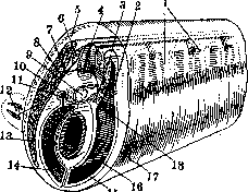
Figure 1. Transverse section of a vertebrate embryo (schematic): 1—primary segment; 2—dermatom; 3—myotom; 4—spinal cord; 5—corium; 6—ganglion; 7—ramus dorsalis; 8—ramus ventralis; 9—chorda; 10—vertebra; 11—pedicle of the primary segment; 12—limb; 13—ventral process; 14—body cavity; 15—visceral mesoderm; 16—parietal mesoderm; 17—ectoderm; 18—sclerotom.
fibers, covering the entire visceral apparatus from below and from the sides and forming its general constrictor (m. constrictor superficialis), as well as from individual deeper muscles attaching to individual visceral arches and setting them in motion (mm. adductores arcuum, interarcuales etc.). These muscles are innervated in the region of the mandibular arch by the trigeminal nerve, in the hyoid region by the facial nerve, in the region of the first gill arch by the glossopharyngeal nerve, and in the region of the following gill arches by the vagus nerve. Corresponding to the great importance of the visceral apparatus, its visceral musculature receives predominant development in the head region, whereas the somatic musculature here recedes into the background. In connection with the significant development of the jaws, the musculature of the first two arches develops especially strongly. The muscles of the mandibular arch break down into the muscle elevating the palato-quadrate cartilage (m. levator palato-quadrati; n. V), the intermandibular muscle (m. intermandibularis; n. V, VII), and the muscle adducting the lower jaw (m. adductor mandibulae; n. V). The latter gives rise in terrestrial vertebrates to all the masticatory musculature, namely the masticatory muscle proper (m. masseter; n. V), the temporal (m. temporalis), and the pterygoid muscles (mm. pterygoidei). From the anterior part of the intermandibular muscle develops the mylohyoid muscle (m. mylo-hyoideus). From the muscle of the hyoid arch in terrestrial vertebrates, the muscle attaching to the posterior angle of the lower jaw and depressing it (m. depressor mandibulae) gains special significance. In mammals, it is somewhat modified and supplemented by a part of the intermandibular muscle (m. digastricus, seu mandibulae; n. V, VII). In terrestrial vertebrates, the superficial constrictor of the hyoid arch region grows strongly, giving rise to the muscle covering the neck from below and from the sides in reptiles (m. sphincter colli; n. VII). In mammals, the superficial subcutaneous layer (platysma myoides) splits off from this muscle. This musculature is innervated according to its origin by the facial nerve, grows over the entire head region, and gives rise to the complex system of subcutaneous facial musculature, including the mimic musculature of monkeys and humans. The muscles of the gill apparatus proper, belonging to the region of the glossopharyngeal and vagus nerves, are reduced with the loss of gill respiration in terrestrial vertebrates, but are partly preserved in the form of the muscles of the hyoid apparatus and especially the laryngeal muscles. The trapezius muscle (m. trapezius, s. cucullaris; n. X), which enters into connection with the shoulder girdle, also belongs to the visceral muscles by its origin. In higher vertebrates, the posterior part of the vagus nerve innervating this muscle separates in the form of an independent accessory nerve (n. accessorius). In mammals, the lower part separates from the trapezius muscle, which receives the name of the sternocleidomastoid muscle (m. sterno-cleido-mastoideus; n. XI). - The somatic musculature of vertebrates is entirely striated, develops at the expense of the muscle layer of the myotome (primary segment), and is innervated by nerves whose fibers emerge from the ventral horns and brain nuclei as part of the ventral roots of the spinal nerves (and the corresponding cranial nerves of the III, IV, VI, and XII pairs). It retains its segmental structure in lower vertebrates throughout life. It is precisely this segmentation of the musculature that ensures sufficient mobility when swimming in water by means of lateral undulations of the body, during which individual myomeres contract sequentially. With the development of the skull and sense organs in vertebrates, however, the mobility of the anterior, head section is lost, and the somatic musculature here is reduced. Only an insignificant part of it is preserved in the form of eye muscles, developing from the anterior three head myotomes, and the hyoid musculature, developing from the ventral processes of the posterior head myotomes. In all vertebrates, there are four rectus muscles and two oblique muscles of the eye, originating: the former from the floor of the orbit on its posterior side, and the latter from the anterior wall of the orbit. The superior, medial, and inferior rectus and the inferior oblique muscles develop from the first head myotome and are innervated by the oculomotor nerve, which is the nerve of this segment. The superior oblique muscle develops from the second myotome and is innervated by the trochlear nerve, and the lateral rectus muscle develops from the third myotome and is innervated by the abducens nerve. In terrestrial vertebrates, a deeper portion separates from the latter muscle, going directly to the base of the eyeball and retracting the latter into the orbit (m. retractor bulbi). In monkeys and humans, this muscle is reduced. - The hyoid musculature sometimes still retains its segmentation in lower vertebrates and represents a longitudinal muscle mass lying on the ventral side of the visceral skeleton and connecting the ventral ends of the latter with the shoulder girdle. In terrestrial vertebrates, this musculature breaks down into a posterior section, extending from the shoulder girdle and sternum to the hyoid apparatus (m. sterno-hyoideus and m. omo-hyoideus), and an anterior section, extending from the hyoid apparatus to the chin (m. genio-hyoideus). From the latter, the intrinsic musculature of the tongue (m. genio-glossus and m. hyo-glossus) also separates. All hyoid musculature is innervated by a complex of occipital and sometimes anterior spinal nerves, which only in higher vertebrates becomes a typical cranial nerve (n. hypoglossus). The musculature of the trunk in lower vertebrates consists of a series of myomeres on the right and left sides, which form the so-called lateral muscles. The latter are divided by a horizontal connective tissue septum into dorsal and ventral muscles. In amphibians, the dorsal musculature still consists of a series of independent myomeres, but in reptiles and higher vertebrates, the myomeres break up into individual muscle bundles connecting individual parts of adjacent vertebrae (mm. interspinales, intertransversales, transverso-spinales, transverso-costales), and partly connect to form longer muscles (m. longissimus dorsi). - The abdominal musculature differentiates already in higher fish and amphibians into sections and layers, differing in the direction of the fibers. In the middle of the abdomen, a longitudinal muscle band separates—the rectus abdominis muscle (m. rectus abdominis), which is sometimes further subdivided into deep and superficial. In the lateral walls, the direction of the fibers changes and, moreover, differently at different depths: in the superficial layers, they are directed obliquely backward toward the abdomen, in the deeper layers—obliquely backward toward the back, and in the deepest layers, they assume an approximately transverse direction. Thus, the external and internal oblique muscles (mm. obliqui externus et internus) and the transverse muscle (m. transversus abdominis) develop and separate from each other. - In the region of the thoracic cage in higher vertebrates, the abdominal muscles are divided into individual sections lying between the ribs and forming the external and internal intercostal muscles (mm. intercostales). In the lumbar region, they correspond to the quadratus lumborum muscle (m. quadratus lumborum). - In front of the ribs, the same musculature appears in the form of the scalene muscles (mm. scaleni). In mammals, the external and internal intercostal muscles also give rise to further products of differentiation: the posterior serratus muscles (mm. serrati postici super. et infer.), which are specifically respiratory muscles. A respiratory muscle of somatic origin is also the diaphragm, which develops only in mammals in the transverse septum, directly behind the heart. The musculature of the limbs develops in the form of a series of epithelial muscle buds growing from the lower ends of the myotomes. The muscle buds each divide into upper and lower secondary buds, which grow into the limb primordium, break down through the loosening of the connection between cells into mesenchyme, and give rise to a continuous layer of myogenic cells on the dorsal and ventral sides of the primordium. In fish, they differentiate into muscle bundles corresponding to the rays of the fin skeleton. In terrestrial vertebrates, the muscle primordium differentiates more complexly and, furthermore, grows in the region of the shoulder girdle onto the back and chest, where it forms the "primary" musculature of the shoulder girdle. In the forelimb, from the dorsal muscle primordium, thus, develop the dorsal muscles of the shoulder girdle (m. deltoideus scapulae in front, i.e., cranially, m. dorsalis scapulae above the scapula, and m. latissimus dorsi behind, i.e., caudally) and the extensor muscles of the limb (m. anconaeus, s. quadriceps, mm. extensores). In mammals, especially in primates, at the expense of the latter, muscles also develop that turn the limb with the palm upward (mm. supinatores). - From the ventral primordium develop the ventral muscles of the shoulder girdle (m. coraco-brachialis in front, i.e., cranially, and m. pectoralis behind, i.e., caudally) and all the flexors of the limb (m. coraco-radialis, s. biceps, mm. flexores). In climbing mammals, at the expense of the latter, muscles also develop that turn the limb with the palm downward (mm. pronatores).
In addition, directly at the expense of the myomeres of the lateral muscle, a "secondary" somatic musculature of the girdle develops later, consisting of the muscle that elevates the scapula (m. levator scapulae) and the anterior serratus muscles (mm. serrati antici). Another visceral muscle, the trapezius (m. trapezius), also enters into connection with the shoulder girdle. There is no secondary girdle musculature in the hind limb. One of the muscles attaching to the free limb, however, has a secondary origin from the tail muscles: this is the piriformis muscle (m. piriformis, s. caudo-femoralis), which lies behind. The remaining muscles develop from primary muscle primordia: the adductor muscles (mm. adductores), the pectineus (m. pectineus), and the pubofemoralis (m. pubo-femoralis) in front and on the inside, and the gluteus (m. gluteus, s. ilio-femoralis) behind and on the outside. Within the free limb, the extensors (m. quadriceps femoris, mm. extensores) represent the result of the differentiation of the primary dorsal musculature, while the flexors (m. biceps femoris, mm. flexores) represent the primary ventral musculature of the limb.
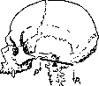
I. Schmalhausen. II. Muscles and their auxiliary apparatuses. Three types of muscle tissue are distinguished: striated (voluntary), smooth (involuntary), and cardiac (see Heart). Smooth muscle is found in the skin and in the walls of internal organs; the muscles of the skeleton are built from striated muscle; in addition, voluntary muscles are present in some other systems: the larynx, the beginning and end of the digestive tract, the end of the urethra, and the muscles of the eye and middle ear. The striated musculature of the skeleton represents a soft, clearly fibrous, reddish-brown mass that covers all parts of the skeleton, such that only a few bony prominences remain free, which are therefore easily palpable through the skin. The musculature as a whole in an adult male constitutes from 35% to 40% (in athletes more than 50%) of body weight, in a woman slightly less (30-35%), in a newborn even less (20-22%), and in an old person 30% or less (25-27%). The muscle mass is not homogeneous and breaks down into a large number (approximately 400) of individual formations-muscles. Each muscle is characterized by a certain size, shape, relationship to surrounding parts, and a definite function. The loose connective tissue located inside the muscle is called endomysium*; it passes into a thin plate that covers the muscle from the outside-perimysium** (Figs. 2 and 3). The blood vessels lying in the endomysium are very numerous and form loops elongated along the length of the muscle bundles. Muscles are so rich in vessels that when the main arterial trunks are occluded (e.g., during ligations), collateral circulation develops mainly at the expense of the muscular arteries. The muscle receives both sensory and motor nerves. The muscle is, as it were, the terminal apparatus of the motor nerve; taken together, they constitute a single whole, a single motor unit. Very often, the nerve and vessels enter the muscle at a specific place-a sort of gate (hilus), the area nervo-vasculosa (Fig. 4). In the vast majority of cases, muscles connect parts of the skeleton-bones, which are brought closer together upon contraction of the muscle; in this process, one of the bones usually does not change its position; here is located the fixed point (punctum fixum) and the origin of the muscle (origo). The second bone performs the movement, which is why the movable point (punctum mobile) and the insertion of the muscle (insertio) are distinguished on it. Mostly at their ends, a muscle has a tendon (tendo), built of dense connective tissue, the bundles of which pass into the periosteum. The tendons of broad muscles have the shape of a plate and are called an aponeurosis (aponeurosis); in long muscles, they approach the shape of a cylinder. The auxiliary apparatuses of muscles represent various types of formations of connective tissue origin that facilitate the work of the muscles in one way or another. These include fasciae, synovial sheaths, mucous and synovial bursae, pulleys and sesamoid bones, fibrous rings, retinacula, etc. In facial muscles, which are mostly embedded in adipose tissue, fasciae and tendons are absent. If muscles are arranged in several layers, the fascia divides into plates, or leaves, which can be connected to each other by so-called intermuscular septa (septa intermuscularia). Fasciae provide the origin and insertion for many muscles, adjoining the skeleton in this respect. According to Bardeleben, 2/3 of the muscles of the human body are closely connected with fasciae, i.e., their fibers either originate or attach to the fasciae. By surrounding the muscles with a dense sheath, the fasciae keep them from useless lateral movements. Fasciae are of significant practical interest: in certain cases, the course of the spread of pathological processes depends on the fascia. In certain places (at the transition from the lower leg to the foot, from the forearm to the hand), the fasciae form thickenings in the form of so-called false ligaments*; the latter, stretching between bony prominences, hold the muscle tendons in their position. It is in such places that synovial tendon channels (vaginae tendinum synoviales) develop. These are tunnels of varying length, closed at both ends, in which tendons move in one direction or the other, moreover with minimal friction, since the inner walls of the channels and the surfaces of the tendons themselves are lined with a smooth, slippery membrane. The slit-like cavity of the channels contains a minimal amount of viscous fluid. In some channels, single tendons pass, in others-two or more. Mucous bursae (bursae mucosae) have the same significance (reduction of friction, facilitation of muscle work) (Fig. 3). The latter arise in the fetal period, are located at the points of greatest mobility of the tendons, usually near the attachment of the muscles, between the tendon and the bone; the intermediate connective tissue located here loosens, and a slit-like cavity with smooth walls is obtained. Organs of movement represent a complex system of levers. Almost all musculoskeletal apparatuses are built according to the type of levers; only in some is the type of lever expressed more clearly, in others it is more complicated. One of the conditions for the action of each muscle is the relative immobility of one of its points of attachment. Thus, each preceding lever must be a support for the subsequent one, and so on. In the human body, levers of both the second and first kind are encountered. Levers of the first kind are encountered quite often in the organism; they are called levers of statics, of equilibrium. The fulcrum in them is located between the points of application of the force of muscle tension and the force of resistance. A similar case can be observed in the articulations of the pelvis with the thigh bones, between the vertebrae, in the joint of the head with the spine (articulatio atlanto-occipitalis); here the fulcrum (a) lies on the frontal axis of the articulatio atlanto-occipitalis, the force (the muscles of the nape, running from the spine to the occipital bone) is applied posterior to the fulcrum (P1), and the resistance (the weight of the head) is placed anterior to it (P) (Figure 5). In many movements, a lever of the 2nd kind takes place, where the point of application of force is located between the fulcrum and the point of resistance (Figs. 6 and 7); thus, during flexion in the elbow joint, the fulcrum lies on the frontal axis of the articulatio cubiti (a); not far from it, in the region of the upper part of the forearm, the point of application of force (Q) is placed (the place of attachment of the flexor, e.g., m. brachialis), and the resistance (the center of gravity of the forearm and hand) is located significantly more distally (P)*. A lever of this type is known by the name of...

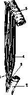
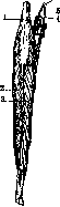
Figure 4.
Figure 2. Muscle fiber (highly magnified): 1-fibrils; 2-Cohnheim's field; 3-perimysium; 4-sarcolemma; 5-connective tissue nucleus; 6-nuclei of muscle fibers. Figure 3. Muscle between two bones: 1-tendinous fibers; 2 and 5-periosteum; 3-bursa subtendinea; 4-perimysium externum. Figure 4. Area nervo-vasculosa (m. rectus femoris): 1-ramus descendens a. circumflexae femoris lateralis; 2 and 3-lateral and medial part; 4-vein; 5-nerve branch; 6-nerve.
Figure 5.
* If the hand is lifting a load at the same time (Fig. 7), then the center of gravity, with significant weighting, will be located in the region of the load.
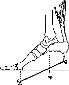
speed lever, because movements here can be performed with great speed. The work of a muscle is primarily influenced by the following factors: the thickness and length of the muscle belly, the structure of the joint, the distance between the latter and the point of muscle attachment, etc. For example, the strength of a muscle depends on its thickness, while the degree of shortening of the muscle (and consequently the height to which a load is lifted) depends on the length of the belly (see Muscles, basic mechanical phenomena). III. Classification of muscles. All the variety of forms of skeletal muscles in the human body can be combined into three main groups—long, short, and broad muscles. In addition, so-called circular muscles can be separated into a separate group. These latter are located around the natural openings of the body and are called sphincters (constrictors). Long muscles, more often with a spindle-shaped muscle belly, are usually found on the limbs, i.e., where parts of the skeleton represent long levers producing large arcs of movement. It is known that the volume
Fig. 6. movement depends, besides the relationships between the socket and the head in the joint, also on the length of the muscle and the bone lever. In those parts of the skeleton where the levers are short and the arcs of movement are very small, short muscles are found (e.g., short muscles of the back and nape). Broad muscles are plates. Their thickness is insignificant. They are found mainly on the trunk (latissimus dorsi, intercostal muscles, broad muscle of the abdomen). The last two groups (muscles of the chest and abdomen) with their mass constitute the walls of the indicated body cavities. The tendons of broad muscles more often have the character of a wide plate as well, called a tendinous expansion, an aponeurosis. In addition to the classification of muscles given above, they can also be subdivided into simple and complex. Simple muscles consist of one muscle belly, a head, and a tail. The course of their muscle bundles is quite uniform. Along with such muscles, more complex ones are also encountered. Complication in such cases is expressed in an increase in the number of bellies (poly-gastric muscles), the number of heads (biceps, triceps, and more muscles), and finally the number of tails. By complicating the form, a muscle achieves great diversity in its activity. In bi- or poly-gastric muscles, the belly (venter) is separated by an intermediate tendon (tendo intermedius) or by several transverse tendinous bridges, which determine the number of bellies, or by layers of connective tissue having a tendinous character (inscriptiones tendineae). These bridges fuse in the muscles with the fibrous sheath surrounding them (fascia propria), which creates a kind of support for each of these bellies individually. The latter, in turn, makes possible their isolated contraction and thereby diversity in work. Examples of digastric muscles in humans can be mm. biventer, omo-hyoideus; of poly-gastric muscles—m. rectus abdominis. In addition, there are also muscles in which the belly consists of several separate longitudinal muscle cones, connected to each other by tendinous layers, wedging into each other (e.g., the deltoid muscle—m. deltoideus). Muscles with several heads have several separate points of origin on one or several bones. Such muscles include, for example, the biceps muscle of the arm, starting from different parts of the scapula (m. biceps brachii), the triceps muscle of the arm (m. triceps brachii), the triceps muscle of the leg (m. triceps surae), etc. Furthermore, a muscle can start with so-called teeth (e.g., serratus muscles of the back, chest, oblique and transverse muscles of the abdomen). The different direction of the course of muscle bundles in some of these teeth makes them different from each other in function as well, and thereby introduces diversity into the work of the entire muscle as a whole. A muscle, by dividing into several tails, thus distributes the force of its pull to several points of attachment, approaching two or more bones, and such a muscle, by the contraction of its belly, causes either the simultaneous action of all its tendons (tails) or, conversely, isolates the work of each of them individually. Examples of such "dexterous" muscles can be the common flexors of the fingers of the hand, the common extensors of the hand. On the leg, the development of isolated contractions of homologous muscles is less pronounced. The process of differentiation of such muscles in phylogenetic development can reach the complete separation of a part of the muscle into an independent unit. An example of this can be the musculature of the thumb of the hand in humans and some monkeys. In animals lower in their organization and not possessing the ability for fine movements of the hand, the muscles of the thumb do not represent separate muscle bodies but are part of the general musculature of all fingers. Thus, in monkeys, m. flexor pollicis longus is not yet separated from the flexor digitorum communis, which, however, as a regressive anomaly, is also observed in humans. Borelli (1710) also introduced the concept of so-called pennate muscles. More often in muscles, the direction of muscle fibers corresponds to the direction of fibers in the tendon. However, there are also such muscles in which muscle bundles run obliquely and attach to the tendon at an acute angle. In this case, the muscle bundles approach the tendon either from one side (unipennate muscles—mm. unipennatae).
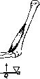
Figure 7. [unipennati] or on both sides (pennate or bipennate muscles - mm. bipennati). Examples of the former can be m. subclavius and flexor hallucis longus (the tendon in them is located on the side of the muscle belly), examples of the latter - m. rectus femoris and other muscles. In the latter case, the tendon lies inside or in the middle on the surface of the muscle. The diverse nature of the direction of muscle fibers in a muscle and their different relationship to the tendinous part complicate the determination of the resultant muscle pull and its application to levers. At the initiative of Richet (M. Ch. Richet; 1879), physiologists and some anatomists began to distinguish between strong muscles, primarily performing the functions of statics, balance of various parts of the body, and agile muscles - dynamic ones. Both differ from each other in their macroscopic structure, the way they relate to levers, and even microscopic structure (Ed. Retterer, A. Lelievre). Each muscle in the apparatus of the musculoskeletal system is the active force which sets bones in motion, while the direction of movements depends on the nature of the connection of these bones with each other, i.e., on the shape of the articular surfaces. It follows from this that the position and grouping of muscles (topographical and functional) must be in certain relationships with the structure of the joints, the number of axes in them, and the existing degrees of freedom of movement. Knowing the structure of a joint, one can predetermine the character and location of the muscles around it and vice versa - on the basis of studying muscle activity - the shape of this or that joint, the number of axes in it, etc. These relationships are functional and very dynamic. Any violation of the activity of one or another group of muscles, especially in childhood (during the period of formation and growth of bone), entails a deviation in the normal course of development of the shape of the articular surfaces. From what has been said, it is clear that the movement of an animal, and consequently the function of the musculature, models and details the shape of the articular surfaces, which arises in the fetal period, and thereby determines the degree of freedom of movement in the joints. - All muscles of the body, according to their relationship to levers and joints, can be subdivided into muscles that cross only one joint, and muscles that cross two or several joints (mono- and multi-articular muscles). Mono-articular muscles act on one joint, i.e., on one bone, on one lever. Multi-articular muscles, acting on several levers, represent by this very fact a complex system. Let us take the lower limb of a human as an example. The musculature of its entire system of levers is distributed in such a way that there are muscles of individual joints and general muscles belonging to several joints. There are muscles which connect the levers only of the free part of the limb, and there are those which connect this free part with the bones of the girdle and even the trunk. The action of the latter muscles is complex. They set in motion not only those bones to which they have a direct relationship, but also change the position of the intermediate links of this chain of levers. The number of muscles in one or another region can be so great that they are arranged in several layers. Thus, there are superficial and deep muscles. Both can in turn be subdivided further. In addition to muscles acting directly on one or another joint between the bones of the skeleton, in the body of humans and other vertebrate animals there are also such muscles which have no direct relationship to joints. To this kind of muscles can be attributed the musculature located above and below the hyoid bone, the muscles of the floor of the oral cavity, the perineum, the facial musculature. As for the relationship between muscles and the skeleton, Virchow already pointed out that muscle activity is a formative factor in relation to bones, and not only the shape of the bone, but even the relief of the outer surface of the bone is largely determined by the action of the force of muscle contraction, resp. muscle pull. In places of attachment of strong muscle tendons on the surface of the bones, tubercles, roughnesses, and processes are noted. On the other hand, muscles with their belly can exert lateral pressure on the surface of the bone, causing the formation of grooves, depressions, and pits. The degree of development of such muscle impressions on bones is in direct dependence on the degree of development of the muscles. They are more pronounced the more strongly the musculature is developed. The bones of people with strong musculature are distinguished by more sharply expressed muscle impressions. A special morphological analysis of these relationships reveals a whole series of very interesting details. IV. Variations of muscles. Variations in the muscular system are very common. On almost every cadaver, one can find one or another anomaly of muscles, and sometimes even several at once. The nature of these variants is very diverse. At the same time, there may be deviations in the shape of the muscles, in the position of their origin and attachment, in size, number, etc. Many researchers have dealt with the question of the origin of these anomalies. Some believed that race might play a role in their occurrence. Testut and others reject this assumption. Of all the classifications, the classification of Wiedersheim has been considered the most worthy of attention until recently. All variants of muscles are subdivided by him into 3 groups, and their occurrence is explained from a phylogenetic point of view. The first group includes so-called regressive muscles - those that are normally weakly expressed in humans - e.g., m. pyramidalis, muscles of the auricle. The presence of strongly expressed aforementioned muscles in a human will indicate a regression in the musculature of this section. To the second group, Wiedersheim assigns muscles of an atavistic character, i.e., cases where a muscle encountered as a variant in a human exists normally in an animal from lower vertebrates (e.g., m. sternalis). Encountered in a human, they, as it were, illustrate a return to the past. The third group - progressive anomalies. The existence of this kind of variation outlines certain prospects for the future musculature of humans. To such muscles can be attributed, for example, extra muscles of the fingers in humans (more often on the hand); strong development of facial muscles can also testify to a step forward in their differentiation. In the opinion of some authors, a variation is the result of the action of some factor in the stage of embryonic life. Knowing the gradual development of a muscle, it is possible to imagine, in the opinion of these authors, what the variation is conditioned by - whether by a simple arrest of development or a more profound change in the primordium. Racial, age, sexual, professional features of the musculature. The question of racial features of the muscular system is presented in a number of separate, unsystematized observations described in various anatomical and anthropological journals. As is known, in addition to the bony skeleton, the shape of the trunk is dependent on the development of muscles and the subcutaneous fat layer. The doctrine of the shape of "soft parts" in general (subcutaneous fat layer and muscles) constitutes one of the interesting chapters of somatology. In this regard, there is rich material in science from the point of view of its racial illumination. One can point to at least an interesting example of racial features of the lips. The matter concerns their mucous part ("Schleimhautlippen"), the development of which is not the same in different races. In some, this protrusion of the mucous membrane represents a real eversion of it. Duckworth and Hauschild see the reason for this in the protrusion of the lowest of the bundles of m. orbicularis oris. This eversion of m. orbicularis oris is weakly expressed in chimpanzees. It is more sharply developed in Melanesians, and especially in Europeans and Negroes, and in the latter it is stronger. In general, the field of studying the external forms of the body in a racial respect from the point of view of the participation in their formation of precisely these or those muscles is at the present time still little developed. - Age anatomy of the muscular system also presents more questions than factual data. It is known that a child is born already with a fully differentiated muscular system. Subsequently, only intensive growth of muscles occurs, which is expressed in an increase in the length and thickness of the primary muscle bundles. Richness in nuclei of its cellular elements is characteristic of muscle tissue of childhood. Muscles of people of senile age are characterized by atrophic changes of their muscle elements (atrophia senilis). The strength of such muscles is exhausted, the latter become flabby, sluggish. - As for the sexual differences of human muscles, only brief remarks of a most general nature are encountered in the literature on this subject. In any case, the skeletal musculature of a woman does not morphologically differ from the muscular system of a man. Due to the peculiarities of the development of the subcutaneous fat layer in a woman, it is often difficult to notice the muscle relief of her body. It is known, for example, how well muscles stand out on the body of a man, especially one who exercises his musculature. The muscles of a woman are generally weaker than those of a man. Until now, little care has been taken about the physical development of girls compared to boys. This could serve as one of the reasons for the relative muscular weakness of a woman. This also finds its reflection on the relief of the female skeleton.
The bones of a woman are less knobby; they bear the imprint of the action of muscular tension force to a lesser extent than male bones. However, women with sufficiently strongly developed musculature are often encountered. In this case, general constitutional properties, as well as professional and living conditions, are of great importance. Various types of women's clothing can deform and disfigure their musculature. For example, tight lacing of the waist causes deformation of the body. Due to tight lacing of the abdominal muscles, the relief of the latter is disturbed. The abdomen hangs down and takes on a pendulous shape. This factor also has a deforming effect on the back muscles. The professional characteristics of the human muscular system are also still a subject of research only just begun in this direction. For now, one can find only the most general indications regarding the strongly developed muscles of athletes, wrestlers, and other persons who specifically exercise their musculature by the nature of their profession; intensive muscular activity and physical labor lead to an intensification of metabolic processes in muscle tissue. As a result of increased functional load, the structure of the muscle changes functionally. The volume increases, and so-called working hypertrophy occurs. There is no doubt about the opposite fact, that, for example, in mental workers, in the absence of physical exercise of the body and general restriction of movement, muscles become flabby and their volume decreases. At the present time, in connection with the growing interest in issues of physical culture and the rationalization of labor, the study of professional characteristics of the muscular system in a functional aspect represents one of the very important and modern tasks of science.
Not all muscles develop evenly and with equal intensity. Muscle fibers thicken significantly up to the 3rd month of intrauterine life; in a newborn, they are approximately 5 times thicker than in a 2-month-old fetus, and in an adult, they are the same number of times thicker than in a newborn. In the lactation period (feeding on mother's milk), the abdominal muscles grow rapidly; in later childhood, the mass of the muscles of the tongue, palate, pharynx, and masticatory muscles begins to increase in connection with the increased function of the corresponding organs. With working hypertrophy of muscles, their fibers thicken, but their number may apparently even decrease. Muscle regeneration can also occur in the postembryonic period.
V. Methodology for examining muscles on a cadaver. Method of preserving muscle preparations. For the examination of muscles on a cadaver, the method of anatomical dissection is used. Even earlier, before the incision of the skin and the underlying layers of subcutaneous tissue and fascia, the corresponding area is studied by inspection and palpation of soft and dense parts through the skin. It is very useful in this regard to learn to project certain muscles onto the skin. An incision of the skin and fascia creates free access to the muscle. During detailed study, the fasciae are dissected separately. In order to preserve muscle preparations for a long time, cadavers are preserved before their dissection. For educational classes in the dissection room of anatomical institutes, preservation of the cadaver for long periods is not required. In these cases, simple and relatively cheap methods are sufficient to obtain the desired results (see Embalming of the cadaver). For the preservation of anatomical preparations in general and muscle ones in particular for a long time, with the preservation of their volume and, if possible, natural color, there existed many different methods even earlier*. Each of them has its disadvantages and advantages in one respect or another. Shorr's method—see Embalming of the cadaver. In 1916, Lysenkov published his method of preserving preparations without liquid, with the preservation of their volume, by impregnating them with hygroscopic substances. He found that glycerin with potassium acetate (Kalium aceticum) turned out to be the best for this purpose. This mixture resembles the Melnikov-Razvedenkov fluid, differing from it in its concentration. Lysenkov's fluid has the following composition: glycerin 500.0, plain water 1,000.0, potassium acetate 500.0 (by weight), formalin 40.0. Either freshly prepared preparations or those previously fixed with formalin for a period of up to 1 month, depending on the size, are placed in this solution. To accelerate the course of embalming, it is recommended to inject the vascular system with the specified fluid before placing the preparation in the solution. When the preparations are sufficiently impregnated with the liquid, they are removed and stored in hermetically sealed vessels or simply under a light cover (e.g., under a glass bell jar). The author's long-term observations on the preservation of the quality of his preparations (some of them were under observation for up to 20 years by the time the method was published) give reason to consider his method worthy of great attention. Muscle preparations preserved by the Lysenkov method serve as a model of anatomical technique in this regard. They are very convenient for the purpose of teaching students, as well as for storage as museum exhibits.
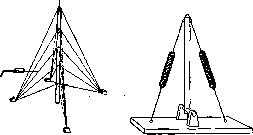
V. Tonkov, V. Dolgo-Saburov. VI. Biomechanics of the human muscular system. 1. Basic principles of the systemic arrangement of human muscles. The emergence and development of the skeletal locomotor apparatus of a vertebrate always proceeds along the path of the joint development of elements working under compression (bones, cartilage) and working under tension (muscles, tendons, ligaments). In this respect, the mechanism of the skeletal system of a vertebrate differs in principle from that of insects or crustaceans. In these animals, the shells covering individual segments are completely rigid structures working both under compression and under tension, so that only a purely locomotor function falls to the share of the muscles. In contrast to this, in vertebrates, muscles are not only a locomotor but also a necessary structural element, without which the strength of the entire skeletal structure differs little from zero; without a clear understanding of this feature, the muscular skeleton of a person is generally impossible to schematize and explain. Conversely, if one considers the human muscular system not in isolation, but in close connection with the entire totality of the structural elements of the skeleton and, moreover, pays decisive attention precisely to the static, and not the locomotor, role of the skeletal muscles, then their arrangement and structure will turn out to be very clear and well explainable. The basic static scheme, which takes place in all parts of the vertebrate skeleton, contains a compressed element (bone) in the middle, and tension elements (muscles and ligaments) on all sides around it. Such an arrangement is analogous to the arrangement of a mast with shrouds (Fig. 8). The static roles of muscles and ligaments are very close to each other*, and can be understood by considering both together. The functional difference between the two begins where the possibility of movements appears, carried out:
Figure 8. The basic structural scheme of the vertebrate skeleton—a mast with shrouds.
Figure 9. Scheme of a link on a uniaxial joint with two muscle tensioners (opposing antagonists). Compare with Fig. 16.
specifically by a muscle, and not by a ligament. Therefore, in the history of development, the processes of specialization of joints, the limitation of their mobility, and the processes of specialization of the muscle-ligamentous apparatus proceed completely in parallel: muscle tissue is replaced by simpler tendon tissue in all those tensioners which, due to the limitation of joint mobility, can no longer perform locomotor functions and are forced into a purely static role. Something similar is observed in individuals in the case of pathological disturbances of joint mobility: a muscle that has lost the ability to perform movements does not atrophy completely, but fully retains its static functions, transforming
Figure 10. Scheme of a link on a biaxial joint with three muscle tensioners.
* Methods of preparing so-called "dry preparations," "wet," "moist," enclosed in liquid, etc., are known.
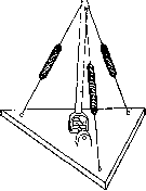
Figure 9. Scheme of a link on a uniaxial joint with two muscle tensioners (opposing antagonists). Compare with Fig. 16.
turning into a tendon-like extension. Thus, among the extensions surrounding a compressed element and its joint, necessarily on all sides, muscular properties are preserved only in some, depending on the conditions and possibilities of the joint's mobility. The cases encountered here are as follows. In a uniaxial joint, mobility is possible only in one of two directions (Fig. 9); in such a joint, we must expect two muscles, the roles of which are necessarily directly opposite. These are the so-called antagonist muscles (see Movement); it is more accurate to call them stable or permanent antagonists. All other extensions in such a joint are converted into the ligamentous apparatus. A very characteristic example of such a case is the brachio-ulnar joint (articulatio brachio-ulnaris): frontally and dorsally, it is provided with two antagonist muscles (m. brachialis int. and the medial and lateral heads of m. tricipitis br.) and two ligaments (septa intermuscularia brachii and lig. lateralia). There is a very common erroneous idea that, for the same reasons, a biaxial joint requires at least two pairs of antagonists, and a triaxial joint requires three pairs. This opinion is not only based on a misunderstanding but is directly refuted by reality (see below, muscles of the forearm). From Figure 10, which schematically depicts a biaxial joint, it is clear that for the exhaustive utilization of the mobility of such a joint, only three muscular extensions are necessary, and in each of the possible movements, one of them is an antagonist to the other two. Similarly, for a triaxial joint, only four muscular extensions are necessary and sufficient (Fig. 11). Indeed, this figure shows that various combinations of these four muscles ensure the possibility of rotation of the bone link around three mutually perpendicular axes AA, BB, and CC: muscles 1 and 2 against 3 and 4 provide rotation around axis AA, muscles 1 and 4 against 2 and 3 around axis BB, and muscles 1 and 3 against 2 and 4 around axis CC. In both of the latter cases (Figs. 10 and 11), we are no longer dealing with stable antagonists; here, depending on the type of movement, each pair of muscles can turn out to be functional antagonists or agonists for each other. From what has been said, it also follows that joints with a smaller number of axes of mobility have a more powerful ligamentous apparatus than triaxial joints (the articular capsule of the scapulohumeral joint does not contain a single auxiliary tendon bundle, and the entire joint is held by muscles), and also that the ligaments are located precisely on those sides of the joints which are free of muscles. The considerations just set forth regarding the minimally necessary number of muscles are not justified for the very important mechanical nodes of the skeleton—the scapulohumeral and hip joints, which have more than 4 muscles each. This is explainable both biologically and mechanically. Biologically (see below), these points are places where independent limb muscles and trunk muscles of the girdles meet, developing from different sources toward each other. Mechanically, this is explained by the fact that the muscles of the shoulder and thigh have to not only move and support these links themselves (this is their quantitatively smallest load) but also serve as a support for the far-removed distal links of these limbs. The hand does not work when the shoulder is loaded, but the shoulder works when the hand is loaded, and moreover, the load on the shoulder muscles in this case (due to the greater length of the levers) is 5-6 times greater than the load on the muscles of the hand itself. In connection with this, in the nodal joints between the limbs and the trunk, there are placed not only muscles ensuring movements in these joints themselves but also very powerful muscles ensuring the possibility of movements and fixations at the periphery. It is interesting that in the shoulder joint, there are 4 muscles of each purpose (see below). 2. Bibl. scheme of the muscles of the trunk tube and girdles. Trunk
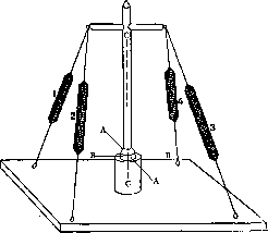
Figure 11. Scheme of a link on a triaxial (ball-and-socket) joint with four muscular extensions.
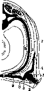
the musculoskeletal system of the human body retains a segmental structure for its entire life. Muscle segments, which originally had a comb-like appearance, retain it in the adult human in the region of the chest (mm. intercostales), having, however, replaced the original tendon intersegmental bridges with bony ones (ribs). The same segmental structure is sometimes revealed quite clearly in the region of the abdominal and back muscles (e.g., m. rhomboideus scapulae). The muscles of each segment are embryologically divided very early into dorsal and ventral groups; the latter, in turn, is stratified into three layers (Fig. 12). The main complication of the segmental - rev™,
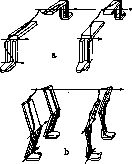
of the limb girdles, built according to a completely similar plan. However, the completely identical transverse arrangement of the fore and hind limbs (with elbows and knees outward), characteristic of reptiles, is replaced in mammals by the rotation of both pairs of limbs into the sagittal plane. In this case, the knees turn forward, and the elbows backward (1st rotation); furthermore, so that the hands of the forelimbs do not end up turned backward, there occurs an additional twisting of both bones of the forearm into a position of permanent pronation (2nd rotation). The mechanism of supination appears only in anthropoids (see Apes). The scheme of the arrangement of the limbs is fully clarified by the Braus figure (Fig. 13). The muscles of the girdles arise in two ways. Some of them begin on the proximal bone of the limb (humerus, femur) and develop in the direction of the girdle bones; these are independent limb muscles (autochtone Muskelblastem, Braus); they, like the limbs themselves, embryologically belong to the ventral wall of the body. Another group of muscles begins from the trunk and grows toward the first, fusing with the bones of the girdles and strengthening them to the trunk; these are the trunk muscles of the girdles (Körpermuskulatur, Braus), arising from the dorsal wall of the body of the embryo. Occurring either only from the ventral or only from the dorsal wall, each of these two groups forms, however, two bundles, functionally being ventral and dorsal. The independent muscles of the upper limb form first of all a pair of short bundles on the ventral and dorsal side of the shoulder (Fig. 14, 1 and 2); in humans, these are mm. infraspinatus and supraspinatus. Further
Figure 12. Schematic cross-section of the muscles of the trunk.
Figure 13. Scheme of the development of the arrangement of the limbs; a - arrangement in reptiles; b - in mammals. Straight arrow - longitudinal axis of the body, its point - head end. Small arrows - main axes of the joints, in the forelimbs the course of the n. radialis is shown schematically, in the hind limbs - n. ischiadicus and n. peronaei. (According to Braus.)
Figure 13. Scheme of the development of the arrangement of the limbs; a - arrangement in reptiles; b - in mammals. Straight arrow - longitudinal axis of the body, its point - head end. Small arrows - main axes of the joints, in the forelimbs the course of the n. radialis is shown schematically, in the hind limbs - n. ischiadicus and n. peronaei. (According to Braus.)
Figure 13. Scheme of the development of the arrangement of the limbs; a - arrangement in reptiles; b - in mammals. Straight arrow - longitudinal axis of the body, its point - head end. Small arrows - main axes of the joints, in the forelimbs the course of the n. radialis is shown schematically, in the hind limbs - n. ischiadicus and n. peronaei. (According to Braus.)
Figure 13. Scheme of the development of the arrangement of the limbs; a - arrangement in reptiles; b - in mammals. Straight arrow - longitudinal axis of the body, its point - head end. Small arrows - main axes of the joints, in the forelimbs the course of the n. radialis is shown schematically, in the hind limbs - n. ischiadicus and n. peronaei. (According to Braus.)
Figure 13. Scheme of the development of the arrangement of the limbs; a - arrangement in reptiles; b - in mammals. Straight arrow - longitudinal axis of the body, its point - head end. Small arrows - main axes of the joints, in the forelimbs the course of the n. radialis is shown schematically, in the hind limbs - n. ischiadicus and n. peronaei. (According to Braus.)
Figure 13. Scheme of the development of the arrangement of the limbs; a - arrangement in reptiles; b - in mammals. Straight arrow - longitudinal axis of the body, its point - head end. Small arrows - main axes of the joints, in the forelimbs the course of the n. radialis is shown schematically, in the hind limbs - n. ischiadicus and n. peronaei. (According to Braus.)
Figure 13. Scheme of the development of the arrangement of the limbs; a - arrangement in reptiles; b - in mammals. Straight arrow - longitudinal axis of the body, its point - head end. Small arrows - main axes of the joints, in the forelimbs the course of the n. radialis is shown schematically, in the hind limbs - n. ischiadicus and n. peronaei. (According to Braus.)
Figure 13. Scheme of the development of the arrangement of the limbs; a - arrangement in reptiles; b - in mammals. Straight arrow - longitudinal axis of the body, its point - head end. Small arrows - main axes of the joints, in the forelimbs the course of the n. radialis is shown schematically, in the hind limbs - n. ischiadicus and n. peronaei. (According to Braus.)
Figure 13. Scheme of the development of the arrangement of the limbs; a - arrangement in reptiles; b - in mammals. Straight arrow - longitudinal axis of the body, its point - head end. Small arrows - main axes of the joints, in the forelimbs the course of the n. radialis is shown schematically, in the hind limbs - n. ischiadicus and n. peronaei. (According to Braus.)
Figure 13. Scheme of the development of the arrangement of the limbs; a - arrangement in reptiles; b - in mammals. Straight arrow - longitudinal axis of the body, its point - head end. Small arrows - main axes of the joints, in the forelimbs the course of the n. radialis is shown schematically, in the hind limbs - n. ischiadicus and n. peronaei. (According to Braus.)
A second, longer pair of independent muscles arises (Fig. 14, 3 and 4); this pair partly extends beyond the bones of the girdle and attaches to the bones of the trunk—the ribs and the spine. In humans, these are the m. pectoralis major and m. latissimus dorsi. The group of trunk muscles of the girdles pushes in between the segmental muscles of the trunk (Fig. 14, 5 and 6) and the independent muscles of the upper limb. In humans, two muscles of this group (m. rhomboideus and m. serratus anterior) support the scapula (Fig. 14, 7 and 8) and one (m. subclavius) (Fig. 14, 9)—the clavicle.
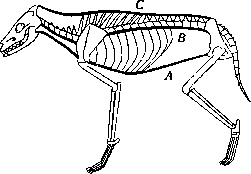
3. Muscular system of the vertebral column and trunk. It was indicated above that, in addition to the trunk muscles proper, numerous muscles of the limb girdles are located on the human trunk. The myostatic meaning of the independent muscles of the trunk is best understood from an examination of them in quadrupedal mammals. The vertebral column in the latter has the structure of an arch (Fig. 15), i.e., a structure in which the compressed part is located at the top and has a convexity facing upward; the stretched part of the structure (the tie-beam of the arch) is stretched between the heels of the arch, preventing them from spreading to the sides. The arch of the spine of a quadrupedal mammal is reinforced in two ways. Firstly, it is equipped with a tie-beam, which was originally served by a long paired muscular band stretching from the pubic symphysis along the ventral side of the trunk to the lower jaw itself (Fig. 15, A). Subsequently, this band was displaced in the thoracic segment by the sternum that developed there, and only the abdominal (mm. recti abdominis) and cervical pieces (mm. sterno-hyoideus, sterno-thyreo-hyoideus, diaphragma oris) survived from it. If the chain-like arc of the spine served only as an arch, then its links—the vertebrae—would have to be firmly wedged, like the compressed stones of an arched bridge. Strength would be achieved by this, but its consequence would be rigidity. Meanwhile, the spine of a mammal is a construction of a flexible arch not yet reproduced by technology. Therefore, the vaulted part of this structure is built not according to the type of a rigid arch, but according to the already mentioned basic type of a chain with cable stays. Such stays envelop the vertebral chain from four sides: abdominal,
dorsal, and both sides, in connection with which each of the vertebrae has an approximately cross-like shape. The abdominal stay (lig. longitudinale ant., Fig. 15, B) is purely tendinous, the other three bundles are predominantly muscular. Spinous and transverse
Figure 15. Diagram of the longitudinal stays of the trunk of a quadruped (dog): A—abdominal tie-beam; B—lig. longitud. anterius; C—tractus medialis m. erectoris trunci. The processes of the vertebrae in this diagram should be considered as ossified layers between the muscle segments; however, in humans, in the vertebral stays (mm. longissimi dorsi), the greater part of the fibers has undergone fusion of the segments and stretches not across 1, but across 2–6 segments. The entire mass of the posterior and lateral stays (mm. erectores trunci, s. longissimi dorsi), split into two cables, stretches along the entire back of a human—from the sacrum and iliac bones to the occipital bone. The posterior, or dorsal, stay (Fig. 15, C) includes the mm. interspinales and spinales (Fig. 16, A and B), running vertically, and the mm. rotatores, multifidi, semispinales (Fig. 16, C), running obliquely, in full similarity with Fig. 8. The lateral stays connect the transverse processes of the vertebrae to each other (mm. intertransversarii, partly m. quadratus lumborum, Fig. 16, D), and partly connect another variety of intersegmental layers—the ribs (mm. iliocostales, Fig. 16, E).
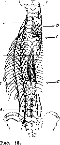
In the thoracic part of the spine, facing with its convexity toward the back, like a real arch, strength is achieved entirely by the abdominal tie-beam (Fig. 15, A), and the dorsal equipment is used only for flexibility; in connection with this, it is purely muscular. In the lumbar and cervical sections, where the spine faces with its convexity toward the abdominal side, constant static braces are necessary: in these sections, the longitudinal muscles of the back are reinforced with thick tendinous plates: fascia lumbo-dorsalis and lig. nuchae; the presence of the latter is also explained by the fact that, statically, the cervical section represents not an arch anymore, but a cantilever with a suspension at the top. Such a statically balanced system carries the entire trunk, built like a case with flexible three-layered walls. These layers and the directions of the fibers characteristic of them can be traced throughout the entire length of the trunk, from the iliac bones to the neck; in the thoracic section, they have a segmental structure, which has already been lost in the abdominal and cervical sections. The deepest layer has transverse fibers and includes the mm. transversi thoracis and transversi abdominis. The next layer has obliquely running fibers; it consists of mm. intercostales interni and mm. obliqui abdominis interni. The outer layer has a reverse oblique direction of fibers. This layer is the most extensive of all; it lies more superficially than the structural elements of Figure 15 and covers the entire neck from behind (mm. splenii), the greater part of the back (mm. serrati post., levatores costarum), the sides and the abdominal side of the trunk (mm. intercost. ext., mm. obliqui abdominis ext.), as well as the lateral sides of the neck (mm. scaleni).
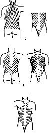
The directions of the layers of the broad muscles of the trunk are best traced in Fig. 17. The static role of these layers is determined entirely by the directions of their fibers; their dynamic functions are extremely diverse, as they exhaust all the muscles of the abdominal wall. The outer layer, which raises and spreads the ribs and tightens the walls of the abdomen, can be used entirely for inhalation; the second and partly the inner layer are suitable for exhalation (lowering the ribs and drawing in the abdominal wall). 4. Muscular system of the shoulder girdle. The structural diagrams of the shoulder and pelvic girdles have a great embryological similarity between them, but the mechanical conditions of their functioning are already very different in quadrupedal mammals.
First of all, the shoulder girdle represents two very flexible half-chains, whereas the pelvic girdle is one almost rigid closed chain. Both the one and the other girdle of a quadruped can be viewed as trestles, in the crotches of which a horizontal beam of the vertebral column is placed. In the shoulder girdle, such a crotch is formed by a chain of both clavicles, manubr. sterni and the associated elements of the muscle system; on this chain, the head end of the trunk lies in such a way that it lies like a student's satchel for which he is suspended. The diagram of the structure of the forelimbs and the shoulder girdle thus has the appearance of the letter M, with the girdle itself being analogous to a suspension bridge on two supports. In the pelvic girdle, instead of a suspension loop, there is an arch, for which the tail part of the spine—the sacrum—serves as a keystone. As befits the keystone of an arch, the sacrum is wedged between two articular surfaces of the pelvic bones diverging upward, thus forming again a crotch, this time already more rigid. The difference in the distribution of forces in both girdles resulting from this difference is quite clear. The stretching of the suspension loop of the anterior girdle tends to bring the suspension points closer together; such a structure needs an upper strut working in compression for stability. For the shoulder girdle, such a strut is the elastic barrel of the chest, against the dorsal side of which the plates of the scapulae press, distributing their pressure over a fairly large surface. The pressure on the vault or arch of the pelvic girdle tends to push its support points—the hip joints—apart; therefore, this vault, like the arch of the spine (see above), needs a lower tie-beam. Such a tie-beam in quadrupeds is the symphysis pubis, located just below the spine; in humans, as a result of upright walking, the mechanical conditions of the work of the pelvis have changed sharply (see below).
As befits a suspension structure working under tension, the shoulder girdle is supported almost entirely by muscles; its bony apparatus is very sparse, and precisely in quadrupeds, in which the shoulder girdle operates under exactly the conditions described above, the clavicles are either completely absent (ungulates) or represented by small cartilaginous inclusions (carnivores). It is worth noting that with the transition to an upright posture, the shoulder girdle gained at least as much in terms of mobility as the pelvic girdle lost. The loop of the shoulder girdle, on which the spine was suspended in the quadrupedal stance, has been freed from the load in humans and has been able to fully utilize all the flexibility inherent to it as a suspension structure. In a quadruped, the forelimb supports the torso; in a human, the torso supports the forelimb; this unloads the shoulder girdle by approximately the factor by which the arm is lighter than a quarter of the torso with the head, that is, by 3-4 times. The suspension of the shoulder girdle of quadrupeds (Fig. 18) is a wide muscle bandage on which the rib cage hangs. Figure 17. Diagram of the directions of the fiber course of the broad muscles of the torso: I—superficial layer; II—middle layer; III—deep layer. This bandage encompasses the rib cage from the ventral side and from the sides and 'fastens' on the dorsal side—at the spinous processes of the spine. To the abutments of the suspension bridge—the limbs—this bandage is suspended by the dorsal (upper) edges of the scapulae, with which it is fused along their entire length; on the ventral side, it begins from the sternum and from the lateral surfaces of the ribs. The same suspension bandage is generally preserved in humans, but due to the reduction in load, it is strongly reduced on the ventral side. It consists dorsally of mm. rhomboidei scapulae, ventrally—of mm. serrati anteriores. Dynamics of the shoulder girdle. The scapula, while it is pressed against the posterior side of the rib cage, has 3 degrees of mobility (see Movements)—adduction-abduction, elevation-depression, rotation inward and outward. Accordingly, we expect 4 muscle stays for the scapula (see above). It indeed has exactly four, arranged in a cross. Two of them, m. rhomboideus and m. serratus ant., which manage the adduction and abduction of the scapula, were described above as a bandage; the other two have anatomically one common name—m. trapezius. In connection with the transition to an upright posture in humans, the role of the suspension has passed to this second pair, which suspends the shoulder girdle to the torso; the first pair serves in humans primarily to press the scapula against the wall of the rib cage, which in quadrupeds was achieved by the weight of the torso itself; functional insufficiency of this pair leads in humans to the formation of scapulae alatae. All movements of the scapula are carried out by redistributing the tensions in the described four stays. * It is possible that the laterally compressed shape of the rib cage of quadrupeds is caused precisely by this lateral pressure of the scapulae. In humans, who do not lean on their arms, this pressure is absent, and the rib cage becomes wider in the transverse direction.
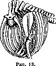
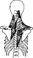
Figure 19.
Figure 18. Diagram of the muscle bandage of the shoulder girdle of a quadrupedal mammal. Figure 19. Semi-diagram of the arrangement and course of the fibers of the scapular suspension: 1, 2, 3—upper, middle, and lower portions of m. trapezii; 4—m. rhomboideus; 5—m. levator anguli scapulae. ...four stays. Adduction is performed by m. rhomboideus and the middle portions of m. trapezii, abduction—by m. serratus ant. Elevation of the scapula is performed by the upper half of m. trapezii, which is more massive than the lower half in accordance with the greater difficulty of elevation; in addition, it is assisted by a piece of the bandage that has migrated to the neck—m. levator anguli scapulae (Figure 19). Depression is performed by
the force of gravity and the lower half of m. trapezii. Biomechanically, the rotation of the scapula plays an incomparably greater role in all movements of the shoulder than its elevation and depression. Therefore, the predominant function of the upper portion of m. trapezii is not elevation, but static support of the scapula. Rotations of the scapula are performed by the coordinated actions of all four stays. Inward rotation, corresponding to raising the arm and therefore more difficult, is carried out by two pairs of pulls: 1) portions 1 and 3 of m. trapezii (see Figure 19) and 2) the lower section of m. serrati ant. together with the upper section of m. rhomboidei. Outward rotation, reinforced by gravity, is provided by one pair of pulls—the upper section of m. serrati ant. and the lower section of m. rhomboidei. All the described muscles originate on the torso and develop from it in the direction
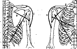
Fig. 20. Directions of the pulls of the long group of muscles of the shoulder joint. Left—view from behind, right—view from the front.
toward the limbs. Toward them, from the humerus, develop independent muscles of the upper limb, which already manage movements in the shoulder joint. Of course, if the position of the limb is fixed (for example, by grasping a stationary object), then the same muscles are capable of moving the scapula as well. Examining this group of muscles biomechanically, we once again encounter the fact (already noted in the characterization of m. trapezii) that anatomical and biomechanical muscle units can differ very significantly from each other. Sometimes what is anatomically a group of muscles is biomechanically one muscle (e.g., m. longissimus dorsi); sometimes, conversely, a single anatomical muscle is for biomechanics a whole group of muscles (e.g., m. trapezius or m. deltoideus). It was mentioned above that the independent muscles of the shoulder are divided into two groups—short muscles, ending on the bones of the shoulder girdle, and long, more massive ones, extending far onto the torso. In humans, the short muscles constitute a complete group, while the long muscles must, for the same biomechanical completeness, be supplemented by one of the short muscles, the most massive of them (m. deltoideus), which functionally does not belong to the group of short ones. The group of long muscles anatomically consists of four muscles, but biomechanically—of five pulls; its diagram is given in Fig. 20. MUSCULAR SYSTEM
Extension of the shoulder (i.e., its movement forward-outward) is performed by the thick, pennate middle portion of the deltoid muscle, a muscle that biomechanically breaks down into three independent units. Mechanically, this extending bundle does not terminate on the clavicle (as the anatomical deltoid muscle does), but continues upward from the clavicle in the form of the anterosuperior edge of the trapezius muscle. In dogs, both exist as an anatomically unified muscle, the levator humeri proprius. Flexion of the shoulder proceeds in the direction of bundle 5 (Fig. 20) of the latissimus dorsi muscle, and the oldest part of this muscle, which is biomechanically unified with it—the teres major muscle. The direction of action of the latissimus dorsi led to its colorful old name—m. aniscalptor (the wiping muscle), which is not entirely correct, as it rotates the shoulder in the direction of hyperpronation, i.e., with the palm away from the back. Another pair of shoulder movements is anteversion (lifting forward-inward) and retroversion (lowering backward-outward). Anteversion is performed by the joint action of bundles 2 and 4 (Fig. 20) (the anterior portion of the deltoid muscle and the pectoralis major muscle). Retroversion occurs with the help of bundle 3 (Fig. 20), which anatomically corresponds to the posterior third of the deltoid muscle and has a mechanical continuation above the scapular spine in the form of portion 2 (Fig. 19) of the trapezius muscle. The third type of shoulder movement, extension and flexion in the frontal plane, represents a mixture of the first two types; for example, extension of this kind is a combination of extension (forward-outward) and retroversion (backward-outward), in accordance with which the muscle pulls of the described group are used for this movement. Finally, rotation of the shoulder around its own longitudinal axis—rotation outward or hypersupination and rotation inward, or hyperpronation, are carried out by bundles 4 (pectoralis major muscle) and 5 (latissimus dorsi muscle), which are antagonists for this movement. During rotation outward or hypersupination, the tendon of the pectoralis muscle winds onto the humerus like a ribbon on a shaft; during hyperpronation, it unwinds back. It is also necessary to briefly consider the role of the elements of the shoulder girdle in shoulder movements. If the scapula were not firmly fixed during each of these movements, then all the muscle pulls just described would perform exactly the opposite of what was said about them: they would move the scapula while the arm hung motionless, since both the mass and the resistance (static moment) of the scapula are significantly less than those of the shoulder. Movements of the shoulder and the entire arm are possible only under the condition of concerted (synergistic) tension of the corresponding scapular muscles. Biomechanically, all muscle pulls that move the shoulder and the entire arm must be traced to truly fixed points of support—the spine and the rib cage. The scapula, which in quadrupeds was a truly fixed structural element held by the weight of the entire torso hanging on it, has in humans effectively turned into a bony inclusion in the thickness of the muscles connecting the shoulder to the spinal system, an inclusion that functionally resembles a kneecap. It is clear that shoulder movements do not presuppose complete, spastic fixation of all scapular muscles. On the contrary, against the background of general (tonic) fixing tension of the scapular musculature, those muscles that move the scapula in a direction concerted with the given direction of shoulder movement tense with particular distinctness (tetanically). These concerted directions are evident from the following table. Shoulder movements Scapular movements Extension (forward-outward) Flexion (backward-medially) Anteversion (forward-medially) Retroversion (backward-outward) Adduction (rotation forward relative to the vertical axis) Abduction (rotation backward relative to the vertical axis) Rotation inward Rotation outward Does not participate Lagging of the lower angle from the rib cage Abduction Adduction. Comparing the data of this table with the indications given above on the functions of the scapular and shoulder muscles, it is easy to trace the synergies encountered here. The second, short group of independent muscles of the shoulder joint is entirely, except for one muscle, connected to the scapula. All muscles of this group lie relatively deep and are almost entirely covered by representatives of the long group. Four muscles from this group, connecting the shoulder to the scapula, form four biomechanical cords; the fifth (pectoralis minor muscle) no longer has a connection to the shoulder (Fig. 21). The functions of these muscles are characterized by the following table (the numbers correspond to the designations in Fig. 21). Name Function Synergist from the long group representatives 1. Supraspinatus muscle 2. Infraspinatus muscle 3. Subscapularis muscle 4. Coracobrachialis muscle Shoulder extension Flexion (and hypersupination) Flexion (and hyperpronation) Anteversion Middle portion of the deltoid muscle Latissimus dorsi muscle (flexion and hyperpronation) Latissimus dorsi muscle Anterior portion of the deltoid muscle. During movements in the shoulder joint, both the long and short groups of muscles work. It is very likely (although not yet strictly proven) that the short group is used primarily for weak, fast, and precise movements; the long group is used for strong movements and for fixations. It should be especially emphasized that one and the same muscle, serving a multi-axial joint, can turn out to be the executor of diametrically opposite functions depending on the initial position of the limb. For example, the latissimus dorsi muscle performs the movement of hyperpronation if the shoulder was previously hypersupinated, and this same muscle rotates it in the opposite direction if the initial position of the shoulder was full hyperpronation. The middle portion of the deltoid muscle can contribute to anteversion of the shoulder if it has already been started by another muscle, and contribute to retroversion if retroversion has begun. By virtue of this, a complete description of the motor functions of the shoulder joint muscles is impossible; schematic figures 20 and 21 can contribute significantly to the analysis of the cases encountered here.
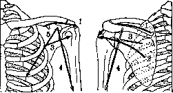
5. Muscular system of the upper limb. The musculature of the humerus, just like that of the femur, illustrates another biologically important function of the Muscular System. Both muscles of the humeroulnar joint—the brachialis muscle and the two short heads of the triceps brachii muscle—overgrow the humerus along the entire surface of its distal half. Meanwhile, to perform movements in the humeroulnar joint, a point attachment to the bones would be sufficient (similar to the attachment of the biceps brachii muscle to the radial tuberosity). Such overgrowth is an example of strengthening the bone itself with stays along the length of its diaphysis. This strengthening has real significance not only in a child with a not yet ossified humerus, but also in an adult individual; it is enough to note how much more frequent fractures are in the upper half of the humerus (surgical neck), which is not strengthened in the same way. The functions of both mentioned muscles are clear from a simple anatomical description; however, it should be noted that both the flexor and extensor of the elbow have clear biomechanical continuations in the region of the shoulder joint. The brachialis muscle begins at the outer edge of the shoulder just where the deltoid muscle attaches; together with this muscle, it forms a kind of unified muscle pull, sewn in the middle to the humerus. In a similar way, the medial head of the triceps muscle merges with the coracobrachialis muscle, which mechanically continues it upward. The biceps brachii muscle spans three whole joints and is nowhere connected to the humerus. When two of the three joints it covers are fixed, this muscle works as a simple single-joint muscle and its functions are uncomplicated. In the shoulder joint, the long head of the biceps brachii muscle causes extension of the shoulder, the short one—anteversion, both heads together—lifting of the shoulder sagittally forward. On the humeroulnar joint, due to the particularly advantageous arrangement, this muscle acts 3-4 times stronger than on the shoulder joint (Braus), causing flexion of the elbow; it develops maximum force in this joint at a right angle between the forearm and the shoulder. In relation to the radioulnar joint, this muscle works as a supinator and, moreover, a significantly stronger one than the forearm's own supinators. It is interesting to point out that the supination of the right hand corresponds in direction to the screwing in of a screw with a normal (right-hand) thread; the choice of precisely a right-hand thread as the normal one is in undoubted connection with the great supinator force of the biceps brachii muscle with a semi-flexed elbow. A persistent antagonist to this movement is the pronator quadratus muscle. The total strength of the elbow flexors in humans is about one and a half times greater than that of the extensors. Here, the same phenomenon is present as in the muscles of the scapula (see above): gravity contributes to extension and counteracts flexion. The structure of the Muscular System of the forearm is revealed with great clarity from an analysis of the mobility of the wrist. The system of wrist joints is generally biaxial, i.e., it gives two degrees of freedom of mobility (see Movements); the radioulnar joint, which determines pronation and supination, is uniaxial, i.e., it possesses
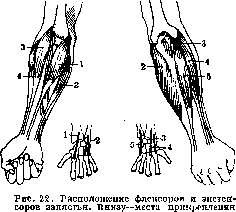
tendons. (According to Mollier.)
possesses one degree of freedom. In total, the forearm muscles, since they displace the radius and the hand, have to carry out three degrees of freedom of movement; in connection with this, they are distributed into 4 clearly expressed pulls (Fig. 22), breaking down into two paired groups. On the ventral side of the forearm and at the medial epicondyle of the humerus is placed the group of flexor-pronators; on the dorsal side and at the lateral epicondyle - the group of extensor-supinators. The tendons of both these groups bifurcate towards the distal end of the forearm and terminate respectively at the radial and ulnar edges of the wrist: the flexor group on the volar, the extensor on the dorsal side. Such an arrangement very closely reproduces the above-cited figure 11. Comparing it with Fig. 22, it is easy to verify the following scheme of action of this system of muscles: Name of muscles : The force of the pro- and supination action of these muscles is small and is used only as synergistic help to the pro- and supination specialists.--To characterize the statics of the hand, it is necessary to indicate the following. It was indicated above that in the direction of movements impossible for a given joint, muscle tissue is replaced by a ligamentous apparatus. For the biaxial radiocarpal joint, rotation of the hand around the longitudinal axis (replaced by pro- and supination) is impossible. In direct connection with this, the wrist is encompassed by a powerful tendon bracelet (apparatus ligamentosus carpi, Fig. 23), the predominant part of the fibers of which runs exactly in the direction of the missing mobility in a ring around the wrist. The intrinsic muscles of the hand do not require special systemic analysis; it is necessary to dwell more carefully only on the functions of the long finger muscles and on their synergies with the short ones (mm. lumbricales). Mm. interossei have their special simple function of abduction and adduction of the fingers and almost do not participate in the continuous flexor-extensor synergy. To analyze the movements of the fingers, they should all be divided into three types. Some of the movements are possible freely-actively, i.e., at the expense of the independent action of the muscles. Others are feasible only forced-actively, i.e., on the condition of using external forces that fix some joints and do not interfere with the freedom of others. Finally, some movements are possible only passively-at the expense of external forces alone. The combinations possible here are most clearly illustrated by the schemes of Fig. 24.-First of all, it is necessary to indicate that the long muscles of the fingers, crossing also the wrist joints, cause, besides movements of the fingers, also flexion and extension of the wrist. Like another similar multi-joint group of muscles (see below), these muscles are too short, or, better to say, have too little variability of length (contractility and extensibility), to simultaneously fully Movement Flexion Extension Abduction Adduction Pronation Supination Muscles 1 and 2 (3+4) and 5 (3+4) and 5 1 and 2 2 and 5 1 and (3+4) 1 and (3+4) 3 and 5 1 and 5 2 and (3+4) 2 and (3+4) 1 and 5 Flexor carpi radialis Flexor carpi ulnaris Extensor carpi radialis longus »
»
»
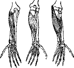
»
Figure 23. Arrangement of the long muscles of the fingers. From left to right: extensor digitorum comm. longus, flexor dig. comm. profundus, flexor dig. comm. superficialis. In all figures, the tendon bracelet (apparatus ligamentosus carpi) is clearly visible. (According to Moller.)
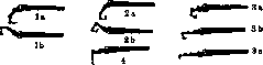
from the opponent's fist a tightly held object, trying to forcibly bend his entire hand. Contraction of the superficial flexor of the fingers causes movements in all joints through which it passes, i.e., flexion of the hand, the proximal and middle phalanges of the fingers, with extended nail phalanges (Figure 24, 1a). Freely-actively (in the air) this movement is almost impossible to perform: the deep flexor, which slightly bends the nail phalanx as well, inevitably intervenes synergistically. Forced-actively, this movement is very commonly used in small, weak movements of the fingers (the movement of striking on a typewriter, a weak piano stroke, holding a bow, rolling pills, twisting yarn, etc.). Movements requiring strength and precision are always associated with the use of the deep flexor, either alone or together with the superficial one. Freely-active contraction of this muscle leads to flexion of the hand and clenching of the fingers into a fist (Fig. 24, 2a). This movement is very difficult to perform in its pure form: the movement of clenching a fist familiar to us is coupled with extension of the hand, i.e., with contraction of the extensores carpi (Fig. 24, 2b, also 1b). This synergy depends on the central nervous system, which habitually includes the entire indicated group of muscles; it was indicated above that such synergy is advantageous for the strength of the fist clench. The actions of the deep flexor include, first of all, all kinds of grips. The strongest grips combine all flexors—both short and long, including here the mm. interossei, which, not being able to start flexion of the proximal phalanges, can, however, fix already bent fingers. Forced-active movements of the fingers under the action of the deep flexor or both long flexors can be very diverse depending on the nature of the influence of external forces. These include, first of all, strong finger strikes (medical percussion), rubbing movements (massage, scratching), strong pressure (pressing strings of musical instruments, movements of modeling, molding, medical palpation, etc.). Furthermore, any pulling with the fingers shaped like a hook is produced by the same muscles (movements of a finger on an ergograph, plucking strings, lifting a weight by a loop, etc.; Fig. 24, 3a, b, c). During all these movements, the fingers are bent in all joints; however, if the pull on the finger hook is very strong, the finger may be forced to straighten in the proximal, and then in the middle phalanges, so that only the nail phalanx remains bent. It is impossible to freely-actively bend only the terminal phalanx of a finger due to the absence of suitable muscles. Rare subjects are capable of producing such flexion, supposedly freely-actively, but in fact, even in them, it is forced-actively: they always possess the ability of hyperextension in the joint between the proximal and middle phalanx, and with such hyperextension, this joint locks in them in a kind of subluxation. Flexion of the fingers in only the metacarpophalangeal joints with the others extended (Fig. 24. 4) is performed freely-actively by the lumbrical (and interosseous) muscles. In forced-active movement (e.g., when pressing on the pistons of brass musical instruments, massage, etc.), this movement is already an undoubted synergy with the long flexors. The extensor of the fingers exists in the singular and therefore performs all types of extensions; it extends first the proximal phalanx, and then pulls up the rest. It should be pointed out that the mass of the finger flexors is greater than the mass of the extensors and therefore
In the resting position, the fingers are (tonically) semi-flexed. If one extends them with an external force or their own extensor, they will elastically return to their previous position due to the tone of the flexors. In connection with this, it should be noted as a little-known circumstance that the majority of the finest flexion movements of the fingers are performed with the help of active extension on the tone of the flexors. This primarily includes rapid writing, insofar as it is not performed by flexions of the entire hand at the wrist. The same passive (tonic) flexion after active extension performs delicate passages on the piano. Experienced teachers know this well when they train the finger extensors for fluency (exercise with high lifting of the fingers). This is one of the most striking examples of using tone for rapid movements. The biomechanics of the thumb muscles are quite clear from their purely anatomical description. 6. Muscular system of the pelvic girdle and lower limb. It was already indicated above that upon the transition to upright walking, the pelvic girdle had to adapt to new biomechanical conditions, completely different from those in which it originated and developed. The change in these conditions boils down to the following. 1) A triple load fell on the pelvic girdle, since the weight of the torso, of which in a quadruped 2/3 falls on the forelimbs and 1/8 on the hind limbs, was transferred entirely to the pelvic girdle, with the addition of the weight of the upper limbs. 2) The wedge of the sacrum, which served as the lock of the pelvic arch and was therefore cut so that its articular surfaces converged toward the abdominal side, ceased to fulfill its purpose as soon as the entire arch of the pelvis turned in the upright human almost at a right angle to the action of gravity. 3) The tie of the symphysis pubis, which lay in quadrupeds just under the sacroiliac joints, in one vertical plane with them and with the hip joints, now turned out to be in front of both, and therefore ceased to be suitable for the role of a tie. A new tie was prevented from forming by the fact that the exit openings of the body are located on the floor of the pelvis: the anus, the urethra, and, most importantly, the birth canal. All the features of the biomechanics of the human pelvis are clarified if one understands that it is a compromise, the most difficult attempt to compensate for the disturbances of the simple biomechanical scheme of the quadruped pelvis, caused by the transition to upright walking, with the help of curvatures and reformations of parts. The human pelvis adapts to new mechanical conditions in three ways: 1) the mutual arrangement of its parts changes; 2) the shape of the bones of the pelvic girdle changes; 3) ligaments are strengthened and developed in those places where this is required by the efforts arising due to the vertical position of the spine. Here it is appropriate to dwell only on the third point of this list. Since the tie of the symphysis pubis in humans lies not under the lock of the arch, but in front of it, a second tie develops behind the arch. These new ties connect the tuber ischii and spina ischiadica of each side with the sacrum (lig. tuberoso- and spinoso-sacrum), thereby depriving the sacrum of any mobility and making it a significant obstacle during the act of childbirth. Along with these ligaments, powerful ligaments also develop, spirally twisting around the hip joints (lig. ilio-, ischio-, pubo-femorale). The reason for their development is that the sacroiliac joints, despite a large compensatory rotation of the pelvis forward, still turn out to be lying 4-5 cm behind the hip axis. Therefore, the torso tends to tilt backward relative to this axis, and this tilting is counteracted by both the mentioned ligaments and the muscles acting in concert with them. The tension of both reaches 20-30 kg during upright standing, and during a tilt
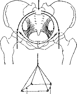
Fig. 25. Scheme of the statics of the human pelvis. Vertical arrows—load of the sacrum from the torso and support reactions of the hip joints. Curved arrows: solid—lines of compression forces, dashed—lines of tension forces. Below is a scheme of the same forces in the form of a tent.
of the torso backward can reach a value ten times greater. Thus, instead of an ordinary ring and an ordinary arch, as in quadrupeds, the human pelvis is biomechanically a double ring and a double arch (Fig. 25), i.e., something like a four-legged tent. Due to the almost complete rigidity of the pelvis as a whole, it has almost no muscular attachments; its stays are almost entirely ligamentous. Despite such a sharp difference in the structure of the shoulder and pelvic girdles, further aggravated in upright walkers by the different conditions of their mechanical functioning, the plan of the muscular equipment of both has preserved unity to an incomparably greater degree than the bony one, and reveals very clear and far-reaching homologies (see Homologous organs). To clarify them, it is necessary to trace first of all the homology of the bony elements of both girdles, indicated in the table. Shoulder girdle: Scapular plate, Clavicle, Proc. coracoid. of the scapula. Pelvic girdle: Iliac plate, Ischium, Pubis. This homology can be traced with no less clarity throughout the limb itself. Upper limb: Ball head of the humerus, Tuberculum majus, Tuberculum minus, Ulna, Proc. olecranon. Lower limb: Ball head of the femur, Trochanter major, Trochanter minor, Tibia, Patella. These bony homologies, in conjunction with the reverse rotation of the forelimbs and hind limbs in mammals indicated above (Fig. 13), will help to easily discover muscular homologies in both girdles and both pairs of limbs. Such homologies are depicted in Fig. 26, explained by the table. According to Fig. 26: Upper Limb: M. latissimus dorsi, M. teres major, M. teres minor, M. infraspinatus, M. supraspinatus, M. deltoideus (clavicular portion), M. deltoideus (scapular portion), M. triceps brachii, M. biceps brachii, M. pectoralis major (clavicular portion), M. coraco-brachialis. Lower Limb: M. psoas major, M. iliacus, M. glutaeus minimus, M. glutaeus medius, M. piriformis, M. quadratus femoris, M. glutaeus maximus, M. quadriceps femoris, M. biceps femoris, Mm. obturatores, Mm. adductores femoris. The mechanical role of the muscles listed here is in most cases fully clarified from their anatomical arrangement and from a comparison with their homologs on the upper limb. Only the adductors of the thigh deserve special illumination, reaching, in comparison with their small homolog—m. coraco-brachialis—enormous development and includ-
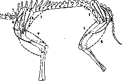
Figure 26. Scheme of homology of the muscles of the girdles and limbs of a quadruped mammal.
which include six anatomical names. By their motor function, these are antagonists to the mm. glutaei medius and minimus: they move the thigh in the frontal plane in a medial direction. However, this movement is rare and does not require great force (since it is performed in the direction of gravity) and cannot explain their enormous development. The power of these muscles becomes understandable only when evaluating their static function. Both legs together with the pelvis again represent an arch, but one already devoid of a tie-beam at the bottom. It is this tie-beam that the adductors replace: they constantly statically counteract the force of gravity of the torso, which tends to push the legs apart to the sides. The homology of the upper and lower limbs extends even further; both the structure and functions of the mm. quadricipitis femoris exactly reproduce those of the m. triceps. However, different conditions of mobility of the hip joint allow us to note in this muscle a peculiarity of multi-joint muscles, already noted above for the long muscles of the fingers, but not manifesting in the m. triceps brachii—its insufficient extensibility to serve both joints. If one bends both knees to the limit (e.g., sits on one's heels), then extending the hip joints, i.e., leaning the torso backward, will prove impossible. To an even greater extent, however, this property is revealed in the group of antagonists of the m. quadricipitis, i.e., in the group of flexors of the thigh and knee. This allows for the exceptional success of using these muscles to measure muscle tone, i.e., their passive resistance to stretching (Vereshchagin). With a flexed hip joint, free-active straightening of the knee is impossible; measurements have shown that forced (passive) straightening of the knee in this posture causes tension in the thigh flexors of up to 70-80 kg. This tension and its relation to the corresponding percentage of muscle stretching can be easily and accurately measured in vivo and can provide the most distinct numerical characteristic of the tone of these muscles. No other muscle of the body can be passively brought to such degrees of tension in vivo et intacto.—The muscular system of the lower leg and foot is very simple in arrangement and biomechanical action and does not require additional description. 7. General conditions of activity of the Muscular System during work. The preceding functional-anatomical review cannot yet give any idea of the conditions and forms of functioning of the Muscular System during actual physical work, in real, and not abstract, conditions. Below, only the most general, fundamental facts relating to such real vital activity of the Muscular System will be indicated. First of all, it is necessary to emphasize that the activity of the Muscular System under working conditions of the whole organism is unusually complex. In every motor (labor) process, the muscles participating in it in one way or another must be counted by the many dozens; at the same time, it is never possible to draw a clear boundary between muscles occupied and unoccupied in a given process. If every working muscle revealed itself, for example, as a glowing point, then no labor process would have the appearance of not only a star, but even of a whole constellation, but would resemble most of all the Milky Way—so abundant are the muscles participating in the process and so vague are the outlines of the muscle areas occupied in the work. It was indicated above that, for example, the lines of force of the m. brachialis internus are traced upward through the m. deltoideus and m. trapezius to the very neck; synergies during standing or during the joint work of the arms and legs are associated with even greater complexity. On the other hand, it is essential that almost never does a movement performed by a multi-axial joint coincide exactly in direction with the line of action of any single muscle, one anatomical or biomechanical unit. Therefore, it would be completely erroneous to imagine that movement is performed at the expense of the tension (or contraction) of one muscle with the simultaneous relaxation (or stretching) of another or others. Such a phenomenon takes place (and even then only approximately) only in the simplest uniaxial joints. In the overwhelming majority of cases, movement in a given joint, even over a small time segment of this movement, is performed at the expense of a very gradual redistribution of tensions in the entire mass of muscles surrounding this joint. If one subjects to consideration not a piece of movement, but an entire more or less prolonged movement as a whole, even a movement that does not change its direction from beginning to end, it will turn out that the leading role in the movement is extremely gradually transferred from one muscle to another, as if crawling across the entire muscle mass like a peculiar muscular relay.—The study of the distribution of static muscle tensions throughout the body (which is significantly simpler than the study of dynamic tensions, and therefore more advanced) and the analysis of the gradual changes in these tensions with increasing load and fatigue lead to a number of general conclusions, which cannot be denied fundamental importance. Under static loading of the body (e.g., holding a weight), the tensing muscles can be divided into three categories according to functional criteria. Firstly, muscles tense whose activity is directly related to holding the weight in the corresponding position—flexors of the fingers when holding weights in the hands, biceps when holding at chest level, etc. Secondly, muscles tense that do not influence the change of posture by themselves, and therefore do not perform any movement when taking the weight, but are necessary for the fixation of the girdles and the torso under conditions of an increased general load. These muscles only carry out an increase in the rigidity of those parts of the body through which the action of the load is transmitted to the supporting parts of the legs. Both described categories can be called, respectively, grip muscles and fixation muscles. Finally, in the case of an asymmetrical position of the weight (on the side or on the back, etc.), when its resultant passes to the side of the center of gravity of the body, a third functional group also tenses—balancing muscles. With a weight on the back, the mm. iliopsoas thus tense, when holding a weight at the side—the m. erector trunci of the opposite side, etc. (Fig. 27). This role usually falls to the powerful muscles of the lower segment of the torso. When studying how one and the same method of physical tension is performed by different subjects or at different moments, it is discovered that some of the muscles necessarily participate in the performance of a given method identically in all cases, while others, on the contrary, sometimes enter into work and sometimes remain inactive, being replaced by neighboring or agonistic ones, thus varying from time to time and from subject to subject. The first category is named obligatory or mandatory, the second—background muscles (Bernstein). For example,
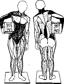
Figure 27. Static muscle tensions when holding a weight under the arm. The increase in the density of the hatching corresponds to the increase in the degree of tension. Unhatched muscles do not show a noticeable difference from unloaded standing.
when holding a weight on a stretcher, the following obligatorily tense: 1) flexors of the forearm (hand and fingers), 2) m. biceps brachii, 3) the anterior portion of the m. deltoideus, 4) m. rhomboideus scapulae, 5) the upper portion of the m. trapezius. It is easy to notice that the general outlines of all these muscles represent a kind of strap thrown over the shoulders and directly pointing to the cut that an artificial strap must have, which is capable of unloading these muscles. Along with these "regular" performers of a given work, there is always present a larger or smaller contingent of non-regular background muscles, usually recruited from among the agonists and synergists of the obligatory muscles (e.g., m. brachioradialis, m. brachialis internus, the middle and posterior portion of the m. deltoideus, mm. obliqui abdominis, etc.).—With an increase in load, as a rule, the following is observed. For any method of holding a weight, at the smallest load, the obligatory muscles enter first—either in isolation or accompanied by individual random background agonists. With an increase in load: 1) tensions in the obligatory muscles increase, reaching maximum values, sometimes accompanied by convulsive tremor; 2) the number of background muscles increases, which are inconstant both in their contingent and in the order of entry and degree of loading. As the load gradually increases, background muscles are recruited first from among the agonists and synergists of the obligatory muscles, and then from a certain moment from among the muscles completely extraneous to the given static scheme. This transitional moment of wide irradiation of background tensions is usually accompanied by the appearance of a large number of muscles tensed to the highest degree (up to tremor). Apparently, such a moment of the beginning of background irradiation can serve as an indicator of the limit load permissible for a given individual.
Related articles
Mentioned in
Cite this page
“Muscular System.” Soviet Medical Encyclopedia. English translation of Bolshaya Meditsinskaya Entsiklopediya, 1st ed. (Moscow, 1928–1936), ed. N. A. Semashko. https://sovietmedicalencyclopedia.pages.dev/article/muscular-system/