Biomechanics
Historical document, translated for reference. It reflects medical knowledge of the 1920s–30s and is not medical advice.
Summary
An overview of biomechanics from the 1920s-1930s Soviet medical perspective, covering its history, mechanical anatomy, physiology of movements, and research methods such as cyclogrammetry.
Encyclopedia article (1928–1936)
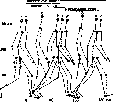
BIOMECHANICS (from Greek bios - life and mechane - machine, tool; synonyms: animal mechanics, biotechnics, physiological mechanics), a branch of general physiology that studies the development, structure, and activity of the motor apparatus of animals and humans. In accordance with these subdivisions, biomechanics falls into: 1) developmental mechanics (in this sense the term biomechanics was first used by Yves Delage), 2) mechanical anatomy, and 3) physiology of movements. The prerequisite for biomechanics is general (theoretical) mechanics, on the basis of which private questions constituting the content of biomechanics are developed. The beginning of biomechanics should be referred to the 16th century (Leonardo da Vinci); the first treatise on biomechanics (Borelli, De motu animalium) was published in Leiden in 1679. The early period of biomechanics, lasting until approximately the mid-19th century, is characterized, on the one hand, by mechanistic apriorism, and on the other, by the collection of very crude and often erroneous observations. In this period, elementary mechanical propositions relating to general physiology are established: the application of the law of the lever to the elementary osteomuscular system, the concept of the center of gravity of the body and the first attempts at its localization, the doctrine of the heart as a force pump, and the like. This period concludes with the works of the Weber brothers (W. and E. Weber), who worked on the distribution of centers of gravity of body parts and published a large monograph on the mechanics of walking (1836), now completely obsolete. The lack of apparatus and research methods retarded the development of biomechanics until the second half of the 19th century, when questions of the study of movements were one of the most energetic impulses for the development of instantaneous photography, later cinematography, on the one hand (Muybridge, Anschütz, Marey), and pneumatic transmission to recording instruments on the other. The emergence of new methods of objective registration of movements with an accuracy far exceeding the accuracy of direct observation gave an energetic impetus to the further development of biomechanics. This time is the beginning of the second period of the development of biomechanics, which can be defined as the period of collecting factual material. To this time belong the brilliant works of Marey and, especially, the important studies of the Leipzig school of biomechanics—Braune and Fischer (O. Fischer) laid the foundation for cyclogrammetry currently cultivated in biomechanical laboratories of the USSR, with its application to the study of walking (see figures 1-5). To this same period of biomechanics should be attributed the theory of joints of the animal organism and the doctrine of the structure and mechanics of bone links (Kuhlmann, Lesgaft, Triepel, Varavin, and others), which received its completion in the hands of A. and R. Fick, H. Strasser, and others. Kino- and cyclographic registration of movements at the beginning of the 20th century acquired an almost exclusively scientific-applied character and almost disappeared from the practice of general physiology laboratories, occupying instead a prominent place in the study of physical labor, sports, the fastest types of professional movements ( stenotypy, pianism), and in wartime—in prosthetic technology. Only in the very latest time is purely research interest in biomechanics and the objective study of the physiology of movements being renewed.-The third period of the development of biomechanics, which is still in its initial stage today, can be started with the methodological works of O. Fischer mentioned above and continues with the studies of E. Fischer, W. Steinhausen, R. Du Bois-Reymond, R. Grammel, N. Bernstein, and others. This period is characterized by the desire to maximize the accuracy of measurements of the movement of studied objects and to study them from the standpoint of higher mechanics, not limiting themselves to first approximations, but trying to embrace all the complexity and originality of living movement. The central question of this newest period of development of biomechanics is the question of the relationships and laws of action of muscles in a normally functioning osteomuscular system. [Figure 1 caption: Cyclogram of walking of a normal man. Right side of the body...]
[Figure 2 caption: Main consecutive phases of normal walking: a - phase of strongest support on the front leg (front push); b - phase of least pressure on the support; c - phase of greatest support on the rear leg (rear push); d - phase of uniform support on both legs.]
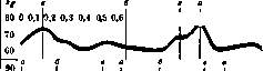
musculoskeletal system. O. Fischer had already investigated the force moments of individual skeletal muscles and the conditions of action of muscles thrown across two or more joints. The formulation of the question characteristic of the works of this period (given the change in the state of the muscle—to find the resulting movement) is beginning in modern times to be replaced by the directly opposite one, which is of incomparably greater practical interest: given real movement (e.g., in the form of a photo document)—to find the form, character, and measure of participation of muscles in this movement. The general solution of this problem was given by N. Bernstein (Abderhaldens Handbuch, Abteilung 5), but research in this direction is still at a very early stage. Direct accounting of the muscular dynamics of living movement, comparison of the picture of this dynamics on normal and pathological material, in a genetic aspect, etc., are of considerable interest, especially in view of the possibility opening up on this path to shed light on the motor role of the central nervous system and its departments. From the standpoint of biomechanics, the organism is a structure subjected to the action of internal and external forces both at rest and in motion. The main internal forces include: a) molecular forces (forces of particle cohesion, surface tension of tissue fluids, viscosity, etc.) and b) forces developed by muscles. External forces include: a) constant forces (gravity), b) variable forces of predominant direction (dynamic reaction forces, environmental resistance, air or water pressure, etc.) and c) forces without a preferential direction (external resistance in physical labor, struggle, etc.). The action of external forces and their interaction with internal ones affect the organism both permanently, exerting their influence on phylogenesis (developmental mechanics), and at each given moment, influencing the course of its movements. External forces of groups (a) and (b) cause stable adaptive changes in organisms that have not yet been studied exhaustively. This includes the distribution of bone septa and Haversian columns of bone, changes in the shapes of bone links as a result of changes in the directions of constant forces; of particular importance is the study of the origin of basic [Figure 3 caption: Curves of support reactions during walking...]
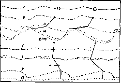
[Figure 4 caption: Pathological walking: cyclogram of a case of disbasiae lordoticae progressivae on the basis of lethargic encephalitis...]
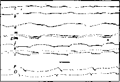
[Figure 5 caption: Pathological walking: cyclogram of a case of ataxiae gravis on the basis of multiple sclerosis...]
of structural-static schemes of the bone-muscular skeleton. The main method of modern physiology of movements is chronophotography, or chronocyclography of movements, created by Muyey and O. Fischer and modified in recent years by Gilbreth (F. Gilbreth), N. Tikhonov, and N. Bernstein. Cinematographic registration, which allows the most perfect apparatus of our time to register up to 240 positions of a moving object per second (Lehmann's 'Zeit-Lupe', A. Debrie's 'Cinema au ralenti', Mod. G. W.), still has only auxiliary value for analytical study, since measurements of cinematographic documents are extremely difficult and inevitably inaccurate. Chronocyclography consists of photographing the movement of several selected points of the organism on a stationary light-sensitive surface. Such points are usually miniature incandescent lamps, fixed above the centers of the joints of the organ being studied. When the organ moves before the open lens of the camera, the trajectories of all the lamps on the organ are photographed on the photographic plate. If the lamps are made to flash with the help of an electric interrupter or (which is much more perfect) a shutter is placed in front of the camera lens, periodically opening and closing it several dozen times per second, then the images of the trajectory will break up into series of points representing a series of instantaneous photographs of the successive positions of the lamps. In this way, frequencies far exceeding the maximum frequency of cinematographs can be easily achieved. The measurement of the scale of the photographic image and the speed of the shutter is currently performed with an accuracy of up to 0.1 mm and up to 0.00001 sec. The difficulty of fixing on a stationary photographic plate the movements of an object that does not fit entirely within the field of view (small, rhythmic movements, slow movements, etc.) led to the design of an apparatus that photographs the simultaneous movements of a series of points of the object on a slowly and uniformly moving film (kymocyclograph), which made it possible to make long-term recordings of movements inaccessible to ordinary chronocyclography (see figure 6). This method of recording proved very convenient for recording pathological disorders of movements [tremors, adiadokokineses (see figure 7), disorders of tonic motor reactions, etc.]. The accuracy of modern biomechanical recording instruments is very significant-see above. The processing of photographic records, which constitutes the content of cyclogrammetry, allows one to determine from a kymocyclogram a whole series of mechanical functions of the movement being studied, giving a very detailed analysis of the latter.
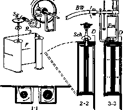
Figure 6. Diagram of a kymocyclograph and its cross-sections (1-1-cross-section, 2-2 and 3-3-longitudinal sections). F-light-sensitive film; Sg-transmission of movement to the film; D, R%-wheels, when the film comes into contact with which it moves; Et-electromagnet, pressing wheel R to й; BSha-flexible shaft connecting the apparatus with the motor; Sck-key, D-shutter of the apparatus. The entire apparatus is inserted into an ordinary camera in place of the cassette.
The difficulty of fixing on a stationary photographic plate the movements of an object that does not fit entirely within the field of view (small, rhythmic movements, slow movements, etc.) led to the design of an apparatus that photographs the simultaneous movements of a series of points of the object on a slowly and uniformly moving film (kymocyclograph), which made it possible to make long-term recordings of movements inaccessible to ordinary chronocyclography (see figure 6). This method of recording proved very convenient for recording pathological disorders of movements [tremors, adiadokokineses (see figure 7), disorders of tonic motor reactions, etc.]. The accuracy of modern biomechanical recording instruments is very significant-see above. The processing of photographic records, which constitutes the content of cyclogrammetry, allows one to determine from a kymocyclogram a whole series of mechanical functions of the movement being studied, giving a very detailed analysis of the latter.
N. Bernstein.
BIONDIOKASKA, see drlsh-Biondi okraska.
Related articles
Mentioned in
Cite this page
“Biomechanics.” Soviet Medical Encyclopedia. English translation of Bolshaya Meditsinskaya Entsiklopediya, 1st ed. (Moscow, 1928–1936), ed. N. A. Semashko. https://sovietmedicalencyclopedia.pages.dev/article/biomechanics/