Joints
Historical document, translated for reference. It reflects medical knowledge of the 1920s–30s and is not medical advice.
Summary
Joints are movable, discontinuous connections between bones in the skeleton, classified by their structure, shape, and degree of mobility. The article describes the basic elements of joints, their mechanical classification based on geometric forms, and various types including ball-and-socket, ellipsoid, saddle, cylindrical, hinge, and flat joints.
Encyclopedia article (1928–1936)
JOINTS. Joint-diarthrosis, diarthrosis (from Greek diarthroo - to separate), synonym articulatio, articulus, a movable, discontinuous connection of bones in the skeleton. Joints are one type of connection between bones (see Articulation) characterized by the presence of a space between the articulating bones, and consequently by the possibility of movement (displacement) of individual bone levers in relation to each other. Together with bones, ligaments, etc., joints are classified as the passive part of the motor apparatus. The main elements present in all so-called true joints are considered: 1) the articular surface (ends) of the connecting bones (fades articularis), 2) the articular capsule (capsula articularis), and 3) the articular cavity (cavum articulare). Each of these main elements, while having a number of common structural features, reveals its own anatomical (macro-microscopic) characteristics in different joints, characteristic only for that joint and determining its functional possibilities. gft In addition to the main elements, joints also have auxiliary (additional) structures of various anatomical construction and functional significance, some of which are more or less constant in almost all joints, while others are observed only in certain joints, and sometimes even in only one. The auxiliary structures should include: ligaments (lig. auxiliaria, s. accessoria), intra-articular cartilages (cartilagines intraarticulares, s. interarticulares), synovial (mucous) bursae (bursae synoviales, s. mucosae), etc.
Figure 1. Formation of an egg-shaped or spindle-shaped body, due to the rotation of a circular segment around a chord. (After Fick.)
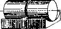
Figure 2. Formation of a cylindrical body. The straight line AB is the generatrix of the cylinder. (After Fick.) From the point of view of mechanics, the shapes of the articular surfaces of the connecting bones deserve special attention, as they play the main guiding role in the movement of individual links of certain parts of the skeleton. The analysis of the mechanics of a particular joint begins with an analysis of the shape of the articular ends of the bones, and one resorts to comparing these forms with the shapes of various geometric bodies, and based on this, in the mechanics of joints themselves (see Biomechanics), laws, formulas, definitions, and terms borrowed from physics are used. But when comparing the shapes of articular surfaces with corresponding geometric figures and
Figure 4. Formation of a block-
like body. The curve AB is the generatrix of a grooved cylinder. (After Fick.) Figure 3. Formation of a spherical body. The black semicircle is the generatrix of the sphere. (After Fick.) using this in the analysis of joint mechanics, corrections should be made, taking into account the compressibility, elasticity of the cartilaginous coverings, intra-articular cartilages, etc. Before us is a living object of study—the human (animal) joint, and not a dead body with its mechanical properties. Various geometric bodies are obtained as a result of the rotation of a certain so-called generatrix (e.g., rotation of a circular segment around a chord gives a body in the form of a spindle, ellipsoid, figs. 1-4). The surfaces of such bodies are called "surfaces of revolution." Each of these bodies, depending on its shape, has a strictly defined number of axes of rotation, and together with this, depending on the degree of its connection with another body or its free position in space, a certain number of so-called degrees of freedom (figs. 5 and 6) (see Movements). Consequently, when analyzing joints, one first notes: 1) the shape of the joint
;i(3*) smn Figure 5.
Figure 6. b Figure 5. Connection of bones by joints: dX (3°) with 3 degrees of freedom and ffll (1°) with 1 degree of freedom=4° (scheme from Fick). Figure 6. Connection of bones by joints: c-gl (3°)+gII (3°)- in total 5 degrees of freedom; б- gl (3°) + flIl(3°)+0lIII (1°)-6 degrees of freedom (scheme from Fick). surfaces, 2) the number of axes of rotation, and 3) the number of degrees of freedom, which determine the degree of mobility, the main movements in a particular joint. In some cases, it is observed that the shape of the articular surface of one of the articulating bones more or less exactly corresponds to the shape of the articular surface of the second (receiving) bone; both surfaces as if fitted to each other, coincide, they are congruent. The most congruent surfaces are the surfaces of rounded bodies, and if on one bone there is a surface in the form of a segment of a sphere, on the other - the corresponding depression, a concave platform. Articular surfaces in some joints, more often of irregular shape, not corresponding to each other, are called incongruent. According to the shape of the articular surfaces, joints are distinguished: 1) ball-and-socket joint (articulatio sphaeroidea) (fig. 7a) with the variety of nut-shaped joint (enarthrosis, s. enarthr. sphaeroidea); 2) ellipsoid, egg-shaped joint (art. ellipsoidea) (fig. 8a); 3) saddle joint (art. sellaris) (fig. 9a); 4) cylindrical joint (art. cylindroidea) (fig. 10a, 11a), with the distinction of wheel-shaped, rotational joint (art. trochoidea); 5) hinge joint, ginglymus (ginglymus) (fig. 12a), with the variety of screw joint (art. cochlearis) (fig. 13); 6) flat joint (art. plana) (fig. 14). According to the number of axes of rotation, joints are divided into multi-axial, primarily tri-axial, bi-axial, and mono-axial. The articular surface can be formed either by one bone or consist of the sum of articular surfaces of two or more adjacent bones. Thus, in some joints, only two bones articulate, in others three or more. The former are called simple joints (art. simplex), the latter complex joints (art. composita, s. complicata). An example of a simple joint can be the shoulder joint (fig. 7a), of a complex joint - the carpometacarpal joint (fig. 8a). The complexity of a joint can also be understood as the complexity of its structure or mechanics, but in this case the word "complex" is used not as a term ("complex joint"), but only as an adjective, S8 According to mobility, joints are divided into two groups: 1) freely movable, so-called arthrodiae (arthrodiae); the term "arthrodiae" is often used to denote only ball-and-socket joints (Rauber)KJiHart. mobilitatis ma-joris (R. Fick); 2) slightly movable, so-called amphiarthroses (amphi-arthrosis) (Rau
Figure 8: -axes of rotation of the ellipsoid joint (wrist joint); b -scheme (from Fick); c -scheme.
Figure 9: a-axes of rotation of the saddle joint (joint between the greater multangular bone and the 1st metacarpal bone); b -scheme (from Fick). ber) or art. mobilitatis minoris (R. Fick). The first group can include most of the joints listed above (when describing the shape of the articular surface), to the second - joints with a strong, tightly stretched articular capsule, with a strong ligamentous apparatus, restrict
a
Figure 10: a-axes of rotation of the cylindrical joint (radioulnar joint); b -scheme (from Fick). ing mobility (e.g., the joint between the lateral surface of the scaphoid bone of the tarsus and the cuboid, between the cuboid and the 3rd cuneiform, between the cuneiform bones, between the bases of the metatarsal bones) (fig. 14). Ball-and-socket joints are those in which the articular surface of one of the articulating bones approaches the shape of a sphere (a larger or smaller segment), while on the other bone there is a corresponding concave articular cavity (cavitas glenoidalis). The surfaces in such joints are congruent. These joints are the most mobile, have three degrees of freedom, and belong to the multi-axial joints. Basically, ball-and-socket joints have three main axes of rotation, intersecting at right angles at one point - the "center of rotation" and located in the anteroposterior direction, transversely, and vertically. The main movements: abduction and adduction, flexion (lifting forward), extension (lowering, abduction backward), rotations (turning) outward and inward. A typical ball-and-socket joint is the shoulder joint (fig. 7a and b) (see Shoulder joint). If in a ball-and-socket joint the articular head and cavity are more than a hemisphere, as for example in the hip joint, such joints are called nut-shaped, enarthroses (fig. 15). In terms of joint mechanics, there is no significant difference between ball-and-socket and nut-shaped joints; the axes of rotation and movements are the same. Ellipsoid joints have on one side a convex, somewhat elongated in length articular surface in the form of a segment of an ellipsoid, formed by one or several bones, and on the other side a correspondingly concave, more or less congruent with the first cavity. Such a shape of articular surfaces allows movement around two axes: one - longitudinal, the second - short, running perpendicular to the first. Ellipsoid joints have two degrees of freedom. The most clearly expressed ellipsoid joint is the wrist joint (fig. 8a, b, c) with movements - flexion and extension, abduction and adduction of the hand. To the bi-axial joints also belong the so-called saddle joints. The articular surfaces of both articulating bones are more or less the same, concave in one (longitudinal) direction and convex in the other (transverse), but are connected to each other in such a way as if two saddles were placed together. Then the concave surface of one of them would receive the convex surface of the other, and the convex surface of the first would be connected
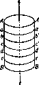
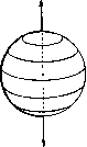
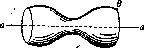
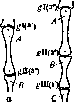
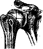
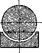
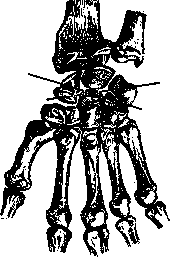
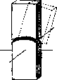
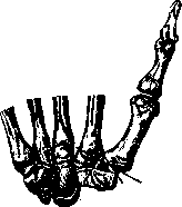
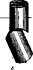
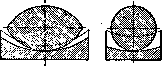
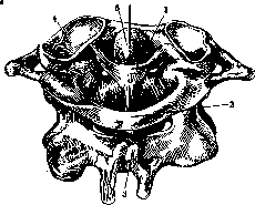
Fig. 10. Axis of rotation of a cylindrical joint (joint between the atlas and the odontoid process of the axis): 1-;transverse ligament; 2-posterior arch of the atlas; 3-axis; 4-superior articular fossa; 5-dens of the axis.
one with a concave surface of the second. A saddle joint has 2 degrees of freedom. As an example, one can take the joint between the large multangular bone of the wrist and the base of the I metacarpal bone (fig. 9a and b) with movements of flexion and extension of the thumb, its abduction and adduction (see Hand). 8» Cylindrical joints, having the shape of the articular end of one bone, comparable to a segment of a cylinder, and a concave articular surface on the other, are uniaxial joints with one degree of freedom, allowing only rotational movement and slight sliding of the cylindrical surface in relation to the articular surface. A striking example of a cylindrical joint can be considered the proximal and distal radioulnar joints (fig. 11a and b") with rotational movement of the radius in relation to the immovably fixed ulna (during pronation and supination of the hand) (see Forearm, Hand). According to the nature of movements, cylindrical joints are also called rotational, some - trochoid. In cases where there is a groove on the cylindrical surface, and a corresponding ridge on the concave surface, the joints are named pivot or trochlear joints (fig. 16a and b). These joints also have one axis of rotation, but the displacement of one bone lever in relation to the other is determined by the sliding of the guiding (movement) ridge in the guiding groove of the pivot; possibilities for lateral sliding, displacements cannot exist. Such joints include, for example, the ankle joint, interphalangeal joints of the foot (see Ankle joint, Foot) and hand (see.) (fig. 12a and b) with movements of flexion and extension. In individual pivot joints, the guiding ridge is not positioned at a right angle to the horizontal plane of the articular surface, but is placed obliquely (at various angles) (and the corresponding direction also has the guiding groove), so that a spiral surface is formed, in joints usually represented only by a small segment of it. Such joints are called spiral. They, as is evident, are merely a variety of pivot joints and therefore have one axis of rotation and one degree of freedom. The movement of one bone in relation to the other will go somewhat obliquely, theoretically with the possibility of translational movement along a spiral surface, but since in joints the spiral surface is represented by a small segment, practically lateral translational displacements can be excluded, all the more so because lateral ligaments will hinder this (see Ligaments). To spiral joints can be included, for example, the elbow joint (fig. 13) in that part where the trochlea of the humerus connects with the greater sigmoid notch of the ulna, with movements of flexion and extension of the forearm (see Elbow joint).
Figure 11: a-axis of rotation of the cylindrical joint (distal and proximal radioulnar joints): 1-semilunar notch; 2-olecranon; 3-coronoid process; 4-tuberosity of ulna; 5-tuberosity of radius; 6-interosseous crest; 7-volar surface; 8-volar margin; 9-styloid process of ulna; 10-styloid process of radius, b-diagram.
only a variety of pivot joints and therefore have one axis of rotation and one degree of freedom. The movement of one bone in relation to the other will go somewhat obliquely, theoretically with the possibility of translational movement along a spiral surface, but since in joints the spiral surface is represented by a small segment, practically lateral translational displacements can be excluded, all the more so because lateral ligaments will hinder this (see Ligaments). To spiral joints can be included, for example, the elbow joint (fig. 13) in that part where the trochlea of the humerus connects with the greater sigmoid notch of the ulna, with movements of flexion and extension of the forearm (see Elbow joint). The last group of joints according to the shape of their articular surfaces are the so-called flat joints. Flat, sometimes somewhat obliquely positioned articular surfaces of the connecting bones allow only slight sliding of the surface of one bone in relation to the other in two directions: up-down and sideways, as well as combined. The possibilities of displacement are determined, on the one hand, by the size of the articular surfaces, on the other hand-by the strength, degree of tension of the joint capsule and ligamentous apparatus. The latter moment in many flat joints is so predominant that practically these joints can
Figure 12: a-axis of rotation of the pivot joint (interphalangeal joints); b-diagram.
be classified as poorly mobile connections, which is why some of them are also called amphiarthroses (see above). To flat joints belong joints between the articular processes of the thoracic vertebrae, between the bones of the tarsus (between the lateral surfaces of the cuneiform bones), between the bases of the metatarsal bones
Figure 13. Spiral joint (joint between the trochlea of the humerus and the notch of the ulna).
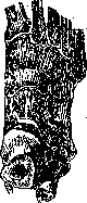
Figure 14. Flat joints - amphiarthroses (horizontal section of the foot). and others (fig. 14). In some joints, the shape of the articular surface cannot be compared with a simple geometric figure, it may be complex, composed of segments of any two bodies, for example, the complex shape of the distal end of the femur (see Knee joint). Joints in which movements occur simultaneously and when moving a bone in one joint, movement necessarily occurs in another, are called combined, for example, the right and left jaw joints, right and left atlanto-occipital joints. The articular surfaces of the connecting bones are covered with a layer of cartilage (cartilago articularis) of varying thickness in different joints (from 0.2 mm to 6 mm). In the jaw joint and sternoclavicular joint, this covering is formed by fibrous cartilage, while in all other joints it is hyaline. An explanation for this fact is given on the basis of data on the development of joints. Joints in which bones participate that in the primordial skeleton consisted of connective tissue receive a covering of articular surfaces from fibrous cartilage; in joints whose connecting bones were prefor
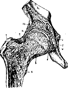
Figure 15. 1-connective tissue in the acetabular fossa; 2-head of the femur; 3-ligament of the head of the femur; 4-transverse ligament of the femur; 6 and 8-zona orbicularis; 6-minor trochanter; 7-major trochanter; 8-glenoidal labrum; 9-iliofemoral ligament.
med by cartilage, a covering from hyaline cartilage is formed. In the articular cartilage of adults, 4 layers are distinguished with different forms of cells and directions of fibers. The most superficial-with tangential arrangement of fibers, with flattened cells, the transitional zone-with round cells included in an arcuate system of fibers, the third layer-zone of radial fibers and groups of cells and the fourth-in which the fibers of the previous layer penetrate into the ossified zone of the bone-cartilage transitional layer between the articular cartilage and the bone tissue of the end of the bone (Benninghoff). In the articular cartilage of a child, the most superficial layer of flat cells is still absent. Articular cartilage is characterized by smoothness, gloss; from a mechanical point of view-elasticity, easy compressibility, ability to deform. The latter properties of cartilage are important for enhancing the congruence of articular surfaces. The physical properties of articular cartilage of children and adults are different (Baer and Gocke). The water-containing child's cartilage deforms more than the "dense cartilage of adults and the elderly.
Figure 16: a-hinge joint (section of the elbow joint); b-hinge (diagram from Fick's). The joint capsule (capsula articularis), connecting the connecting bones and encompassing their articular ends, together with the latter closes the joint cavity. In it, two
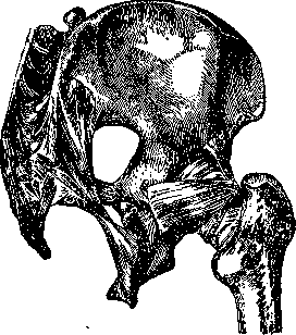
Figure 17. Extracapsular ligaments (shoulder and hip joint, view from behind).
layers are distinguished: 1) the outer layer of varying density of connective tissue elastic and collagen fibers (stratum fibrosum) and 2) the inner layer lining the capsule from the side of the joint cavity, the so-called synovial layer (stratum synoviale), synovial membrane. The first layer in most joints is attached along the edges of the articular surfaces of the connecting bones, interweaving its fiber bundles into their periosteum. The joint capsule in almost all joints is reinforced by auxiliary ligaments (see Ligament) (fig. 17 and 18) interweaving into it. In some joints, bundles of TENDON FIBERS of nearby muscles are attached to the fibrous capsule. Thickness, resistance to stretching and rupture vary in different joints of the body, fibrous fibers are usually arranged circularly and longitudinally, partly oriented in directions determined by movements in this joint. The second layer of the joint capsule-the synovial membrane-lines the inside of the fibrous capsule, tightly fusing with the latter, and covers areas
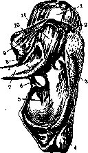
Figure 18. Bracing ligament - shoulder joint ligament (lig. coraco-acromiale): 1-humerus joint; 2-acromion; 3-
spine of the scapula; 4-infraspinous fossa; 5-superior transverse ligament of the scapula; 6-clavicle; 7-trapezoid ligament; 8-coracoacromial ligament; 9 and 10-coracoclavicular ligament.

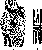
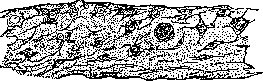
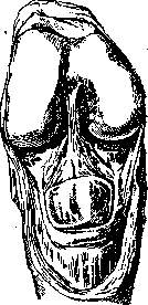
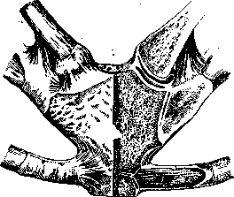
bones located within the J, but free from cartilage covering. The articular cartilage is not covered by the synovial membrane. This membrane also lines the intra-articular ligaments. Macroscopically, the synovial layer is characterized by smoothness, gloss, and is constantly moistened by fluid secreted into the cavity of the J, called synovia (synovia, syn. smegma, serum articulare). Synovia is a serous fluid that exudes into the joint cavity with desquamated and fatty degenerated cells of the articular cartilage and synovial membrane. It was previously believed that synovia was the product of special glands, taking the fatty lobules of the synovial membrane or the so-called synovial villi (fig. 19) for glands. These glands were called Haversian glands (glandulae Haversianae). Microscopically. 19. Synovial layer - cross-section. (According to Fick.) The synovial layer is composed of connective tissue with an admixture of elastic fibers, in the spaces between which are found individual fat cells, rarely cartilage cells. The synovial layer has a large number of thread-like, microscopic-sized projections, so-called synovial villi (villi synoviales), which, when detached, contribute to the formation of free bodies 'mice' in the cavity of the J (see Articular mouse). The surface of the synovial membrane is not lined with endothelium, as was previously thought, but with connective tissue (fig. 20). Into the cavity of the J from the synovial layer also project with a fatty lining (plicae adiposae), folds (plicae synoviales) (fig. 21 and 22). In many joints, the synovial membrane forms special protrusions, more often between bundles of fibers of the fibrous layer, usually located between the bone and the adjacent muscle. These protrusions of the cavity, which remain in communication with the general cavity of the J, are called synovial, mucous bursae (bursae synoviales, s. mucosae) (see Mucous bursa). Small protrusions of the synovial membrane are also called ganglia. The joint cavity (cavum articulare) appears as a narrow slit remaining between the tightly pressed articular surfaces and somewhat increasing due to the above-mentioned cavities of the synovial bursae. Cavum articulare is filled with synovial fluid, the amount of which can vary. The presence of fluid in the cavity of the J creates even greater conditions for congruence ('liquid meniscus' - Lesgaft), as well as for the adhesion of articular surfaces. To the anatomical formations, which are not observed in all joints, but are revealed as features of this or that J, should be attributed the so-called intra-articular cartilages (cartilagines intraarticulares, s. interarticulares). Two kinds of intra-articular cartilages are distinguished: discs (discus articularis) and menisci (see Articular menisci) (meniscus articularis). The disc is a cartilaginous or dense connective tissue plate of greater or lesser uniform (throughout its entire length) thickness, attached to the joint capsule and dividing the cavity of the J into two isolated chambers. Such J are called two-chambered. Discs are present in the jaw J and in the sternoclavicular J (fig. 23). Intra-articular cartilages are built from dense connective tissue or fibrocartilage. From the point of view of functional significance, discs and menisci increase or create congruence of the articular surfaces of the articulating bones, act as movable articular surfaces, soften shocks, impacts, etc. in weight-bearing joints (knee J and others), serving as springy pads between the articular ends. In mechanics
Fig. 21. Synovial folds - cross-section. (According to Fick.)
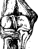
Fig. 22. Synovial and fatty folds. (Knee joint - opened from the front.)
Fig. 23. Intra-articular cartilage - disc; two-
chambered joint (sternoclavicular joint - sawn through). To the mechanics of joints, intra-articular cartilages play an essential role (especially if we consider their physical properties: compressibility, elasticity, etc.). A special adaptation for increasing the articular surfaces and for enhancing congruence is the so-called cartilaginous lip (labium, s. labrum glenoidale, cartilagineum) - a dense connective tissue rim (with cartilage cells included in places), attached to the edge of the concave articular surface of one of the articulating bones. On the side facing the cavity of the J, the cartilaginous lip is covered with a cartilaginous covering common with the rest of the articular surface. Examples: labrum glenoidale of the shoulder and hip J (fig. 15).
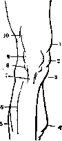
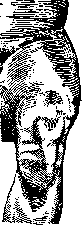
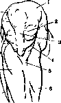
In strengthening the J, in enhancing the connections between the articulating bones, together with the joint capsule, a number of other anatomical formations and factors play an essential role: 1) Ligaments of this J, which are divided into external, extracapsular (lig. extra-capsularia; fig. 17) and intra-articular (lig. intraarticularia, s. interarticular.). Intra-articular ligaments are observed only in some J, for example lig. teres femoris in the hip J (fig. 15), lig. cruciata in the knee J (fig. 22) and others. At the points of attachment of intra-articular ligaments, the articular surfaces are devoid of cartilage covering and are woven into the periosteum. 2) Muscles surrounding that or their Fig. Fig. 2 Fig. 25. Traction of the shoulder joint capsule due to atmospheric pressure - when the head of the humerus is pulled out of the glenoid cavity of the scapula (scheme from Fick). Fig. 25. External forms of the area of the right knee joint - view from the front: 1-m. vastus medialis; 2-internal condyle of the femur; 3-internal condyle of the tibia; 4 and 5-m. gastrocnemius; 6 - extensor of the big toe; 7-lig. patellae; 8-fatty lining; 9-patella; 10-m. vastus lateralis. (According to Shadow.) tissues of the J area, such as fasciae, subcutaneous tissue, skin, etc., 4) Atmospheric pressure (fig. 24). 5) Adhesion of surfaces due to the presence of synovia in the joint cleft. For a physician, it is important to be familiar with the external forms of the area of this or that J, with the so-called configuration of the J. The protruding parts of the articulating bones (their shape, size, etc.), external ligaments, surrounding muscles with their tendons, subcutaneous fatty tissue, etc. determine the relief of the surfaces of the J area. At the same time, some J (knee, fig. 25, 26a, b and c, 27a and b, 28a and b, elbow, etc.) are more clearly configured, while others (hip,
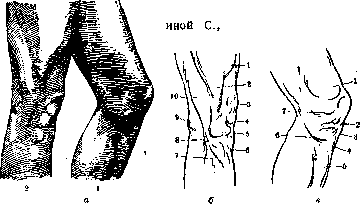
Fig. 26: a-relief of the knee joint area: 1-in strong extension; view from the outside; 2-in slight flexion; view from the inside (from Kollmann). b and c-schemes explaining fig. a; b: 1-extensor of the leg external; 2-tractus iliotibialis; 3-patella; 4-fatty capsule; 5-patellar ligament; 6-tuberosity of the tibia; 7-fibular muscle; 8-head of the fibula; 9-fat; 10-biceps muscle, c: 1-m. vastus medialis; 2-internal condyle of the femur; 3-internal condyle of the tibia; 4-tuberosity of the tibia; 5-anterior surface of the tibia; 6-m. gracilis and m. semitendinosus; 7-internal condyle of the femur. with constant tension (tonus), and even more so with contraction, actively strengthen the J, often having almost no auxiliary ligaments (for example the shoulder J). 3) Soft tissues of the
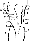
Fig. 27 a-external areas of the left knee joint (view from the front) in a semi-flexed position (from Kollmann); b-scheme explaining fig. a; b: 1-m. vastus lateralis; 2-external condyle of the femur; 3-m. biceps; 4-tibia; 5-head of the fibula; 6 and 7-m. gastrocnemius; 8-mm. gracilis and sartorius; 9-internal condyle of the tibia; 10-joint; 11-internal condyle of the femur; 12-m. vastus medialis; 13-m. adductor. shoulder and others) due to their deep location under well-developed musculature do not have a clear relief. Nevertheless, in pathological conditions (inflammatory process, dislocation, etc.) the configuration of each J, the relief of the surface, to a greater or lesser degree should change. In the configuration of J, significant age, sex, type, professional, and individual features can be noted. Joints, as movable connections of bones, play an essential role in the statics and dynamics of the body, especially of the limbs. Corresponding to the axes of rotation in J and the degrees of freedom, the muscle groups acting on this J are distributed (see Muscular system). Movements in J are divided into active and passive (see Movements). The range of motion is determined in degrees of angles, which are formed by individual bone levers, links of the skeleton, in relation to each other. The moments limiting mobility, besides pathological conditions of J, are: the presence of bony protrusions on one of the articulating bones, which at a certain moment of movement press against the other bone (e.g. limitation of flexion and extension in the elbow J, fig. 16a), the presence of restraining ligaments (fig. 18 and 13), tension of the joint capsule, etc. J have a number of features in blood supply. Most J receive arterial branches from several nearby arteries. These branches approach the joint capsule from arteries located above and in the area of this J and finally below the J [the latter have an ascending direction and are represented by the so-called recurrent arteries (aa. recurrentes)]. All arterial branches in the joint ~^
capsule form a large number of anastomoses, Iplb
and together--joint-

an arterial network (rete arteriosum articula-"V »
re) with great possibilities of collateral \ \ blood circulation. With the lymph. \ ' system, Joints stand in their own /l? t* g 13- i И- ь л ю 4 £i# 7-
O
b
Figure 28: a-external forms of the knee joint area with strong bending-view from the front (from J. Kollmann); b-diagram explaining figure a; b: 1-m. vastus medialis; 2-fatty capsule and femoral condyle; i-patellar ligament; 4-edge of the tibia; 5-inner surface of the tibia; c-calf muscles; 7-extensor hallucis longus; S-arteria tibialis anterior; 9-m. peroneus longus; 10-extensor hallucis longus; 11-edge of the tibia; 12-fatty capsule; 13-tendon of m. vasti medialis; 14-edge of the femoral condyle. figurative relationships. Some authors consider joint cavities as wide lymphatic clefts, from which networks of lymphatic capillaries begin, and then larger carrying vessels (see Lymphatic system).- Joints are the most sensitive structures in the body. Nerve endings are found in the form of numerous Vater-Pacini corpuscles. In addition to true joints between bony links of the skeleton, similar ones are also found between the auditory ossicles and between the cartilages of the larynx. In the formation of Joints, which occurs in the early embryonic stage (in the 3rd month of embryonic life) and in a 10-week embryo leads to the formation of a joint cleft, initially in the area of more centrally located and larger, later in that of smaller parts of the skeleton, there is a noticeable thickening of mesenchyme. A slit appears in the mesenchyme, which will divide this mesenchymal area into two parts, directly adjacent to the future joint surfaces of the bones to be connected. These coverings of the joint surfaces (perichondrium) later disappear, and the cartilaginous ends of the future bones lie directly against each other, enclosed in a mesenchymal sac, which then differentiates into the joint capsule. Ligaments, joint capsule, etc. in the cartilaginous stage of the skeleton represent a continuation of the perichondral layer from one segment of the skeleton to another. In cases where two cavities develop in the mesenchyme between the joint surfaces, a two-chamber Joint is formed with a cartilaginous disc between the two clefts. Underdevelopment of the central part of the disc leads to the development of menisci. Joint surfaces comparatively early take on the forms they have in various Joints of the formed organism. By the time movements begin, Joints are more or less ready, and the further influence of muscles acting on a given Joint on the formation of joint ends and other structures of the joint is insignificant.-The study of Joints is carried out by simple anatomical dissection on cadavers or after preliminary filling of the joint cavity with a colored hardening mass (see Blood vessels) or air; by making series of topographic saws of frozen cadavers, made in different planes through joint areas; by the method of fluoroscopy and radiography. Pathology of joints-see individual joints, as well as Arthritis, Gonitis, Coxitis, Synovitis, Bursitis, Rheumatism.
Related articles
Cite this page
“Joints.” Soviet Medical Encyclopedia. English translation of Bolshaya Meditsinskaya Entsiklopediya, 1st ed. (Moscow, 1928–1936), ed. N. A. Semashko. https://sovietmedicalencyclopedia.pages.dev/article/joints/