Mastication
Historical document, translated for reference. It reflects medical knowledge of the 1920s–30s and is not medical advice.
Summary
Mastication refers to the mechanical processes that break down and pulverize food in the oral cavity. This article details the anatomy, mechanics, and physiology of chewing, including the role of teeth, muscles, and the nervous system.
Encyclopedia article (1928–1936)
Mastication, the aggregate of mechanical processes that lead to the crushing and pulverization of food in the oral cavity. The main factor in mastication is the cutting and grinding of food between the teeth; the movements of the tongue and cheeks have only an auxiliary function. - Anatomy of the masticatory apparatus. The immovable upper jaw plays only a passive role in mastication, and the mechanics of mastication reduce to movements of the lower jaw. The bone-muscular system of the lower jaw can be considered as a lever of the second kind, in which the point of application of the acting force is located between the fulcrum and the resistance to be overcome. It should be noted that due to the extreme smoothness of the articular surfaces, the energy expenditure for their friction during movement is negligible. The movements of the jaw are carried out mainly by the masticatory muscles (see below). Perfect grinding of food is achieved because each cusp of an upper tooth fits into the corresponding depression of a lower tooth. Due to the fact that, starting from the front incisors, the upper teeth are shifted backward by half the width of a tooth, each upper tooth (except incisors) contacts two lower teeth. This position of the teeth is called occlusion. During chewing, the position of the teeth relative to each other changes, their articulation occurs, which is all the more perfect the more complete the contact between the upper and lower teeth. Mechanics of mastication. The lower jaw can move in three directions: up and down, forward and backward, and sideways. The first movement is carried out by the contraction of the mm. pterygoid. ext. and partly by the muscles of the base of the tongue. The closing of the jaws occurs partly due to the relaxation of these same muscles, but mainly by the contraction of mm. temporales, masseter. and pterygoid. int. It is possible that the tone of mm. pterygoid. ext. plays the role of a brake, preventing excessively strong closure of the jaws. When lowering and raising the lower jaw, its head, in addition to rotational movement, in the joint capsule, lowers (raises) somewhat, so that each point of the jaw (and therefore the teeth) moves along a curve that is not the same for all points and not concentric. With improper biting, the configuration of the articular surfaces undergoes changes, which affects the entire mechanics of mastication. The forward and backward movement of the jaw is carried out by the same muscles that lower and raise the jaw; forward movement by the simultaneous action of mm. pterygoid. ext., backward movement by the posterior third of mm. temporales. This movement of the jaw consists of the displacement of the jaw head forward and of the guiding movement which is inherent in the sliding of the front surface of the lower incisors along the back surface of the upper ones. In the ideal case during this movement, there is only sliding of the jaw head forward and, accordingly, complete contact of all the lower and upper teeth. Usually, however (especially after tooth replacement), rotational movements of the jaw head are mixed in, and the articulation of the teeth turns out to be incomplete. The degree of articulation during the forward and backward movement of the jaw can serve as a measure of the completeness of the masticatory apparatus. Lateral movements of the jaw are carried out by the contraction of mm. masseterum. In this case, the jaw head on the side toward which the jaw moves produces only a slight rotational movement, while the head on the other side lowers and shifts to the side. Processing of food by mastication. In mastication, several moments should be distinguished: biting off, cutting, and grinding food. Biting off food occurs between the incisors and is carried out mainly by the closing of the jaws, sometimes accompanied by a slight backward movement of the jaw. At the moment of biting, the greatest pressure develops between the teeth, and it is at this moment that dental prostheses easily come loose. Soft food is usually bitten off only between the incisors; harder food is shifted somewhat sideways, and in its biting off, the canines also participate, and in the case of very hard food, sometimes the lateral teeth as well. Mastication in the proper sense of the word begins after a piece of a certain size has been separated from the rest of the food. Usually, mastication begins with several rhythmic movements of the jaw, consisting of slight separation and very strong closing. This is the type of so-called cutting mastication. Almost always to a greater or lesser degree, food is ground when it is ground between the teeth. This grinding is produced by sideways movements of the jaw. Between these two types of mastication, all sorts of combinations are observed: mastication of a small piece of food occurs only on one side, of a large piece on both sides of the mouth. Whatever the type of mastication, at the end of it, the grinding of food always predominates. During mastication, very significant pressure develops between the teeth. Theoretically, this pressure (calculated from the thickness of the working muscles) equals 230 kg. However, the pressure observed in experiments was always significantly lower, though still reaching 120 kg. The pressure usually developed during chewing is even lower (for mixed food 20-30 kg). In persons with artificial teeth, Black (Black) registered with a special dynamometer during chewing of fried meat a pressure of 9-15 kg (pork), 13-18 kg (veal), during chewing of boiled meat 18-36 kg, when biting off sugar 20-54 kg and when chewing dry bread over 109 kg. When grinding food, the pressure decreases, since in this case the pressure is resolved into two forces: vertical pressure and horizontal traction. Due to this, in this type of mastication, the pressure in the mouth does not exceed 7-8 kg. With inadequate mastication, caused by loss of teeth, the efficiency of mastication sharply decreases; for example, when only 4 premolars and 4 anterior molars remain in the mouth, the efficiency of mastication decreases by 50%. Christiansen (Christiansen) developed the following method for assessing the efficiency of mastication. The subject chews 50 times a certain amount of coconut kernels of the same size and shape and then spits them out, after which the mouth is rinsed. After drying, the crushed grains are shaken through sieves of different mesh sizes, and it is determined what percentage (by weight) of the chewed coconut kernels passed through the sieves with different hole widths. The fewer teeth in the mouth, the greater the loss of the ability to finely grind food. Research conducted by the indicated method showed that best suited to their purpose are prostheses made on gold bridges, in which it is extremely important that the shape of the prostheses allows for complete contact of the teeth not only in occlusion but also in articulation. Schreder and Rumpel (Schreder, Rumpel) succeeded in preparing such prostheses. An important, although passive, role in mastication is played by moistening the food bolus with saliva, thanks to which the food bolus easily slides further down the esophagus. - Innervation of mastication. The masticatory muscles are innervated by ram. mandibularis n. trigemini. Irritation of the centripetal branches of n. trigemini coming from the oral cavity reflexively causes mastication. In the lateral part of the frontal lobe of the brain, the mastication center is described.
g. konradi.
Masticatory muscles. The masticatory muscles, numbering four, arise from one common rudiment and are derivatives of the first visceral (mandibular) arch, which explains their innervation from the nerve of this arch - the trigeminal (third branch), whereas the remaining muscles located on the skull are supplied by the facial nerve. The masticatory muscles are as follows: 1. M. masseter, the masseter muscle (fig. 1); its superficial portion begins with a powerful tendon from the zygomatic processes of the temporal and zygomatic bones and covers the anterior part of its deep portion, which originates from the zygomatic arch; they end on the roughness of the outer surface of the angle of the lower jaw (tuberostas masseterica), the body and partly its ramus (sometimes both of these portions are independent); the masseter muscle is covered by an extension of the fibrous capsule of the parotid salivary gland (fascia parotideo-masseterica). 2. M. temporalis, the temporal muscle, begins from the planum temporale of the temporal fossa and fascia, part of its bundles - and from the zygomatic arch (sometimes the origin of the muscle extends b. or m. high on the crown), its strong flat terminal tendon ends on the coronoid process of the lower jaw (processus coronoideus mandibulae; fig. 2). During its growth, the temporal muscle splits the periosteum of the temporal fossa, the superficial part of which in turn splits again, in which
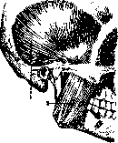
Figure 1. Fascia temporalis et m. masseter, right side: 1-fascia temporalis; 2-m. masseter. (After Spalteholz.)
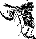
Figure 2. Mm. pterygoidei right side, from the outside: 1 - tuberc. articulare; 2 - 3-m. pterygoideus ext. et int.; 4-rhaphe pterygo-mandibularis; 5 - m. buccinator. (After Spalteholz.)
the most superficial, very dense sheet attaches to the zygomatic arch and is separated by fatty connective tissue from the proper muscular fascia. 3. M. pterygoideus externus (lateralis), the lateral pterygoid muscle (fig. 2), covered by the ascending branch of the lower jaw, originates with one head from the outer surface of laminae lateralis processus pterygoidei, the other-from crista infratemporalis, with its terminal tendon attaches to the neck of the articular process of the lower jaw, to the capsule of the jaw joint and to the intraarticular disc. 4. M. pterygoideus internus (medialis) originates from ossa pterygoidea and ends on the inner surface of the angle of the lower jaw (tuberostas pterygoidea).
p. Kiaruzin.
Related articles
Mentioned in
Cite this page
“Mastication.” Soviet Medical Encyclopedia. English translation of Bolshaya Meditsinskaya Entsiklopediya, 1st ed. (Moscow, 1928–1936), ed. N. A. Semashko. https://sovietmedicalencyclopedia.pages.dev/article/mastication/