Orthodontics
Historical document, translated for reference. It reflects medical knowledge of the 1920s–30s and is not medical advice.
Summary
Orthodontics is a branch of dentistry that studies the diagnosis, etiology, and therapy of dental position and occlusion anomalies. It aims to restore normal function of the masticatory apparatus and has both therapeutic and cosmetic significance.
Encyclopedia article (1928–1936)
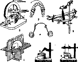
Orthodontics (from Greek orthos- straight, odous- tooth), a branch of dentistry studying the diagnosis, etiology, and therapy of anomalies of tooth position and their occlusion. In view of the fact that occlusion anomalies are accompanied by anomalies in the development of the jaws (see Teeth, pathology) and other bones of the skull, and orthodontic tooth movement contributes to changes in the shape of the jaws and face, the accepted term O. is incomplete and is often replaced by the terms "jaw orthopedics" and "facial orthopedics." Anomalies of tooth position should be distinguished from anomalies of form, size, structure, etc. of the teeth themselves (see Teeth, pathology); the latter anomalies are not the subject of O. The practical significance of O. is multifaceted. By restoring normal anatomical relationships of the parts of the masticatory apparatus, O. creates conditions for the normal physiological existence of the masticatory apparatus as a whole. With occlusion anomalies, overload and traumatization of individual teeth occur during the act of chewing. Orthodontic tooth movement, creating conditions for balanced articulation, eliminates traumatizing factors and increases the chewing force of the dental arches. In oral breathing, orthodontic treatment creates favorable conditions for the restoration of nasal breathing, since here mechanical intervention achieves expansion of the upper jaw and nasal passages and straightening of the nasal septum. The clarity of speech significantly suffers with occlusion anomalies, especially with open bite (see Teeth, pathology). Orthodontic intervention, correcting the form of the organs of the oral cavity, improves conditions for phonation. Occlusion anomalies are a factor predisposing to disease of the tissues surrounding the teeth. Filling overlapping teeth is difficult. The natural cleansing of teeth by the act of chewing and subsequent movements of the tongue, as well as their cleaning with a brush, occurs unsatisfactorily with incorrect tooth position. These conditions contribute to the deposition of dental calculus, which provides favorable ground for the multiplication of bacteria. Traumatization of tissues in abnormal interdental spaces by calculus and food particles leads to an inflammatory condition of the gums and their atrophy. Destruction of the circular ligament and formation of gingival pockets in the presence of a large number of bacteria are predisposing factors for the development of gingivitis and alveolar pyorrhea. The incorrect form of the alveolar processes, remaining untreated throughout life, also has significance after the loss of teeth. The mismatch of alveolar arches not only makes prosthetics difficult but also reduces the functional value of prostheses. In addition to all this, orthodontic measures also have cosmetic significance. Thus, the task of O. is to create conditions during the period of growth for the normal function of the entire masticatory apparatus. The latter in turn promotes the normal growth and development of the masticatory apparatus and the parts of the facial skeleton surrounding it. Until the end of the 19th century, O. had no scientific basis. In this empirical period, physicians invented apparatus of the most diverse constructions. Regulation of teeth was performed for cosmetic purposes with the help of strongly and rapidly acting apparatus. From the end of the 19th century, O. began to exist as an independent branch. Restoration of functions gradually became the main task of orthodontists. The first scientific foundations of O. were given by Angle. Teaching of O. began in 1900 with the opening of Angle's school. His orthodontic arch (see below), suitable for correcting various anomalies, received the widest distribution. Since 1909, there have been two main directions in O. The first, led by Angle, sets itself the task of correcting occlusion anomalies by means of bodily, i.e., parallel to the physiological axes, tooth movement. The second trend, led by Mershon, is called biological, since it considers the problem of O. as a problem of growth and in correcting occlusion anomalies takes into account and maximally utilizes the natural forces of the masticatory organ. Slowly and gently acting apparatus gradually replace the previous tissue-traumatizing constructions. Diagnosis of occlusion anomalies requires prior determination of what form of the masticatory apparatus should be considered normal. Forms that are correct for all times and races do not exist. With the change in the human type over centuries, the form of the human masticatory apparatus also changes. For primitive peoples, pronounced prognathia is characteristic. The diversity of forms of the masticatory apparatus depends not only on phylogenetic changes, but also on racial characteristics, but even among representatives of one race there is diversity in the forms of dental arches. During the life of an individual, the masticatory apparatus also undergoes a number of changes: during the period of growth, when changing from deciduous to permanent teeth, during the wearing down of teeth, their loss, and in senile atrophy of the jaws. On the form of the masticatory apparatus for representatives of the Caucasian race, eruption of teeth and their statics--see Teeth. The masticatory apparatus is considered normal if its individual parts are in a certain correspondence with each other. To determine the correctness of the relationship of the parts of the masticatory apparatus, it is necessary 1) to select its parts that can serve for the required calculations, and 2) to develop certain measurement methods. As the quantities to be measured, the transverse diameters of certain teeth and the distances between certain points of the jaws usually serve. The principles of determining the norm in O. are based on variation statistics and are similar to the principles of general clinical anthropometry. The most common, average variants are taken as the norm, and the extreme variants are considered anomalies. The absolute numerical values of teeth and jaws obtained are insufficient for determining the norm or anomaly. The indicators of the latter are relative values, or indices. At present, the indices of Pont (1907) have the greatest practical significance for orthodontic diagnosis: one for premolars, the other for molars. The first determines the ratio of the diameter of the four incisors at their maximum width to the distance between the right and left premolars. Pont multiplies the sum of the transverse diameters of the incisors by 100 and obtains the numerator of the fraction. The distance between the centers of the first premolars is the denominator of the fraction. When the fraction is reduced, a number is obtained that is the index for premolars. The same is done for molars. The normal index for premolars = 80, and for molars = 64. Pont developed a table according to which, knowing the total width of the incisors, one can find the normal distances between premolars and between molars for a given case. Before starting orthodontic treatment, according to Pont's table, one can make a drawing of the dental arch normal for the given case. In addition to Pont's index, to determine the normal individual dental arch, indicators developed by a number of authors are also used. They all consider the dental arch taken in isolation from the head. Izard, considering it erroneous to solve the problem of the normal dental arch without taking into account the form and size of the face, measures a significant number of skulls and finds that in individuals possessing teeth of the same transverse diameter, dental arches have different forms, but between measurements of the latter and measurements of the face there is a constant correspondence. The latter applies both to the width of the arch and face, and to their length (the depth of the face is implied). Izard came to the conclusion (1924) that the maximum width of the dental arch usually equals half the distance between paired zygomatic points. Maximum width of the dental arch × 100 / Maximum distance between paired zygomatic points = index of transverse measurement of the dental arch and face. During the period of growth, this index changes somewhat, but remains around 50. As for the length of the dental arch, Izard found (1927) that it usually represents half the incisor-ear radius, measured in projection on the occlusal plane. The index of longitudinal measurement of the dental arch and face thus also equals 50 (approximately). Based on these measurements, Izard, with the help of an ellipsograph, draws the dental arch in the form of a part of an ellipse, the minor axis of which equals half the distance between paired zygomatic points, and the major axis equals the incisor-ear radius. Izard's indices show the relationship between the form of the face and the form of the dental arch, but most modern researchers are not satisfied with this and seek ways, based on three dimensions, to accurately determine the position which the dental arches and their parts occupy in the skull. These measurements are made from planes intersecting the skull and located perpendicular to each other (Figure 1). These planes are as follows: 1) the sagittal plane, passing along the sagittal suture and determined in a living person by the raphe. 2) The Frankfurt horizontal, which is obtained as follows: the infraorbital point, easily palpable under the skin, is connected with the tragion point, and thus the Frankfurt horizontal line is obtained. Through the right and left horizontal lines, a plane is drawn. With the head in normal position, this plane is parallel to the earth's horizon.
To judge the normality of occlusion, the patient's head must be in this position. 3) The frontal, orbital plane, proposed by Simon, which passes through both orbital points and descends perpendicularly to the horizontal plane. Based on the sagittal plane, the distance between corresponding dental points in the right and left halves of the dental arches is established. Thus, the presence of transverse symmetry or asymmetry, constriction or expansion of the dental arches (according to Pont's index), and consequently their shape is determined. Based on the horizontal plane, the following are established: 1) the distance from it of certain points of the occlusal surfaces along a vertical line, 2) the angle between the horizontal plane and the occlusal surface, and 3) the form of the occlusal curve. Based on the orbital plane, the following relationships are established: 1) the intrajaw sagittal relationship. We have sagittal symmetry if corresponding points of the right and left sides of the jaw lie on the same transverse line, as for example the orbital plane normally passes approximately through the tips of the upper canines (Simon). 2) The interjaw sagittal relationship is expressed in the antagonism of individual teeth or dental arches (see Teeth, eruption of teeth and their statics). It is the only measure of occlusion anomalies for many authors, including Angle. 3) The tooth-cranial sagittal relationship. The degree of prognathism of the dental and alveolar areas can be judged by what part of the dental arches is intersected by the orbital plane. Of modern orthodontic diagnostic methods, the most practically valuable is Simon's method (1922). With the help of Simon's instruments, based on the three planes of the skull, cephalic measurements are made, establishing deviations from the norm in the masticatory apparatus. These measurements are made on gnathostatic models and photostatic images. Gnathostatics is called Simon's method of reproducing occlusion, and photostatics is his method of applying photography for orthodontic diagnosis. The following instruments are used to obtain gnathostatic models: 1) gnathostat (see), the arch of which is located in the horizontal plane; the upper gnathostatic model is made in such a way that its base corresponds to this plane; 2) the orbital bar (fig. 2), which is fixed to the gnathostat and serves for marking the orbital plane on the model; 3) the symmetrograph (fig. 3), equipped with a pin which, based on the raphe, marks the sagittal plane on the upper and lower models. The symmetry of the dental arches in the sagittal and transverse directions is checked by means of a horizontal sliding caliper. Guided by Pont's index, one can establish the presence of transverse symmetry or asymmetry, constriction or expansion of the dental arches. Other measurements on gnathostatic models are made using Simon's 'diameter', which is used in combination with two sliding calipers: horizontal (fig. 4) and vertical (fig. 5). With the help of the horizontal caliper, the distance between certain dental points of the dental arch, projected on the horizontal plane, i.e., on the horizontal bar of the caliper, is measured. With the help of the vertical caliper, the direct distances between the same dental points and the horizontal plane (i.e., the base of the upper model) are measured. Simon's method makes it possible to reproduce the individual occlusion curve (occlusal curve), as well as the curve of the palate in the form of diagrams. Diagrams successively drawn during the stage of orthodontic treatment serve as a visual indicator of its success.
Simon's method makes it possible to reproduce the individual occlusion curve (occlusal curve), as well as the curve of the palate in the form of diagrams. Diagrams successively drawn during the stage of orthodontic treatment serve as a visual indicator of its success.
The photostatic method fills the gaps in gnathostatic research; for example, the inability to determine the position of the jaw in the skull using only the gnathostatic method is such a gap. Before photographing, small pieces of black tape are applied to the face at the points necessary for measurements (tragus, orbital, and mandibular). Photographs should always be taken with the same orientation relative to the three cranial planes. Special photostatic equipment is used for this purpose. On the resulting negatives, the necessary lines are marked by scratching and measurements are made. - Some authors propose various other methods instead of photostatics, which complement gnathostatics. Among them, the method of making 'profile masks' proposed by Trifus has attracted particular attention. He saws the gnathostatic model along the sagittal plane and fixes it to the 'profile mask' according to the position occupied in this case by the dental arches in the head. In orthodontic diagnosis, so-called graphic methods are also common. Their proponents believe that for certain tooth sizes, there should be a certain arch shape. Representatives of the graphic method, based on measurements of teeth and other mathematical data, draw diagrams of the upper dental arch, and determine the lower arch based on the upper one. For practical application of the graphic method, Herbst has ready-made diagrams of different sizes printed on transparent celluloid plates. To compare the individual dental arch with the diagram on the model, the incisal edges and buccal cusps of the teeth are marked with a colored pencil, and then the diagram is placed over it to compare the dental arch with it. Bogue applied the graphic method to deciduous teeth. - Among the methods of orthodontic diagnosis, the method of Angle, which appeared in the 1890s, has been and still is the most popular. The diagnostic basis of Angle, i.e., the invariably correct position of the upper first molars, is now considered unsatisfactory according to anthropological data. The upper jaw along with its teeth is no exception to the other organs of the human body, and cases of its abnormal development are not uncommon; they occur both with normal and with abnormal conditions of the lower jaw. A number of authors have proposed classifications of malocclusion. The basis of almost all of them is the morphological principle. The classifications of Angle (1889) (see Teeth, pathology) and Simon (1922) are of the greatest importance. Angle's classification is based on the mesio-distal relationship of the dental arches, while all transverse displacements, including narrowing of the dental arches, observed in 90% of all malocclusion cases, as well as vertical displacements, such as open bite, according to Angle belong to his Class I, i.e., they remain essentially unsystematized. As for Simon's classification, it is based on a more rigorous system. Its scheme is as follows. A. Deviations measured from the sagittal plane: 1) narrowing and 2) widening. B. Deviations measured from the orbital plane: 1) mesial position and 2) distal position. C. Deviations measured from the horizontal plane: 1) high position of the masticatory apparatus (i.e., too close to the horizontal plane) and 2) low position (i.e., too far from the horizontal plane). From the above, it is clear that in orthodontic diagnosis, main attention is paid to objective indirect research, primarily the study of models, by means of which the presence of malocclusion, its type, location, and size are established. Indirect research also includes the study of facial photographs (see above) and radioscopy, which reveals the condition of the roots, alveolar processes, the position of unerupted teeth, etc. Direct examination, which gives a clinical picture of the anomaly, is also of essential importance. All parts of the masticatory apparatus are examined, and the role of deciduous teeth in the development of the jaws is taken into account. Further attention is paid to the condition of adjacent cavities. Usually there is a connection between anomalies of the masticatory apparatus and pathological conditions of the nose, nasopharynx, and pharynx. Oral breathing is most often observed between the ages of 3 and 14 years, i.e., during the period of development of the jaw apparatus. With oral breathing, the normal growth of the upper part of the facial skull is disrupted, and if nasal breathing is restored in adulthood, the developmental anomalies of the jaws nevertheless remain. In clinical examination, it is necessary to take into account the constitution (see) and physical development of the individual, and one can be guided by indices (see Indices of physical development). - There is a close connection between malocclusion and the general condition of the body, and often these anomalies are a symptom of the main general disease. Such cases require the orthodontist to cooperate with a pediatrician, rhinologist, sometimes a surgeon, and other specialists. During questioning, it should be clarified whether the child has the habit of sucking fingers or tongue. From the habitual position during sleep, for example, tucking a fist under the cheek, as well as from the mentioned habits, displacement of a group of teeth can occur. From the above, it is clear that malocclusion is the result of the combined action of a number of conditions. In cases where the main role in the origin of the anomaly belongs to external, mechanical factors, the prognosis is favorable. In such cases, the teeth usually take an incorrect position primarily, and the jaws are secondarily involved in the anomaly. In cases where the decisive role in the occurrence of the anomaly belongs to internal factors, the prognosis largely depends on the possibility of curing the underlying disease. The prognosis also depends on the age at which treatment is undertaken. The most favorable period for orthodontic treatment is the period of intensive growth, i.e., the age of 6 to 9 years; between 9-12 years, the use of orthodontic appliances is difficult, because at this age the teeth are not very stable: the roots of deciduous teeth are already resorbed, and the roots of permanent teeth are not yet fully developed. In the period between 12 years and the onset of maturity, orthodontic appliances can promote the development of underdeveloped jaws, but here greater use of mechanical forces is required than in early childhood. In orthodontics, as in other fields of medicine, the preventive approach and the replacement of individual forms of treatment with mass treatment forms are most appropriate. The essential period of formation of the jaw apparatus belongs to intrauterine life, hence the importance of dietetics of pregnancy for the development of the infant's jaws. Further, the hygiene of infancy (see Social hygiene, Protection of motherhood and infancy) is important. The physician who improves the general condition of the child, cures him of rickets, eliminates oral breathing, etc., at the same time eliminates factors that interfere with the normal growth of the jaws. In addition to the general measures mentioned, there are a number of local preventive measures. Children at the age of changing from deciduous to permanent teeth especially need the supervision of a specialist. Sometimes simple measures lead to spontaneous, physiological regulation of teeth, for example, timely extraction of deciduous teeth or timely grinding of cusps or incisal edges of those deciduous teeth that hold the jaw in an incorrect position. Local preventive measures also include the preservation of deciduous and permanent teeth from destruction by caries, and in cases where their extraction is necessary, the preservation of natural spaces by means of preventive fillings, as well as the eradication of habits that contribute to the formation of anomalies. The custom of consuming softened food that requires little chewing contributes to the weakening of the jaws. Hence the importance of propagating a reform of nutrition. For preventive purposes, the function of the masticatory muscles can be strengthened through exercise. Where an anomaly has already occurred, it should be prevented from further developing at the earliest possible stage. During the period of intensive growth, minimal use of appliances with maximum use of natural forces is possible, for example, simultaneous expansion of both dental arches when an expanding appliance is applied to one jaw. Facial muscle gymnastic exercises, as well as the shape of the tooth cusps, which promotes the movement of antagonists, help in this. The technique of exercises for raising, protruding, and other facial muscles has been most fully developed by Rogers. Clinical and histological observations have established that under the influence of orthodontic appliances, a dual process occurs in the alveolar process: 1) its bending, which is easier the more elastic the bone, i.e., the younger the subject, and 2) resorption of bone substance under the influence of pressure on one side of the root and the formation of new bone on the other. In childhood, when the alveoli are not yet fully formed and are more spacious and elastic, the second process is hardly observed. Teeth move easily, and the process forms according to the correctly positioned roots. The principle of orthodontic appliances is based on the action of levers.
As a point of support, stable teeth that are not to be moved are usually chosen. The point of support may be on the same side of the jaw, on its other side, on the opposite jaw (intermaxillary traction; see Wecker's reinforcement), and in rare cases on the chin or on the back of the head in the form of a helmet (see Extraoral and intraoral arches). The point of application of force is the tooth to be moved. As a force, at present, the elastic properties of metal wire (fig. 6) and rubber are most commonly used (see Wecker's reinforcement). Wire and rubber provide a more or less constant force. Previously, systems of screws and nuts were common, which provide intermittent force, almost not used at present. Correct calculation of the direction and magnitude of force is of essential importance. It should be directed perpendicular to the surface on which it acts and 597 can develop in two directions: to move the tooth away from the point of force application (in a lingual appliance) and to move it toward the point of force application (in a vestibular arch). A number of requirements are made of the orthodontic appliance. It should not interfere with the growth of the jaw and the natural tooth movements associated with it. Hence the contraindication to the use of rubber or metal splints that tightly fit the teeth. The appliance should not interfere with the lowering of the palatal vault (contraindications for palatal plates). The appliance should not interfere with the physiological mobility of teeth, which plays a role in the formation of the alveolus and is necessary for the involvement of the bone tissue surrounding the tooth in function. The appliance should not, by its size and shape, interfere with the action of muscles and hinder the acts of chewing, speaking, and cleaning the oral cavity (hence contraindications to the use of screws and other complex appliances). The occlusion of the tooth surfaces should also not be disturbed (except in rare cases requiring tooth lengthening) due to its regulatory significance, which was mentioned above. The appliance should not exert significant pressure on the tissues and cause sensations stronger than a certain tension. By experimental method, the norm for tooth movement has been established: the magnitude of movement should not exceed 1 mm per month. As for the effect produced by orthodontic treatment on the tissues, it turns out that appliances that exert light constant force (elastic wire) have all advantages over appliances that provide intermittent force (screws, which act in jolts). The former normally cause changes in the tissues similar to the processes of physiological development, while the latter easily cross the boundaries of physiological tissue irritation and traumatize them. Clinically, this manifests as pain and loosening of the tooth. Histological examinations reveal in such cases destructive changes in the periodontium, cementum, and bone tissue, and radioscopic examinations reveal a significant percentage of root resorption. The orthodontic appliance should allow selective movement of individual teeth in any direction, which cannot be achieved by splints that allow only group movements of teeth. The appliance should be stable, strong, and made of material that is harmless to health and does not change in the fluids of the oral cavity. The following metals are most suitable: platinum-gold, iridium-platinum, gold, nickel silver. Steel, despite its springiness, is almost not used, because when soldered it loses this property and in addition quickly changes in the fluids of the mouth. Orthodontic appliances can be removable and non-removable. The former have rather extensive palatal plates or splints as integral parts, which rest in part on soft tissues. In addition to the aforementioned disadvantages of appliances of this construction, the action of the latter is unreliable, as the child can remove them himself. Non-removable orthodontic appliances better satisfy the above requirements. Special advantage should be given to those that interfere least with the physiological processes that help in correcting anomalies. The main components of non-removable appliances are arches and rings, and in the latest type of appliances, in addition to arches and rings, auxiliary springs and locks. At present, the appliances of Angle, Mershon, and Ainsworth are most common. Angle's orthodontic arch (fig. see Extraoral and intraoral arches) is used for correcting various anomalies of occlusion. It is made of springy wire of noble or base metals. According to the size of the jaws, the arches are of different sizes. The arches are used in combination with Angle's rings. The latter, like the arches, are prepared by factory methods. The rings are provided on one side with a screw for tightening them around the supporting tooth, and on the other side with a horizontal tube for holding the end of the arch. The ends of the arch have a screw thread. The length of the arch is regulated by nuts, which are placed in front of the tubes of the supporting rings. The technique of using Angle's arch is quite simple. The arch is placed in the mouth on the vestibular side and is held, in addition to the supporting rings, by tying it to the teeth with bronze-aluminum ligatures. The acting force of the arch consists of its springiness. It is given the shape of a normal dental arch and tied to the teeth with ligatures. Due to its springiness, the arch tends to return to its original shape and carries with it in its movement the incorrectly positioned teeth. It corrects sagittal deviations by its spring action in the horizontal plane. In horizontal deviations, it regulates the teeth by its spring action in the vertical plane. In frontal deviations, it can lengthen the dental arch by the action of the screw and nut. Despite its universal purpose, it is called an expanding arch because 90% of all orthodontic cases require expansion. The disadvantage of Angle's arch is that ligatures are inevitable with it, creating the danger of caries and gum irritation. In addition, ligatures have the property of quickly weakening, so that a child wearing Angle's arch must visit the doctor 2-3 times a week. In his later appliances ('pin and tube appliance' and 'ribbon arch'), Angle eliminated ligatures. Thanks to a special system of connecting the working arch with rings fitted on all teeth to be moved, the latter undergo bodily movement ('bodily movement'), i.e., they move parallel to their physiological axes, whereas almost all orthodontic appliances cause deviation of teeth from their original axes. It has been clinically established that teeth artificially deviated from their physiological axes spontaneously straighten over time, whereas bodily movement, which exerts pressure in the apical area, very often leads to resorption of the apex of the root or alveolus, as shown by X-ray films. From a biological point of view, Angle's later appliances are inferior to his classic arch, because in bodily movement the fixed system of connection of teeth with the arch completely excludes the action of functional forces. The technique of using Angle's later appliances is extremely difficult. Mershon's appliances (fig. 6) and Ainsworth's appliances do not require ligatures. From a biological point of view, they are flawless, as they preserve the teeth complete freedom of function. While Angle's vestibular arches are the acting force, Mershon's lingual arch serves as the point of force application, while the active role belongs to the auxiliary springs ('auxiliary springs'). With their help, it is possible to move any tooth in all possible directions; at the same time, they touch only those teeth that need to be moved. These springs are made of very elastic wire 0.5 mm in diameter. Therefore, they exert very light pressure, and treatment with Mershon's appliances lasts for several years, much longer than the more powerful Angle's arches, which have a diameter of 0.9 to 1.29 mm. Mershon's appliances are difficult to obtain, because 1) they can be made only of noble metal, 2) they require very complex locks, as they are held exclusively by locks fixed on the supporting rings. In Ainsworth's appliances, the active role belongs to the vestibular arch (fig. 7), the ends of which are bent at a right angle and enter the vertical tubes soldered to the buccal sides of the supporting rings. On the lingual side at the level of the necks of the teeth, a wire covering the teeth to be pulled to the sides is soldered to the rings. The rings are fitted on the teeth requiring maximum movement, usually on the second premolars. Ainsworth's arch can be used in combination with auxiliary springs, which are soldered to the supporting rings on the lingual side. Ainsworth's arch is an excellent expanding appliance. When used without auxiliary springs, it can be made of base metal. A child wearing Ainsworth's appliance can visit the doctor approximately once every 2 weeks, while Mershon's appliance requires visits approximately once every 2 months.
A significant portion of the diverse modern apparatuses represents modifications built on the basis of the principles indicated. After the rapid completion of orthodontic treatment, teeth mostly tend to return to their original incorrect position, and this phenomenon is expressed all the more sharply, the faster the treatment was carried out. This occurs because the tissues do not have time to adapt to the new position. Therefore, in extremely slow treatment (according to Mershon), the aforementioned recurrence is not observed at all. Usually, however, it is necessary to prevent secondary displacement of teeth with retention appliances. The form of the latter is established by comparing models obtained before and after treatment. After expansion of the dental arch, a retainer is usually applied, which represents a wire arc tightly fitting against the lingual surfaces of the teeth. Depending on the elimination of the cause of the anomaly, age, method of treatment, correctness of the restored occlusion, and a number of other factors, the retainer is worn from several months to 2 years, i.e., until all tissues affected by the anomaly return to normal. With gymnastic exercises according to Rogers, it is possible to strengthen the results achieved by the use of orthodontic appliances. In some cases, when anomalies cannot be corrected by orthodontic appliances alone, correction is achieved by various types of surgical intervention. The latter also includes the extraction of healthy teeth. There are no direct indications for performing such an extraction. Its application is permitted in exceptional cases, for example, when it is necessary to perform distal displacement of the dental arch in the presence of two permanent molars. The main contraindication to extraction is the reduction in the total chewing surface, and consequently, the lowering of function. The previously used forcible repositioning of incorrectly positioned teeth (redressement force) (see Teeth, operative surgery), which represented a crude trauma, has now been abandoned. In cases requiring the elimination of an interdiastema, it is sometimes necessary to perform preliminary incision of a well-developed labial frenulum. For the purpose of correcting alveolar protrusion, alveolectomy, i.e., resection of a part of the alveolar process, is sometimes indicated. In cases of particularly pronounced micro- or macrognathia, for the purpose of lengthening or shortening the lower jaw, plastic operations are performed on various of its parts.
Related articles
Mentioned in
Cite this page
“Orthodontics.” Soviet Medical Encyclopedia. English translation of Bolshaya Meditsinskaya Entsiklopediya, 1st ed. (Moscow, 1928–1936), ed. N. A. Semashko. https://sovietmedicalencyclopedia.pages.dev/article/orthodontics/