Prognathism (PROGNATHISM, PROGNATHIA, is secondary as a)
Historical document, translated for reference. It reflects medical knowledge of the 1920s–30s and is not medical advice.
Summary
This article from the 1928-1936 Soviet Medical Encyclopedia describes prognathism, its classification as physiological, developmental, or pathological, and discusses anthropological measurements and surgical treatments for the condition.
Encyclopedia article (1928–1936)
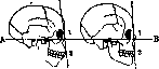
PROGNATHISM, PROGNATHIA, is secondary as a result of retrognathia (see). True prognathia can be 1) physiological, when it is a racial trait (e.g. in Negroes), in which case both jaws are equally well developed and functions are performed normally. 2) Prognathia can represent a developmental anomaly of one or both jaws; due to such an anomaly, functions are disturbed to a greater or lesser degree. 3) Prognathia can have a pathological origin, e.g. in acromegaly. In prognathism, the anomaly can extend to the entire jaw or to its parts. The latter in turn can be either developed beyond the norm or can be displaced in the sagittal direction. Thus, cases of protrusion of the intermaxillary bone or the alveolar process are observed. Cases where the anomaly is caused by protrusion of the teeth forward are more correctly called prodentia. The types of anomalies mentioned rarely have exclusively a sagittal direction, usually they are mixed, so for example protrusion of the upper incisors most often combines with narrowing of the jaw in the premolar area. Displacement of teeth can occur either parallel to their physiological axes (corpus protrusion) or at an angle to them (coronal protrusion). Fig. 1 shows the normal inclination of the physiological axes of the incisors in a 5-year-old child (a-orthognathic position), in an adult representative of an orthognathic race (b-more prognathic position than in the child) and in a representative of a prognathic race (c). Establishing the presence, degree and type of prognathia pursues different purposes. Anthropology, conducting racial studies, studies the facial profile. The latter is determined by the facial angle or the prognathism angle. The profile angle, according to Martin, is formed by the sagittal line of the profile (nasion-prosthion) and the Frankfurt horizontal line (Fig. 2). Measurements, according to Martin, are made with a sliding compass with an angle meter. The smaller the facial angle, the greater the degree of prognathism. The following degrees of prognathism are distinguished: hyperprognathia-69.9°, prognathia 70.0-79.9°, mesognathia 80.0-84.9°, orthognathia 85.0-92.9°, hyperorthognathia 93.0-∞. The degree of prognathism on the skull is also indicated by the alveolar, or jaw index: the ratio of the distance from basion to prosthion to the distance from basion to nasion, the latter distance being taken as 100. The following variations of prognathism are distinguished: 1) complete prognathism of both jaws (observed in lower races); 2) prognathism of the nasal and alveolar areas (in Negroes); 3) prognathism of the alveolar area (in Chinese, etc.). As for European nationalities, they are characterized by an orthognathic profile. Orthodontics, establishing the presence of prognathia, has in mind mainly anomalies of occlusion. The latter affect chewing, breathing,
Fig. 2. a - prognathic profile, b-orthognathic profile; AB - Frankfurt horizontal; 1-nasion; 2-prosthion.
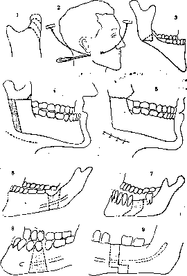
phonation and the external appearance of the subject, and orthodontics takes into account first of all the influence on function. Therefore, establishing the presence of prognathia is for the orthodontist only a partial indicator, since the characteristics of occlusion are not determined by it. With significant prognathism of both jaws, function can be performed normally - here orthodontic treatment is not needed. On the other hand, a small degree of P. can lead to significant disturbances of function, especially in the presence of retrognathia - then treatment is needed. G. Bervovich. Treatment of prognathism and prognathia. Surgical correction of these deformations is indicated only in cases where conventional orthodontic treatment becomes inapplicable: with extremely severe degrees of deformation and in individuals over 20 years of age. The goal of treatment is to restore the correct contours of the face and a satisfactory functional occlusion of the teeth. The most suitable age for surgical treatment is from 20 to 30 years. Correction of jaw deformations is one of the most complex branches of maxillofacial surgery, as it requires the combined application of special treatment methods. The entire treatment plan must be carefully thought out and is based on measurement data. The relationship of the dental arches of the upper and lower jaws is checked on plaster models made with the help of Simon's gnathostat. The models are sawed at the site of the planned osteotomy, individual parts are moved to a new position, and the possibility of teeth closure is checked. If with such a trial movement the dental arches of the upper and lower jaws do not correspond to each other, then it is necessary first to correct the entire arch by orthodontic means or to change the position of individual teeth in order to immediately achieve correct teeth closure during the operation after osteotomy or bone resection and after displacement of the segments. After completion of orthodontic treatment, wire splints are made, fixing the teeth in their new position in the dental arch and at the same time serving as a support for intermaxillary traction with rubber rings, for fixing the entire displaced jaw segment as a whole. The splints are fixed to the teeth on the eve of the operation. Depending on the individual characteristics of each case, various osteoplastic operations can be applied. The largest number of various plastic operations has been proposed for the correction of prognathism. Ziepfel, Jaboulay and Berard, Berger, Dufourmentel proposed to perform bilateral resection of the articular heads with subsequent displacement of the entire lower jaw backward (Fig. 3). Lane, Babcock, Lindemann, Ernst, Pichler perform bilateral horizontal osteotomy of the ascending rami. It is necessary to cut the bone in the area of the upper third of the ascending ramus, above the mandibular canal. Lindemann makes a skin incision near the earlobe and saws the bone with a hacksaw. Pichler and Kostecka pass a Gigli saw through two small skin incisions. In severe prognathism, when surgical treatment is especially indicated, the above methods do not provide sufficient scope for shifting back. Therefore, it is better to expose the ascending ramus through a skin incision below the angle of the lower jaw and after horizontal osteotomy in the place indicated above, resect the posterior edge of the ascending ramus below the bone intersection line, so as not to press on the soft tissues and keep the angle of the lower jaw in place (Limberg). Harsha, Cryer, Ernst propose plastic osteotomy in the area of the angle of the lower jaw. Blair, Eiselsberg, Pichler, Pickerill, Schultz and others applied bilateral resection of pieces from the horizontal ramus. The latter methods are difficult to perform without damaging the integrity of the vascular-nervous bundle. After the operation, immobilization of the lower jaw is necessary for 1-2
Fig. 3. Osteoplastic operations for correction of prognathism: 1-Dufourmentel; 2-Kostecka; 3-Ernst; 4 and 5-Limberg; 6-Harsha; 7-Pichler; 8-Angle; 9-Faderspiel.
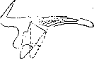
months. Untimely application or insufficient strength of fixation of the jaw segments can be the cause of severe inflammatory complications with subsequent bone necrosis (Angle) and the formation of pseudoarthroses. With any large displacement of the horizontal part of the lower jaw backward, it is necessary to take into account the resulting obstruction of all soft parts of the floor of the mouth and together with this the root of the tongue and the epiglottis, as a result of which difficulties in the function of these organs may arise. Surgical correction of prognathia is rarely applied. Sometimes it is possible to perform a fracture of the anterior part of the alveolar process with subsequent displacement of it into a more correct position. The canine or small molar tooth is removed on both sides, the sockets are resected in a wedge shape and then the base of the alveolar process is broken. Conn-Stock drills the base of the alveolar process on the right and left with a drill and saws it in vertical planes with a Gigli saw. Then the cortical plate is chiseled off and a fracture is made and the entire anterior part of the alveolar process is displaced inward (Fig. 4). The technical difficulties consist in not damaging the maxillary sinuses and the roots of the teeth, not crushing the bone and maintaining sufficient blood supply to the fragment through the surrounding soft tissues. The unsightly protrusion forward of the anterior part of the alveolar process and the teeth of the upper jaw can be more safely corrected by removing the teeth and subgingival resection of the edge and anterior wall of the alveolar process with subsequent insertion of a dental prosthesis.

Related articles
Cite this page
“Prognathism (PROGNATHISM, PROGNATHIA, is secondary as a).” Soviet Medical Encyclopedia. English translation of Bolshaya Meditsinskaya Entsiklopediya, 1st ed. (Moscow, 1928–1936), ed. N. A. Semashko. https://sovietmedicalencyclopedia.pages.dev/article/prognathism-2/