Orthopedic Appliances
Historical document, translated for reference. It reflects medical knowledge of the 1920s–30s and is not medical advice.
Summary
Orthopedic appliances are mechanical devices used to treat orthopedic conditions. This article describes their historical development, classification, manufacturing techniques, and various types including corsets and joint mechanisms.
Encyclopedia article (1928–1936)
Orthopedic Appliances, mechanical devices for the treatment of orthopedic diseases. The widespread use of mechanical principles in the treatment of orthopedic diseases led to the application of orthopedic appliances as early as ancient times. Already in 210 A.D., Caelius Aurelianus first described splints for paralysis. A. Pare (1501) in France and simultaneously Arceus in Spain proposed rather complex orthopedic appliances for the treatment of clubfoot. Fabricius of Aquapendente (1619) used a universal iron corset resembling knightly armor for the treatment of various deformities. In 1660, Glisson's work was published describing a series of supporting appliances, of which his loop is still widely used today. Le Vacher (1772) elaborates in detail on mechanical methods for treating rachitic curvatures and proposes a 'jury-mast'. Venel (1780) invented a bed for traction and a shoe for clubfoot, the further improvement of which was made by the Italian Scarpa (1803). Further mention should be made of Heine (1812), who, being a convinced advocate of the exclusive use of mechanics in orthopedics, contributed greatly to the widespread use of numerous types of orthopedic appliances, but at the same time earned criticism that because of him orthopedics fell into the hands of instrument makers. The introduction of gymnastic methods of treatment into orthopedics led to the formation of two extreme camps - adherents of dynamic and mechanical treatment, who were reconciled by Guerin and Bouvier (1837) in France and Berend (1861) in Germany, who developed specific indications for the use of various methods. In 1857, Delacroix proposed to replace the functions of paralyzed muscles with elastic traction. Mention should also be made of Hessing (1870), son of a poor potter, a self-taught man who invented appliances (see Hessing's appliance) that make it possible to use the limb in joint diseases. The era of Lister is characterized by the flourishing of surgical orthopedics, which significantly limited the excessive enthusiasm for orthopedic appliances and transformed apparatus therapy from a dominant basic method to an auxiliary one. Further accumulation of experience in the use of orthopedic appliances led to a reduction in their large number, which now have only historical interest, and contributed greatly to the improvement of the main types of orthopedic appliances, which in their various modifications are used at certain stages of treatment for almost all orthopedic diseases. All orthopedic appliances can be divided into 3 groups according to their purpose: 1. Fixing or unloading orthopedic appliances, which aim to hold the trunk or limbs in a certain position or relieve them of load. 2. Reducing orthopedic appliances, which restore the lost normal shape to the limbs or trunk by traction or pressure. 3. Replacing orthopedic appliances - to replace the function of paralyzed muscle groups or to replace a limb defect; in the latter case, they are called prostheses (see). Orthopedic appliances can be portable, when the patient wears them on himself, or bed-type, when their use requires staying in bed; in such cases, the points of application of forces, in addition to the patient's body, are also outside it.
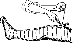
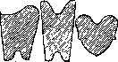
The main requirements for orthopedic appliances are as follows: precise fit, simplicity of design, low cost, durability, and lightness. While yielding to plaster casts in accuracy of modeling, accessibility, and low cost, orthopedic appliances have the advantage of being lightweight, and above all, removable, which is important for observation and carrying out therapeutic procedures. As a rule, orthopedic appliances are made of leather, less often of fabric, and reinforced with metal splints. A substitute for leather can be fabric (knitwear, canvas) impregnated with glue, gelatin, emadetine, etc. Orthopedic appliances should be made individually from special plaster models. The model is obtained as follows: the required part of the body is smeared with vaseline, lard, or some oil, then along the midline of the trunk or limb a strong cord or wire is pulled, and over it circular turns of plaster bandage are applied in 2-3 layers. The plaster cast should be carefully modeled according to bony protrusions. Without letting the cast dry completely, the end of the cord is pulled, as a result of which a longitudinal ridge rises on it, along which the cast is cut (fig. 1). After minimally separating the edges of the cast, it is removed and immediately the edges are brought together again, securing the cast with several turns of gauze bandage. After the negative cast dries, its inner surface is smeared with some fat (Fridland recommends a solution: one stearin candle per 400.0 kerosene) and filled with plaster paste or gypsum (cheaper). After the negative is removed, the positive is opened, which accurately reproduces the shape of the trunk or limb. Subsequently, the positive undergoes additional correction - in those areas where pressure needs to be achieved, a more or less thick layer of plaster is removed, and conversely, on those areas that need to be relieved of pressure (iliac crests, condyles, etc.), a layer of plaster should be applied. Then the manufacture of the leather sheath from calfskin or cowhide begins, which is cut according to a paper 'pattern' (fig. 2), previously made from the model. The leather is first soaked and hardened in a solution of glue or potassium bichromate, and before use it is soaked in warm water and stretched on the model, where it is fixed with small nails. For greater strength or to connect several sheaths into one, metal splints are fitted on the model itself, placed in a known direction according to the laws of mechanics. Subsequently, the sheath is removed from the model, fastened with rivets to the splints, padded inside with flannel or suede, and equipped with lacing.
Figure 2. Patterns for cutting sheaths: a-hip; b-leg; c-foot.
For movable connection of splints, the ends participating in the joint are processed in the form of
Figure 3. Hinge constructions: 1-overlay; 2-fork-shaped or through; 3-block-shaped (brake shoulders are indicated by dotted lines); 4-petal. These hinges allow movement in only one plane, where movement in several planes is necessary, either a ball joint or a combination of the two aforementioned hinges with mutually perpendicular axes of movement (fig. 4 and 5) are used. To
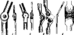
Figure 4.
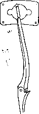
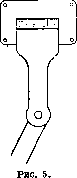
Figure 4. Combined hinge for rotation with abduction. Figure 5. Combined hinge for flexion with abduction. obtain a greater range of motion, Stillman combines three overlay hinges, any of which can be
Figure 6. Triple hinge of Stillman with key.
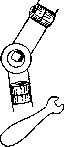
Figure 7. Schematic drawings of splints: a-sector splint of Stillman for gradual redressation; b-sector splint of Braatz for straightening and stretching the knee joint; c-clamps for fixing the hinge.
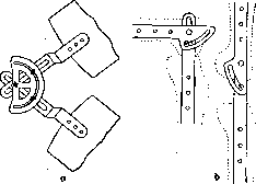
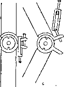
obtainable changes in the relative position of the splints participating in the hinge joint, sector splints of Stillman, Braatz, or a device in the form of clamps are used, the application of which is clear from fig. 7. For the purpose of locking the hinge for a certain period of time, 'locks' of various designs are used, which mainly consist of a latch fixed on one hinge, penetrating into the corresponding recess of the second hinge (fig. 8). Among the fixing orthopedic appliances, the corset is most widely used. Its task is the immobilization and unloading of the affected part of the spine. Immobilization of the spine is achieved by precise fitting of the corset, which at the bottom covers the pelvic girdle, and at the top is fixed with the help of crutches on the shoulder girdle (see Corsets, Volume XIV, article 20, figure 6). Unloading of the spine is achieved by transferring the weight of the body through crutches located in the axillary fossae, and vertical metal splints fixed to them directly to the pelvis. The main splints in the corset are arranged as follows: the upper semicircle along the upper edge of the corset (behind and on the sides), the lower circular splint (buckle)-one finger above and parallel to the crista ossis ilii. On both sides along the middle axillary line, vertical splints are fixed, at the bottom resting against the lower pelvic splint, at the top connected to the upper semicircle and ending with crutches. When taking measurements for a corset for tuberculosis of the spine, the latter is reclined (lordosed), as a result of which the affected vertebral bodies are unloaded due to the transfer of the main load to their arches. Reclination is achieved by suspension using Glisson's loop and
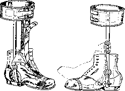
with a key (fig. 6). Braking devices in the form of shoulders on hinges (fig. 3, 3) serve to limit the range of motion. For gradual, periodic simultaneous abduction of the lower limbs backward behind the frontal plane. Usually the corset is made of leather or the mentioned surrogates thereof and for better perspiration is provided with numerous holes. In cases requiring less strict immobilization, one can use fabric corsets, and when there is no need for unloading, equipping the corset with crutches is superfluous. In lesions of the cervical spine, the so-called 'jury-mast' proposed by Levashev has been used since long ago, which is now replaced by a more convenient and less unsightly collar (see vol. XIV, art. 19, figs. 3 and 4). By supporting the head in the chin and occipital regions, it transfers its entire weight to the shoulder girdle. An asymmetrically constructed collar is used in torticollis. The next most common among orthopedic appliances are splints intended for immobilization of joints and fixation of limbs over a period. They are splint-sleeve appliances without hinges, which distinguishes them from other fixation appliances. For immobilization of the hip joint, a splint is used consisting of a high half-corset belt continuing into a sleeve covering the thigh (fig. 9). The splint for the knee joint is a conical sleeve reinforced with side splints, starting from the groin fold and ending above the ankles (fig. 10 a). A stirrup attached to the lower end of the splint or a narrow pelvic girdle secured to the upper end prevents the splint from slipping. Splints for the leg-foot and elbow joints are shown in fig. 10 (b and c). The other orthopedic appliances are splint-sleeve appliances equipped with hinges at the joint level and are used both for fixing segments of limbs in a certain position while simultaneously preserving motion in the joints, and for limiting lateral motion in loose joints. A typical example is the Gessing apparatus (see Gessing apparatus). A complete fixation appliance for the lower extremity consists of thigh and leg sleeves as well as a shoe for the foot, usually connected by external hinges. Often for better fixation of the appliance, a narrow pelvic girdle articulated with the upper edge of the outer thigh splint by means of a double hinge is used, which increases the range of motion in the hip joint. In case of looseness of one hip joint, one can limit oneself to just the thigh sleeve, but the pelvic girdle must be considerably wider. The appliance for looseness of the knee joint consists of thigh and leg sleeves, and the boot used for the ankle-foot joint consists of a leg sleeve and a shoe.
Figure 9.
Figure 10.
Figure 9. Splint for immobilization of the hip joint or so-called 'spine'. Figure 10. Splints: a-for the knee joint; b-for the elbow joint; c-for the leg-foot joint.
in the form of a half-ring surrounding the patella from the outside (fig. 12). The introduction of the principle of unloading into fixation appliances for the lower extremity belongs to Thomas (Thomas). Unloading is accomplished by means of a seat fixed to the upper end of the side splints of the appliance, the length of which exceeds the length of the limb by 1-2 cm. When bearing weight on the limb in such an appliance, the entire weight through the ischial tuberosity and perineum is transferred to the seat, and from the latter through the side splints to the metal sole of the appliance, while the limb hangs freely in the appliance, not reaching its bottom by the mentioned 1-2 cm (fig. 13). By using such an appliance, unloading of the affected joint is achieved during walking, and by locking the hinge and immobilizing it, while the other healthy joints of the limb function simultaneously. Reduction appliances exert their corrective action using the force of a spring, lever, elastic plate with a screw passing through it, traction, or finally body weight. By rotating the screw, the degree of pressure is regulated.-In the treatment of kyphosis in growing patients, some reduction of the rib cage can be achieved by applying a pressing pelvic support in the area of protrusion. In the corset, an oval opening is cut out corresponding to the apex of the rib hump, filled with a pelvic support (a metal plate padded with felt), over which a steel rod is thrown. For the correction of deformities of the spine, so-called straighteners are used. The Nyrop straightener consists of a steel belt padded with soft material, put around the pelvis and fixed by two arches passing over the crests of the pelvic bones. From the middle of the belt at the back, a metal plate rises upward along the spine, to which at the level of the axillary pits a horizontal semicircle is riveted, reaching on the sides to the anterior axillary lines and ending with steel tabs. From the latter begin straps thrown over the shoulders and fastened in front. The plaster bed of Lorenz (Lorenz), used in deformities of the spine (see Plaster casts, corsets, beds). In adults in lesions of the spine, one often can do without a plaster bed, using a wooden flat
Figure 11. Fixation appliance for habitual dislocation of the shoulder joint.
Figure 12. Haudek's bandage for habitual dislocation of the patella (externally).
Figure 13. Appliances for immobilization and unloading of the knee joint: a-more expensive model; b-simplified model.
p\
Figure 16.
Figure G Figure 19.
bed. For the correction of congenital clubfoot (pes equino-varus congenitus), various apparatuses are used. The most common is the apparatus of Lorenz (Lorenz), consisting of a metal sole with a heel raise and a metal rod attached to it, which is bent in such a way that when the foot is placed on it, the foot is brought into a position of dorsiflexion and eversion. The apparatus is fixed by straps passing over the shoulders and around the chest. For the correction of congenital flatfoot with heel valgus (pes calcaneo-valgus congenitus), an apparatus is used consisting of a metal sole with a depression for the heel and a metal rod attached to it, which is bent so that when the foot is placed on it, the foot is brought into a position of plantar flexion and inversion. The apparatus is fixed by straps passing over the shoulders and around the chest.
Figure 20.
Figure. 21. Splint for correction of pes equino-varus congenitus.
Figure 22. Splint for correction of pes equino-varus congenitus: a-application; b-bandaging.
Figure 24. Splint for correction of pes calcaneo-valgus congenitus: a-application; b-bandaging.
Figure 23. Splint for correction of pes calcaneo-valgus congenitus.
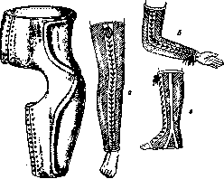
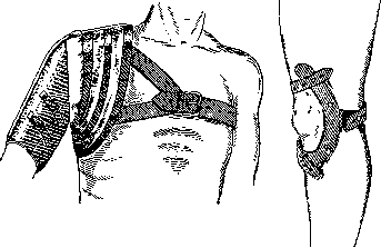
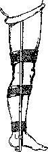
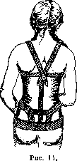
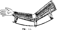
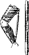
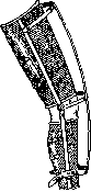
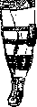
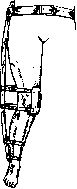
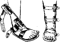
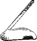
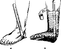
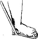
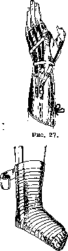
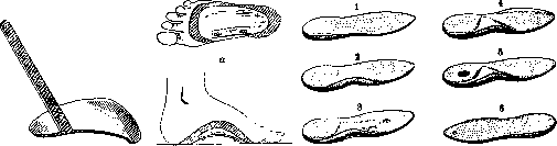
Figure 25. Metal spring insole supporting the arch of the foot: a-from below; b-from inside. Figure 26. Types of insoles: 1-supinator, raising the inner edge of the heel; 2-the same and forming the arch of the foot; 3-the same with a Seitz roll and a depression for a corn; 4-the same as 2, but with simultaneous raising of the outer edge in the anterior part of the foot; 5-the same as 2, but with a depression for a heel spur; 6-pronator, raising the outer edge of the heel and the middle part. passing over the shoulders and fastening to the back plate (fig. 14).-Severe kyphoses, as for example in ankylosing spondylitis, often yield to the straightening effect of strong steel spring-plates attached below to the posterior surface of the corset, and above to the forehead band, the so-called diadem. To reduction appliances, but of the accommodating type, belongs the reclining shield, placed under a thin hair mattress, under which the hump is brought with bags filled with sand and hair, or as Vreden suggests, with flax seed.-For the treatment of contractures, splint-sleeve appliances are used, equipped as a driving force with a spring, elastic traction or a screw. Success can be expected only in cases of not particularly severe contractures without marked destructive changes in the articular ends. For flexion contracture of the elbow in a splint-sleeve appliance (fig. 15) on its extensor side is inserted a flexible steel blade, the ends of which are drawn by straps to the sleeves. To increase the range of flexion, to the appliance on its flexor surface are attached two crossing elastic tractions to the ends of the sleeves. To eliminate flexion contracture of the knee joint, the splint-sleeve appliance has long been equipped with a screw, one end attached to the posterior surface of the thigh sleeve, and the other end screwed into a tube secured to the lower end of the leg sleeve. Unwinding the screw gave straightening of the contracture. However, later the screw was replaced by a spiral spring, providing continuous action and giving better results (fig. 17). One can use a straight band spring, located on the anterior surface of the appliance, the upper end of which is secured to the thigh sleeve, and the lower end is drawn by a strap to the leg sleeve (fig. 17). In children suffering from genu valgum of moderate degrees, appliances consisting of an outer splint with hinges in the hip, knee, and ankle joints and straps-pelvic, femoral, knee, and leg; at the bottom the splint is secured to a boot (fig. 18). By gradual pulling of the knee strap the deformation is corrected. A more simplified type is the night splint without hinges and therefore it can be made of wood (fig. 19). For genu varum similar splints are used, but they are applied along the inner surface of the lower extremities.
For the treatment of clubfoot, numerous boot appliances of very complex construction have been proposed, which are now abandoned. Only night splints have retained practical significance. Their action is based on the lever principle. The Taylor appliance consists of a metal sole with a lever attached to its inner side, inclined inward and backward (fig. 20 a). First the foot is fixed by straps, and then by setting the lever parallel to the leg and securing it with straps, the foot is brought into the position necessary for correction (fig. 20 b). The simplest modification of this splint is shown in fig. 21 and 22, where it is seen that for convenience of application its lever has been moved to the outer side, and to prevent the toes from being adducted, a rim is made along the inner edge. The boot used for clubfoot is equipped inside with an insole raising the outer edge of the foot, and has an outer splint with a hinge in the ankle joint and a strap encircling the leg at the level of its upper third. The task of the splint-to abduct the foot.
For the treatment of congenital flatfoot, splints (fig. 23 and 24) are used, the lever of which is located along the inner edge and is inclined inward and backward. Orthopedic insoles, which are widely used, are a device which, due to the relief of its surface, using the force of gravity, sets the foot in the required position. Materials for making insoles are metal, cork, leather, rubber, etc. In Germany, Seitz insoles for flatfoot (fig. 25) are widely used. A steel spring plate is equipped below with a braking device to limit the degree of flattening, and above is covered with leather. The most reasonable should be considered cork insoles (fig. 26). In initial
degrees of flatfoot in the supinator insole only its inner edge under the heel is raised (fig. 26,2), in more advanced cases the arch of the foot is also filled (fig. 26,2). When transverse flatfoot exists simultaneously, a roll raising the metatarsal bones is used, and a depression is made for the painful corn (fig. 26,3). When to correct the position of the heel it is necessary to raise its inner edge more than 11/2 cm, and the foot tends to slide off the insole outward, then to hold it up a depression is made corresponding to the place of a heel spur (fig. 26,5). For clubfoot a pronator insole is used, raising the outer edge (fig. 26,6), so-called pronator insole and a pressure pad on the dorsal surface of the bent joint. Of replacement appliances here it is necessary to note only those which replace the function of paralyzed muscles. The driving force in them is elastic traction or more rarely a spring. For paralysis of the flexors of the forearm, the splint-sleeve appliance is equipped on its flexor surface with crossing rubber tractions, which when actively relaxing the preserved function of the extensors flex the* arm at the elbow. For paralysis of the finger extensors the appliance (fig. 27) consists of a sleeve covering the forearm, wrist, and metacarpus, and five leather rings, one for each finger. Rubber tractions running from the fingers along the dorsum replace the traction of the paralyzed flexors. On the lower extremity in paralysis of the quadriceps muscle its function is replaced by a strong rubber band (one or two), attached in a stretched state to the anterior surface of the thigh and leg sleeves of the fixation appliance. During walking the rubber helps to extend the knee joint, and at the moment of support on the leg it prevents it from buckling. In paralysis of muscle groups of the foot a boot with side splints is used, ensuring correct positioning of the foot. To eliminate foot drop in paralysis of the extensors, under the tongue of the boot a rubber traction is passed, secured in the toe and fastening to a ring encircling the leg (fig. 28). For the same purpose Goldschmidt uses a spiral spring setting the foot in the position of dorsal flexion (fig. 29). In weakness of the peroneal muscles to eliminate clubfoot a rubber traction is used, attached to the outer side of the anterior part of the boot (fig. 30).
. III. For osteotomy, one of the most frequent orthopedic operations, numerous chisels and osteotomes have been proposed. Figure 53 shows a chisel of simple form, fig. 32 and 33-a Bergmann chisel (12, 15 and 20 mm width), the blade of which directly passes into the handle. A very convenient osteotome was proposed by Mac Ewen (fig. 34). It has an octagonal handle and an expanded platform for striking with a hammer. On the narrow face of the blade there are graduations, which allows to monitor the depth of penetration of the osteotome into the bone; the width of the blade is 9, 11 and 15 mm. In fig. 35 the same osteotome is shown, but of grooved form. Practically the Gocht chisel, made from a single strip of steel (fig. 23); the handle is inconvenient to hold, but it allows to control the direction of the blade. For subcutaneous
34 35
40 41
O 51
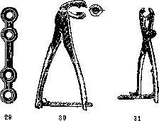
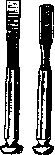

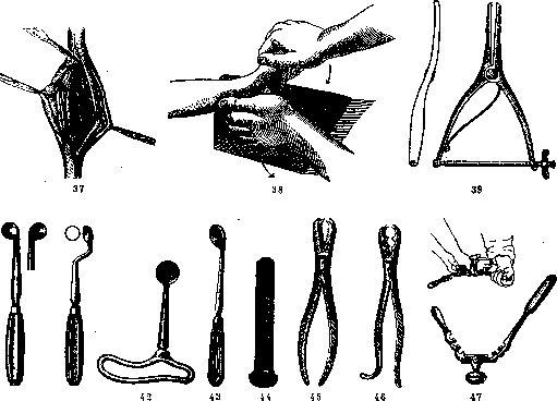
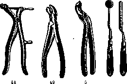
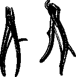
For osteotomies, the chisel proposed by Lorenz (Lo-renz; fig. 3) is used. Its length is 17 cm, handle 10 cm, width of blade 1 cm. 'Elevators' and 'protectors' (Schiitzer) greatly facilitate the performance of osteotomies. By fixing the bone, they push aside soft tissues, protect them from the blade, and reduce bleeding from soft tissues by tissue tension. In Vol. XIV (p. 169), fig. 9 shows the Lange elevator. For femoral osteotomy according to Schantz (palliative operation for congenital dislocation of the hip in adults), steel screws are used, which are screwed into the bone before its incision (fig. 70). With the help of screws, the angle between bone fragments can be controlled, which is important in this operation (fig. 71). On the free end of the screw there is a face for placing a key, by means of which the screw is screwed into the bone. After the operation, the screws are immobilized in the cast until the consolidation of bone fragments begins, therefore they must be made of stainless steel. Bone suture can be performed besides wire with the help of metal plates of various lengths according to Leno (fig. 29), aluminum plates (fig. 28), celluloid plates according to Lange, or metal staples (Vol. XIV, p. 126, fig. 5). The plates are fixed with screws. For connecting bones, metal nails or pins made of ivory or ox bone are also used. Bone fixation is performed with the simplest bone forceps of Langenbeck (fig. 45), Ollier (fig. 46) or more complex but more convenient forceps, for example, of the Lambotte type (Lambotte) (fig. 30, 31), which allow to firmly fix the bone by means of a lock. Franke proposed a clamp (fig. 63) for holding bone fragments until the hardening of the plaster cast, which allows to do without bone suture. For laminectomy, many models of forceps have been proposed, for example, forceps of Hoffmann (Hoffmann; fig. 54), Kerven (Quervain; fig. 55), sharp ones according to Horsley (Horsley; fig. 50). The 'crocodiles' of the Lannelongue type (Lannelongue; fig. 49) or simple forceps of Luer (fig. 11) are also convenient. Krause's laminectomy is shown in fig. 48. Operations on joints sometimes require special instruments. For smoothing rough bone surfaces, files according to Scheppelmann (fig. 50 and 51) are used. For scraping cartilage from the acetabulum during arthrodesis, Lorenz proposed a special sharp spoon (fig. 40). A somewhat different appearance has the spoon for the same purpose according to Hoffa (Hoffa; fig. 41). For deepening the acetabulum, Hoffa proposed a short spoon (fig. 42), and Doyen - a special drill (fig. 44). For the bloodless reduction of hip dislocation, Codivilla uses a lever, the action of which is clear from fig. 69. For expanding the joint capsule during reduction of hip dislocation, Calot proposed a 'subcutaneous' dilator (fig. 39, 67). For operations on the formation of the hip joint, Murphy proposed a round milling cutter and a concave file in the shape of a hemisphere (fig. 59). IV. Equipment for bloodless orthopedic operations. The simplest device is a loop, used for the redressation of certain types of clubfoot. Very simple and convenient for redressation of clubfoot are the wooden wedge of König, covered with felt and oilcloth (leather) (fig. 38), and the apparatus of Schulze (fig. 73). The latter consists of two boards connected by a hinge. The leg is fixed in the apparatus with straps. For the treatment of clubfoot in children, what is listed is quite sufficient. For adults, many kinds of redressators have been proposed. Fig. 58 shows the 'Thomas wrench' - Thomas' osteoclast, which can also be used as a redressator. It is modified by Stille. Its 'claws' can come together (when gripping the limb) by a screw located in the handle. Fig. 74 shows the Stille redressator-osteoclast, fig. 74a shows redressation with the help of this apparatus, fig. 47 shows the Turner redressator, fig. 64 shows the Gocht redressator, fig. 22 shows the Phelps-Gocht osteoclast-redressator, fig. 78 shows it in action, fig. 75 shows the Lorenz-Stille osteoclast. The limb is fixed in it with a screw. Traction with a leather loop is also performed with a screw. Fig. 66 shows osteoclasis, and fig. 68 shows redressation by means of the Lorenz-Stille apparatus. As in any redressator, felt or rubber pads, sponges are used to protect the skin from damage. The Rizzoli osteoclast (Rizzoli; fig. 62) is very simple. The limb is held with rings, pressure is applied with a screw. For the treatment of clubfoot and flatfoot, the Schede redressator (fig. 79, 57, 65) and the Gorman redressator (fig. 60) have been proposed recently. The construction and action are clear from the figures. Extension tables, frames, etc., can also be included in the group of redressating appliances, which (especially recently) have been offered in many designs. The Abbott bed (fig. 26) is used for the correction of scoliosis in children before applying a plaster corset. For the correction of deformities of the spine, the Wullstein frame (Wullstein; fig. 27) is used, for the correction of the spine in kyphosis - the Schede table (fig. 72). Fig. 76 shows the Lorenz apparatus for redressation of contractures of the hip joint by screw traction. The healthy leg rests on a special stand, the knee is fixed. Fig. 77 shows an apparatus for redressation of contracture of the knee joint. For bloodless reduction of congenital dislocation of the hip joint, the 'Muskel-quetscher' of Gocht (fig. 1) is used, an instrument designed for lengthening the contracted adductor muscles of the thigh. It is placed transversely to the direction of the muscle fibers at their attachment to the pubic bone, after which sawing movements are made with it. The skin, rubbed with alcohol and covered with sterile gauze, should not be damaged at this time. Gocht also proposed levers (fig. 2) for fixing the pelvis during reduction. The method of use is indicated in fig. 56. Lit.-em. lit. to the article Orthopedics and Prostheses. V. Blokhin.
Related articles
Mentioned in
Cite this page
“Orthopedic Appliances.” Soviet Medical Encyclopedia. English translation of Bolshaya Meditsinskaya Entsiklopediya, 1st ed. (Moscow, 1928–1936), ed. N. A. Semashko. https://sovietmedicalencyclopedia.pages.dev/article/orthopedic-appliances/