Splints
Historical document, translated for reference. It reflects medical knowledge of the 1920s–30s and is not medical advice.
Summary
Splints are devices used to immobilize limbs, jaws, and joints, particularly for fractures. This article describes various types of splints, their historical development, and their application in emergency care, transportation, and treatment.
Encyclopedia article (1928–1936)
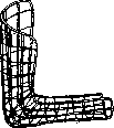
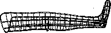
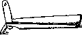
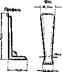
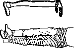
Splints, a device used to create immobility, most often in fractures of long bones of the limbs, jaws, and in joint injuries, for the purpose of first aid, transportation, and treatment. Excavations of Egyptian tombs testify that 4,000-5,000 years B.C., Egyptians already used various kinds of splints for fractures, admittedly very simplified—in the form of wooden planks, bark, or even dried animal skins—for the purpose of fixing the fracture site. At present, one can count hundreds of various types of simple fixation splints made from various materials: from cardboard, bast, wood, metal, plate-like, wire, mesh, sometimes pre-prepared for various parts of the body. The most common fixation splints used for 'laying' the upper and lower limbs were wire or mesh splints of Mayor—wide, suitable for any arm bent at a right angle or extended leg (fig. 1 and 2), and tin, grooved for the leg with a foot part—the splint of Volkmann and Cabot (fig. 3 and 4). To make a fixation splint
Figure 1. Wire splint for the arm of Mayor. so that it could be easily fitted to one or another part of the body and to the bends of joints, Cramer proposed the use of strips of 'wire ladder' (fig. 5), and Filbry—metal
Figure 2.
Wire splint for the leg of Mayor.
Figure 3. Volkmann splint.
mesh fabric (fig. 6 and 7), obtained by stretching frequently and in a certain order perforated sheets of aluminum or galvanized iron. When used, strips of the required length and width are cut from them, to which the necessary bends are given. When bandaged with soft bandages, they, due to their springiness (especially during transportation, when shaken), gradually loosen the bandage and fixation is not achieved. More satisfactory fixation is obtained when these splints are well fitted to the bends of the body with reinforcement by starch or plaster bandages. The great experience with treating fractures in the imperialist war, in addition to the abundant splint equipment that entered textbooks, added a large number of new variations of fixation splints. For example, among Russian splints, the wire splint of Sapeshko is known, then the sliding wire splint, with support under the pelvis, the splint of Krause-Tarnovsky (fig. 8), the splint of Faltin, Sozon-Yaroshevich. Like most old ones, they proved to be little effective. The rich museum collection composed of them, as this for example is poss-POzholИooбforago5eepiеТСЯetplace in the Military-Sanitary Museum of the Military Medical Academy (from which some objects were taken for the accompanying figures), has more historical than practical interest, since the vast majority of this arsenal of splints cannot fulfill the requirements of rational immobilization in first aid, transportation, and treatment of fractures. To an even lesser extent are suitable, at least only for primary immobilization of a fracture, the primitive improvisations from various materials, ^ found under hand in various conditions (rifle, sticks, boards, bast, plywood, tied twigs, bundles of straw,
Figure 4. Posterior wire splint of Cabot; &Ш)ПШ Figure 5. Splint of Cramer. bags with sand, etc.). Such primitive immobilizations are permissible for use only in extreme cases of hopeless situations. But even in such cases they
Figure 6. Sheets
of metal fabric Filbry—stretched. can provide some service in the matter of immobilization only in combination with a plaster or starch bandage. The imperialist war showed that the best immobilizing means in first aid and for transportation is a properly applied plaster cast, with preliminary setting of fragments by means of forced traction in the so-called physiological position. But under the conditions of first aid not only in war but also in peacetime, it is difficult to make a plaster cast, and in view of possible complications (hematomas, infections) and risky. It was recognized that it should be replaced by a splint, but not a simple fixation splint, but a splint that ideally performs fixation together with traction. The principle of fixation with traction in the treatment of fractures is a fairly old principle, known even from antiquity—from the time of Hippocrates. Its broader practical application was carried out 200 years ago in the form of a double inclined plane, when Petit (1719) demonstrated it at the Paris Academy of Sciences (fig. 9) and recommended it for the treatment of leg fractures. With relaxed muscles at rest, the leg and thigh, due to their weight, are passively pulled on the movable bend of the inclined plane. The idea of the apparatus of Zuppinger (1905) (fig. 10) repeats the idea of the double inclined plane of Petit for passive traction by one's own weight of a leg bent at the knee. Subsequently, Zuppinger himself abandoned the idea of treating fractures with passive traction. On these same apparatuses in the position of semiflexion, but with active adhesive traction according to the principle of Bardenheuer, with early active movements and massage, Zuppinger tried to implement the so-called Рве. 7. Sheets of metal fabric Filbry—perforated.
Figure 8. Splint of Krause-Tarnovsky.
functional treatment, the principle of which was proclaimed by Hennequin. According to the type of Zuppinger was built the splint of Heusner, who instead of plaster for active traction recommended the use of cotton strips with the help of glue (see Traction). Wegener performed active traction of the leg bent at the knee on pillows instead of a splint (fig. 11). Codivilla and Steinmann replaced plaster traction with nail traction: the first—behind the heel, the second—behind the condyles of the femur. However, the idea of the double inclined plane with
Figure 9. Splint of Petit.
passive traction did not die, and, having undergone significant modification, during the imperialist war played a role not only in hospitals but also at the front—on evacuation routes. Thus, in a fracture of the femur Fritz Lange proposed for transportation a rather cumbersome splint-cast apparatus on a metal basis with a cardboard cast on straps. Criticizing this splint in a post-war report, K. Franz says that cardboard from pus and moisture becomes soft and the splint was not successful with German surgeons. A similar splint bandage, built on the principle of the double inclined plane with fixation and passive traction on the bent knee, Rummel proposed to make by bending its frame from three Cramer splints. K. Franz warns against using it for transportation, since, not being reinforced by plaster, Cramer strips STRONGLY Pr-
Fig. 10. Splint of Zuppinger. ing, loosen bandages and the entire bandage comes apart. Thus, fixation with passive traction did not justify itself in transportation during the imperialist war on any of the splints used. A much greater role in transportation was played during the last war by fixation with active traction by the most various methods. For example Weis-'', _
„
senstein (fig. 12) in Austria for this purpose in fractures of the femur adapted ordinary stretch- Fig. 12. Stretcher-splint in fracture of the femur (according to Weis-''senstein). ers, and in winter ski stretchers (fig. 13). In the lower half on each side, along the sides of the poles, in the canvas of the stretchers, 6 slits are made the length of the width of a palm, through which with circular bandaging the leg and pelvis are secured to the pole. The foot is bandaged to the crossbar of the stretcher. Counter-traction is conducted through the groin and secured
Fig. 13. Ski stretchers (manual according to U.S. army splints).
at the head end. These stretchers went with the wounded to the place of his stationary treatment. A hindrance to the widespread application in Austria of the interesting idea of Weis-''enstein was the insufficient quantity and bulkiness of stretchers used as splints. The old splint of Liston-Esmarch (fig. 14) also served the idea of fixation with traction. The leg, fixed with circular bandaging through holes in a long wooden board, was pulled by the movable foot part, which is fixed to the side splint with the help of a screw. Counter-traction is elastic traction,
Fig. 14. Splint of Liston-Esmarch.
conducted through the groin, secured at the upper end of the side board. The splint of Liston-Esmarch, widely, almost exclusively used in the first year of the war by the English in fracture of the femur, was subsequently completely condemned on all fronts, when the huge mortality among wounded with fracture of the femur was discovered. 'The rifle gave better results,' says'
Figure 15. Splint of Diterichs.

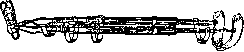
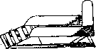
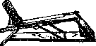
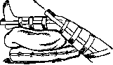
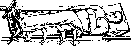
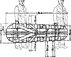
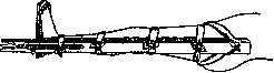
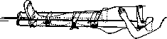
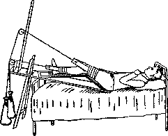
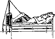
Bowlby. Based on the Liston-Esmarch splint, Diterichs splint (fig. 15) was constructed with two lateral wooden strips and a movable foot. With its weight and bulkiness, it can only compete with the wooden Volkovich splint (fig. 1B), which the author also intended for walking in cases of femoral fracture. Based on the principle of fixation with active traction, the Stubenrauch splint was constructed in Germany, consisting of two Kramer splints: a posterior one embracing below the knee, the shin, and the sole, and a lateral one extending to the foot from the lateral surface of the trunk and pelvis. The excess 12 cm of the foot end of the lateral splint were bent for the purpose of attaching to it a traction made through the skin on the sides of the knee and shin. As in the splints described above, elastic countertraction was also conducted through the groin. However, all these splins insufficiently implement the principle of 'fixation and active traction.' The latter is insufficient due to the small weight that such splint bandages allow. This circumstance prompted the search for combinations of plaster and traction. The so-called extension plaster cast of Lange appeared; however, plaster and traction have completely opposite actions. The plastered extension traction ceases to exert traction and becomes meaningless. Of all methods of splinting, the idea of the double inclined plane with active traction survived the imperialist war. But not for first aid or transport, but for treatment in a hospital, the idea of the double inclined plane developed into a separate method and entered the postwar situation for treating fractures in peacetime. Even during the war (in Germany), so-called bed splints with active adhesive or nail traction appeared. Braun (fig. 17) was the first during the war, in 1916, to propose an iron frame for treating femoral fractures, into which the thigh and shin are placed with the hip and knee joints bent. Subsequently, during the war, the Vienna surgeon Bohler (fig. 18) improved it by adapting the blocks for active traction of the thigh and shin, which are usually located on the bed, directly onto the splint itself. For this purpose, Bohler attached several metal arches to the splint: one arch as an extension of the upper horizontal bar of the frame for traction of the shin, and two arches for the thigh bent at the knee in various positions. Thanks to the work of Bohler during and after the war, bed splints gained great popularity for hospital treatment of femoral and shin fractures. In addition to the Braun and Bohler types, other modifications of bed splints appeared—Beyer's, Schede's (Schede), completely similar to the Braun bed splint for the leg, as well as more complex and cumbersome apparatuses of Tikhomirov and Anzin (fig. 19) with traction and simultaneous movement in the joints. The folding bed splint (for transport) of Osten-Saken (fig. 20) and two wooden triangles of Colmers (fig. 21), placed under the bent knee, deserve attention. When dressing a wound, one of the triangles is removed. For hospital treatment, several authors also proposed so-called suspension, or hanging splints, for example, the Smith splint; traction by suspending the leg on slings to a long crossbar of a wooden frame, for example, the method of Florschütz. Only one or two of the many slings are removed when dressing, and the limb continues to hang on the remaining ones. Hanging splints were used during the war in all countries. English authors called this method a hammock. However, for severe fractures and infections, suspension splints, or the method of air suspension, are little suitable in terms of fixation, as they create a certain mobility of the fractured and infected limb. Nevertheless, the method of suspension has to be used, mainly for severely torn wounds that not only prevent the application of a plaster cast but also a constant splint. The suspension itself is best accomplished by means of the so-called Balkan frame, which is also good for suspending various types of extension splints. Of all splints that provide fixation and traction and are suitable for first aid and transport in cases of femoral and shin fractures, only one splint survived the imperialist war—the Thomas splint (fig. 22). The latter was constructed by Thomas shortly before the Franco-Prussian War of 1870/71 and was then offered to the French army; however, the French refused this offer at the time. For half a century, the Thomas splint was included in surgical manuals, finding little practical application among surgeons. It was used mainly by prosthetists in the form of the so-called Thomas apparatus, wishing to provide relief to one or another of the three diseased joints of the lower limb (fig. 23, 24). Seeing the appalling mortality from complications in gunshot fractures of the femur in the first year of the imperialist war, the great English orthopedist R. Jones persistently recommended the use of the Thomas splint at the site of the wounded person's location—over the trousers and boot—with traction through a spike conducted through an incision in the boot along the inner plane of the sole. Subduction of the shoulder. Detail. In the second half of the war, the Thomas splint became mandatory for first aid and transport in cases of femoral fractures not only in the English but also in the American army. Several authors, such as Sinclair, modifying the Thomas splint, applied it for further treatment of femoral and shin fractures, as well as for fractures of the upper limb (fig. 25, 26). Thus, for English and American orthopedists, it has now become to a certain extent universal. The arm rarely needs splinting compared to the leg, as in many cases of first aid and transport, the trunk can replace the splint, to which the arm is bandaged. For splinting fractures of the upper limb, there are many variations of the method based on the principle of the double inclined plane. Thus, Middeldorf, in fractures of the humerus, proposed fixing the downward-hanging arm, abducted at the shoulder and bent at the elbow, on a triangle. However, this position of the arm with sharp inward rotation of the shoulder cannot provide correct alignment of the fragments of the humerus. Based on the same principle, but with a more correct physiological position of the shoulder and forearm, the cardboard triangular splint of Falten is also constructed. In addition, there is a folding metal Turner splint (fig. 27) and the so-called ambulatory crutch splint of Borchgrevink, applied according to the type of a femoral splint for the shoulder. (According to Zuppinger). The upper end of the splint, a 'fork,' is inserted under the support in the armpit. The shoulder and forearm hang down and rest on the plane of the splint. A strip of adhesive plaster applied along the length of the shoulder and forearm produces traction by means of a rubber tube. Tikhomirov and N. N. Petrov added abduction of the shoulder to the Borchgrevink splint (fig. 28). In M. I. Sitenko, this splint with abduction is made folding (fig. 29). Of the extension splints used in Germany during the imperialist war for fractures of the shoulder, Müller particularly notes the improvised splint by Porzelt, which when applied to the extended shoulder was bandaged to the trunk along its length. When the Kraemer splint appeared, the rectangular extension splint by Zuppinger (fig. 30) came into wide application. Furthermore, according to Müller, the adjustable angular extension splint of Braun with a spring and screw proved to be very rational (fig. 31).-For fixation of the limb after resection of a joint, various types of so-called resection splints are used. The two parts of the splint, usually wooden, bandaged to the limb above and below the diseased joint, are connected by a 'bridge'—a metal arch thrown over the site of joint resection for dressing the wound (fig. 32). In fractures of the jaw, at present, splints of the Tigerstedt type are most often used, consisting of a main aluminum wire 2 2/3 mm thick. Each tooth individually is
Fig. 16. Volkovich splints. an splints
^
Figure 17. Braun splint. nia, made through the skin on the sides of the knee and shin. As in the splints described above, elastic countertraction was also conducted through the groin. All these splints, however, insufficiently implement the principle 'fixation and active traction.' The latter is insufficient due to the small weight that such splint bandages allow. This circumstance prompted the search for combinations of plaster and traction. The so-called extension plaster cast of Lange appeared; however, plaster and traction have completely opposite actions. The plastered extension traction ceases to exert traction and becomes meaningless. Of all methods of splinting, the idea of the double inclined plane with active traction survived the imperialist war. But not for first aid or transport, but for treatment in a hospital, the idea of the double inclined plane developed into a separate method and entered the postwar situation for treating fractures in peacetime. Even during the war (in Germany), so-called bed splints with active adhesive or nail traction appeared. Braun (fig. 17) was the first during the war, in 1916, to propose an iron frame for treating femoral fractures, into which the thigh and shin are placed with the hip and knee joints bent. Subsequently, during the war, the Vienna surgeon Bohler (fig. 18) improved it by adapting the blocks for active traction of the thigh and shin, which are usually located on the bed, directly onto the splint itself. For this purpose, Bohler attached several metal arches to the splint: one arch as an extension of the upper horizontal bar of the frame for traction of the shin, and two arches for the thigh bent at the knee in various positions. Thanks to the work of Bohler during and after the war, bed splints gained great popularity for hospital treatment of femoral and shin fractures. In addition to the Braun and Bohler types, other modifications of bed splints appeared—Beyer's, Schede's (Schede), completely similar to the Braun bed splint for the leg, as well as more complex and cumbersome apparatuses of Tikhomirov and Anzin (fig. 19) with traction and simultaneous movement in the joints.
Figure 18. Bohler splint.
Figure 19. Tikhomirov and Anzin splint.
received greater popularity for hospital treatment of fractures of the femur and shin. In addition to the Braun and Bohler types, other modifications of bed splints appeared—Beyer's, Schede's (Schede), completely similar to the Braun bed splint for the leg, as well as more complex and cumbersome apparatuses of Tikhomirov and Anzin (fig. 19) with traction and simultaneous movement in the joints. The folding bed splint (for transport) of Osten-Saken (fig. 20) and two wooden triangles of Colmers (fig. 21), placed under the bent knee, deserve attention. When dressing a wound, one of the triangles is removed. For hospital treatment, several authors also proposed so-called suspension, or hanging splints, for example, the Smith splint; traction by suspending the leg on slings to a long crossbar of a wooden frame, for example, the method of Florschütz. Only one or two of the many slings are removed when dressing, and the limb continues to hang on the remaining ones. Hanging splints were used during the war in all countries. English authors called this method a hammock. However, for severe fractures and infections, suspension splints, or the method of air suspension, are little suitable in terms of fixation, as they create a certain mobility of the fractured and infected limb. Nevertheless, the method of suspension has to be used, mainly for severely torn wounds that not only prevent the application of a plaster cast but also a constant splint. The suspension itself is best accomplished by means of the so-called Balkan frame, which is also good for suspending various types of extension splints. Of all splints that provide fixation and traction and are suitable for first aid and transport in cases of femoral and shin fractures, only one splint survived the imperialist war—the Thomas splint (fig. 22). The latter was constructed by Thomas shortly before the Franco-Prussian War of 1870/71 and was then offered to the French army; however, the French refused this offer at the time. For half a century, the Thomas splint was included in surgical manuals, finding little practical application among surgeons. It was used mainly by prosthetists in the form of the so-called Thomas apparatus, wishing to provide relief to one or another of the three diseased joints of the lower limb (fig. 23, 24). Seeing the appalling mortality from complications in gunshot fractures of the femur in the first year of the imperialist war, the great English orthopedist R. Jones persistently recommended the use of the Thomas splint at the site of the wounded person's location—over the trousers and boot—with traction through a spike conducted through an incision in the boot along the inner plane of the sole. Subduction of the shoulder. Detail. In the second half of the war, the Thomas splint became mandatory for first aid and transport in cases of femoral fractures not only in the English but also in the American army. Several authors, such as Sinclair, modifying the Thomas splint, applied it for further treatment of femoral and shin fractures, as well as for fractures of the upper limb (fig. 25, 26). Thus, for English and American orthopedists, it has now become to a certain extent universal. The arm rarely needs splinting compared to the leg, as in many cases of first aid and transport, the trunk can replace the splint, to which the arm is bandaged. For splinting fractures of the upper limb, there are many variations of the method based on the principle of the double inclined plane. Thus, Middeldorf, in fractures of the humerus, proposed fixing the downward-hanging arm, abducted at the shoulder and bent at the elbow, on a triangle. However, this position of the arm with sharp inward rotation of the shoulder cannot provide correct alignment of the fragments of the humerus. Based on the same principle, but with a more correct physiological position of the shoulder and forearm, the cardboard triangular splint of Falten is also constructed. In addition, there is a folding metal Turner splint (fig. 27) and the so-called ambulatory crutch splint of Borchgrevink, applied according to the type of a femoral splint for the shoulder. (According to Zuppinger). The upper end of the splint, a 'fork,' is inserted under the support in the armpit. The shoulder and forearm hang down and rest on the plane of the splint. A strip of adhesive plaster applied along the length of the shoulder and forearm produces traction by means of a rubber tube. Tikhomirov and N. N. Petrov added abduction of the shoulder to the Borchgrevink splint (fig. 28). In M. I. Sitenko, this splint with abduction is made folding (fig. 29). Of the extension splints used in Germany during the imperialist war for fractures of the shoulder, Müller particularly notes the improvised splint by Porzelt, which when applied to the extended shoulder was bandaged to the trunk along its length. When the Kraemer splint appeared, the rectangular extension splint by Zuppinger (fig. 30) came into wide application. Furthermore, according to Müller, the adjustable angular extension splint of Braun with a spring and screw proved to be very rational (fig. 31).-For fixation of the limb after resection of a joint, various types of so-called resection splints are used. The two parts of the splint, usually wooden, bandaged to the limb above and below the diseased joint, are connected by a 'bridge'—a metal arch thrown over the site of joint resection for dressing the wound (fig. 32). In fractures of the jaw, at present, splints of the Tigerstedt type are most often used, consisting of a main aluminum wire 2 2/3 mm thick. Each tooth individually is
Figure 20. Folding—for transport and treatment—Osten-Saken splint.
of the many slings are removed when dressing, and the limb continues to hang on the remaining ones. Hanging splints were used during the war in all countries. English authors called this method a hammock. However, for severe fractures and infections, suspension splints, or the method of air suspension, are little suitable in terms of fixation, as they create a certain mobility of the fractured and infected limb. Nevertheless, the method of suspension has to be used, mainly for severely torn wounds that not only prevent the application of a plaster cast but also a constant splint. The suspension itself is best accomplished by means of the so-called Balkan frame, which is also good for suspending various types of extension splints. Of all splints that provide fixation and traction and are suitable for first aid and transport in cases of femoral and shin fractures, only one splint survived the imperialist war—the Thomas splint (fig. 22). The latter was constructed by Thomas shortly before the Franco-Prussian War of 1870/71 and was then offered to the French army; however, the French refused this offer at the time. For half a century, the Thomas splint was included in surgical manuals, finding little practical application among surgeons. It was used mainly by prosthetists in the form of the so-called Thomas apparatus, wishing to provide relief to one or another of the three diseased joints of the lower limb (fig. 23, 24). Seeing the appalling mortality from complications in gunshot fractures of the femur in the first year of the imperialist war, the great English orthopedist R. Jones persistently recommended the use of the Thomas splint at the site of the wounded person's location—over the trousers and boot—with traction through a spike conducted through an incision in the boot along the inner plane of the sole. Subduction of the shoulder. Detail. In the second half of the war, the Thomas splint became mandatory for first aid and transport in cases of femoral fractures not only in the English but also in the American army. Several authors, such as Sinclair, modifying the Thomas splint, applied it for further treatment of femoral and shin fractures, as well as for fractures of the upper limb (fig. 25, 26). Thus, for English and American orthopedists, it has now become to a certain extent universal. The arm rarely needs splinting compared to the leg, as in many cases of first aid and transport, the trunk can replace the splint, to which the arm is bandaged. For splinting fractures of the upper limb, there are many variations of the method based on the principle of the double inclined plane. Thus, Middeldorf, in fractures of the humerus, proposed fixing the downward-hanging arm, abducted at the shoulder and bent at the elbow, on a triangle. However, this position of the arm with sharp inward rotation of the shoulder cannot provide correct alignment of the fragments of the humerus. Based on the same principle, but with a more correct physiological position of the shoulder and forearm, the cardboard triangular splint of Falten is also constructed. In addition, there is a folding metal Turner splint (fig. 27) and the so-called ambulatory crutch splint of Borchgrevink, applied according to the type of a femoral splint for the shoulder. (According to Zuppinger). The upper end of the splint, a 'fork,' is inserted under the support in the armpit. The shoulder and forearm hang down and rest on the plane of the splint. A strip of adhesive plaster applied along the length of the shoulder and forearm produces traction by means of a rubber tube. Tikhomirov and N. N. Petrov added abduction of the shoulder to the Borchgrevink splint (fig. 28). In M. I. Sitenko, this splint with abduction is made folding (fig. 29). Of the extension splints used in Germany during the imperialist war for fractures of the shoulder, Müller particularly notes the improvised splint by Porzelt, which when applied to the extended shoulder was bandaged to the trunk along its length. When the Kraemer splint appeared, the rectangular extension splint by Zuppinger (fig. 30) came into wide application. Furthermore, according to Müller, the adjustable angular extension splint of Braun with a spring and screw proved to be very rational (fig. 31).-For fixation of the limb after resection of a joint, various types of so-called resection splints are used. The two parts of the splint, usually wooden, bandaged to the limb above and below the diseased joint, are connected by a 'bridge'—a metal arch thrown over the site of joint resection for dressing the wound (fig. 32). In fractures of the jaw, at present, splints of the Tigerstedt type are most often used, consisting of a main aluminum wire 2 2/3 mm thick. Each tooth individually is
Figure 21. Colmers triangles.
attached to this wire. For fractures of the ribs, so-called rib splints are used—wide strips of adhesive plaster applied over the chest in a transverse direction, covering several ribs at once. For fractures of the spine, so-called spinal splints are used—various types of frames and corsets. For fractures of the pelvis, so-called pelvic splints are used—various types of belts and bands. For fractures of the skull, so-called cranial splints are used—various types of bandages and apparatuses. For fractures of the facial bones, so-called facial splints are used—various types of bandages and apparatuses. For fractures of the mandible, so-called mandibular splints are used—various types of bandages and apparatuses. For fractures of the clavicle, so-called clavicular splints are used—various types of bandages and apparatuses. For fractures of the scapula, so-called scapular splints are used—various types of bandages and apparatuses. For fractures of the humerus, so-called humeral splints are used—various types of bandages and apparatuses. For fractures of the radius and ulna, so-called radial and ulnar splints are used—various types of bandages and apparatuses. For fractures of the bones of the hand, so-called digital splints are used—various types of bandages and apparatuses. For fractures of the femur, so-called femoral splints are used—various types of bandages and apparatuses. For fractures of the tibia and fibula, so-called tibial and fibular splints are used—various types of bandages and apparatuses. For fractures of the bones of the foot, so-called digital splints are used—various types of bandages and apparatuses.
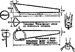
Fig 22. Thomas splint and one of its modifications—for first aid and transport in fractures of the femur.
constructed by Thomas shortly before the Franco-Prussian War of 1870/71 and was then offered to the French army; however, the French refused this offer at the time. For half a century, the Thomas splint was included in surgical manuals, finding little practical application among surgeons. It was used mainly by prosthetists in the form of the so-called Thomas apparatus, wishing to provide relief to one or another of the three diseased joints of the lower limb (fig. 23, 24). Seeing the appalling mortality from complications in gunshot fractures of the femur in the first year of the imperialist war, the great English orthopedist R. Jones persistently recommended the use of the Thomas splint at the site of the wounded person's location—over the trousers and boot—with traction through a spike conducted through an incision in the boot along the inner plane of the sole. Subduction of the shoulder. Detail. In the second half of the war, the Thomas splint became mandatory for first aid and transport in cases of femoral fractures not only in the English but also in the American army. Several authors, such as Sinclair, modifying the Thomas splint, applied it for further treatment of femoral and shin fractures, as well as for fractures of the upper limb (fig. 25, 26). Thus, for English and American orthopedists, it has now become to a certain extent universal. The arm rarely needs splinting compared to the leg, as in many cases of first aid and transport, the trunk can replace the splint, to which the arm is bandaged. For splinting fractures of the upper limb, there are many variations of the method based on the principle of the double inclined plane. Thus, Middeldorf, in fractures of the humerus, proposed fixing the downward-hanging arm, abducted at the shoulder and bent at the elbow, on a triangle. However, this position of the arm with sharp inward rotation of the shoulder cannot provide correct alignment of the fragments of the humerus. Based on the same principle, but with a more correct physiological position of the shoulder and forearm, the cardboard triangular splint of Falten is also constructed. In addition, there is a folding metal Turner splint (fig. 27) and the so-called ambulatory crutch splint of Borchgrevink, applied according to the type of a femoral splint for the shoulder. (According to Zuppinger). The upper end of the splint, a 'fork,' is inserted under the support in the armpit. The shoulder and forearm hang down and rest on the plane of the splint. A strip of adhesive plaster applied along the length of the shoulder and forearm produces traction by means of a rubber tube. Tikhomirov and N. N. Petrov added abduction of the shoulder to the Borchgrevink splint (fig. 28). In M. I. Sitenko, this splint with abduction is made folding (fig. 29). Of the extension splints used in Germany during the imperialist war for fractures of the shoulder, Müller particularly notes the improvised splint by Porzelt, which when applied to the extended shoulder was bandaged to the trunk along its length. When the Kraemer splint appeared, the rectangular extension splint by Zuppinger (fig. 30) came into wide application. Furthermore, according to Müller, the adjustable angular extension splint of Braun with a spring and screw proved to be very rational (fig. 31).-For fixation of the limb after resection of a joint, various types of so-called resection splints are used. The two parts of the splint, usually wooden, bandaged to the limb above and below the diseased joint, are connected by a 'bridge'—a metal arch thrown over the site of joint resection for dressing the wound (fig. 32). In fractures of the jaw, at present, splints of the Tigerstedt type are most often used, consisting of a main aluminum wire 2 2/3 mm thick. Each tooth individually is
Figure 23. Thomas splint in the treatment in a hospital of a wounded person with a fracture of the femur.
Figure 24. Two types of Thomas leg splint for first aid (manual on army splints of the USA).
the wounded person's location—over the trousers and boot—with traction through a spike conducted through an incision in the boot along the inner plane of the sole. Subduction of the shoulder. Detail. In the second half of the war, the Thomas splint became mandatory for first aid and transport in cases of femoral fractures not only in the English but also in the American army. Several authors, such as Sinclair, modifying the Thomas splint, applied it for further treatment of femoral and shin fractures, as well as for fractures of the upper limb (fig. 25, 26). Thus, for English and American orthopedists, it has now become to a certain extent universal. The arm rarely needs splinting compared to the leg, as in many cases of first aid and transport, the trunk can replace the splint, to which the arm is bandaged. For splinting fractures of the upper limb, there are many variations of the method based on the principle of the double inclined plane. Thus, Middeldorf, in fractures of the humerus, proposed fixing the downward-hanging arm, abducted at the shoulder and bent at the elbow, on a triangle. However, this position of the arm with sharp inward rotation of the shoulder cannot provide correct alignment of the fragments of the humerus. Based on the same principle, but with a more correct physiological position of the shoulder and forearm, the cardboard triangular splint of Falten is also constructed. In addition, there is a folding metal Turner splint (fig. 27) and the so-called ambulatory crutch splint of Borchgrevink, applied according to the type of a femoral splint for the shoulder. (According to Zuppinger). The upper end of the splint, a 'fork,' is inserted under the support in the armpit. The shoulder and forearm hang down and rest on the plane of the splint. A strip of adhesive plaster applied along the length of the shoulder and forearm produces traction by means of a rubber tube. Tikhomirov and N. N. Petrov added abduction of the shoulder to the Borchgrevink splint (fig. 28). In M. I. Sitenko, this splint with abduction is made folding (fig. 29). Of the extension splints used in Germany during the imperialist war for fractures of the shoulder, Müller particularly notes the improvised splint by Porzelt, which when applied to the extended shoulder was bandaged to the trunk along its length. When the Kraemer splint appeared, the rectangular extension splint by Zuppinger (fig. 30) came into wide application. Furthermore, according to Müller, the adjustable angular extension splint of Braun with a spring and screw proved to be very rational (fig. 31).-For fixation of the limb after resection of a joint, various types of so-called resection splints are used. The two parts of the splint, usually wooden, bandaged to the limb above and below the diseased joint, are connected by a 'bridge'—a metal arch thrown over the site of joint resection for dressing the wound (fig. 32). In fractures of the jaw, at present, splints of the Tigerstedt type are most often used, consisting of a main aluminum wire 2 2/3 mm thick. Each tooth individually is
Figure 25. Manual Thomas splint.
Figure 26. Hinged manual transport Thomas splint (modification by Murray).
and American orthopedists it is now to a certain extent universal. The arm significantly less often needs splinting than the leg, since in many cases of first aid and transport, the trunk can replace the splint, to which the arm is bandaged. For splinting fractures of the upper limb, there are many variations of the method built on the principle of the double inclined plane. Thus, Middeldorf, in fractures of the humeral bone, proposed fixing the downward-hanging arm, abducted at the shoulder and bent at the elbow, on a triangle. However, this position of the arm with sharp inward rotation of the shoulder cannot provide correct alignment of the fragments of the humeral bone. Based on the same principle, but with a more correct physiological position of the shoulder and forearm, the cardboard triangular splint of Falten is also constructed. In addition, there is a folding metal Turner splint (fig. 27) and the so-called ambulatory crutch splint of Borchgrevink, applied according to the type of a femoral splint for the shoulder. (According to Zuppinger). The upper end of the splint, a 'fork,' is inserted under the support in the armpit. The shoulder and forearm hang down and rest on the plane of the splint. An adhesive strip applied along the length of the shoulder and forearm produces traction by means of a rubber tube. Tikhomirov and N. N. Petrov added abduction of the shoulder to the Borchgrevink splint (fig. 28). In M. I. Sitenko, this splint with abduction is made folding (fig. 29). Of the extension splints used in Germany during the imperialist war for fractures of the shoulder, Müller particularly notes the improvised splint by Porzelt, which when applied to the extended shoulder was bandaged to the trunk along its length. When the Kraemer splint appeared, the rectangular extension splint by Zuppinger (fig. 30) came into wide application. Furthermore, according to Müller, the adjustable angular extension splint of Braun with a spring and screw proved to be very rational (fig. 31).-For fixation of the limb after resection of a joint, various types of so-called resection splints are used. The two parts of the splint, usually wooden, bandaged to the limb above and below the diseased joint, are connected by a 'bridge'—a metal arch thrown over the site of joint resection for dressing the wound (fig. 32). In fractures of the jaw, at present, splints of the Tigerstedt type are most often used, consisting of a main aluminum wire 2 2/3 mm thick. Each tooth individually is
Figure 27. Turner metal splint—for fractures of the shoulder.
Figure 28. Tikhomirov-Petrov splint.
Figure 29. Sitenko shoulder splint.
rectangular extension splint by Zuppinger (fig. 30) came into wide application. Furthermore, according to Müller, the adjustable angular extension splint of Braun with a spring and screw proved to be very rational (fig. 31).-For fixation of the limb after resection of a joint, various types of so-called resection splints are used. The two parts of the splint, usually wooden, bandaged to the limb above and below the diseased joint, are connected by a 'bridge'—a metal arch thrown over the site of joint resection for dressing the wound (fig. 32). In fractures of the jaw, at present, splints of the Tigerstedt type are most often used, consisting of a main aluminum wire 2 2/3 mm thick. Each tooth individually is
Figure 30. Rectangular extension splint based on the Thomas splint for treating fractures of the shoulder on the move.
bandaged to the trunk along its length. When the Kraemer splint appeared, the rectangular extension splint by Zuppinger (fig. 30) came into wide application. Furthermore, according to Müller, the adjustable angular extension splint of Braun with a spring and screw proved to be very rational (fig. 31).-For fixation of the limb after resection of a joint, various types of so-called resection splints are used. The two parts of the splint, usually wooden, bandaged to the limb above and below the diseased joint, are connected by a 'bridge'—a metal arch thrown over the site of joint resection for dressing the wound (fig. 32). In fractures of the jaw, at present, splints of the Tigerstedt type are most often used, consisting of a main aluminum wire 2 2/3 mm thick. Each tooth individually is
Figure 31. Braun extension splint for the shoulder with an adjustable angle.
For fixation of the limb after resection of a joint, various types of so-called resection splints are used. The two parts of the splint, usually wooden, bandaged to the limb above and below the diseased joint, are connected by a 'bridge'—a metal arch thrown over the site of joint resection for dressing the wound (fig. 32). In fractures of the jaw, at present, splints of the Tigerstedt type are most often used, consisting of a main aluminum wire 2 2/3 mm thick. Each tooth individually is
Fig 32
attached to this wire. For fractures of the ribs, so-called rib splints are used—wide strips of adhesive plaster applied over the chest in a transverse direction, covering several ribs at once. For fractures of the spine, so-called spinal splints are used—various types of frames and corsets. For fractures of the pelvis, so-called pelvic splints are used—various types of belts and bands. For fractures of the skull, so-called cranial splints are used—various types of bandages and apparatuses. For fractures of the facial bones, so-called facial splints are used—various types of bandages and apparatuses. For fractures of the mandible, so-called mandibular splints are used—various types of bandages and apparatuses. For fractures of the clavicle, so-called clavicular splints are used—various types of bandages and apparatuses. For fractures of the scapula, so-called scapular splints are used—various types of bandages and apparatuses. For fractures of the humerus, so-called humeral splints are used—various types of bandages and apparatuses. For fractures of the radius and ulna, so-called radial and ulnar splints are used—various types of bandages and apparatuses. For fractures of the bones of the hand, so-called digital splints are used—various types of bandages and apparatuses. For fractures of the femur, so-called femoral splints are used—various types of bandages and apparatuses. For fractures of the tibia and fibula, so-called tibial and fibular splints are used—various types of bandages and apparatuses. For fractures of the bones of the foot, so-called digital splints are used—various types of bandages and apparatuses.
Figure 34.

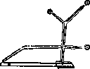
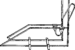
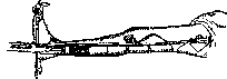
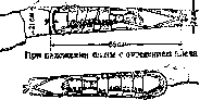
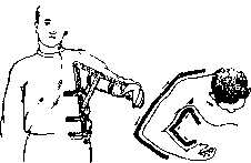
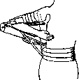
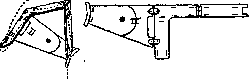
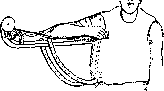
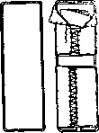
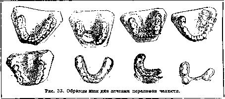
is tied to it with thin ligature wire 0.3-0.4 mm in diameter. The only difference in Sauer's dental splints is that they are made not of aluminum but of nickel silver. Schröder's dental splints differ in that on the end teeth they have metal rings soldered to the splints, which are fixed to the teeth with cement. The rest of the splint is fixed to the teeth with thin ligature wire (fig. 33).
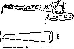
If the historical course of the development of the idea of splinting a fracture in first aid led to traction on a splint, then the development of this principle based on the experience of the imperialist war and the modern stage leads to the use of splints of the type of hoop frames - both for treatment purposes and for first aid and transportation. Modern requirements for a field splint are as follows: 1) the splint should be portable, inexpensive, simple and convenient to use; it should provide both fixation and traction, allow bandaging of the wound without removing the splint, and in extreme cases should serve not only for first aid and transportation but also for treatment, and be suitable for decontamination. The imperialist war proved the need for standardization of splints for all these purposes with a limitation of types. In this respect, in the USA, 5-7 types of army metal frame splints have been chosen: 1. The Thomas manual splint in two types: one (fig. 25) with a fixed ring, the other - modified by Murray (Murray), hinged - for fractures of the shoulder, elbow and forearm (fig. 26). 2. The Thomas leg splint also in two types: one with a complete fixed ring (fig. 24), the other - with a movable (flipping 180°) half-ring - for fractures of the thigh, knee and shin (fig. 24). 3. The Cabot posterior frame (Cabot) - for injuries of the lower end of the shin and foot (fig. 4). 4. The Bradford frame (Bradford) (fig. 34), providing convenience and facilitating care in fractures of the spine and pelvis, i.e., for the most severe and most painful fractures, serves as that necessary stable surface on which the corresponding wounded can be transported without being moved to the place of treatment. The ideal means of transport for these fractures, according to Wilson's admission during the imperialist war, were stretchers (fig. 13); in them the wounded can be tied (swaddled) so firmly that in narrow turns in trenches they can be placed on their side - on edge. 5. In addition to these hoop metal frames, among the army splints of the USA, the long wooden Liston splint (fig. 14) is recommended - only for cases of injury to the hip joint area, in which the ring of the Thomas splint cannot get support (in the pelvis).
Related articles
Mentioned in
- Bandages (a269)
- CORSETS
- DENTAL PROSTHESES
- Dental Technical Instrumentation
- Dupuytren's Fracture
- First Aid Kit
- Hammond Splint
- Hessing Apparatus
- Immobilization
- Injuries
- Kienböck's Disease
- Operating Tables
- Orthopedic Appliances
- Plaster Casts, Corsets, and Beds
- Sanitary Orderly
- Sanitary Transport
- Stomatology
- Traumatology
- Vladimirova-Mikulicz Operation
- Volkmann
Cite this page
“Splints.” Soviet Medical Encyclopedia. English translation of Bolshaya Meditsinskaya Entsiklopediya, 1st ed. (Moscow, 1928–1936), ed. N. A. Semashko. https://sovietmedicalencyclopedia.pages.dev/article/splints/