Skin
Historical document, translated for reference. It reflects medical knowledge of the 1920s–30s and is not medical advice.
Summary
This article provides an overview of the anatomy and physical characteristics of human skin, including its layers, surface patterns, and structural variations. It also discusses historical perspectives on dermatological study and methods for calculating skin surface area.
Encyclopedia article (1928–1936)
SKIN (integumentum commune), a complex organ constituting the outer layer of the entire body and performing a number of functions, namely: protection of the organism from harmful external influences, participation in thermoregulation and metabolism, and the perception of stimuli coming from the outside. In accordance with these tasks, the skin is equipped with a number of accessory organs (hair, glands, nails, etc.). The study of the skin began a very long time ago. The most ancient sources of writing of various peoples already mention the skin and its diseases. However, a more detailed study of the fine structure of the skin and its physiology began in the 17th–18th centuries (see Dermatology). I. Anatomy. Macroscopically, the skin can be divided into 3 layers: the epidermis (cuticle, epidermis), the dermis (skin proper, cutis, corium), and the hypodermis (subcutaneous tissue, tela subcutanea, or subcutis) [see color table (pp. 743–744), fig. 12]. The epidermis—the outermost layer of the skin—represents a stratified squamous epithelium undergoing cornification. By putrefaction, maceration in water, or weak solutions of acids, it can be separated from the skin proper. The dermis represents the dense fibrous base of the skin. Processed in a known manner and stripped of the epidermis, the dermis of animals goes into the production of various leather goods. The hypodermis is an accumulation of fat lobules separated by denser connective tissue layers. The shape of the skin corresponds to the shape of the musculoskeletal base of the body, but not entirely. It mainly, thanks to the presence of subcutaneous adipose tissue, lines numerous depressions, rounds out forms, and in turn provides independent protrusions and folds. At natural openings (mouth, nose, urethra, vagina, anus), the skin transitions into the corresponding mucous membrane. The skin is in a state of constant tension and presses on the underlying tissues. Only on the scalp, on the palms, and on the soles is this tension weakly expressed. The thickness of the skin without subcutaneous tissue varies in different areas within the range of 0.5 mm to 4 mm. The thickness of the subcutaneous tissue varies more strongly. In some places, the subcutaneous tissue is completely absent; in areas subject to frequent compression and friction, it is very thick. The weight of the skin of a healthy 22-year-old woman was 3,175 g, and the subcutaneous adipose tissue was 15,670 g. In a 33-year-old healthy man, the skin weighed 4,850 g, and the subcutaneous adipose tissue was 12,570 g (Bischoff). In a newborn girl, the skin weighed 337 g, and the subcutaneous adipose tissue was 405 g. According to Vierordt, the weight of the skin and subcutaneous adipose tissue constitutes 19.74% of the body weight in a newborn, and 17.77% in an adult. The weight of the cuticle alone was determined by Moleschott to be 488.5 g (cited by Rauber).
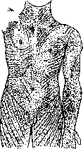
The surface of the skin of an adult varies on average from 1.5 m2 to 2 m2 depending on height, stoutness, sex, and age. A number of special formulas have been proposed for determining the surface area of human skin. Meeh's formula: S = 12.3 3√p2 for adults and S = 11.9 3√p2 for children. For obese individuals, the constant value decreases. For children, in the same formula, they suggest a constant: 9.5 (Michel, Perret), 10.5 (Lassabliere) (p = volume, which can be replaced by weight). Lassabliere's formulas: S = 2.3 · P2 (P = chest perimeter) and S = 0.92 · T2 (T = height). There are also special tables that allow one to determine the skin surface graphically by weight and height. The surface of the skin is not smooth but is equipped with numerous elevations, or folds, and depressions. The skin throughout its extent is covered with the finest grooves with a width of fractions of a millimeter. These grooves, intersecting many times, divide the entire surface of the epidermis into a number of fields, mostly of a triangular or rhombic shape. The excretory ducts of the sweat glands open on the surface of these fields, while hairs emerge from the grooves. The pattern of triangular and rhombic fields is not expressed equally everywhere. It is well developed on the back of the hands. In many other areas, a magnifying glass or smearing with iodine tincture, which spreads along the grooves and thereby reveals the entire pattern, is needed to study it. The direction of the long axis of the fields is characteristic for each individual area of the skin. In many skin diseases accompanied by infiltration of the skin, the pattern of rhombic fields becomes especially strongly expressed (see separate table, fig. 2). Upon recovery, it returns to normal (Nikolsky). On the palms and soles, the described pattern is replaced by a more regular pattern of papillary ridges. The latter is caused by the regular arrangement of connective tissue papillae in paired rows on long, mostly parallel-arranged connective tissue protrusions. On the palmar surface of the hands and feet, there are also 10 small cushion-like elevations, so-called tactile pads (toruli tactiles). They are present on the terminal phalanx of each finger and in the amount of 5 on each palm and sole. As the name implies, they have special significance in touch due to the abundance of nerves in them and the presence of a special fat pad, which increases the surface area of contact of the skin with the object being felt during touch. The pattern of papillary ridges on the pad is more complex than outside it. Giving a huge number of almost non-repeating variants, the pattern of these ridges is used for the purpose of establishing the identity of a criminal (see Dactyloscopy). Large permanent folds include such skin formations as the eyelids, ears, labia, foreskin, etc. Folds of the skin also form over joints as a result of the constant work of muscles. These folds are located in a direction transverse to the long axis of the limb (Figure 1). Similar transverse folds to the direction of the muscles form on the face as a result of the constant work of facial muscles. In the young, these are transient folds; in the elderly, as a result of the loss of skin elasticity, they are permanent wrinkles. The wrinkling of the scrotum depends on the contraction of its smooth muscles. It changes depending on the degree of their excitation. Among pathological folds, one should note the thick folds on the sides of obese women. Depressions and large furrows are very numerous. These include, for example, the opening of the ear canal, the axillary cavity, the sulcus naso-labialis, and many others. Deep furrows on the palms and inconstant dimples on the lower back and in the gluteal fold (fovea sacralis et coccygea) deserve special mention, caused both by the weak development of subcutaneous adipose tissue in these places and the denser adhesion of the skin to the underlying fascia, and probably also by muscle work. Hair cover. The skin is covered almost throughout its entire extent, with the exception of the red border of the lips, palms, soles, etc. Figure 1. Direction of skin folds.
Figure 1. Cutis rhomboidea hypertrophica cervicis. Figure 2. Sharply defined pattern of triangular and rhombic fields in ichthyosis. Figure 3. Section through the sweat glands of the sole: 1-membrana basilaris; 2-secretory epithelium; 3-myoepithelial cells; 4-beginning of the excretory duct. Figure 4. Part of the stratum spinosum of the palm: 1-stratum spinosum; 2-intercellular bridges; 3-intercellular spaces; 4-nuclei with nucleoli. Figure 5. Psoriasis vulgaris: 1-parakeratosis; 2-granular layer, absent directly under the area of parakeratosis and well-defined above the normal horny layer; 3-excretory duct of the sweat gland; 4-appearance of the granular layer under the parakeratosis as a manifestation of the healing of the epidermis; 5-miliary abscess. Figure 6. Acantholysis. Blister in pemphigus foliaceus. Figure 7. Section of the Malpighian layer at the border with eczema vesicles: Sp-status spongoides; Ac-alteration cavitaire; L-leukocytes; V-vesicle with an epithelial cell. (Figures 1, 2, 5, 6, and 7 are from the collection of the Skin Clinic of the North Caucasian State University; figures 3 and 4 are from Kyrle.) (To the illustration of the article Skin) with hair (see). Already in a newborn, one can distinguish several types of hair: hair of the head, eyebrows, eyelashes, and vellus hair. Thin, weakly pigmented vellus hair covers the entire skin, except for the indicated areas, until the onset of puberty. Later, part of it turns into thicker, longer permanent hair, while part retains its vellus character forever. This transformation and the further growth of all hair are in close dependence on the function of the endocrine glands. Among the permanent hair that forms, a group is distinguished that is particularly closely related to the sex glands (Sexualbehaarung), namely: hair of the beard, mustache, armpits, around the nipples, and the genital organs. Beard and mustache hair are characteristic of men. They begin to form at the age of about 18-20 years. Their quantity varies within wide limits. The white race, as well as the Ainu and Australians, possess a strongly developed beard; among Mongols and Negroes, it is developed much more weakly. The growth of a beard and mustache is possible as an exception in women as well. However, small local hypertrichosis in the form of thicker vellus or a small amount of pigmented hair on the upper lip, cheeks, and chin is encountered not infrequently, especially in old women; Trotter and Danforth found it in 27%, and Dupre and Duclos in 38% of women. Armpit hair is relatively short and weakly pigmented, except for brunettes, in whom it can be intensely black. In women, they are sharply demarcated, while in men they often transition into chest hair without a clear boundary. Genital hair covers the pubis, labia majora, inguinal folds, the root of the penis, the perineum, and the anal region. There is less hair on the scrotum, and it is thin. Genital hair is thick, usually curly, and sometimes forms loops. It develops in women earlier than in men: in women sometimes from 10-12 years old, in men from 12-15 years old. The upper border of pubic hair in women is more often a straight transverse line. In men, however, the hair rises along the midline to the navel. In hairy men, abdominal hair connects with pubic hair without a clear boundary. In addition to the described hair, thick pigmented hair is present on the trunk and limbs. Most men have such hair on the chest and the extensor surfaces of the forearms and shins. In women, they more often develop on the lower limbs (thighs, shins). The remaining areas of the skin more often retain their vellus cover but can also be covered with thick, real hair. Head hair, having replaced the vellus hair, continues to grow for many years at approximately 1 cm per month. On the eyebrows, hair grows especially slowly. In old people, they thin out, but individual eyebrow hairs can become strongly hypertrophied. The color of the skin is connected with many conditions. The degree of blood supply, the color of the blood, the color of the epidermis and connective tissue, and various degenerative processes in them influence the coloration of the skin. The greatest importance belongs to the pigment of the basal layer of the epidermis and the papillary layer of the skin itself, as well as the presence and thickness of the keratohyalin layer. The degree of density of the elastic tissue also plays a considerable role; its rarefaction makes the skin transparent, and capillary vessels begin to show through in these places. The color of the skin, mainly determined by the genotype (see below - inheritance of skin color), also depends on age and sex. Some areas of the skin are usually more pigmented than others. These include the nipple with its areola, the armpit, and the genital organs. In summer, under the influence of sunlight, increased pigmentation of exposed parts occurs (suntan). In persons with delicate white or freckled skin, pigmentation from sunlight is negligible, which is often accompanied by weakness of the whole organism. II. Histology. The epidermis is a multilayered squamous, keratinizing epithelium on the surface, varying in its thickness in different areas of the skin. Cell reproduction in it occurs in the deepest layer, adjacent to the basement membrane. Newly formed cells are pushed away from the membrane, gradually rise to the surface of the epidermis, undergo a series of changes in their structure and chemical composition, and finally turn into a dense horny layer. Changes in cells located at the same level occur approximately simultaneously, due to which the entire epidermis breaks down into a series of regularly alternating layers. The following layers are distinguished: basal, spinous, granular, glassy, and horny [see color table (art. 743-744), fig. 11]. The basal layer (stratum basale, stratum cylindricum) is the lowest layer. It consists of fairly tall cylindrical cells arranged in a palisade-like manner, perpendicular to the basement membrane. The cell nuclei are large, round or oval in shape, rich in chromatin, and located mainly in the upper half of the cells. The cells are separated from each other by intercellular spaces, remaining in connection with each other by means of the finest protoplasmic bridges. From the lower surface of the cells, protoplasmic processes extend, entering the basement membrane. Inside the cells of the basal layer, there is a granular pigment of a light brown color. The amount of pigment depends on race, skin area, exposure to sun, wind, and other causes. Pigment is also present in special dendritic cells located both between the described epithelial cells and in the papillary layer of the skin itself (so-called chromatophores). The cells of the basal layer divide karyokinetically and thereby compensate for the constant loss of the epidermis on the surface. As an exception, cell division can occur under normal conditions in 1-2 overlying layers as well. Above the basal layer lies a thick layer of spinous cells (stratum spinosum). These are rounded or polyhedral cells of large size, with a large round vesicular nucleus with 1-2 nucleoli. In the protoplasm of the cells, with certain processing methods, a large number of fine fibrils can be distinguished, surrounding the nucleus and passing from one cell to another along the already described intercellular bridges (see separate table, figure 4). The latter are especially strongly developed in this layer. The cells equipped with them appear covered with small spines, hence the name of the layer - spinous. In the middle of the bridges, one can sometimes see thickenings. In adjacent bridges, they are located at the same level. If one connects the thickenings on the bridges of one cell, one gets a continuous line outlining the contours of the cell. The indicated system of fibrils is given great importance, considering it a support for epithelial cells, a kind of skeleton for them. The system of intercellular spaces, extending up to the granular layer, serves the purposes of nutrition of the epithelial cells. Through it, nutrient juices come into the epidermis, and metabolic products are excreted into it. In these same slits, migrating leukocytes are located, normally single, but in inflammatory phenomena encountered in large numbers and even in the form of large clusters. Recently, Kolosov, who worked a lot on intercellular bridges, came to the conclusion that the described intercellular spaces do not actually exist. The true boundary of the cell is the line drawn along the thickenings of the bridges. The bridges themselves are the result of the wrinkling of the outer layer of protoplasm. The nutrition of the epidermis, like all epithelia, proceeds through special juice channels. This view requires confirmation. The described two layers are combined under the name of the Malpighian layer (rete Malpighii), and also under the name of the germinal layer (stratum germinativum). However, it would be more correct to call only the basal layer, in which cell reproduction occurs, the germinal layer. The boundary of the Malpighian layer with the skin itself appears on preparations as a convoluted line due to more or less long outgrowths of the epithelium penetrating into the thickness of the connective tissue. These outgrowths are called interpapillary extensions (processes) of the epithelium. The spaces between them are filled with connective tissue papillae, equipped with vessels and nerves. Such a structure ensures better nutrition of the epidermis.
Cells of the Malpighian layer, as they move upward, gradually flatten and appear spindle-shaped in a preparation. The intercellular spaces between them narrow. In their protoplasm, granules of keratohyalin (see) of various sizes begin to appear. At first, they are located around the nucleus, and then they fill the entire cell. Simultaneously, the fibrils disappear from the cells. The cell nuclei shrivel but are still clearly distinguishable. The layer of these cells is called the granular layer of Langerhans (strat. granulosum). This is the beginning of the keratinization process. Above the granular layer lies the glassy, or otherwise, clear layer (stratum lucidum of Oehl) consisting of 2-3 rows of elongated cells filled with a light-refracting substance. In this layer, keratohyalin turns into eleidin. The nuclei in this layer are either not distinguishable at all or appear as shriveled lumps. Still higher, eleidin turns into keratin; the cells become flat; the nuclei in them are not distinguishable; the cell membrane thickens significantly. Inside the cell, one can distinguish remnants of protoplasm in the form of a reticular lump. This is the horny layer (stratum corneum). There is another view on the process of keratinization (Kromayer, Unna), according to which it begins in the upper cells of the Malpighian layer and consists of the keratinization of the surface layer of cells (mainly fibrils), while keratohyalin and eleidin are by-products of this process. The cells of the horny layer on the surface gradually desquamate and fall off, maintaining normal desquamation, which is invisible to the eye. The layer of desquamating cells is sometimes distinguished under the name stratum disjunctum. When the horny layer is treated with osmic acid, it partially stains black, which indicates the presence of fat in it, arising from 2 sources: from the fat of the sebaceous and sweat glands and as a result of fatty degeneration of the cells of the horny cover itself. - The thickness of the various layers of the epidermis varies depending on the area of the skin. The prickle cell layer is usually the most powerful; the granular layer is usually thin—1-2 rows of cells, and the glassy layer is barely noticeable and sometimes seems absent, precisely where the horny layer is also thin, e.g., in the joint flexures. Conversely, the glassy layer is clearly distinguishable in places where there is a powerful horny cover, e.g., on the palms and soles. - Between the epidermis and the connective tissue part of the skin lies the basement membrane (membrana basilaris). At low magnifications, this is a structureless membrane separating the epidermis and all organs of epithelial origin from the connective tissue. At high magnifications, it appears as a dense felt-like plexus of the finest connective tissue fibrils with protoplasmic processes of the basal cells of the epidermis. The skin proper (corium, cutis) consists of dense connective tissue, in which various epithelial appendages of the skin (hair, glands), vessels, muscles, and nerves are embedded. The connective tissue itself consists of relatively few cells (fibroblasts, lymphocytes, mast cells, melanoblasts, melanophores) and a large number of collagenous and elastic fibers. The number of cells can fluctuate significantly, especially along the vessels of the subpapillary network, where loose lymphocytic perivascular cuffs are often found even in normal conditions. In the skin proper, 2 layers are distinguished: pars papillaris and pars reticularis. The pars papillaris is characterized by a dense plexus of finer connective tissue fibers. Bundles of fibers are directed partly irregularly, partly perpendicular to the surface. The number of cells in this layer is more significant. In its upper part, the already mentioned connective tissue papillae with blood and lymphatic vessels are formed. In some papillae, there are also nerve terminal corpuscles. The pars papillaris corii transitions indistinctly into the deeper pars reticularis. The latter is distinguished by thicker bundles of collagenous fibers and a small number of cells. Despite the apparent disorder, the arrangement of connective tissue bundles in this layer is distinguished by great regularity. They are directed for the most part parallel to the surface, a smaller part of them runs perpendicular to it. The fiber bundles running parallel to the skin surface usually intersect each other at an acute angle, forming rhombic figures. The latter are clearly visible on skin sections made parallel to its surface. The direction of the long axis of these rhombi, and consequently the main direction of the fibers, is characteristic for each given area of the skin. Due to this, the skin's ability to stretch is not the same in different directions. Specifically, in the direction of the long axis of the bundles, it is the least, and in the transverse direction, it is the greatest. For the same reason, the shape of a puncture wound after the removal of the piercing object does not remain round but becomes elongated in the direction of the main flow of the connective tissue fiber bundles. The direction of these flows was carefully studied by Langer, which is why it received the name Langer's lines (Fig. 1). In some areas where several flows converge, a puncture into the skin leads not to a linear, but to an irregular shape of the wound. Langer's lines also have practical significance, as an incision of the skin in a direction perpendicular to them leads to greater gaping of the wound and, consequently, to worse cosmetic scars. Besides collagenous fibers, the skin is rich in elastic fibers. The latter do not form bundles but form networks and plexuses. Starting from the fasciae, thick elastic fibers, gradually thinning, pass in an oblique direction through the subcutaneous tissue and the skin proper. Under the papillae, they form a dense plexus running parallel to the surface. From it, thin networks and individual fibrils extend into the papillae. In the skin proper, elastic fibers entwine the bundles of collagenous fibers and form basket-like plexuses around the hair and sebaceous glands. The glomeruli and excretory ducts of the sweat glands, smooth muscles, and blood vessels are also surrounded by dense plexuses of fine elastic fibrils. - Elastic fibers are the main support for the entire skin. They maintain its normal shape, return hair and muscles to their initial position, and resist pressure and stretching of the skin. Elastic tissue is most strongly developed in areas of the skin frequently subjected to mechanical irritation—pressure, friction, stretching (palms, soles, penis, skin over joints). The subcutaneous tissue is characterized by a large number of fat lobules. These are clusters of fat cells located in the loops of connective tissue. Moderately dense bundles of the latter, starting from the fasciae or periosteum, pass through the thickness of the subcutaneous tissue, heading into the skin proper. They are called retinacula cutis. Along the way, these strands give off branches that intersect with each other and thus form loops filled with looser tissue. The latter is filled with fat cells and is supplied with a well-developed network of blood vessels (see Adipose tissue). Around the lobules, elastic tissue is also well-defined. - The thickness of the subcutaneous tissue fluctuates within wide limits both in different individuals and in the same individual. On the skin of the scalp, its usual thickness is about 2 mm, on the back—5-10 mm. On the female breasts, buttocks, and abdomen, it can reach very large dimensions. The skin glands belong partly to holocrine (sebaceous glands), partly to merocrine (sweat) (see Glands); the sweat glands of the axillary fossae and some other parts of the body belong to apocrine glands (see). - Sweat glands are simple tubular glands, coiled at the end into a glomerulus located in the deep layers of the skin proper or even in the upper layer of the subcutaneous tissue; they consist of a secretory part and an excretory duct. The secretory part is a repeatedly convoluted tube forming the glomerulus of the gland. A section through it in a preparation appears as numerous round, elongated, or curved segments of the glandular tube. Its walls consist of a single layer of cylindrical or cuboidal secretory cells. Small round nuclei are located approximately in the middle of these cells. The protoplasm contains numerous inclusions in the form of granules of various sizes, fat droplets, and pigment granules. The shape of the secretory cells is in close dependence on their functional activity. At rest, these are tall cylindrical, or rather, prismatic cells; after the secretion is released, they flatten. Outside the described secretory cells are long spindle-shaped cells with a rod-shaped nucleus. These are the so-called myoepithelial cells (myoepithelium), located obliquely in relation to the long axis of the glandular tube, capable of contracting and thus taking an active part in the process of excreting sweat outward. In preparations, they appear either as small triangular protrusions adjacent to the basement membrane (see separate table, Fig. 3) or, in sections passing along the surface of the secretory tube, as long spindle-shaped formations. The excretory duct of the gland, slightly winding, rises almost perpendicularly upward to the epidermis. Its wall consists of 2 rows of epithelial cuboidal cells. There are no contractile elements in it anymore. The inner layer of cells is provided with a thick cuticle.
Approaching the epidermis, the excretory duct enters it at the apex of one of its interpapillary extensions. In the epidermis, it twists repeatedly like a corkscrew and opens outward in the form of a small funnel, always located at the apex of one of the skin ridges. The excretory duct has no proper wall inside the epidermis, but the adjacent epithelial cells are arranged around it in concentric layers. The process of keratinization in the cells surrounding the duct begins much deeper than in the epidermal cells lying nearby. Such a structural feature of the excretory duct wall, as well as the presence of the aforementioned cuticle in its cells located in the thickness of the skin proper, apparently has significance in protecting the epithelial cells from constant wetting and irritation by the secreted sweat. Sweat glands are located on the entire skin with the exception of the red border of the lips, the glans penis, and the inner layer of the prepuce. The total number of glands reaches 2 million. According to Krause, per 1 cm2 on the palm there are 373 sweat glands, on the sole—366, on the flexor side of the forearm—157, on the forehead—172, on the chest and abdomen—255, on the thigh and lower leg—79, on the back and buttocks—57 (cited by Rauber). Sebaceous glands are simple or branched alveolar glands. They are distributed over the entire skin. They are absent on the palms and soles. Most of them are associated with hairs, and only on the lips, the mucous membrane of the mouth and nose, the mammary nipples, the genital organs, and near the anus can free sebaceous glands be observed. There is no regular dependence between the size of the glands and the hairs, although it is noted that the largest glands on the nose and in the auricle are associated with the smallest hairs, while the glands of large hairs on the beard or the scalp are relatively small. Each hair is provided with 1-2 or a large number of sebaceous glands. In large hairs, they are an appendage of the hair follicle, into the cavity of which their excretory duct opens. Small hairs, on the contrary, are themselves appendages of the sebaceous glands, emerging onto the surface of the skin through the excretory duct of the latter. Sebaceous glands are located in the upper half of the skin proper and represent an overgrown and modified invagination of the epithelium of the hair follicle [see color table (p. 743–744), fig. 12]. The multilayered epithelium of the follicle gives rise, on the side of the obtuse angle formed by the hair with the epidermis, to a sac-like protrusion, which divides into a certain number of gland lobules. The latter are lined along the periphery with cuboidal, intensively dividing cells. A portion of the newly formed cells is pushed away from the wall of the alveolus. Increasingly numerous fat droplets begin to appear in them. The nuclei shrivel. The cells, swollen from the accumulated fat, burst, and the fat, together with cell debris, enters the excretory duct of the gland, which opens into the follicle cavity next to the hair shaft. The excretory duct is lined with multilayered epithelium, which thins as it approaches the alveoli. Meibomian glands of the eyelids are close to the sebaceous glands. Tyson's glands, located on the inner layer of the prepuce and producing an odorous lubricant—smegma—also belong to the sebaceous glands. Muscles of the skin. There are smooth and striated muscles in the skin. The latter are distributed in the skin of the face in the form of ramifications of the facial muscles. Most of the smooth muscles are associated with hairs (see Arrector pili). The hairs of the eyelashes, nasal openings, and lips are devoid of these muscles. In the axillary cavities, they are few or may be absent altogether. On the scrotum and near the mammary nipple, there are bundles of smooth muscle fibers not associated with hair. Vessels of the skin. More or less large arterial trunks coming from the fascia pierce the subcutaneous adipose tissue, giving off branches along the way for its deep parts. At the border between the subcutaneous adipose tissue and the skin proper, these arteries anastomose with each other, forming a deep cutaneous network [see color table (p. 723–724), fig. 6]. It gives off branches for the upper parts of the subcutaneous adipose tissue, for the hair papillae, and the glomeruli of the sweat glands. From this same network, fairly large arteries also depart upward. Having passed in an almost vertical direction through almost the entire thickness of the skin proper, the latter again break up into branches that anastomose with each other and form the subpapillary arterial network. From this network, small arterioles depart, nourishing the hair follicles, sebaceous glands, excretory ducts of the sweat glands, and muscles. Upward from this same network, small, no longer anastomosing terminal arterioles depart. They run for some distance parallel to the epidermis and break up into capillaries, which head into the papillae. The size of the area nourished by such a terminal artery is calculated on average at 0.16 mm2. The capillaries departing from them are distinguished by significant thickness ("Hoyer's derivation channels"); they twist in the papillae, form loops there, and return back already in the form of wider venous capillaries. No networks are formed in the papillae. The structural features of the skin capillaries, in particular the close contact and even direct transition of arterial and venous branches (in the papillae), indicate the special function of the skin vessels mainly as a thermoregulatory and excretory organ. The arterial network is developed unevenly in different areas of the skin. Its most powerful development is found in areas subject to pressure, e.g., on the palms, soles, and buttocks. Venous capillaries collecting from the papillae, hairs, glands, and muscles form the first subpapillary venous network. Directly below it lies the second, even lower—the third, and finally, at the border of the skin proper and the subcutaneous tissue—the fourth network. The veins emerging from the latter network pass through the subcutaneous tissue and empty into large deep veins. The large number of venous (and lymphatic) networks in the skin is attributed significance in the nutrition of the connective tissue of the skin, which does not have independent branches. In addition, it is believed that the numerous venous vessels filled with blood possibly soften any trauma. Lymphatic vessels begin in the middle of the papillae and form a network in the subpapillary layer. A second network of larger lymphatic vessels lies at the border of the skin proper with the subcutaneous tissue. Nerves of the skin. The skin is supplied with a huge number of nerves—sensory, motor, vasomotor, secretory, and, possibly, trophic. Sensory nerves are especially numerous. The skin of the palms, soles, lips, and genital organs is richest in nerves. Sensory nerves enter the skin proper in the form of bundles of medullated nerve fibers. Rising upward through the skin proper, they divide into individual fibers, lose their medullary sheath, and terminate in the form of free nerve endings or in specially adapted nerve corpuscles. Free nerve endings are formed in such a way that the axis cylinder, having lost its medullary sheath, enters the epidermis and divides repeatedly. Its finest fibrils pass between the cells, reaching the granular layer, and terminate in button-like thickenings. The latter touch the epithelial cells and sometimes form indentations on them. Free nerve endings in the form of tree-like figures are also found in the skin proper. In the epidermis, there are also so-called Merkel tactile cells. Merkel cells are located in the deep layers of the epidermis and even in the skin proper. The number of Merkel cells in humans is relatively small. Terminal nerve corpuscles are distributed in the skin proper, subcutaneous tissue, and in deeper organs. Meissner's tactile corpuscles (figure 2) are located in
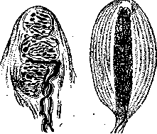
Figure 2. Meissner's corpuscle.
Figure 3. Vater-Pacini corpuscle. [They are located in the] papillae, have an elongated egg-like shape [see color table (pp. 743-744), fig. 11]. The greatest number of them is found on the palmar surfaces of the fingers of the hands and feet. In smaller numbers, they are scattered in other areas of the skin. Krause end-bulbs are distributed in the mucous membranes and, in smaller numbers, in the skin proper. The sensation of cold is associated with them. Closely related to the Krause end-bulbs are the so-called genital nerve corpuscles. They are larger than ordinary bulbs, reaching up to 400 μ. Vater-Pacini corpuscles (Fig. 3) are the largest of the nerve corpuscles, up to 2-4 mm in length. These corpuscles are located in the subcutaneous tissue, especially on the palms and soles, and in smaller quantities in other areas of the skin and in deeper-lying organs (bones, periosteum, tendons, organs of the abdominal cavity). The sense of pressure is associated with them. The sensation of warmth is attributed to the Ruffini corpuscles. Pinkus described additional special hair discs (Haarscheibe). These are areas of the epidermis with a smoother surface than the surrounding skin. They are located near hairs and are supplied with numerous nerves. Pinkus also classifies these discs as sensory organs. The methods of termination of motor (for smooth muscles), secretory, and vasomotor nerves in the skin are less studied. All of them belong to the sympathetic nervous system. III. Embryology. For the first two months of embryonic life, the skin is a thin epithelial covering of ectodermal origin. It is separated from the connective tissue derived from the mesenchyme by a straight line. Already at this time, 2 layers of cells are distinguished in the epidermis: the lower one, consisting of cells of irregular shape, and the upper one, consisting of polyhedral or flattened light cells. In the lower layer, continuous cell division occurs; in the upper one, it soon ceases. The upper layer is called the periderm. Soon, at different times in different areas of the skin, an intermediate layer (stratum intermedium) appears between these two layers. At first, it is an intermittent layer, then it gradually thickens and becomes the most powerful part of the entire epidermis. Its cells are light, of irregular polyhedral shape, without mitoses. The cells of the lower layer at this time change their shape and turn into tall cylindrical cells. Their nuclei are pushed to the upper pole of the cells. The reproduction of cells in this basal layer occurs continuously. The newly formed cells do not yet undergo complete keratinization. They only flatten like the epithelium of the oral mucosa, rise to the surface of the epidermis, and gradually fall off, becoming part of the so-called vernix caseosa. In some animals, these cells separate in a solid sheet. Thus, a very thin membrane can form, under which the hair shines through, which is why this layer of cells received the name epitrichium. From the second half of embryonic life, normal keratinization of the epidermis begins with the formation of keratin. In the formative layer at this time, a fibrous structure is revealed. Intercellular bridges begin to be distinguished between the cells. Simultaneously, the boundary between the epidermis and the skin proper becomes convoluted due to the formation of papillae. The epidermis takes on its usual appearance. Pigment in the epidermis is formed only after birth. Hair begins to develop at the beginning of the 4th month, and in some areas (eyebrows, upper lip, chin) even earlier. The process of hair formation begins with the intensified reproduction of cells in a limited area of the epidermis. The increased number of epithelial cells protrudes at the lower boundary of the epidermis in the form of a crescent-shaped projection. As a result of intensified cell reproduction, the projection turns into a long, compact strand running at an angle to the epidermis. At the apex of this strand, first a flattening and then a depression forms, in which the connective tissue papilla with blood vessels is located. The epithelial cells lying at the apex of the papilla continue to divide intensively. Flattening and keratinizing, they form a pointed cone that rapidly moves upward. This cone is the newly formed hair with its internal epithelial sheath. By the time the hair is formed, in the upper part of the former compact epithelial strand, due to the keratinization of the central cells, a hair canal is formed, through which the growing hair is directed. Such a canal is also formed in the thickness of the epidermis. Most hair reaches the surface of the skin only in the 5th month. Even at the time when the hair germ is a long, compact epithelial strand, 2 epithelial projections appear on it; the upper one grows even more in the future and turns into a sebaceous gland; the significance of the lower one is still unclear; it is only known that it corresponds to the attachment site of the arrector pili muscle. Sweat glands also go through the stage of a dense epithelial strand growing into the skin proper in a direction perpendicular to the surface. Sweat and sebaceous glands are formed in the 5th month of embryonic life. The cavity in the sweat glands appears only in the 7th month. The skin proper in the first months is an accumulation of cells, first round and then spindle-shaped, immersed in a jelly-like mass. In the 3rd month, collagenous fibers form between the cells, and somewhat later, elastic ones. In the 3rd month, the subcutaneous tissue begins to differentiate. Regarding the mechanism of the formation of the latter, there is no generally accepted opinion yet. Some authors admit the possibility of the formation of subcutaneous tissue from ordinary connective tissue. Others consider this transition impossible. In their opinion, adipose tissue is formed from an independent germ in the mesenchyme. Wassermann speaks of a special primary organ, the origin of which he links to embryonic capillaries. In his opinion, subcutaneous adipose tissue is close to bone marrow in origin and function. IV. Comparative anatomy. The skin of almost all invertebrates (except coelenterates) consists of a single-layered epidermis, to which only in some of them adheres a poorly delimited connective tissue base of the skin. The single-layered epithelium can be flat, cuboidal, or cylindrical. It can be equipped with diverse adaptations for the purposes of movement (cilia, rowing plates), perception of sensations (sensory cells), and protection from harmful external influences (cuticle with various bristles, spines, hairs, scales). All the latter formations, despite the apparent analogy with some appendages of the skin in vertebrates (scales, hair, needles), differ from the latter in the mechanism of their formation. All of them, like the cuticle itself, are merely a product of the secretion of the epithelium, and not the transformed epithelial cells themselves, as is the case in vertebrates. The epidermis, covered on the outside by such a supercellular cuticle, is called the hypodermis. The cuticle can reach a very significant thickness and, being built of chemically stable and dense substances (for example, chitin), is a good protection for the animal (e.g., the carapace of arthropods). This dense framework is simultaneously a good support for the entire organism. It often gives off processes that go inside the animal's body and support the internal organs, which is why it is often called an ectodermal skeleton, or exoskeleton. In acraniate vertebrates, the epidermis still consists of one layer of prismatic ciliated cells, while in the rest of the vertebrates, it becomes multi-layered. In this case, the basal layer retains a cylindrical shape, while the cells lying above it first become polyhedral and then flatten. In fish, all cells of the epidermis still retain their plasmatic character; starting from amphibians, its upper layers begin to produce keratin, lose their nuclei, and keratinize. The epidermis, thus having lost its cuticle, remains adapted to protect the organism from harmful physical, mechanical, and chemical influences. Horny cells are short-lived formations subject to frequent replacement. This replacement in amphibians and reptiles is periodic in the form of molting, while in birds and mammals, the horny layer begins to peel off in small patches and even individual scales. The epidermis in vertebrates forms various outgrowths and thickenings that serve the purposes of better protection of the organism. In reptiles, these are scales, which are flat outgrowths of the skin with a thickened horny layer on them and connective tissue papillae at their base. These outgrowths are inclined backward and cover each other like tiles. The scales found on the legs of birds and on the legs and tails of some mammals (rat, beaver) are compared to this type of scale. Pinkus also sees the rudiments of such scales in human skin in the form of a small, poorly delimited skin fold with a smooth surface, sometimes noticeable near a hair. The feathers of birds are apparently genetically related to the described scales. Their development begins, like scales, with an outgrowth of the epithelium upward, followed by its reverse immersion into the thickness of the skin with the formation of a sheath. In contrast to this, hair never begins with an outgrowth of the epithelium upward, but from the very beginning is a dense epithelial strand immersing itself into the thickness of the skin proper. Hair growth begins from the bottom of this strand.
The hair papilla forms late, while the feather papilla arises at the very beginning, causing the very outgrowth of the epidermis upwards. Therefore, hair and feathers are considered phylogenetically distinct formations. The origin of hair still remains unclear. They are derived from special sensory organs or from the cutaneous teeth of Selachii, but there is no sufficient evidence in favor of one theory or another. The arrangement of hair in regular groups, alternating in a checkerboard pattern, suggests that they formed behind the scales that were present in the ancestors of mammals.
The skin of vertebrates produces a number of other dense formations: claws, hooves, nails, beaks, horns. The epidermis of invertebrates is rich in glands. The most common are unicellular mucous glands, but glands that secrete poisonous substances, odorous fluids, silk glands, and stinging capsules are also frequent. Multicellular glands are also not uncommon in invertebrates.
Skin glands in fish are almost exclusively unicellular, mucous. In amphibians, besides unicellular ones, there is also a large number of multicellular glands, both smaller mucous ones and larger ones that secrete a special, often poisonous, secretion. In amniota, unicellular skin glands disappear completely. The skin of reptiles contains no glands, and birds have only the uropygial gland, which secretes an oily substance intended for lubricating feathers in order to protect them from getting wet by water. Conversely, in most mammals, the number of skin glands is very large. The glands here are of two types: tubular (sweat) and alveolar (sebaceous). The former correspond to the mucous glands of amphibians, the latter to their granular poisonous glands. And among mammals, there are species that are very poor in glands or even devoid of them (cetaceans).
Sweat glands are poorly developed in rodents and insectivores; sebaceous glands are poorly developed in sloths and insectivores. Specially differentiated mammary glands are also skin glands, close in origin to apocrine glands (see Mammary gland, Apocrine glands). Under the epidermis in some invertebrates and in all vertebrates, a special layer of connective tissue develops, the so-called skin proper (dermis). In echinoderms, for example, this is a gelatinous or fibrous ground substance with stellate connective tissue cells. In vertebrates, this layer receives powerful development and consists of numerous intersecting connective tissue bundles, the majority of which are directed parallel to the surface. In this skin proper, a vascular network and numerous nerves with their terminal corpuscles develop. Starting with cyclostomes, vascularized skin papillae are formed, which penetrate into the epidermis and facilitate its nutrition. These papillae, which are not connected with various skin appendages, reach their maximum development in mammals, especially on hairless areas of the skin. In many fish, reptiles, and some mammals, bony plates form in the thickness of the skin proper, constituting the so-called dermal skeleton, or external mesodermal skeleton. This includes the scales of fish, the bony shields of crocodiles and turtles, and the bony plates of the armadillo shell.
V. Physiology. Sweating. Sweat is a cloudy, odorous fluid secreted by sweat glands. At a low ambient air temperature and low humidity, sweat is secreted in negligible quantities and manages to evaporate in the openings of the glands, remaining invisible to the eye. Increased sweating is observed with an increase in the ambient temperature (in a bath, a dry-air bath, on hot summer days), with hydremia (drinking fluids, especially warm ones), and during intense muscular work. Psychic factors (fright, mental strain) can also lead to increased sweating. An increase in the ambient temperature causes sweating by reflex irritation of the nerve centers, which is evident from the fact that general sweating can occur even in the case of strong heating of a small part of the body. Direct heating of the blood in the isolated carotid artery of an animal also leads to irritation of the sweat centers in the brain and increased sweating despite a normal body temperature (measured in the rectum). Cutting the nerves destroys this reflex sweating. Increased sweating is usually accompanied by local hyperemia. Skin with red dermographism is also prone to hyperhidrosis. However, increased sweating can also be observed on pale skin, as, for example, during fright or hyperhidrosis of the hands and feet (the so-called "cold sweat"). Certain substances have the property of acting on the peripheral endings of secretory nerves in the sense of exciting them (pilocarpine) or depressing them (atropine). Different areas of the skin sweat unevenly. The scalp, forehead, armpits, genital area, palms, and soles sweat particularly heavily.
The chemical composition of sweat is not constant. It depends on numerous conditions: on the method of obtaining sweat, the intensity of metabolism in the body, the functional activity of the kidneys, etc. All fluctuations associated with this are still little studied despite their high theoretical and practical interest. The amount of sweat secreted per day is 500-900 cm3. It increases in summer to 2-3 liters with a corresponding decrease in the amount of urine excreted. When the body is overheated in a bath or a dry-air bath, the amount of sweat can reach 2-2.5 liters within 1.5-2 hours. The specific gravity of sweat fluctuates within the range of 1.004-1.008. The reaction of sweat collected under normal conditions is weakly acidic, possibly due to the admixture of fatty acids from sebum. With careful removal of the secretion of sebaceous glands from the skin, it was possible to obtain sweat with an alkaline reaction. In recent years, a weakly acidic reaction of sweat has been found again (pH=6.6-6.8). Apocrine glands possess sweat of an alkaline reaction. In various skin diseases, the reaction of sweat changes either towards a more acidic one (pruritus, seborrhea) or towards an alkaline reaction (eczema). The amount of solid substances in sweat fluctuates within the range of 1-2%. Most of them are inorganic products, the smaller part is organic. The largest amount in sweat is NaCl, namely 0.2-0.7% (according to various authors). Besides it, there are ammonia, phosphoric acid anhydride, potassium, calcium. Of organic substances, there is mostly urea (0.02-0.14%); uric acid—from traces to 0.03%. In addition, there are sugar, creatinine, volatile fatty acids, cholesterol, aromatic sulfo- and hydroxy acids, traces of serum protein. Of the volatile fatty acids, there are formic, acetic, butyric, propionic, and caproic acids. In pathological conditions, acetone, bile pigments, cystine, etc., can be excreted. Metabolic disorders and the retention of their excretions through the kidneys can manifest as a high content of certain products in sweat. Occasionally, in such cases, it was possible to observe the deposition of urea and uric acid crystals on the skin. Tonian's studies showed significant fluctuations in the composition of sweat in skin diseases. Pruritus, for example, is often accompanied by increased excretion of NaCl, urea, and uric acid. A number of medicinal substances are excreted with sweat: iodine, bromine, phenol, salicylic acid, quinine, arsenic, mercury, chloroform, etc.
The activity of sweat glands is in close dependence on the nerve centers located in the lateral horns of the spinal cord, in its thoracic and cervical parts. From there, secretory fibers pass through the anterior roots and rami communicantes into the sympathetic nerves and reach their ganglia. Postganglionic fibers return through the gray rami communicantes to the spinal nerves and go to the periphery together with sensory nerves. Higher centers are located in the medulla oblongata and in the diencephalon in the region of the central gray matter of the third ventricle. The cerebral cortex can also influence sweating. Besides sweat, the skin also excretes water by way of the so-called perspiratio insensibilis.
Sebum secretion. Sebum is a mass that thickens in the air. It consists of neutral fat, cholesterol ester, free fatty acids, protein (casein and albumin), and some little-studied extractive substances. Inorganic substances are represented by phosphates and chlorides. Under a microscope, sebum consists of numerous fat droplets and undissolved fatty-degenerated cells. The total amount of sebum secreted per week fluctuates in the range from 100 to 300 g. The largest amount of sebum is secreted by the skin of the face, back, upper half of the chest, and the pubis. In other areas of the skin, sebum is secreted in small quantities. Sebum secretion is easily studied by the Arnozan method. Water is poured into a degreased plate, and tiny pieces of camphor are shaved into it, which immediately begin to spin intensively. The area of the skin being studied is rubbed with a thin glass rod, and then it is touched to the surface of the water with the camphor. If there is sebum on the rod, the movement of the camphor stops. Sebum secretion reaches its maximum during puberty. It also depends on the amount of fat in the diet and the nutritional status of the subject.
The endings of secretory nerves in the sebaceous glands, as well as the nerve centers of sebum secretion, have been little studied to date. - Desquamation of the epidermis. The significance of epidermal desquamation in metabolism is often underestimated. The amount of horny scales shed per day is determined to be 6 g according to Funke and 14 g according to Moleschott. In some skin diseases, it can reach 100 g and higher (dermatitis exfoliativa). The organism loses a large amount of S, N, and O in this process. Skin respiration. Gas exchange through the skin is of great importance in lower animals. Frogs remain alive after the ligation of their lungs and perish if their cutaneous respiration is eliminated by immersion in oil. In warm-blooded animals, the presence of the horny layer and the negligible surface area of the skin in relation to the pulmonary respiratory surface (in humans 1/50-1/70) reduce skin respiration to a negligible value. A human being, at an air temperature of 30°, excretes 8-10 g of CO2 and 3-4 g of O2 through the skin per day. Skin respiration thus constitutes about 1% of total gas exchange. It increases with rising air temperature and during digestion and muscular work. At a temperature of 38.4°, 29.5 g of CO2 is excreted per day. Perspiratio insensibilis is detected by a sensitive hygrometer applied to the skin, provided that sweating is excluded by the injection of atropine. Perspiratio insensibilis depends on the diffusion of water through the epidermis and, consequently, on its thickness and, primarily, on the thickness of the horny layer. Hyperemia of the skin during an increase in local temperature enhances perspiratio insensibilis. - Absorption by the skin. The skin of lower animals, up to and including amphibians, is a membrane that easily allows water and substances dissolved in it to pass through. In mammals, with their horny layer, absorption by the skin is strongly hindered. Water is not absorbed because the horny layer, impregnated with fat, is impassable to it. The absorption of salts and alkaloids dissolved in water, if it occurs at all, is in minimal quantities. Substances that dissolve lipoids, such as alcohol, ether, chloroform, acetone, iodine, methyl salicylate, pyrogallol, naphthol, guaiacol, etc., are absorbed by the skin much more easily. Certain gases dissolved in water, such as H2S, can pass through the intact epidermis, as was recently proven experimentally by Maliwa and Kryukov. Various injuries to the epidermis, and even more so its absence, make the absorption of most soluble substances possible. Maceration of the epidermis by water also somewhat facilitates absorption. From ointments applied to the skin, volatile products are absorbed under normal conditions. With intensive and prolonged rubbing of ointments, other substances are also absorbed, such as mercury. This occurs as a result of the penetration of the smallest particles of the substance into the openings of glands and hair follicles, and partly due to mechanical damage to the horny layer. The significance of the skin in heat exchange. The skin does not have a constant temperature. Various conditions, such as the temperature of the external environment, the presence and nature of clothing, and the work of vasomotor nerves, change the local skin temperature within wide limits. At the same time, the skin takes a very large part in the entire heat exchange of the organism, as evidenced by some details of the structure of the vascular system (see above). - The heat expenditure of the skin constitutes about 80% of the total heat expenditure of the organism, i.e., for a person doing little work, about 2,000 large calories out of a total of 2,400 calories. The heat expenditure of the skin depends on clothing, the temperature of the outside air, the presence or absence of wind, air humidity, and the functional capacity of the skin. There is a close dependence between heat production and skin surface area. And since volume increases faster than surface area, children and small animals lose or produce particularly large amounts of heat. Heat production, when converted to a unit of surface area, turns out to fluctuate within relatively narrow limits for various species of mammals: 900-1,200 calories per 1 m2 of surface area. Of all the heat released by the organism under normal conditions, 31% is lost through the skin by direct heat conduction and 44% by radiation. Being a barrier between internal organs with a constant temperature and the external environment, the skin possesses numerous adaptations for protecting the organism from sharp temperature fluctuations. These include the horny layer, hair, and a thick layer of subcutaneous adipose tissue. All these formations hinder heat exchange between the external environment and internal organs. At low temperatures, this is highly expedient, but at elevated air temperatures, an abundance of hair and a thick subcutaneous layer are already burdensome. In the latter case, a number of dynamic adaptations come to the fore. Skin vessels dilate. The blood-rich skin becomes warmer and strongly increases heat loss through conduction and radiation. Simultaneously, perspiratio insensibilis increases. Then, increased sweating begins. The secreted sweat, by evaporating, removes a mass of heat from the skin. To evaporate 1 cm3 of sweat, about 0.6 large calories are required. Consequently, the excretion of 1 liter of sweat is associated with a loss of up to 600 calories by the organism. High humidity of the surrounding air retards the evaporation of sweat. Heat losses by conduction and radiation in humid air increase but cannot compensate for the delay in sweat evaporation. Therefore, humid heat is tolerated much worse than dry heat. The significance of sweating also drops sharply in cases where sweat does not evaporate on the skin but is absorbed by clothing. In the presence of a cold external environment, vessels constrict, and sweating ceases. An animal or human curls up, trying to reduce the skin surface area and thereby reduce heat loss. As a result of the contraction of the arrector pili muscles, so-called goosebumps appear on the skin. This phenomenon, which has relatively negligible significance in humans, undoubtedly also leads to heat conservation in animals covered with fur. Bristled hair with an abundant amount of air between them is a particularly poor conductor of heat. The heat generated by the contraction of a huge number of cutaneous muscles also has some significance. The functional capacity of the skin for thermoregulation is in direct connection with the central and autonomic nervous system, with the activity of vasomotor nerves and muscles, which finds its reflection in the phenomena of dermographism. A person easily excitable by red dermographism easily develops general hyperemia and increased sweating in response to high external temperature. Skin with white dermographism, on the contrary, cannot sufficiently increase heat loss, which sometimes leads to overheating of the organism. At low ambient temperatures, the opposite phenomena occur: the skin of white dermographics, with vessels prone to spasm, protects the organism well from heat loss; skin with red dermographism loses much heat and easily leads to colds. Adaptive functions of the skin. One of the main functions of the skin is the protection of the entire organism from harmful mechanical, physical, and chemical influences and from the penetration of microbes from the outside. A number of anatomical and functional adaptations of the skin serve this purpose. The horny layer with its keratin (see) stands in the first place here. In relation to acids and alkalis, Unna divided the skin horn into 3 types of keratin and easily digestible albumoses: dense shells of horny cells are built from keratin A. It is insoluble in either acids or alkalis and does not give the xanthoproteic reaction. Keratin B is found inside the cells. It dissolves in alkalis and concentrated acids and gives the xanthoproteic reaction. Keratin C does not dissolve in either alkalis or acids but gives the xanthoproteic reaction. Hair is built from this keratin. The horny layer of the epidermis, according to Unna's calculation, contains 13% keratin A, 10% keratin B, and 77% albumoses. Keratin protects the organism well from many chemical substances. The horny layer also hinders the exchange of water between the external environment and the organism. Dense and impregnated with skin sebum, it prevents the deeper tissues from becoming saturated with water in a humid atmosphere or upon contact with water. In dry air, on the contrary, it protects the organism from excessive loss of fluid. The horny layer is also a poor conductor of heat and electricity. - Mechanical impacts, e.g., impact, friction, pressure, are significantly weakened by the horny layer. In areas subjected to repeated trauma, it reactively thickens (occupational callosities). Any trauma is also weakened due to the mobility of the skin over the underlying tissues. The subcutaneous tissue, filling all free spaces, is a good elastic cushion, which in turn protects internal organs from bruises and pressure. To protect against solar and artificial ultraviolet rays, the skin produces a large amount of pigment. Finally, the skin hinders the penetration of various microorganisms. A dry, dense horny layer is an environment unsuitable for their development. Only in the case of a breach in its integrity do many microbes get the opportunity to develop in the Malpighian layer. There are more opportunities for the penetration of microbes deep into the skin in the openings of hair follicles and the excretory ducts of glands. Sweat glands are protected in this regard by the tortuosity of their duct.
Furthermore, the constant flow of sweat and sebum from the inside out, just like the continuously occurring desquamation of the horny layer, carries microorganisms that have already penetrated deep into the surface. The skin thus cleanses itself. VI. Anatomical-physiological features of the skin on various parts of the body. Skin of the head. Poorly taken into folds. Abundance of coarse hair. Relatively small sebaceous glands. Weakly expressed papillae. The pattern of triangular and rhombic fields is absent. Prone to alopecia and seborrhea. - Skin of the face. Large sebaceous glands. Abundant sebum secretion. In the skin proper - branches of striated muscles. Subjected to frequent stretching from the work of facial and masticatory muscles and the irritating influence of wind, sun, and temperature fluctuations. Prone to the formation of senile wrinkles and diseases such as seborrhea, erythema, acne rosacea. - Skin of the eyelids - the thinnest. Subcutaneous tissue is absent. - Skin of the auricles. On the inner side, it is fused with the cartilage and cannot be taken into a fold. Large sebaceous glands. In the external auditory canal - ceruminous glomerular glands, which secrete earwax. - Skin of the red border of the lips. Absence of hair and sweat glands. Sometimes the presence of free sebaceous glands. The red color comes from the abundance of blood vessels and the absence of the granular layer. - Skin of the chest and back. Thin vellus hair. Large sebaceous glands. Well-expressed vasomotor (dermographism) and pilomotor reflexes. Tendency to seborrhea. - Skin of the nipple and areola. Large papillae. Free sebaceous glands. Apocrine glands. A layer of smooth muscles not connected to hair. In women - a tendency to eczema. - Skin of the axillary cavities. Apocrine glands. Weakly developed hair muscles. Tendency to hyperhidrosis and hidradenitis. - Skin of the pubis. Apocrine glands. Thick, often curled hair. Thick fatty lining. Skin of the penis - thin, easily stretchable. On the raphe penis, there may also exist sac-like crypts, causing the presence of so-called "gonorrheal exofolliculitis". - Skin of the glans penis. On the posterior edge, free sebaceous glands (Tyson's). Large papillae. No sweat glands. Connective tissue fibers of the skin proper directed radially in relation to the urethral opening, diverging during erection and forming folds on the surface of the glans in the resting state. - Skin of the scrotum. Easily stretches. Contracts under the influence of cold. In place of subcutaneous tissue - a thick layer of smooth muscles (tunica dartos). Tendency to skin itching. - Skin at the anus. Apocrine glands. Tendency to pruritic processes. - Skin of the labia majora. On the inner surface, it mostly does not contain hair, but only free sebaceous glands. - Skin of the extensor surfaces of the limbs. Thicker, drier, hairier in comparison with the flexor surface. The former is prone to lichen pilaris and is a favorite site for prurigo and squamous lichen. - Skin of the palms and soles. Thick horny layer. Powerfully developed papillae, arranged in regular paired rows on connective tissue ridges. Absence of sebaceous glands and hair. Abundance of sweat glands. Abundance of nerve endings. Of 400 papillae on the tip of the index finger, when counted, 108 contained Meissner's corpuscles. Well-developed network of blood vessels. Tendency to hyperhidrosis and excessive keratinization. Many skin processes on the palms and soles proceed with the picture of increased keratinization of the skin (horny syphilitic papules, eczema keratodes, and other processes).
P. Kozhevnikov. VII. Anatomical-physiological features of the skin in children. In its external appearance, the skin of a newborn and an infant differs from the skin of an adult by its softness and velvety texture, because the horny layer is very thin and consists of cells that adhere less densely to each other. The pars papillaris corii is poorly developed, but its vessels are relatively wide, which, given the thin epithelial layer, causes the pinkish color of the skin of a normal child. The elastic network of the skin is weakly developed, as are the muscle fibers (absence of "goosebumps"). These features are the cause of the skin's easy vulnerability to mechanical, physical, and chemical influences (abrasions, maceration, irritation by urine, saliva, sweat, products of improper metabolism, etc.). In the skin of children, hemorrhages occur easily, and edema appears easily. On the other hand, these same features are the cause of the skin's insufficient adaptability to changes in the ambient temperature, which, given the relatively large surface area of the child's body compared to its mass, acquires great importance in the regulation of heat exchange. The thinness and tenderness of the epidermal layer also cause the poor function of the skin in relation to the penetration of microbes, especially in places where maceration of the epidermis easily occurs (intertrigo). The insufficient development of the child's immunity is the cause of frequent inflammatory processes in the skin under the influence of staphylococcus (dermatitis, folliculitis, pyoderma, etc.). The skin glands are not fully developed in the newborn. Sebaceous glands often represent single formations; they take on a racemose form during the first 4-5 months of life. Sweat glands have not yet finished their development (Korolev); the openings of their ducts are often covered by scales of the epidermis. Sweating up to 4 months is weakly expressed; perspiratio insensibilis, on the contrary, is greater in the first months, but decreases with the increase in the child's height and weight. The subcutaneous fatty layer contains a lot of loose tissue, but there are relatively few fat cells, and they are small; the protoplasm is in a comparatively large quantity. The color of the fatty tissue is significantly whiter than in adults. The chemical composition of subcutaneous fat is distinguished by a higher content of palmitic and stearic acids at the expense of oleic acid. Per 100 parts of insoluble fatty acids, an adult has 89.8 of oleic, 8.16 of palmitic, 2.04 of stearic; in a 6-month-old child - 67.75 of oleic, 28.87 of palmitic, 3.28 of stearic. With age, the subcutaneous fat of children becomes richer in liquid fatty acids, which continues at least until 4 years of age (Dobatovkin). Hair on the head in newborns is usually well developed within the limits of individual variations. The entire body is covered with soft vellus hair (lanugo), which falls out during the first weeks of the child's life, persisting longest above the upper lip and on the pubis.
g- Speransky. VIII. General pathology of the Skin. Hyperemia of the Skin can be active and passive. Active hyperemia manifests as spots of bright red color or as a continuous redness of the same color. Upon pressure with a finger, the redness disappears and quickly reappears after the pressure is removed. Subjectively, burning or itching may be felt (see Hyperemia). Red dermographism (see) is also an example of active hyperemia. The Skin of people with red dermographism is especially prone to reacting with active hyperemia in response to various external influences. Active hyperemia is the beginning of many skin diseases, for example, a large group of erythemas (see). Congestive hyperemia is characterized by continuous or patchy redness with a bluish tint and a decrease in the temperature of the skin. - Edema of the Skin is observed frequently and is characterized by an increase in the volume of the tissue, sometimes by its pallor and a decrease in turgor (a pit remains upon pressure). It is observed during disturbances of blood outflow (heart diseases, thrombophlebitis) and lymph (lymphadenitis and lymphangitis). Inflammatory, angioneurotic, or toxic edema of the Skin is included in the clinical picture of many skin diseases. - Hemorrhages in the Skin have the appearance of spots of bright red or dark red color. Upon pressure, they do not change their color at all. One distinguishes petechiae—small hemorrhages (from the size of a point to a lentil), ecchymoses—large spots, and vibices—linear hemorrhages. Hemorrhage spots disappear slowly, changing their color sequentially to violet, greenish, yellowish. - Embolism of the vessels of the Skin is possible but occurs relatively rarely, because due to the small size of the cutaneous arteries, the embolus does not reach them. Bacterial embolism occurs as a rule in certain infections and lies at the basis of many infectious rashes. Atrophy of the Skin manifests in the thinning of the epidermis and the disappearance of its interpapillary processes, due to which the boundary between the epidermis and the Skin proper appears on preparations as a straight line. The Skin proper also thins. The elastic tissue becomes sparse, tears into separate fibrils, and in places may disappear completely. The hair and glands atrophy. The Skin becomes very thin and wrinkles like a sheet of cigarette paper. - Senile atrophy of the Skin (cutis senilis) is characterized by thinning of the epidermis, the Skin proper, and the subcutaneous tissue. In the thinned Malpighian layer, a significant amount of yellow-brown pigment appears. The connective tissue of the Skin proper becomes sclerotic. The elastic fibers in the superficial layers thicken strongly and become basophilic. The vessels become sclerotic. The sebaceous glands atrophy. Sometimes retention cysts are noted. Clinically, senile Skin is characterized by thinning, pigmentation, dryness, and scaling. Due to degenerative changes in the elastic tissue, it becomes covered with wrinkles. The greatest changes are observed on exposed areas of the body and over joints. Senile Skin is prone to the appearance of telangiectasias, seborrheic warts, and cancerous tumors. The activity of glands, muscles, and vasomotor nerves is weakened. The hair turns gray and falls out. Among degenerative processes in the Skin, hyaline, amyloid, and mucous degeneration occur (see Myxedema). Calcium deposition in the Skin occurs in various forms. Small nodules are known on the shins of old people, which are calcified fat lobules. The altered wall of varicose veins, certain tumors (fibromas, dermoids, epitheliomas, sarcomas), and necrotic areas of various origins (tuberculosis, trauma, etc.) can also undergo calcification. Cases of calcium deposition in Skin affected by scleroderma have been observed. In all these cases, calcium is deposited in degenerated or even necrotic tissue. Rarer are cases of the formation of multiple nodules with calcium among apparently healthy tissue. Such cases have sometimes been observed in connection with trauma. Calcium is deposited in the form of dense nodules containing calcium carbonate and calcium phosphate in the Skin proper and subcutaneous tissue and has a tendency to impregnate elastic fibers. Nodules with calcium can open, sometimes giving the picture of a cold abscess. Calcium deposition in the Skin is related either to an increased content of calcium in the blood or to a disturbance in the solubility of calcium salts in the blood (see Calcareous deposits, metastases). - Elastic tissue is particularly easily subjected to degenerative changes. In areas of inflammatory infiltrate and in tumors, it is preserved only in the form of torn, thin fibrils. Its fibers may lose the ability to be selectively stained with orcein. Sometimes they swell or form spheres; also, a rupture of elastic fibers is observed, and sometimes their twisting. Gangrene of the Skin can be caused by trauma (severe contusion), prolonged pressure (poorly applied bandages, especially plaster ones, bedsores), chemical substances (strong acids and alkalis, compresses from carbolic solution, even a weak 1-2% one, or a solution of mercuric chloride), electric current, X-rays, and radium. Circulatory disorders on the basis of embolism, arteritis (e.g., syphilitic), or vascular spasm (Raynaud's disease, ergotism) can also lead to gangrene of the Skin and deeper-lying organs. Gangrene is also possible on the basis of trophic disorders as a result of damage to the nervous system. Diabetes quite often leads to cutaneous gangrene, the mechanism of which is not yet entirely clear. Some cases of cutaneous gangrene must be attributed to the action of microbes (anaerobes; symbiosis of spirilla and Vincent's fusiform bacillus). Gangrene of the skin in some cases begins with the formation of a blister, the content of which soon becomes hemorrhagic. The blister then bursts. The gangrenous area of the Skin is dark brown or black, and sometimes initially light yellow in color, insensitive to pain, heat, and pressure. Initially, it is somewhat depressed; the temperature of the Skin on it is lowered. Around it, an inflamed red ridge soon forms (demarcation inflammation). Subjectively, gangrene of the Skin sometimes proceeds almost painlessly, and sometimes is accompanied by severe pain. Sometimes multiple gangrene of the Skin is encountered (see Gangrene, gangraena cutis multiplex). In addition, a large number of secondary gangrenous processes are known, joining the primary disease (ulcus molle gangraenosum, herpes zoster gangraenosus, etc.). - Hypertrophy of the Skin. Hypertrophy of individual tissues and organs included in the composition of the Skin is possible. Hypertrophy of connective tissue takes place in elephantiasis, hypertrophy of sebaceous glands in rhinophyma, of hair in hypertrichosis, of elastic tissue in pseudoxanthoma elasticum. Hypertrophy of the connective tissue of the Skin develops during any prolonged stasis of blood and lymph. - Inflammation of the Skin is characterized by swelling and redness, which does not fully disappear upon pressure due to the presence of exudation. The local temperature is initially elevated. Subjectively, pain, burning, and itching are felt. The manifestations of inflammation in the Skin are distinguished by very great variety both pathologically-anatomically and clinically. The different localization of the inflammatory process—sometimes predominantly in the papillary layer (eczema), sometimes in the subcutaneous tissue (erythema nodosum), sometimes around sweat glands (hidradenitis), or sebaceous glands (acne)—gives a number of forms characteristic both in clinical picture and in pathological-anatomical substrate. - In the majority of cases, the epidermis is involved in the process, in which both proliferative and degenerative processes are possible. Many acute inflammations proceed with the formation of small and large vesicles in the epidermis. - The etiology of skin inflammations is extremely diverse. Here, many factors play a role that have no significance in the pathology of internal diseases. The reaction to them is often limited only to the skin. These factors include the action of light, small fluctuations in air temperature, many chemical substances, etc. As a result of the process of sensitization, the skin can react with inflammatory phenomena to a number of substances harmless to healthy skin. Pathological-histological processes in the epidermis. Defects of cell division. Amitotic cell division and irregularities in the distribution of chromatin are encountered. Sometimes nuclear division is not accompanied by protoplasm division, which leads to the formation of multinucleated cells (e.g., in mild frostbite). Defects of cell division are known in skin cancers, X-ray dermatitis, herpes zoster, etc. - Cloudy swelling is encountered in the Malpighian layer. Its cells increase in size. Their contours smooth out. The nuclei also increase and begin to stain poorly. The protoplasm becomes finely granular. Hyperacanthosis, or simply acanthosis—hyperplasia of the Malpighian layer of the epidermis. It is caused by the increased multiplication of cells of the basal as well as the overlying layers. As a result, the thickness of the Malpighian layer sometimes increases several times. Simultaneously, the regularity of the arrangement of epithelial cells is disrupted. Sometimes the part of the prickle cell layer lying above the papillae grows particularly strongly (lichen ruber), sometimes the hyperplasia captures predominantly the interpapillary processes, compressing the papillae and penetrating deeply into the Skin proper (psoriasis). The connective tissue papillae can either be pushed aside by the growing epidermis or, in turn, grow upward.
With their excessive proliferation upward, a papillomatous structure is formed (condylomas, warts). Hyperacanthosis can be combined with hypertrophy of the remaining layers of the epidermis, but it can also be independent. Epidermal cells can undergo degenerative changes, produce a distorted staining reaction, but can also remain normal (simple acanthosis). Hyperacanthosis develops in very many skin processes in response to irritations of both exogenous and endogenous nature. Hyperkeratosis is a thickening of the horny layer. Sometimes it proceeds in parallel with acanthosis and with hyperplasia of the granular and glassy layers. In such a case, hyperkeratosis is a manifestation of hyperplasia of the entire epidermis (lichen ruber). In other cases, the remaining layers remain normal in the presence of hyperkeratosis in the horny layer. Such hyperkeratosis reduces to a strongly increased adhesiveness of the horny cells. As a result of structural and chemical changes unclear to us, the cells of the horny layer do not exfoliate, but accumulate and form, for example, a callus or a cutaneous horn, sometimes reaching 10-15 cm in length. The underlying layers of the epithelium may even be atrophied. A distinction is made between diffuse hyperkeratosis (ichthyosis) and localized hyperkeratosis. The latter can sometimes involve only the openings of the hair follicles (lichen pilaris) and sweat glands. Parakeratosis is an anomaly of keratinization consisting in the loss by epidermal cells of the ability to produce a normal horny layer. At the basis of parakeratosis lies a disturbance in the vital activity of the cells of the Malpighian layer, in which cloudy swelling, edema, and acanthosis are discovered. The damaged cells lose the ability to produce keratohyalin, eleidin, and keratin. As a result, the granular and glassy layers disappear, and the upper layers of cells only flatten, retaining a little protoplasm and flattened (rod-shaped) intensely staining nuclei. The altered horny layer becomes abnormally loose, peels, and often gives a distorted staining reaction. The appearance of a granular layer under a section of the nuclear horny layer signifies the beginning of the healing of the epidermis. A normal horny layer is already formed from the cells of the granular layer. Parakeratosis is very widely prevalent in diseases of the skin, but is especially characteristic of psoriasis and psoriasiform processes [see separate table (cols. 211-212), fig. 5]. Dyskeratosis is an anomaly of keratinization (see Dyskeratosis). Alteration cavitaire (type I of serous inflammation of the skin epithelium), a term proposed by Leloir to denote vacuolization of the cells of the Malpighian layer. A vacuole forms near the cell nucleus, occupying the region of the endoplasm. Increasing in volume, it pushes the nucleus to the periphery, compressing it and sometimes giving it a sickle shape (see separate table, fig. 7). In this form, epidermal cells can remain for a long time, undergoing normal keratinization later or leading to parakeratosis. Alteration cavitaire is encountered in single cells in many skin processes. In some, however, it involves large groups of cells. The enlarging vacuoles can thereby lead to the rupture of the cell membrane. By the fusion of several such unicellular cavities, small vesicles can form. Vesiculation interstitielle (Leloir), status spongiosus, or spongiosis (type II of serous inflammation of the skin epithelium) denotes intercellular edema of the Malpighian layer. As a result of the increased accumulation of serous fluid between the cells, the latter are pushed away from each other. Intercellular bridges become clearly visible. Upon further separation of the cells from each other, the bridges burst, whereby a small vesicle is formed. More often, the process begins above the papillae. It is especially characteristic of eczema, the vesicle of which is predominantly (but not exclusively) formed due to spongiosis. Ballonierende Degeneration is a special type of degeneration of the epidermis, described by Unna. Epithelial cells round off, at first without increasing in size. Their protoplasm becomes cloudy and begins to resemble fibrin in color. The nucleus enlarges, divides amitotically, forming multinucleated epithelial cells. Individual cells can reach giant sizes and contain up to 20-30 nuclei. The degenerated cells lose their intercellular bridges, and consequently their connection with surrounding cells, and in the form of peculiar spheres float in the surrounding serous fluid or, as specifically heavier, fall to the bottom of the vesicle forming thereby. This vesicle is single-chambered. This type of degeneration is especially characteristic of herpes zoster. The indicated 3 types of degeneration of the epidermis, leading to the formation of vesicles, can be combined with each other in various ways. In eczema, for example, status spongiosus is characteristic in the cells adjacent to the vesicle, but as a rule, alteration cavitaire is also encountered. The fluid of the vesicles formed in these types of degeneration is initially transparent, then becomes cloudy due to leukocytes emigrating into the cavity of the vesicles. Acantholysis is a concept introduced by Auspitz to denote a disturbance of the usual strong connection between the epithelial cells of the Malpighian layer. Poorly connected cells can be torn from each other under the influence of an increased influx of lymph. In this case, a large vesicle filled with transparent lymph is formed. Bubbles of this kind are present in some types of pemphigus (see separate table, fig. 6). Plasmolysis filaris is a change in epithelial cells in which, without any special processing of the preparation, the fibrillar structure of the protoplasm is visible. Nikolsky observed it in one case of congenital ichthyosis accompanied by vesicles. Hyperpigmentation and hypopigmentation are anomalies of pigmentation (see Hyperchromia, hypochromia). Pigment either disappears from the skin or is deposited in large quantities in the deep layers of the epidermis and in the chromatophores of the skin proper. Each of the accessory organs of the skin has its own pathology. They can be affected both independently and be involved in a general process involving the entire skin as a whole. In hair, phenomena of atrophy (various alopecias, atrophy after exposure to X-rays) and hypertrophy (hypertrichosis) are possible. Among degenerative phenomena in hair, cloudy swelling and vacuolar degeneration are encountered. Monilethrix is characterized by the alternation of swellings and constrictions on the hair as a result of the rhythmic fluctuation of the process of normal growth and atrophy of the hair. Already on a formed hair, the formation of nodules of various origins is possible (piedra, trichorrhexis nodosa). Of great importance in the pathology of hair are numerous fungi that grow through it and cause inflammatory phenomena in the adjacent connective tissue. In favus, they lead to cicatricial changes of the skin and the death of the hair. In sweat glands, atrophy in the form of flattening of secretory cells and hyperplasia of them, reaching the closure of the lumen of the gland, are possible. We are dealing with compensatory strengthening of the gland function in ichthyosis, where hyperhidrosis of the glands of the axillary cavities is sometimes noted with a decrease in sweating in other areas of the skin. Cloudy swelling and vacuolar degeneration are encountered. Obstruction of the excretory duct of the gland in the horny layer can lead to the formation of vesicles in it (sudamina) or to the breakthrough of sweat into the Malpighian layer with the formation of a deeper vesicle (see Dyshidrosis). In sebaceous glands, phenomena of atrophy and hypertrophy (rhinophyma) are known. Degenerative changes in them have been little studied. Obstruction of the excretory duct can lead to the formation of retention cysts (comedo, milium). Hair and glands can be the starting point for the growth of both benign and malignant tumors (trichoepithelioma, hidradenoma, atheroma, etc.). A disturbance of the function of skin glands is possible even without clear anatomical processes in them. Hypofunction of this kind exists, for example, on plaques of psoriasis. With the healing of the skin, the function of the glands is also restored. Obviously, here we are dealing with processes in the secretory nerves and corresponding centers. The disturbance of the thermoregulatory function of the skin affects the entire organism very strongly. Disturbances in the work of vasomotor nerves or in sweat glands (anhidrosis), disrupting the usual thermoregulatory adaptations of the skin, can easily lead to overheating of the organism or to excessive heat losses. Overheating can be observed in patients with white dermographism and especially in patients with ichthyosis. In the latter, phenomena of hyperthermia are detected already after taking hot liquid (Nikolsky). Patients with sharp red dermographism and especially with inflammatory hyperemia of the entire skin lose a large amount of heat, which leads to constant chilliness and colds. Phenomena of this kind are present in universal eczema, psoriasis, and in dermatitis exfoliativa. Varnishing of the skin. Experiments with varnishing the skin, begun by Fourcault in 1838, had the goal of proving the importance of the excretory function of the skin. By covering the skin with varnish, thickened oil, or tar, the authors believed that they were stopping the activity of the skin as an excretory organ. Animals with thin skin covered with varnish begin to tremble, lose their appetite; their temperature drops.
Shortness of breath, cardiac dysfunction, signs of renal irritation, and a number of nervous phenomena (paresis, paralysis, convulsions) appear. Animals die within 1–3 days. Autopsy reveals hyperemia of internal organs, subserous hemorrhages, and degenerative changes in parenchymal organs. The cause of death in animals has not yet been definitively clarified; in all likelihood, it consists of a profound disturbance of heat exchange. Humans tolerate the varnishing of the entire skin or the smearing of it with various ointments; however, a decrease in the amount of urine, drowsiness, and a lowering of cutaneous sensitivity are noted. Congenital anomalies of the skin. In a number of cases, the skin and its appendages take part in genotypic defects (“malformations” and deformities) of other organs, for example, in malformations of the hands (syndactyly, polydactyly, amniotic bands), in facial deformities, numerous anomalies of the auricle, malformations of the genital organs, and many others. Sometimes skin anomalies are abortive forms of more serious malformations. Webbing between the fingers corresponds to initial forms of syndactyly; a small, limited hypertrichosis on the sacrum may be the only clinical manifestation of a hidden spina bifida. In the same relationship, supernumerary nipples (hyperthelia) stand to supernumerary mammary glands (hypermastia). Neurofibromatosis, in which the skin suffers along with other organs (multiple soft fibromas and pigment spots), must also be classified among malformations. Here, too, abortive forms are possible, manifesting in solitary tumors or even only in the appearance of pigment spots. There are a number of anomalies in the structure of the skin itself and its accessory organs. These include pigmentation anomalies in the form of depigmentation (see Albinism), diffuse or limited hyperpigmentation (congenital pigment spots), and abnormal pigmentation. The latter includes blue spots found on the sacrum in children of Mongoloid tribes, and occasionally in Europeans. Regarding hair, congenital hypertrichosis (see Hairiness) or hypotrichosis on the skin of the face, axillary cavities, and genital organs is observed. Hair growth on the pubis in women according to the male type, and in men according to the female type, doubled hair whorls on the nape, additional whorls on the forehead and on unusual areas of the trunk and limbs, and unusual hair streams (for example, fan-shaped diverging hair streams on the forehead) also relate to congenital skin anomalies. Cutis hyperelastica is a congenital abnormal extensibility of the skin. A skin fold from the chest can be stretched to the chin and even higher. Upon cessation of tension, the skin immediately returns to its original position, sometimes with a certain noise. Cutis verticis gyrata (cutis plicata) is characterized by congenital redundancy of the skin of the scalp. Due to this, folds with deep, permanent furrows between them form on the head. Cutis rhomboidea hypertrophica cervicis consists of the appearance of deep furrows on the back of the neck, dividing the skin into areas of rhombic shape [see separate table (cols. 211–212), figure 1]. It is observed more often in old age, but is encountered even in children. Histologically, it presents with atrophy of the epidermis, degenerative changes of the elastic tissue, and sclerosis of the connective tissue. A large group of birthmarks (see Naevus) also belongs to skin anomalies. Besides the mentioned anomalies of the anatomical structure of the skin, a number of its functional anomalies are known. This includes epidermolysis with its disrupted connection between epithelial cells, due to which even a negligible trauma to the skin can lead to the formation of blisters. Ichthyosis is based on an anomaly of keratinization (hyperkeratosis) combined with congenital functional weakness of the cutaneous glands. Hydroa vacciniforme and xeroderma pigmentosum are characterized by a congenital abnormal sensitivity of the skin to solar rays. A large number of cases of congenital and even hereditary idiosyncrasy of the skin to various food substances (berries, crayfish, etc.) are known. Some skin malformations are transmitted by inheritance. Defectus (aplasia) cutis et subcutis congenitus is a rarely encountered developmental anomaly, manifesting in the underdevelopment of all layers of the skin in limited areas of the skull. It is described only in children, and more often in non-viable ones. Clinically, it manifests in the form of hairless areas of round or oval shape, usually located on the crown. The skin on them is thinned, and their center is sunken. The thinned skin is sometimes fused with the periosteum. In the case of Sarajev, there were cavities under the skin filled with fluid that communicated with the intracranial cavity. Histologically, thinning of the epidermis and the absence of papillae, hair, glands, muscles, and subcutaneous tissue are noted. The mechanism of formation of this anomaly has not been clarified. Cutis laxa pendula—see Dermatolysis. Skin tumors (see also Adenoma sebaceum, Adenoma sudoriferum, Acanthoma, Angiokeratoma, Dermoid). Fibroma. Soft fibroma, fibroma molle, s. molluscum, is encountered frequently. These are round or elongated tumors, usually sitting on a stalk. Their consistency is soft. The surface is often wrinkled. The color is that of normal skin or slightly pigmented. It is usually encountered on the trunk and neck, especially in the elderly. Multiple soft fibromas are observed in Recklinghausen's disease (neurofibromatosis). Here they either protrude above the skin, or are located on a stalk, or are enclosed in the skin, revealing themselves by a bluish coloration. In the latter case, upon palpation, the finger seems to sink deep into the skin at the site of the tumor. The number of tumors is large, up to several hundred and even thousands. Sometimes the fibrous tissue of the tumor grows through a large extent of the skin proper, stretches it, and, hanging down, forms the so-called cutis laxa. Simultaneously, there are numerous small and large pigment spots on the skin. The small ones are round, resembling freckles; the large ones are of irregular shape, the color of coffee with milk. Hard fibroma is rarer. These are dense, sharply limited nodules fused with the skin. Their growth is slow, and their size varies. They consist of fibrous connective tissue poor in cells, without elastic fibers. After surgical removal, they do not recur. Keloid (see) is close to fibromas. Lipoma. Usually multiple nodules of various sizes. Upon palpation, they are elastic-resilient and lobulated. They are located in the subcutaneous tissue. The skin over them is mobile and normally colored. Myxoma. A rare tumor forming soft nodules consisting of stellate spider-like cells and intercellular mucus, which gives characteristic mucin reactions (see Myxoma). It is more often encountered in the form of mixed tumors—myxolipoma, myxosarcoma. Sometimes, under the name of myxoma, mucous degeneration of the connective tissue of the skin proper in elephantiasis and myxedema is described. Osteoma is observed very rarely in the form of small bony plates in the skin proper and subcutaneous tissue. Xanthoma. Straw-yellow spots, papules, or nodules depending on the appearance of xanthoma cells in the skin proper (see Xanthoma). Three clinical forms of xanthoma are distinguished. 1. Xanthoma palpebrarum, the most frequent form. Straw-yellow spots barely protruding above the skin surface on the eyelids. They are usually located symmetrically, more often at the inner corner of the eye. It is encountered in middle and old age, in women slightly more often than in men. 2. Xanthoma tuberosum multiplex—a tumor ranging in size from a millet seed to a pea and larger. Sometimes large lobulated tumors form. Preferred localization: elbows, knees, extensor surfaces of the hands, scalp, but it is also encountered on other areas of the skin. The color of the tumors is from light yellow to dark red. Subjective sensations are either absent, or itching, tingling, and pain are observed. 3. Xanthoma diabeticorum—acute eruptions of xanthoma tumors in diabetics. Inflammatory redness of the tumors themselves and the adjacent skin is noted. It may disappear in parallel with the improvement of the diabetes. Myoma. In the skin, 4 types of myomas are distinguished. 1. Myomes dartoiques of French authors, growing from the muscle layer of the scrotum (tunica dartos), the labia majora, or the nipple. They are solitary and can reach large sizes (up to a fist). 2. Solitary myomas from the muscle elements of vessels. 3. Multiple myomas, the so-called myomes simples. 4. Mixed tumors—fibromyomas, angiomyomas. All of them are built from smooth muscle fibers (leiomyomas). Myomes dartoiques are the most frequent. Multiple dermatomyomas are rare. In total, about 60–70 cases have been described. They represent small nodules, the size of a lentil and only rarely up to a bean or a hazelnut. They are located in the thickness of the skin proper and are easily mobile over the underlying tissues. Their color is flesh-colored, reddish, bluish. The nodules do not coalesce with each other. Painful sensations upon cooling, changes in weather, and mechanical irritations are characteristic. The pains sometimes have the character of paroxysms. Under the influence of artificial irritation by friction, electric current, or cold, the tumors contract (the so-called physiological sign of Sobotka). Multiple skin myomas develop more often from the arrector pili muscles, less often from the muscle walls of vessels. Angioma.
True tumors of the angioblastoma type in the skin are rare. The majority of skin angiomas belong to naevi. - Angioma simplex. A simple, or flat, angioma is spots of bright red or bluish color. The size is from a point to 10-15 cm in diameter. The shape is sometimes small round spots, sometimes large surfaces of irregular shape (naevus flammeus). They are sometimes arranged systematically, i.e., along the course of peripheral nerves, or involving areas innervated by specific spinal nerve roots. Angiomas of the face are especially common. Angiomas usually appear shortly after birth, but they also occur before birth and at a later age. In the elderly, numerous small angiomas often appear on the trunk and extremities (angioma senile). The basis of simple angiomas is the dilation of capillaries, capillary ectasia. In senile angiomas, degenerative changes are found in the surrounding connective tissue, which is seen as the starting point for the dilation of the capillaries. - Angioma tuberosum - a tuberous tumor. The color is either intensely red when they are located superficially, or pale bluish in the case of localization of the tumor in the depth of the skin. In the first case, the surface is more granular; in the second, it is smoother. Sometimes serpentine, tortuous blood vessels are visible (angioma racemosum). Botryomycosis and angiokeratoma (see) are close to angiomas. - The treatment of angiomas consists of excision, electrolysis, freezing with CO2, or cauterization with a galvanocautery. Also, scarification, X-rays, and radium. - Lymphangiomas appear as granular elevations consisting of vesicle-like formations shining through the epidermis. Upon puncture, a transparent serous fluid flows out of them. Histologically, wide lymphatic vessels are located in the papillae, bulging the epidermis. Deep lymphangiomas form nodules or cause diffuse enlargement of the tissue. It is necessary to distinguish lymphangioma from varicose dilations of lymphatic vessels, which sometimes give a similar clinical picture (lymphangiectasia). The latter are formed as a result of lymph stasis in elephantiasis, sometimes in syphilis, tuberculosis. The treatment of lymphangiomas consists of freezing with solid CO2, excision, and electrolysis. IX. Cancer and sarcoma of the skin. Skin cancer is genetically associated more often with the covering epithelium, less often with the epithelium of hair follicles or skin glands. It manifests either in the form of small, translucent, hard nodules that disintegrate in the center and reform at the periphery and in the base of the ulcer, or in the form of large nodules with deep disintegration. It is localized more often on the face, less often on the genitals, and even less often on the trunk and extremities. On the face, the lower lip is most often affected, followed by the nose, cheeks, the area around the eyes, the forehead, and the ears. It is often found on old scars, chronic ulcers of the lower legs, lupus discs, near fistulous tracts of various origins, and at the site of X-ray dermatitis. Some occupational hazards can also lead to cancer. Sometimes cancer develops from a birthmark (naevocarcinoma) and quite often from senile seborrheic warts. Skin cancer is sometimes multiple. Most skin cancers are relatively benign. Their growth is often slow. The ulcers formed as a result of disintegration can remain superficial for a long time and even partially scar over. Metastases are rare. However, there are exceptions when skin cancers show rapid growth, extensive destruction, and rapid generalization. - There is no generally accepted classification of skin cancers. It is based either on clinical or histological signs. According to the clinical picture, flat, deep, and papillary cancer are distinguished. Flat cancer grows primarily along the surface. Its course is usually slow (see Ulcus rodens). - Deep cancer presents as a nodule of very dense consistency, ranging in size from a pea to a hazelnut. It is located in the thickness of the skin and is initially mobile along with it, but soon, by fusing with the underlying tissues, it becomes immobile. As it grows, it bulges the skin, which is colored either normally or reddish or bluish. The shape of the nodule is initially round; then, as a result of uneven growth, it becomes irregular, and the surface becomes tuberous. Sometimes an umbilicated depression is noticed at the apex of the cancerous nodule. Soon the nodule disintegrates in the center. The resulting ulcer is usually deep and crater-like. The bottom and sides of the ulcer are uneven, pitted with additional depressions. Sometimes there are numerous fistula-like tracts; from the latter, one can squeeze out grains consisting, under a microscope, of cancer cells. The edges and bottom of the ulcer are as hard as wood. Deep skin cancer is much more malignant than flat cancer, due to the speed of growth and the destruction it causes. - Papillary cancer is characterized by the fact that the atypically proliferating epithelium grows not only deep into the tissues but also bulges upward in the form of papillary formations. It often arises from a senile wart or from an initially flat cancer. The general appearance of papillary cancer is often compared to a cauliflower. The papillary outgrowths are especially large when the cancer is localized on the red border of the lips and the external genitalia of both sexes. On the rest of the skin, papillary cancer is usually covered with bloody crusts or significant horny layers, forming horn-like formations. Sometimes the newly formed atypical epithelium grows upward so much that it forms large mushroom-like protrusions above the surface of the skin (fungating form of cancer). The boundaries between all the indicated forms are not sharp; a transition from one form to another is often encountered. According to histological structure, skin cancers are divided into keratinizing and non-keratinizing. Squamous cell keratinizing cancer is built of cells copying the structure of stratified squamous epithelium. In particular, their tendency to keratinize is preserved, which leads to the formation of numerous "cancer pearls." Between the cells, one can often find intercellular bridges. This type of cancer more often manifests as deep cancer and is especially prone to localize at the site of the transition of the mucosa to the skin (lips, genitals); it is encountered less frequently on other areas of the skin. Besides the described form, there is another form, distinguished by Krompecher under the name Basalzellenkrebs (carcinoma basocellulare). It is built of cells of small size, of irregular cubic, polyhedral, and even spindle-shaped form. These cells stain well, contain chromatin-rich nuclei, and are prone to forming gland-like cords. The ability to keratinize is absent in these cells. In their general appearance, they resemble the cells of the basal layer of the epidermis, from which the name basal cell cancer, or basalioma (see), originates. This form of cancer is more frequent. Clinically, it most often proceeds as superficial cancer (ulcus rodens). A separate form is also distinguished under the name naevocarcinoma. This is cancer developing from various types of birthmarks. It is often distinguished by great malignancy and may contain pigment. Finally, metastatic cancer is encountered in the skin. The diagnosis of skin cancer in many cases can be made clinically on the basis of the presence of dense, semi-translucent cancerous nodules in the edges of the ulcer, but often one has to resort to histological examination of the biopsied area. - Treatment comes down to the destruction of cancerous nodules by all available means. This includes excision, galvanocautery, electrolysis, X-ray or radiotherapy, diathermy coagulation, etc. Sarcoma of the skin is encountered much less frequently than skin cancer. Primary and metastatic sarcoma of the skin are distinguished. Primary sarcoma begins as a nodule located in the thickness of the skin proper or in the subcutaneous tissue. The consistency of the nodules can be both soft and hard, depending on the histological structure. Their color is also very diverse. The sarcoma nodule, initially mobile, then fuses with the surrounding tissues and disintegrates. The primary nodule is usually followed by generalization of the process. Microscopically, in the skin, spindle-cell sarcoma (the most benign form), round-cell sarcoma, and angiosarcoma are distinguished. - Treatment - early excision. In some cases, treatment with ARSENIC has a good effect (Pospelov).
P. Kozhevnikov. X. Skin Parasites. Parasites of the skin in humans belong to protozoa, worms, mites, and insects. The skin can be a place of temporary or permanent residence for parasites. Parasites either settle on the skin to suck blood (thereby causing a special inflammatory reaction) or live in various layers of the integuments themselves. In the pink layer of the skin lives Sarcoptes scabiei, in the Malpighian layer live fertilized females of the sand flea—Sarcopsylla penetrans (see Fleas) and the I phase of the larva of the stomach botfly (Gastrophilus equi, see), which causes a special disease—creeping disease (see Larva migrans, oestrosa); the latter can also be caused by larvae of nematodes, Ankylostoma, Uncinaria stenocephala, and others (larva migrans nematosa). In the sebaceous glands of the skin lives Demodex folliculorum (see). In the corium and subcutaneous tissue there can be larvae of the skin botfly (Hypoderma bovis) and the human botfly (Dermatobia), as well as guest-parasites and false parasites in the form of larvae of various flies, e.g., the Wohlfahrtia fly (see) and other species. Among worms, cysticerci, i.e., bladder worms of the tapeworm (Taenia solium), echinococcus cysts (rarely), Guinea worm (see), filariae—Onchocerca volvulus (causing the formation of dense tumors) and Loa-loa—are found under the skin. As "stray" parasites, the liver fluke (Fasciola hepatica) and various species of ascarids have been found in the skin. Among protozoa, a typical parasite is the causative agent of oriental sore—Leishmania tropica (see Leishmaniasis), which nests in the cells of granulations. Cases of skin amebiasis have been noted in the form of ulcers, in the discharge of which mobile amebae are found; their clusters can also be seen in the cutis. The skin also serves as a "gateway" for the introduction into the body of various parasites, predominantly worms, which either reach the skin themselves or are carried onto it by various vectors. Larvae of nematodes—Strongyloides stercoralis, Ankylostoma duodenale (see Ankylostoma), Necator americanus, possibly ascarids, and cercariae of all species of dioecious flukes of the genus Schistosomum reach the skin of humans themselves upon contact and pass through it. Larvae of filariae are carried onto the skin by various vectors: Wuchereria bancrofti by mosquitoes, Onchocerca volvulus by blackflies (Simulium damnosum), Loa-loa by deer flies (Chrysops silaceus and Chrysops dimidiatus), Acanthocheilonema perstans by Culicoides austeni. All these vectors are temporary ectoparasites of the skin, as they settle on it for a very short time, required only for sucking blood. A number of parasites from protozoa also enter the body through the skin, which is wounded either by the vector itself or by the host (scratching); such parasites are the malaria plasmodium (see Malaria), the trypanosome of sleeping sickness (see Glossina), Trypanosoma cruzi, cutaneous leishmanias, spirochetes of cosmopolitan and tick-borne relapsing fever (see Lice), and others. Among external blood-sucking parasites, the head louse and the pubic louse live on the skin, i.e., its appendages—the hair. Ticks of the superfamily Ixodoidea attach themselves for a relatively long time, piercing deeply with their proboscis into the skin integuments. The larvae of Gastrophilus and itch mites feed on the actual tissue of the epidermis. The skin integuments are used for the production of certain special immunobiological reactions, e.g., for the skin reaction in intestinal worms [rubbing of worm antigen into skin scratches, injection of echinococcus fluid (intradermal test)], and others.
E. Pavlovsky. XI. Heredity of Skin Color. Various human races differ in skin color, which is determined by the ratio of four colors: black, red, yellow, and white. The ratio of the indicated colors can be easily determined with the help of a fairly simple device—a color wheel, the various sectors of which can be decreased and increased. When crossing different human races, skin color is usually intermediate. A classic example: mulattoes—descendants of the white and black races. Davenport showed that skin color is also inherited according to the laws of Mendel, while intermediate inheritance is conditioned by polymeria (see). When examining the skin of whites, Negroes, and their hybrids (mulattoes), the whole matter boils down to the content of black pigment (the amount of red and yellow pigment in this case is insignificant). In whites, the amount of black pigment is 0-9%; in Negroes—37-78%; in mulattoes of the first generation (according to Davenport's research)—20-49%; on average—35%. The second generation (mulatto on mulatto) is not at all as homogeneous as was previously assumed. Even within one family, fluctuations in skin color are noticed in children. The variation curve of black pigment content in the second generation turns out to be very stretched, covering classes from 0% to 80%, and multi-peaked. Davenport constructed a hypothesis that the amount of black pigment depends on two genes. Each of them is incompletely dominant and increases the percentage content of pigment by a certain amount. Then the maximum pigment content (56-78%) will be observed in a person homozygous for both genes (AABB). He will be black—a pure Negro. Replacing gene A with gene a (resp. B with b) reduces the amount of black pigment by a certain amount. The homozygous double recessive (aabb)—white (percentage of black pigment no more than 9). When crossing white and black, the first generation is heterozygous (AaBb), and the amount of pigment will be intermediate (26-40%). In the second generation, dihybrid segregation will occur, and forms very different in genotype (AaBB, AABb, etc.) may appear. Therefore, the degree of dark coloration of second-generation mulattoes will also be very diverse. Davenport's hypothesis was tested in a number of cases and proved to be quite correct. Thus, the laws of Mendel also cover this relatively complex phenomenon of human heredity.
p.
XII. Hygiene of the Skin. The condition of the skin can serve as an indicator of the culture of a society and its well-being. The low cultural level of a given society, its material insecurity, and housing overcrowding—all this leads to a significant spread among it of various skin diseases: scabies, pediculosis, fungal and pyodermic processes (see Skin diseases). Therefore, the basis of skin hygiene must be measures for the improvement of the social and living conditions of the life of society. These measures include the improvement of housing and clothing, the provision of the population with inexpensive, well-maintained public baths, disinfection measures, and periodic physical examinations, primarily of the child population, the isolation of the sick, and the fastest possible neutralization of the latter. Sanitary education work and the cultivation of hygienic habits, starting from childhood, must be well established. The child population requires special attention, since children suffer from skin parasites more often than adults and spread them among those around them. The health of the skin of each individual is inseparable from the health of the entire organism. Many pathological processes in the latter lead in one way or another to various diseases of the skin. In turn, skin diseases can strongly affect the health of the entire organism. Therefore, skin hygiene is closely connected with other branches of hygiene and, in particular, with nutrition hygiene and the hygiene of the nervous system. Insufficient nutrition leads to a weakening of the vital activity of the skin and to a tendency toward pyodermic and other parasitic processes. Excessive nutrition and defects in nutrition, especially in children with certain constitutional characteristics, can lead to eczema and other skin diseases. It is also necessary to monitor the activity of the intestine, since autointoxication originating from it is often the main cause of some skin diseases. It is necessary to avoid all abnormal irritants of the nervous system—strong emotions, excessive mental work, abuse of alcohol, tea, coffee, nicotine, etc. It is necessary to monitor the metabolism and, in general, the state of all internal organs. Skin hygiene itself boils down to monitoring its cleanliness and maintaining the normal function of its vascular-secretory-muscular apparatus. Organic and inorganic dust particles and numerous microbes, both pathogenic and saprophytic, adhere to the skin. Mixing with the desquamating cells of the epidermis and the secretion of the skin glands, they form "dirt," which accumulates in large quantities, especially in the grooves of the skin and in the openings of the glands and hair follicles. This dirt irritates the skin mechanically by its presence and chemically by the products of the decomposition of sebum and the toxins of microbes. It must be removed periodically, and the more often, the faster a given area of skin gets dirty; this is achieved by washing with warm water and soap with simultaneous rubbing with a washcloth or sponge. Soap removes sebum and facilitates the access of water to the epidermis; it also acts keratolytically on the stratum corneum with its alkali. The surface cells of the latter swell, separate from each other, and come off in large layers, carrying away all the dirt. The more alkali in the soap and the softer the water, i.e., the less it contains calcium and magnesium salts, the more completely the skin is cleaned. Green soap cleans the skin well, but with frequent use, it leads to excessive degreasing and dryness of the skin. Therefore, for daily washing, it is better to use ordinary toilet soaps. For delicate skin, one can recommend glycerin or so-called superfatted soap. Widely advertised antiseptic soaps (carbolic, mercuric chloride, etc.) do not have any special advantages. Their antiseptic properties are doubtful in view of the negligible concentration of disinfecting substances, most of which lose their disinfecting properties in a fatty base. At the same time, in some individuals, the skin itself turns out to be sensitive to them, reacting with dermatitis. Washing should be combined with a change of underwear, which is a reservoir for the dirt with its microbes that irritates the skin. It is sufficient to perform soap washings of the entire skin once every 7 days, except in cases where the nature of the work leads to its particularly strong and rapid contamination. The face, neck, and hands must be washed with soap daily. Cleanliness and the intensity of washing must be regulated depending on the condition of the skin and its secretory organs. Oily skin, shiny from hypersecretion of the sebaceous glands, is subject to more frequent washing with soap. Conversely, skin with reduced sebum secretion loses its smoothness and elasticity and becomes covered with cracks after frequent soap washings. For such skin, it is advisable to use superfatted soap (Unna's soap), as well as lubrication with fatty substances after washing. Lanolin, which is close in its composition to sebum, is especially suitable for this purpose, as well as various creams (see Cosmetics). In addition to caring for the cleanliness of the skin, one must set as one's task the maintenance of its normal functional activity. The health of the skin is the result of the normal structure and normal function of all the tissues and apparatuses that make up its composition. A violation of the integrity of the epidermis, abrasions, and cracks on it lead to the penetration of microbes into the skin and the appearance of various diseases (pyodermic processes, syphilis, erysipelas). Abnormal activity of the sebaceous glands manifests itself in the form of hypersecretion with symptoms of seborrhea or in the form of hyposecretion with dryness of the skin and a tendency for it to form cracks. Hyperhidrosis leads to maceration of the epithelium and rashes of prickly heat and lowers resistance to the penetration of microbes. Hypohidrosis causes abnormal dryness of the skin and hinders the thermoregulation of the organism. The latter also suffers when the work of the vasoconstrictors is disrupted. Reduced working capacity of the skin muscles leads to stagnation of sebum and to stagnation in the blood and lymph vessels of the skin itself. It is necessary to monitor the work of all these organs, maintaining their tone at a normal level. Especially important in this regard are the natural irritants of the skin: wind, sun, and fluctuations in the temperature of the surrounding air. Hardened skin resists various harmful influences much better than pampered skin. Therefore, it is important from childhood to avoid excessive bundling up in winter. In the summer period, it is desirable to spend sufficient time in the air without clothes and to moderately use swimming and sun baths, sports, and gymnastics (see Aerotherapy, Heliotherapy, Gymnastics, Sport). In summer, clothing should be as light as possible. Showers and moist rubdowns of the skin also have a beneficial effect; the water temperature in summer is room temperature, in winter it is slightly warm; it can be gradually lowered. Under the influence of such showers and rubdowns, the vessels of the skin first constrict from the cold, and then, when rubbing the skin with a towel, they expand strongly. Thus, the vasoconstrictors of the skin and its muscles are trained, and its cleanliness is maintained. The skin becomes more resistant to temperature fluctuations and mechanical irritations. At the same time, a good effect of such rubdowns on the entire nervous system is noted.
P. Kozhevnikov. Skin transplantation—see Transplantation.
Related articles
Mentioned in
Cite this page
“Skin.” Soviet Medical Encyclopedia. English translation of Bolshaya Meditsinskaya Entsiklopediya, 1st ed. (Moscow, 1928–1936), ed. N. A. Semashko. https://sovietmedicalencyclopedia.pages.dev/article/skin/