Dracunculiasis
Historical document, translated for reference. It reflects medical knowledge of the 1920s–30s and is not medical advice.
Summary
Dracunculiasis is a parasitic skin disease caused by the nematode Dracunculus medinensis, endemic in parts of Africa and Asia. The article describes the parasite's biology, clinical manifestations, diagnosis, treatment, and prevention efforts, particularly noting its eradication in the Soviet Union.
Encyclopedia article (1928–1936)
DRACUNCULIASIS, or dracunculiasis, is a parasitic skin disease caused by the nematode Dracunculus medinensis (L. 1758), belonging to the family Dracunculidae Leiper, 1912, suborder Filarata Skrj., 1915. Synonyms of the causative agent: Gordius medinensis (L. 1758); Vena medinensis (L. 1758), Gallandant 1773; Dracunculus graecorum Gruner 1777; Filaria medinensis (L.) Gmelin 1790; Furia vena medinensis (L.) Modeer 1795; Filaria aethiopica Valenciennes 1856; Dracunculus aethiopicus (Val.) Schneidernh 1896; Vermiculus capsularis Dunglison 1895; Dracunculus persarum Kaempfer; Filebornius medinensis (L.) Leiper 1926; guinea worm, medinensis worm, medinensis filaria. Dracunculiasis is endemic in Africa and certain points of Asia (Arabia, Persia, India, Afghanistan), and has been introduced to Brazil. In the USSR, only one endemic focus was established in the city of Old Bukhara (see below). The male has not yet been described. The mature female, reaching up to 120 mm in length with a thickness of up to 1.7 mm (fig. 1), is located in the subcutaneous tissue, most often in the lower extremities, frequently found under the skin of the hands, trunk, neck, genital organs, and rarely on the head. Experimentally, dracunculiasis has been transmitted to dogs and monkeys. On the head there is a cuticular shield (fig. 2) bearing around the mouth 8 sensory papillae. The mouth leads to a short esophagus, which passes into a rudimentary intestine with an obliterated, non-functioning anus. There is a pair of cervical papillae and a pair of asymmetrical postanal papillae. The tail ends in a spine-like process. The internal part of the body is filled with the uterus containing numerous microfilariae. The vulval opening is obliterated, so the emergence of larvae into the external environment occurs through the rupture of the parasite's cuticular coverings in the head region. This rupture occurs at the moment of contact of the parasite's head with water.
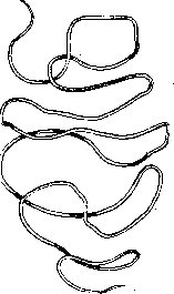
Figure 1. Female dracunculus (natural size).
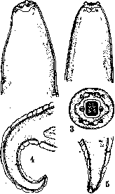
The vulval opening is obliterated, so the emergence of larvae into the external environment occurs through the rupture of the parasite's cuticular coverings in the head region. This rupture occurs at the moment of contact of the parasite's head with water.
Biology. The larvae of dracunculiasis reach 0.5-0.75 mm in length; further development depends on the presence in water of the smallest cyclops crustaceans, which are intermediate hosts for this parasite, as shown by the research of Fedchenko as early as the 19th century. Studies by Isaev, developed on the basis of the Bukhara Tropical Institute, clarified the details of the mechanism of penetration of dracunculus larvae into the cyclops body. It turns out that the cyclops swallows the larva, which, entering its intestine, becomes active, breaks through the intestinal wall and penetrates into its body cavity. In its new location, the dracunculus larva undergoes 2 molts, after which it becomes invasive for humans and other definitive hosts. The latter can become infected with dracunculiasis only if they swallow a cyclops with water. The path of penetration of the dracunculus larva from the intestine into the human subcutaneous tissue has not yet been traced at all: the intermediate stages between the larva and the mature female are unknown. The female dracunculus reaches its full development approximately one year after infection. Most often, only one specimen of the parasite is found in the patient, but cases have been described where the subcutaneous tissue of a person was invaded by 17 and even 50 specimens of dracunculiasis.
The clinical picture is most vividly revealed from the moment of full maturity of the female of this parasite, when it begins to make its way, piercing the skin coverings with its head end to release larvae into the external environment. At this time, in the area of localization of the head, the epidermis swells, forming a fairly large blister, which bursts after about 4 days, exposing the subcutaneous passage in which the head of the worm gapes. Due to the obliteration of the vulva in dracunculiasis, the release of larvae occurs through the rupture not only of the parasite's cuticular covering but also of the uterine wall. Some authors believe that the formation of the blister is associated with the release of special toxic products by the dracunculus. Confirmation of this are observations speaking of the appearance of urticaria, nausea, and other symptoms in patients in whom the blister has not yet burst. On the basis of violation of skin integrity, secondary infections can be observed with the subsequent development of various complications: abscesses, phlegmons, arthritis, ankylosis, and gangrene. The disease can end in spontaneous healing provided complete resorption of the dracunculus. In individual cases, the parasite can remain in the subcutaneous tissue without causing perforation of the skin covering: it is sometimes accidentally discovered during X-ray examination in a petrified state.
Diagnosis of dracunculiasis does not present difficulties, as the parasite is well noticeable due to the relief it forms on the skin surface. In case the blister has already burst, the diagnosis is made by finding the head end of the female parasite. Treatment of dracunculiasis is mainly surgical. The head of the worm is grasped with tweezers, clamped, and gradually wound onto some object (a stick, a piece of gauze, etc.). Extraction of the parasite has to be done extremely slowly over a series of sessions so as not to break the body of the female, which can cause complications. Some authors propose a special method of sterilizing the female dracunculus by injecting into its body through a syringe various disinfectants: mercuric chloride, chloroform, etc. Prevention of dracunculiasis should correspond to all details of the biological cycle of the parasite. First of all, it is necessary to protect the definitive host from infection with dracunculiasis through the intermediate host. For this purpose, it is necessary: 1) to destroy cyclops in various ways in water bodies located in endemic foci of dracunculiasis; 2) to regulate water supply, providing the population with good quality water; 3) to promote personal prevention so that the population in endemic dracunculiasis foci does not consume raw water. An essential element of prevention should be considered the protection of water sources and the intermediate hosts-cyclops living there from contact with dracunculiasis-infested definitive hosts, and first of all, with humans. In Old Bukhara-the focus of dracunculiasis on the territory of the USSR-there was a constant infection with dracunculiasis from special water carriers who supplied the population with water; to fill their 'tursuks' with water, these water carriers descended the steps of Bukhara's reservoirs, immersing the lower parts of their legs, affected by dracunculiasis, in the water. Contact with water activated the female dracunculus, which ejected into the pool an enormous number of larvae. These larvae were eaten by cyclops living in the reservoirs, which became invasive for humans, thus closing the biological cycle of dracunculiasis. Dracunculiasis in Old Bukhara was defeated by the efforts of the local Tropical Institute (L. M. Isaev), which persistently applied a systematic set of preventive measures. First of all, all dracunculiasis patients were registered and mass therapy was applied to them either by extraction of parasites or sterilization of female dracunculi. By thus excluding the definitive host from the biological-epidemiological cycle, the Tropical Institute began to fight the intermediate host. For this purpose, the Bukhara reservoirs were alternately and periodically subjected to emptying and drying by exposing their bottoms to direct sunlight-this achieved the mass death of cyclops. On the other hand, all approaches to the reservoirs in the form of stone steps were subjected to careful repair, as a result of which the cracks in which the smallest crustaceans-intermediate hosts of dracunculiasis-found shelter were eliminated. A huge role in the successful fight against dracunculiasis was played by the extensive sanitary-educational work that the Tropical Institute in Bukhara carried out among the native population. The final blow to Bukhara dracunculiasis was dealt by the construction of a water supply, thanks to which the local population no longer needed to use water from stagnant, polluted reservoirs. Dracunculiasis in the USSR has been defeated; the last dracunculiasis focus in the USSR is now eliminated. This achievement is the first case in the history of medicine of the complete elimination of a helminthiasis in the territory of a particular state-this is a triumph of Soviet medicine. In connection with the growth of culture in the republics of Central Asia, with the complete elimination of the religious pilgrimage of the population to the main focus of dracunculiasis-Arabia-no new endemic foci of dracunculiasis should be created in the USSR.
Related articles
Cite this page
“Dracunculiasis.” Soviet Medical Encyclopedia. English translation of Bolshaya Meditsinskaya Entsiklopediya, 1st ed. (Moscow, 1928–1936), ed. N. A. Semashko. https://sovietmedicalencyclopedia.pages.dev/article/dracunculiasis/