Nematodes
Historical document, translated for reference. It reflects medical knowledge of the 1920s–30s and is not medical advice.
Summary
This article from the 1928–1936 Soviet medical encyclopedia provides a comprehensive overview of Nematodes, also known as roundworms. It details their classification, morphology, life cycles, and parasitic relationships, including their role as human parasites.
Encyclopedia article (1928–1936)
NEMATODES, Nematoda Rudolphi, 1808 (from Greek nema-thread), or roundworms, are, from the standpoint of those authors who recognize the type Vermes, a separate class of the subphylum Nemathelminthes; many recent researchers abolish Vermes as a type, considering Nematodes a class of an independent type Nemathelminthes. Nematodes are organisms not divided into metameres (in contrast to annelids; see Annelids); the body is in most cases cylindrical or spindle-shaped; they are dioecious. Some authors join Acanthocephala (see Acanthocephala) as a separate class to Nemathelminthes. Nemathelminthes are divided into two classes: Nematoda and Gordiacea. Nematodes are either free-living organisms or parasites of animals or plants. Free-living Nematodes inhabit fresh waters and seas, in moist soil, in decomposing organic substances, etc. Many species are capable of inhabiting heterogeneous substrates; for example, freshwater forms can settle in soil and mud; soil Nematodes can migrate to decaying matter and grow and multiply there and even migrate for temporary parasitism on plants or rarely on animals. Examples of such Nematodes are species of Rhabditis and Diplogaster; representatives of these genera can multiply on decaying plants or animals (fungi, earthworms, mollusks, etc.); some Nematodes are registered as facultative and rare parasites of humans. Some free-living Nematodes in the larval stage can settle in invertebrate animals, in which however they mostly do not develop further and therefore are not true parasites. In these cases, free-living Nematodes use their "hosts" mainly for their further dissemination; at the same time, the "hosts" into which Nematodes penetrate provide them with abundant nutrient material after their death. For example, Rhabditis pellio settle in earthworms, Leptodera appendiculata in snails, Rhabditis obtusa in the hindgut of one of the beetles. Such infestation is an example of the transition from a free mode of life to parasitism. On the other hand, among parasitic Nematodes there are some groups of forms which spend part of their life as free-living organisms. For example, in Mermithidae the adult form is free-living, while the larva passes through a parasitic stage of development in insects; Strongylata, on the contrary, spend part of the larval development in the external environment; finally, a form such as Strongyloides is characterized by the alternation of parasitic and free-living generations. In terms of structure, free-living and parasitic Nematodes do not present essential differences from each other. Parasitic Nematodes can be parasites of vertebrates and invertebrate animals or plants. They are organisms with an elongated body of thread-like or spindle shape, from 1 mm to 1 m in length; mostly dioecious. The body is covered on the outside by a cuticle, inside of which a single-layered epithelium (hypodermal layer of the cuticle) is located; even further inside there is muscle tissue. According to the structure of the muscle tissue, Schneider (1860) divided Nematodes into three groups: 1. Megomyarii, muscular cells in the circumference of the body (at each transverse section) of Nematodes, 8 in total, 2 in each sector formed by the dorsal, ventral and lateral fields of the body. 2. Polymyarii, with more numerous muscular cells in each sector of the transverse section of the body. 3. Holomyarii, muscular cells are closely adjacent to each other, forming a continuous layer (Fig. 1). In the vast majority of cases, muscular cells lie in one annular layer and are elongated in the longitudinal direction; during general contraction they shorten the length of the Nematode body, while during unilateral action they bend the body in the corresponding direction. The majority of Nematodes have a straight through digestive tube opening at the anterior end by a mouth, often surrounded by cuticular outgrowths - "lips"; behind the mouth sometimes follows a buccal capsule or in other cases a "pharynx" leading to the esophagus, which may consist of two sections - muscular and glandular; in some forms of Nematodes (mainly Oxyurata) at the end of the esophagus there is an expansion, so-called "bulb", which may include a special chewing apparatus in the form of chitinous plates. The intestine opens ventrally in front of the posterior end of the body by an anal opening (Fig. 2). The nervous system consists of a nerve ring surrounding the anterior end of the esophagus; from it nerve trunks go forward and backward, sending their branches to the peripheral nervous apparatus - "papillae", located mainly at the anterior end ("head papillae"), in the region of the esophagus ("cervical papillae") and on the tail, mainly in males ("genital papillae"). The excretory system consists mainly of two symmetric channels, merging in the anterior part of the body and opening here by an excretory opening. The sexual organs have the form of convoluted tubes, and all parts of the sexual apparatus, both in males (testis, vas deferens) and in females (ovary, oviduct, uterus, vagina), are only certain sections of one continuous tube. The female sexual organs are often double and open by a vulval opening. In males, near the genital duct opening, located ventrally at the tail end, auxiliary elements are also located: spicules, gubernaculum, representing chitinous formations playing a role in the act of copulation (Fig. 3). Reproduction of Nematodes occurs mostly by females laying eggs in various stages of development - from presegmentation stage to the ready larva; viviparity is also encountered in Nematodes belonging to the most diverse suborders. Of human parasites, viviparity occurs in the guinea worm. Post-embryonic development of Nematodes occurs in most cases by a direct path (without intermediate hosts); in such cases, infection of an animal is accomplished either by the entry into its digestive tract of eggs or larvae or by the active penetration of larvae through the integument; sometimes development is complicated by so-called heterogony, for example in Strongyloides stercoralis. In other cases, development of Nematodes occurs with the help of intermediate hosts, which are mainly invertebrates (mainly insects, see Mosquitoes). Development of Nematodes in the host's body sometimes occurs by complex paths, with the help of so-called migration (see Invasion, Ascaris, Human Helminthiases). Localization of Nematodes in the definitive host's body serves mainly as the digestive tract, as well as subcutaneous tissue (various Filariata), muscles (Trichinella and others), blood, abdominal cavity (in humans Dipetalonema perstans, syn. Acanthocheilonema perstans), conjunctiva of the eyes and lacrimal canals (Thelazia), etc. Systematics of Nematodes is at present studied still relatively weakly. It is based mainly on morphology (structure of lips, cuticular elements, external elements of the male sexual apparatus) and to some extent on anatomy (esophagus, sexual apparatus, etc.). Nematodes are divided into 9 suborders: Filariata, Spirurata, Dioctophymata, Ascaridata, Oxyurata, Strongylata, Rhabdiasata, Trichocephalata, Mermithata, of which all nine have their representatives among human parasites. For larval and immature forms, which are not amenable to exact determination, collective names have been proposed, such as: Agamonema, or Agamonematodum for Nematodes in general; Agamofilaria (for adult immature Filariata); Microfilaria (for larvae of Filariata with localization mainly in the blood); Agamospirura (for immature Spirurata); Agamascaris (for immature Ascaridata), Agamomermis (for immature Mermithata). Nematodes constitute the numerically richest group of parasitic worms of humans, of which about 70 are registered. Some of human Nematodes are cosmopolitans, occurring in all parts of the world (ascaris, pinworm, whipworm). The geographical distribution of others is limited by certain climatic conditions; these are also forms whose larvae spend a certain stage of their development in the external environment (Ankylostomidae, Strongyloides); the third group of Nematodes has an even narrower area of distribution, which also depends on the presence of corresponding intermediate hosts (filariae, guinea worm).
J21
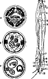
Figure 1. Figure 2. Figure 1. Various types of structure of the muscular system in nematodes: a Polymyarii; b-Megomyarii; c-Holomyarii. Figure 2. Scheme of structure of nematodes: 1-labial papillae; 2-nervous ring; 3-cervical papillae; 4-esophagus; 5-intestinal tube; 6-sexual papillae; 7-spicules; 8-preanal pedunculated papillae; 9-gubernaculum; 10-cloacal opening; 11-postanal pedunculated papillae; 12-caudal wings; 13-posterior sessile papillae; 14-preanal pedunculated papillae; 15-vulval opening; 16-intestinal tube; 17-lateral wings; 18-uterus; 19-ovaries; 20-seminal receptacles; 21-anus. Rhabditis pellio settle in earthworms, Leptodera appendiculata in snails, Rhabditis obtusa in the hindgut of one of the beetles. Such infestation is an example of the transition from a free mode of life to parasitism. On the other hand, among parasitic Nematodes there are some groups of forms which spend part of their life as free-living organisms. For example, in Mermithidae the adult form is free-living, while the larva passes through a parasitic stage of development in insects; Strongylata, on the contrary, spend part of the larval development in the external environment; finally, a form such as Strongyloides is characterized by the alternation of parasitic and free-living generations. In terms of structure, free-living and parasitic Nematodes do not present essential differences from each other. Parasitic Nematodes can be parasites of vertebrates and invertebrate animals or plants. They are organisms with an elongated body of thread-like or spindle shape, from 1 mm to 1 m in length; mostly dioecious. The body is covered on the outside by a cuticle, inside of which a single-layered epithelium (hypodermal layer of the cuticle) is located; even further inside there is muscle tissue. According to the structure of the muscle tissue, Schneider (1860) divided Nematodes into three groups: 1. Megomyarii, muscular cells in the circumference of the body (at each transverse section) of Nematodes, 8 in total, 2 in each sector formed by the dorsal, ventral and lateral fields of the body. 2. Polymyarii, with more numerous muscular cells in each sector of the transverse section of the body. 3. Holomyarii, muscular cells are closely adjacent to each other, forming a continuous layer (Fig. 1). In the vast majority of cases, muscular cells lie in one annular layer and are elongated in the longitudinal direction; during general contraction they shorten the length of the Nematode body, while during unilateral action they bend the body in the corresponding direction. The majority of Nematodes have a straight through digestive tube opening at the anterior end by a mouth, often surrounded by cuticular outgrowths - "lips"; behind the mouth sometimes follows a buccal capsule or in other cases a "pharynx" leading to the esophagus, which may consist of two sections - muscular and glandular; in some forms of Nematodes (mainly Oxyurata) at the end of the esophagus there is an expansion, so-called "bulb", which may include a special chewing apparatus in the form of chitinous plates. The intestine opens ventrally in front of the posterior end of the body by an anal opening (Fig. 2). The nervous system consists of a nerve ring surrounding the anterior end of the esophagus; from it nerve trunks go forward and backward, sending their branches to the peripheral nervous apparatus - "papillae", located mainly at the anterior end ("head papillae"), in the region of the esophagus ("cervical papillae") and on the tail, mainly in males ("genital papillae"). The excretory system consists mainly of two symmetric channels, merging in the anterior part of the body and opening here by an excretory opening. The sexual organs have the form of convoluted tubes, and all parts of the sexual apparatus, both in males (testis, vas deferens) and in females (ovary, oviduct, uterus, vagina), are only certain sections of one continuous tube. The female sexual organs are often double and open by a vulval opening. In males, near the genital duct opening, located ventrally at the tail end, auxiliary elements are also located: spicules, gubernaculum, representing chitinous formations playing a role in the act of copulation (Fig. 3). Reproduction of Nematodes occurs mostly by females laying eggs in various stages of development - from presegmentation stage to the ready larva; viviparity is also encountered in Nematodes belonging to the most diverse suborders. Of human parasites, viviparity occurs in the guinea worm. Post-embryonic development of Nematodes occurs in most cases by a direct path (without intermediate hosts); in such cases, infection of an animal is accomplished either by the entry into its digestive tract of eggs or larvae or by the active penetration of larvae through the integument; sometimes development is complicated by so-called heterogony, for example in Strongyloides stercoralis. In other cases, development of Nematodes occurs with the help of intermediate hosts, which are mainly invertebrates (mainly insects, see Mosquitoes). Development of Nematodes in the host's body sometimes occurs by complex paths, with the help of so-called migration (see Invasion, Ascaris, Human Helminthiases). Localization of Nematodes in the definitive host's body serves mainly as the digestive tract, as well as subcutaneous tissue (various Filariata), muscles (Trichinella and others), blood, abdominal cavity (in humans Dipetalonema perstans, syn. Acanthocheilonema perstans), conjunctiva of the eyes and lacrimal canals (Thelazia), etc. Systematics of Nematodes is at present studied still relatively weakly. It is based mainly on morphology (structure of lips, cuticular elements, external elements of the male sexual apparatus) and to some extent on anatomy (esophagus, sexual apparatus, etc.). Nematodes are divided into 9 suborders: Filariata, Spirurata, Dioctophymata, Ascaridata, Oxyurata, Strongylata, Rhabdiasata, Trichocephalata, Mermithata, of which all nine have their representatives among human parasites. For larval and immature forms, which are not amenable to exact determination, collective names have been proposed, such as: Agamonema, or Agamonematodum for Nematodes in general; Agamofilaria (for adult immature Filariata); Microfilaria (for larvae of Filariata with localization mainly in the blood); Agamospirura (for immature Spirurata); Agamascaris (for immature Ascaridata), Agamomermis (for immature Mermithata). Nematodes constitute the numerically richest group of parasitic worms of humans, of which about 70 are registered. Some of human Nematodes are cosmopolitans, occurring in all parts of the world (ascaris, pinworm, whipworm). The geographical distribution of others is limited by certain climatic conditions; these are also forms whose larvae spend a certain stage of their development in the external environment (Ankylostomidae, Strongyloides); the third group of Nematodes has an even narrower area of distribution, which also depends on the presence of corresponding intermediate hosts (filariae, guinea worm).
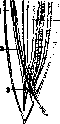
Figure 3. Sagittal section of the tail end of Ascaris: 1-main retractors of spiculae; 2-spicula; 3-m. protractor spiculae; 4-vas deferens; 5-intestine.
development of Nematodes occurs in most cases by a direct path (without intermediate hosts); in such cases, infection of an animal is accomplished either by the entry into its digestive tract of eggs or larvae or by the active penetration of larvae through the integument; sometimes development is complicated by so-called heterogony, for example in Strongyloides stercoralis. In other cases, development of Nematodes occurs with the help of intermediate hosts, which are mainly invertebrates (mainly insects, see Mosquitoes). Development of Nematodes in the host's body sometimes occurs by complex paths, with the help of so-called migration (see Invasion, Ascaris, Human Helminthiases). Localization of Nematodes in the definitive host's body serves mainly as the digestive tract, as well as subcutaneous tissue (various Filariata), muscles (Trichinella and others), blood, abdominal cavity (in humans Dipetalonema perstans, syn. Acanthocheilonema perstans), conjunctiva of the eyes and lacrimal canals (Thelazia), etc. Systematics of Nematodes is at present studied still relatively weakly. It is based mainly on morphology (structure of lips, cuticular elements, external elements of the male sexual apparatus) and to some extent on anatomy (esophagus, sexual apparatus, etc.). Nematodes are divided into 9 suborders: Filariata, Spirurata, Dioctophymata, Ascaridata, Oxyurata, Strongylata, Rhabdiasata, Trichocephalata, Mermithata, of which all nine have their representatives among human parasites. For larval and immature forms, which are not amenable to exact determination, collective names have been proposed, such as: Agamonema, or Agamonematodum for Nematodes in general; Agamofilaria (for adult immature Filariata); Microfilaria (for larvae of Filariata with localization mainly in the blood); Agamospirura (for immature Spirurata); Agamascaris (for immature Ascaridata), Agamomermis (for immature Mermithata). Nematodes constitute the numerically richest group of parasitic worms of humans, of which about 70 are registered. Some of human Nematodes are cosmopolitans, occurring in all parts of the world (ascaris, pinworm, whipworm). The geographical distribution of others is limited by certain climatic conditions; these are also forms whose larvae spend a certain stage of their development in the external environment (Ankylostomidae, Strongyloides); the third group of Nematodes has an even narrower area of distribution, which also depends on the presence of corresponding intermediate hosts (filariae, guinea worm).
Related articles
Mentioned in
Cite this page
“Nematodes.” Soviet Medical Encyclopedia. English translation of Bolshaya Meditsinskaya Entsiklopediya, 1st ed. (Moscow, 1928–1936), ed. N. A. Semashko. https://sovietmedicalencyclopedia.pages.dev/article/nematodes/