Rudolphi
Historical document, translated for reference. It reflects medical knowledge of the 1920s–30s and is not medical advice.
Summary
Karl Rudolphi (1771-1832) was the founder of helminthology, known for his comprehensive works on parasitic worms. His major publications included 'Entozoorura, sive vermium intestinalium historia naturalis' and 'Entozoorum synopsis', which cataloged hundreds of helminth species and established foundational classifications for the field.
Encyclopedia article (1928–1936)
RUDOLPHI Karl (Rudolphi Karl Asmund, 1771-1832), founder of helminthology; graduated from the philosophical and medical faculties at Greifswald, and for 22 years held the chair of anatomy and physiology in Berlin. He became interested in helminthology during his student years; his first two helminthological works served as his dissertations for the degrees of Doctor of Philosophy and Doctor of Medicine. In 1808-1810, his two-volume monograph 'Entozoorura, sive vermium intestinalium historia naturalis' (Amsterdam) was published, in which 603 species of helminths were described. This major work by R. had for helminthology the same significance that Linnaeus's 'Systema naturae' had for zoology at its time. It concentrated all material on helminthology, described many new forms, and gave a critical analysis of all helminthological literature, which consisted of 629 bibliographical items. R. divided all helminths into 5 orders: Nematoidea, Acanthocephala, Trematoda, Cestodes, and Cystica. The last group included bladder-like helminths (echinococcus, cysticercus), the cestode nature of which had not yet been deciphered. In 1819, his second remarkable work 'Entozoorum synopsis' (Berlin) was published, in which data on 981 species of helminths were summarized. Being the founder of helminthology, Rudolphi left behind many works in the fields of anatomy, physiology, anthropology, botany, veterinary medicine, and even mineralogy. He was a corresponding member of the Russian Academy of Sciences and was invited to St. Petersburg as an academician.
Figure 1. Left - hand of Aran-Duchenne. Right - hand of a preacher.
Accordingly, anomalies occur more frequently in the latter than in the former. According to Starkov, developmental defects can be considered as phenomena of absence or duplication of one of the 5 rays, of which, according to Gegenbaur, the skeleton of R. consists. The main ray, according to Gegenbaur, consists of the humerus, radius, part of the trapezoid part of the hamate, V metacarpal, and three phalanges of the little finger; the 1st lateral ray consists of the radius, scaphoid, trapezium, I metacarpal, and two phalanges of the thumb; the 2nd lateral ray consists of the lunate, trapezoid, II metacarpal, and three phalanges of the index finger; the 3rd lateral ray consists of part of the trapezoid and capitate bones, III metacarpal, three phalanges of the middle finger; the 4th lateral ray consists of part of the hamate bone, IV metacarpal, and three phalanges of the ring finger. In the 1st lateral ray, complete absence is more often observed, but partial absence also occurs when the radius is present but the thumb is absent. With absence of the 2nd, 3rd, or 4th lateral rays, there is absence of the index, middle, and ring fingers. Underdevelopment of the 3rd lateral ray gives a 'fourchue' hand (main fourchue). Absence of the main ray leads to complete absence of the hand or hands, which may be combined with absence of the lower limbs (see Amelia). The opposite developmental defects consist in duplication of one or several rays, usually expressed only in the distal parts (see Polydactyly). As a rarity, complete duplication of a ray is described. The weakest manifestation of splitting is duplication of the nail phalanx, most often occurring on the thumb. Partial fusion of two rays with each other gives syndactyly (see). Diseases of the hand. Shoulder, Forearm, Hand, individual joints and nerves of the hand, as well as: Clubhand, Kienböck's disease, Panaritium, Tenosynovitis, Aran-Duchenne hand, Morvan's disease, Dupuytren's contracture, Contractures, Dactylomegaly, Paralysis agitans, Childhood paralyses, Athetosis, Amyotrophic lateral sclerosis, Syringomyelia, Arthritis, Rheumatism. Professional injuries and diseases of the hand are very numerous.
Figure 2. Main simienne with cubital claw.
Figure 3. Dropping hand in hemiplegia.
Figure 4. Hand
of the radius, scaphoid, trapezium, I metacarpal and two phalanges of the thumb; 2nd lateral ray - of the lunate, trapezoid, II metacarpal and three phalanges of the index finger; 3rd lateral ray - of part of the trapezoid and capitate bones, III metacarpal, three phalanges of the middle finger; 4th lateral ray - of part of the hamate bone, IV metacarpal and three phalanges of the ring finger. In the 1st lateral ray, complete absence is more often observed, but partial absence also occurs when the radius is present but the thumb is absent. With absence of the 2nd, 3rd, or 4th lateral rays, there is absence of the index, middle and ring fingers. Underdevelopment of the 3rd lateral ray gives a 'fourchue' hand (main fourchue). Absence of the main ray leads to complete absence of the hand or hands, which may be combined with absence of the lower limbs (see Amelia). The opposite developmental defects consist in duplication of one or several rays, usually expressed only in the distal parts (see Polydactyly). As a rarity, complete duplication of a ray is described. The weakest manifestation of splitting is duplication of the nail phalanx, most often occurring on the thumb. Partial fusion of two rays with each other gives syndactyly (see). Diseases of the hand. Shoulder, Forearm, Hand, individual joints and nerves of the hand, as well as: Clubhand, Kienböck's disease, Panaritium, Tenosynovitis, Aran-Duchenne hand, Morvan's disease, Dupuytren's contracture, Contractures, Dactylomegaly, Paralysis agitans, Childhood paralyses, Athetosis, Amyotrophic lateral sclerosis, Syringomyelia, Arthritis, Rheumatism. Professional injuries and diseases of the hand are very numerous.
Figure 5. Dropping hand in paralysis of the radial nerve.
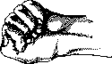
Figure 6. Hand clenched into a fist.
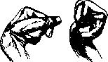
Fig. 7. Hand clenched into a fist (hand of a fakir).
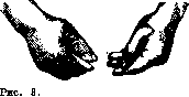
Hand rolling pills.
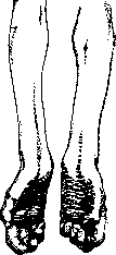
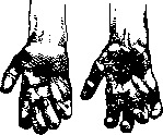
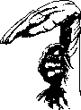
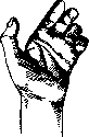
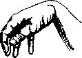
Various deformities of the hand. Various types of hand deformity. Cause of deformity. Clinical manifestation. In which diseases it occurs. Monkey hand (main de singe). Claw hand, synonym: talon hand (main en griffe). Simian hand with claw (main simienne avec griffe). Skeleton hand (main de squelette). Obstetrician's hand (main d'accoucheur). Hammer hand (main en battoir). Hand in flexion (main en flexion). Trident hand (main en trident). Cubital claw hand (main en griffe cubitale). Median claw hand + monkey hand (main en griffe médiane + main de singe). Morvan's hand (rhumatisme analgesique de Morvan). Edematous hand (main succulante). Drooping hand (main tombante). Flat hand. Atrophy of thenar muscles. Atrophy of hypothenar and interosseous muscles. Atrophy of thenar, hypothenar and interosseous muscles. The thumb is in the same plane as the other fingers and cannot oppose them. The fingers are adducted, the first phalanx is extended, II and III are strongly flexed. Combination of changes. In monkey hand and in claw hand (figs. 1 and 2). In far-advanced cases of the previous form. Contracture in the small muscles of the hand. Increase in the bones of the hand and fingers. Paralysis and contracture of the muscles of the hand, forearm and shoulder. Insufficient development. Paralysis of the muscles of the forearm and hand innervated by the ulnar nerve. Paralysis of the muscles of the forearm and hand innervated by the median nerve. Trophic disorders. Vasomotor disorders. Paralysis and atrophy of the muscles innervated by the radial nerve. Paralysis and atrophy of the muscles of the hand, forearm, shoulder. Without contractures. The thumb is in a state of forced adduction, all fingers are tightly pressed against each other in a semi-flexed state, the palm forms a depression due to the approximation of its edges. Increase in the hand without any deformities: the hand is wide and thick, without change in length, the fingers are short, thick, in the form of sausages (doigts en saucisse). The shoulder is adducted, the forearm, hand and fingers are in a state of flexion. Various variations (fig. 3). II, III and IV fingers are little separated from each other, as a result of which the hand resembles a trident. Only IV and V fingers are in a state of flexion (griffe), II and III remain extended, as their lumbrical muscles are innervated by the median nerve. Sensory disturbance on the palmar and dorsal surface of the hand (fig. 4). Changes in monkey hand, flexion of III and II fingers, inability to flex and pronate the hand. Sensory disturbance. Absence of one or two phalanges on one or several fingers, thickening of the skin, disturbance of all types of skin sensitivity. The hand is edematous, cold, bluish in color. Hypertrophy of subcutaneous tissue, often with muscle atrophy. Flexion of the hand and fingers and inability to extend them: sometimes II and V fingers, which have their own extensors, remain extended - the patient seems to make horns (fig. 5). Severe emaciation of the muscles, so that all the elevated places and roundnesses on the hand are smoothed out a) In various diseases of the spinal cord, in lesions of the anterior horns at the level of C8 and D1 (place of departure of fibers for innervation of the corresponding muscles) - poliomyelitis chronic and acute, syringomyelia, hematomyelia, tumors, amyotrophic lateral sclerosis, traumatic diseases of the spinal cord; b) peripheral diseases of the roots, plexus and peripheral nerves - median and ulnar nerves, observed in diseases of the spine, in cervical ribs, diffuse paralyses, in neuritis - traumatic, infectious, intoxication. In tetany. In acromegaly. In central hemiplegias. Achondroplasia. In diseases of the ulnar nerve on the forearm or above - infectious, toxic, professional neuritis (in work requiring support on the inner half of the elbow or on the hypothenar - bakers, shoemakers, printers). Diseases of the median nerve - trauma, nerve transection, toxic and infectious neuritis. Syringomyelia, leprosy (leprosy). Syringomyelia, hemiplegia, myopathy. Diseases of the radial nerve - various neuritis (more often mercury poisoning); disease of the spinal cord (D1), brachial plexus. In central hemiplegias (more often in childhood forms).
Various types of hand deformity. Cause of deformity. Clinical manifestation. In which diseases it occurs. Preacher's hand (main de prédicateur). Hand with large thumbs (chiromegalie, s. macrochirie). Hand with fingers in the form of drumsticks (doigts en baguettes de tambour). Hand with reduced fingers (sclerodactylie). Hand clenched in a fist (main en poing fermé). Fakir's hand (main de fakir). Hand rolling pills. Paralysis and atrophy of the muscles innervated by the median and ulnar nerves. Trophic disorders of the bones of the hand. Trophic disorder of the bones. Trophic disorders of the bones of the hand. Contracture in the flexors of the fingers. Contracture of interosseous and lumbrical muscles. Flexion of the last two phalanges and hyperextension of the first and hand (fig. 1). Increase in the hand, especially the fingers (one or several), which become thick, round. The hand is increased in width, thickness and length, the fingers are elongated, flattened, and expanded at the ends. The fingers are thinned, reduced in volume, change in the bone-articular apparatus of the joints. Resorption of the III phalanx is possible. Hyperflexion of all phalanges of the fingers to such an extent that the nails cut into the skin (figs. 6 and 7). Slight flexion of the I phalanx, extension of II and III; adduction of the fingers, the thumb in a state of extension and hyperadduction - rests on the outer surface of the II finger (fig. 8). The change is characteristic for pachymeningitis cervicalis hypertrophica, but occurs in syringomyelia, poliomyelitis, central hemiplegias and in lesions of nerves at the elbow level. In syringomyelia. In chronic diseases of the lungs. Scleroderma. In paralysis agitans, hysteria, syringomyelia.
In paralysis agitans.
Related articles
Mentioned in
Cite this page
“Rudolphi.” Soviet Medical Encyclopedia. English translation of Bolshaya Meditsinskaya Entsiklopediya, 1st ed. (Moscow, 1928–1936), ed. N. A. Semashko. https://sovietmedicalencyclopedia.pages.dev/article/rudolphi/