Human Helminthiases
Historical document, translated for reference. It reflects medical knowledge of the 1920s–30s and is not medical advice.
Summary
Human helminthiases are diseases caused by parasitic worms that infect various organs and systems of the human body. This article covers the classification, localization, pathogenesis, clinical manifestations, diagnosis, treatment, and prevention of these infections.
Encyclopedia article (1928–1936)
Human Helminthiases. Contents: Localization of parasitic worms......432 Pathogenesis. ;..................435 Clinic.....................438 Diagnosis.....................438 Therapy.....................439 Prevention..................43 9 Helminthiases in children...............440 Helminthiases (from Greek helmins-worm), morbid conditions of animals (rarely plants) caused by parasitic worms that have settled in them. H. h.-diseases that are caused by the parasitization in the human body of parasitic worms, localized in various systems or organs, stationary or transit. The pathogenic effect of parasitic worms is expressed in one form or another, on the one hand, depending on the species of parasite, i.e., on all its anatomical-morphological and ecological-biological peculiarities, and on the other-from the reaction of the host organism. H. of man represent a complex of diseases consisting of as many separate nosological units as there are different species of parasitic worms that constitute the helminthofauna (see) of man. Counting about 130 forms of parasitic worms in humans, it must be recognized, with the current state of knowledge, about 130 separate helminthiases. The names of H. are derived from the generic name of the parasite. In accordance with the belonging of parasitic worms to different classes, H. are divided into the following main groups: trematodases (Trematoda-flukes), cestodases (Cestoda-tapeworms), nematodases (Nematoda-roundworms), acanthocephaloses (Acanthocephala-thorny-headed worms, thorny-headed worms). Of these groups, H. in our geographical zone have the greatest significance for humans: nematodases and cestodases, lesser significance: trematodases; acanthocephaloses have no practical significance in medicine. The spread of parasitic worms among the population of the USSR is extremely great, and inhabitants of different geographical regions, representatives of different professional, national and other groups may be characterized by their specific helminthofauna (see). Living conditions, dietary regimen, domestic peculiarities, climate, terrain and soil of the area, character of the surrounding fauna-all these are among the factors determining the nature and degree of human infection with parasitic worms and thus play a role in the epidemiology of helminthiases. Localization of parasitic worms. Parasitic worms, localizing mainly in the lumen of the intestine and choosing certain zones of it (ankylostoma, trichostrongylides-upper part of the small intestine, ascaris-small intestine, oxyuris-lower part of the small intestine and large intestine, trichuris-blind intestine, etc.), can affect all organs and tissues of man. Thus, in the liver can settle Fasciola hepatica, Dicrocoelium lanceatum, Opisthorchis felineus, Clonorchis sinensis, Echinococcus granulosus, etc.; in the pancreas-O. felineus, C. sinensis, echinococcus, cysticercus, etc.; in Lieberkühn's glands-Strongyloides stercoralis; in the tissues of the intestine-Hymenolepis nana, etc. Systems not related to the digestive tract can also be affected; thus, in the respiratory organs can parasitize Paragonimus westermani, Metastrongylus apri (stationary) and larvae of ascarids, ankylostomas, etc. (transit); in the urinary organs-Dioctophyme renale, Schistosoma haematobium, echinococcus, cysticercus, larvae of ascarids, ankylostomas, etc.; in the circulatory system-schistosomes, larvae of many nematodes and cestodes. In the lymphatic system can inhabit the causative agent of 'elephantiasis'-filaria (Wuchereria bancrofti); in the central nervous system occur as stationary parasites larvae of cestodes Cysticercus cellulosae, Coenurus cerebralis, echinococcus and transit-larvae of nematodes; in the eyes and periocular tissue Loa loa, L. extraocularis, Thelazia callipaeda and a number of larval forms; in the skin and subcutaneous tissue parasitize dracunculus-Fullebornius medinensis, Ancylostoma braziliense, there can also be found Fasciola hepatica, etc.; on the skin and mucosa-Enterobius vermicularis, under the mucosa-Gongylonema, in bone tissue-echinococcus, in muscle tissue-Trichinella spiralis, echinococcus, cysticercus, etc.-Each species of parasitic worm in most cases has its own specific localization; the latter is closely related, on the one hand, to the physico-chemical conditions of the environment necessary for the parasite's development and reaching sexual maturity, and on the other-to its methods of reproduction. Larval stages in relation to localization are less demanding; thus, Echinococcus granulosus and Cysticercus cellulosae can inhabit any tissues of man and other animals, but the corresponding mature forms require certain localization and a specific host, e.g., Echinococcus granulosus-small intestine of the dog, Taenia solium-small intestine of man. The most frequently observed localization of the parasitic worm in the final host, ensuring the attainment of sexual maturity and reproduction, is called normal in H, in contrast to aberrant, when the parasite settles in an organ or tissue not characteristic of it, in which it cannot reach sexual maturity (Ascaris in the subcutaneous tissue,
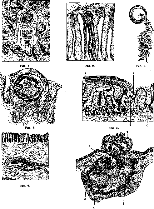
Figure 1. Nematode-Ankylostoma duodenale, having a powerful oral capsule and chitinous teeth, seizes a section of tissue of the small intestine (after Brumpt). Figure 2. Nematode-Strongyloides stercoralis, having penetrated into the lumen of a gland and coiled up (after Oudendal'io).
Figure 7. Figure 3. Nematode-Trichuris trichiurus,
possessing a smooth and thread-like anterior end, becomes fixed by piercing the intestinal mucosa of humans (according to Leuckart). Figure 4. Trichocephalus trichiurus fixed in the human intestine in cross-section (according to Lewinson). Figure 5. Cestoda - Hymenolepis fraterna, attached to the intestinal mucosa of a rat: 1-cestode strobila; 2-suckers; 3-rostellum with hooks; 4-gland. Figure 6. Trematoda - Schistosoma mansoni in a venous vessel of the submucosa lies freely, without becoming fixed (according to Piraja da Silva from Brumpt). Figure 7. Acanthocephalan - Macracanthorhynchus hirudinaceus, having penetrated with its rostellum into the thickness of the intestinal wall of a pig: 1-parasite's rostellum; 2-parasite's muscles; 3-eosinophilic infiltration; 4-fatty tissue; 5-necrotized tissue with eosinophils (according to Brumpt). Enterobius vermicularis in the nose, Fasciola hepatica in the lungs, etc.). or when the parasite changes its localization after reaching maturity (ascaris in the liver). Aberrant localization is observed significantly less frequently than normal, but plays a considerable role in human pathology, causing severe forms of diseases, mostly very difficult to diagnose. Pathogenesis. The effect of parasitic worms on the host organism is very multifaceted; basically it can be reduced to two points: mechanical influence and toxic. Mechanical influence can manifest as a result of direct action of adult parasites or their larvae (traumatization of tissue during the movement of parasites, destruction during fixation, perforations, ruptures, blockages, atrophy from pressure) or of egg elements, which can themselves traumatize tissues (eggs of schistosomes, with their spines destroying the integrity of blood vessels and other tissues). Finally, one or another pathological effect can also be caused by concretions that form around the eggs (Opisthorchis felineus in the bile ducts of the liver and gallbladder, Schistosoma haematobium in the bladder, Enterobius vermicularis - fecal concretions in the vermiform appendix, etc.). Mechanical effect on the host organism produced by adult parasites and their larvae deserves separate consideration. The majority of parasitic worms secure for themselves more or less permanent residence in an organ by attaching to tissues, which often undergo significant destruction as a result. The nature of these destructions will depend entirely on the method of fixation, resp. on the structure of the fixing apparatus. (Methods of fixation of parasitic worms - see figures 1-7). In nematodes, the oral end most often serves this purpose. In some, there is a strongly developed oral capsule with cutting, grinding or piercing cuticular elements: in such forms, the strength of attachment is achieved by capturing a plug-shaped area of tissue into the capsule and sucking onto it or suspending with the help of special 'teeth' (Ankylostoma duodenale); the result is sometimes more superficial, sometimes deeper destruction, with rupture of blood vessels, etc. Other species, having movable lips and tooth-like formations, pinch the tissue (Abreviata). In a third group, in the absence of such structures, a thin and completely smooth anterior end is found; in these cases, fixation occurs by more or less deep penetration of the anterior end into the thickness of the tissues (Trichocephalus trichiurus pierces the intestinal mucosa, Trichinella spiralis deeply penetrates the mucosa, Gongylonema completely goes into the tissue). Trematodes are equipped with a special muscular apparatus - suckers - with which tissue is captured; in most cestodes, in addition to four similarly arranged suckers, there is often one or many rows of chitinous hooks. In addition to all the above, in some forms the body is covered with spines, which play a significant role in the traumatization of the host's tissues (Fasciola hepatica, etc.). Numerous facts speak of the possibility of blockages of the intestinal lumen (by balls of ascarids or tapeworms), bile ducts (ascaris, Fasciola) or the pancreatic duct. Adult nematodes Wuchereria bancrofti block the lymphatic vessels, causing elephantiasis; they, for example, can be the cause of inflammatory infiltrations on the wall of the thoracic duct, which can result in thrombosis of the duct (Nörr, 1927). Finally, tissue atrophy from pressure can be observed in any organ in echinococcosis; the presence of Dioctophyme renale in the kidneys or renal pelvis leads to the disappearance of specific tissue; cysticercus gives severe phenomena from compression of areas of the brain and sudden death when closing the communication between the brain ventricles. A relatively new and important chapter is the question of the effect of larval forms of nematodes, and precisely those which, before settling for stationary parasitism in one or another organ (mainly the intestine), must pass a complex path in the host's body. The phenomenon of nematode migration, long noted for hookworm (see) by Loos (1898), has also been established for ascaris (see) (Stewart, Ransom, Fulleborn) and some other forms. The migration of larval forms must be evaluated, first of all, from the point of view of tissue traumatization: wandering larvae act, on one hand, as foreign bodies and, on the other hand, as living organisms possessing their own movement. The effect of larvae will vary depending on many factors, and first of all on their size. The reaction of the organism (Nörr, 1927) manifests as cellular infiltration in the tissue around the larvae. Where the larva gets stuck inside a capillary, it is surrounded by leukocytes, mainly eosinophils. Bleeding, which is caused by larvae, mainly in the lungs, kidneys and brain membranes, is caused either by blockage of capillaries and subsequent rupture by blood pressure, or is actively caused by the drilling movements of the larva. Some migrating larvae can encapsulate in different organs, and in some cases the organism thereby puts an obstacle to the further normal development of the parasite (encapsulation of ascaris), in others - encapsulation is a necessary link in the development cycle of the worm (Trichinella spiralis). - Inoculation by worms of bacteria. Another aspect from which the migration of larvae of parasitic worms must be considered is the possibility of inoculation by larvae of microbial flora. This question was first raised by Mechnikov (1901), who pointed to the connection between the disease of appendicitis and the parasitism in the intestine of certain nematodes; now the problem is posed more broadly, and the role of inoculators is attributed not only to mature individuals traumatizing the intestine (particular importance is attributed to Trichocephalus trichiurus, Enterobius vermicularis), but also to migrating larvae: at least twice during their wandering they violate the integrity of tissues (in the intestine 'entrance gate' and in the lungs 'exit gate' for larvae) and thus twice can open 'entrance' gates for infection. There are also forms that have the ability to penetrate through the skin and, thus, can introduce infection from outside through the skin (Experiments of Malvoz and Lambinet with tubercle bacilli inoculated by larvae of Ankylostoma duodenale); finally, parasitic worms breaking through the skin from the inside equally open wide access for infection (dracunculus phlegmon). - A certain role in the traumatization of tissues during migration, as well as in the inoculation of microbes, may also belong to parasitic worms of different animals, i.e., those which are not counted as parasites of humans, because, not reaching maturity in his organism, they are not detected. It is known that many such worms can complete a complete migratory cycle in the human body and are thrown out into the external environment only after completion of migration (Ankylostoma caninum). Intoxication. The toxic influence of parasitic worms is no less important. The secretion of toxic substances has been experimentally proven in representatives of different classes of worms. The question has been most fully developed in relation to nematodes, and especially ascarids, which contain within their skin-muscle sac strongly acting toxic substances (see Ascaris, ascaridotoxin). Hookworms secrete substances acting hemolytically and preventing blood clotting. The secretion of various toxic substances (in the form of secretions or excretions of worms) has also been established in most other human nematodes, e.g., whipworm (see), trichostrongylids, pinworms, etc. Among cestodes, the broad tapeworm - Diphyllobothrium latum - is distinguished by particular toxicity, causing such changes in the blood that approach the type of true pernicious anemias (Pavroza, 1913). Hemolytic substances have also been isolated from a number of other cestodes: Taenia solium, T. saginata, Dipylidium caninum, Hymenolepis diminuta, etc. The secretion of various toxic substances and poisoning of the host organism by them has also been proven in relation to many trematodes (Schistosomatidae, Fasciola, etc.). - The pathogenic effect of parasitic worms on the host organism mostly consists of the combined effect of the mechanical shocks they produce and intoxication.
The latter is caused not only by toxins secreted by the worms themselves, but also by products of the host's tissue decomposition, released as a result of the action of parasitic worms. In various cases, either the mechanical or the toxic influence of the worms comes to the forefront. Even local tissue destruction is mostly due to the combined effect of both factors. Bleeding caused by parasitic worms may sometimes be of significant importance in the pathogenesis of helminthiases; such bleeding can be especially harmful when parasites secrete anticoagulants. Clinics. Clinical manifestations of helminthiases are extremely diverse and depend, first, on the nature of the relationship between the parasite and the host, and second, on the intensity of the invasion, i.e., the numerical composition of the parasitic population. The pathological processes in helminthiases may be either more local or of a general nature, and all organs and systems can be involved. The digestive system is most commonly affected: intestinal disorders are observed in the form of diarrhea, constipation, spasmodic conditions, obstructions, etc. The effect on the blood is clinically manifested in various forms of anemic conditions up to the type of true pernicious anemia. Nervous disorders are observed not only with local lesions of the central nervous system by the parasite (cysticercosis, echinococcosis, etc.), but (and more often) as a result of the absorption of helminthotoxins and manifest as headaches, neurasthenia, insomnia, fatigue, decreased mental abilities, and in severe cases - convulsions, epileptoid seizures. Furthermore, helminthiases can lead to various skin disorders (as a result of local lesions or intoxication from the intestines), eye diseases, etc. Observations show that some helminthiases can lead to delays in physical and mental development, to a decrease in intellectual abilities, and affect the function of the endocrine glands. The clinical picture of helminthiases mostly does not present any specific phenomena that would be pathognomonic for certain types of helminthiasis or would distinguish helminthiases from similar diseases of other etiologies; helminthiases can simulate various diseases that fall within the competence of various specialists. For more details - see individual helminthiases. Diagnosis cannot in most cases be based solely on the clinical picture, but should, mainly, be established with the help of various laboratory research methods. Most often, it is necessary to resort to examinations of feces for worm eggs, their larvae, fragments, or even mature individuals (see Helminthological research methods). In helminthology, the examination of duodenal juice and bile, obtained by duodenal tube, for detecting worms located in the upper part of the small intestine, pancreas, gallbladder and liver (Strongyloides stercoralis, Trichostrongylidae, Ankylostomatidae, liver flukes, pancreatic flukes, etc.) is of great importance. The examination of gastric juice is of less importance due to the rarity of parasitic worms inhabiting the human stomach. In helminthiases of other organs, it is advisable to examine the corresponding secretions: sputum - for lung flukes and nematodes, migrating larvae, echinococcus; urine - in helminthiases of the urinary tract (for example, schistosomiasis, etc.). The changes in physico-chemical and biological properties, as well as the morphological composition of blood, caused by parasitic worms, are used for diagnostic purposes. The Ascoli meyostagmin reaction, based on changes in the physical conditions of blood serum, is used in echinococcosis, ankylostomiasis. For the diagnosis of those helminthiases that are difficult to determine by other methods (e.g., echinococcosis), various immunobiological methods are of greatest importance, such as: complement fixation reaction (Ghedini, Weinberg), precipitation reaction (Fleig et Lisbonne), subcutaneous and intradermal reactions (Casoni). The latter is probably the most valuable method for the diagnosis of echinococcosis in terms of both specificity and simplicity of technique. Morphological examination of blood (eosinophilia) is often helpful. Finally, in some cases, anamnestic data, palpation [echinococcosis, cysticercosis, trichinelliasis (old name for trichinosis), dracunculiasis], ophthalmoscopy (cysticercosis), X-ray examination (echinococcosis, calcified cysticerci, trichina, dracunculus, etc.) provide useful indications. Therapy of helminthiases should first be radical, i.e., aimed at removing the parasite (deworming) or, if this is not possible, at killing it in the body. Pharmacotherapy, with a large arsenal of anthelmintic agents, is of greatest importance in helminthology; the latest research is aimed at establishing specific drugs that act on certain species or genera of worms (see individual helminthiases, also Deworming, Anthelmintic agents). Among physical methods of treatment, radiotherapy should be mentioned (in echinococcosis). Introduction of warm solutions (up to 45°) into the intestine kills many intestinal parasites, however, this method of treatment is not yet sufficiently tested. In certain cases, surgical intervention is indicated either for the purpose of removing parasites (echinococcosis, cysticercosis, extra-intestinal ascariasis, ascariasis ileus, dracunculiasis, etc.), or for creating pathways for subsequent pharmacotherapy (for example, appendicostomy, typhlostomy in trichocephaliasis, etc.). Sometimes it is possible to directly extract parasites (for example, dracunculus - through a skin wound punctured by it, ascaris - when it penetrates into the upper respiratory tract). Prevention. When conducting preventive measures, it must be remembered that the source of infection for some forms are elements of dead nature - soil, water, etc., and for others - objects of living nature - intermediate or final hosts or mechanical vectors. Intermediate hosts infect humans either by serving as their food (beef, pork, fish, crustaceans), or by passively entering the digestive tract, or finally, by actively attacking humans (blood-sucking insects). Thus, for each type of helminthiasis, its specific prevention must be carried out. The main preventive measure against most helminthiases is the improvement of general sanitary-hygienic living conditions of the population, such as: the construction of sewerage and water supply; strict supervision of food products, especially meat (expansion and improvement of veterinary-sanitary supervision), in order to protect them from contamination with the invasive element. In the fight against helminthiases, close contact between medical and veterinary organizations should be established - only their joint and coordinated actions can actually put the matter of fighting against such helminthiases as taeniasis, echinococcosis, trematodiasis, etc. Taking into account that the domestic dog is a source of infection for humans and domestic animals with a whole range of different parasitic worms, it should be given serious attention in the fight against helminthiases: first of all, the fight against stray dogs and rational care for domestic dogs (periodic deworming). The possibility of exchange of parasitic worms between humans and domestic rodents (Trichinella spiralis, Hymenolepis diminuta, etc.) dictates the need to combat them. The main prerequisite for all preventive measures is the raising of the cultural level of the population and, in particular, vigorous sanitary education on helminthological issues. (See also individual helminthiases.)
r. Schultz.
Helminthiases in children. The degree of spread of parasitic worms among children in the USSR is enormous. For Leningrad, Rogovina gives 76% out of 500 children aged 1-15 years who attended the outpatient clinic, for Moscow Charushin - over 70%, Zeiss, Gerle, Sutterlin - about 50%, Pod'yapol'skaya - about 40%, Rabinovich and Krashioshchekova for Kharkov - 64%. In the Caucasus, in some places almost 100% of children are infected with intestinal parasites. Most authors found that residents of children's homes were more infected than the rest of the child population. Drigalski and Koch point out that in asthenics they more often found intestinal parasites (73%) than in pycnics (16%). Among children of different ages, the degree of spread of intestinal parasites is not the same - they are most often found in preschool and school age. In children under 1 year, they are very rare. In my children's clinic, there were only individual cases of ascariasis and enterobiasis (Enterobius vermicularis) in children under 1 year. - The clinical picture of helminthiases in children has been developed extremely poorly. It must be borne in mind that the presence of one or another parasitic worm in the child's body does not always cause any subjective or objective pathological phenomena; often parasites can be found only as an accidental finding during a detailed examination of the child. The reason for such different attitude of the body to the parasites living in it is not yet fully clear; it is also not fully clear why sometimes the presence
HELMINTHOLOGICAL
The course of parasitic worm infestations can be asymptomatic for a long time, and then suddenly they begin to cause various pathological manifestations. Among all types of parasitic worms, intestinal ones are most common in children. Although various types of parasites can be found in a child's intestine, the clinical pictures they produce have many features in common. Both subjective and objective signs of helminthiases in children are essentially the same as in adults. One should only remember that children, especially small ones, can rarely describe their painful sensations with sufficient accuracy, therefore their subjective complaints are very vague. To the subjective complaints listed in the main article, one can add frequent complaints about changes in appetite - its decrease and variability, unpleasant sensations or pain in the abdomen, especially on an empty stomach. Objectively, one can notice vomiting, irregular stool of a dyspeptic nature, sometimes with a positive reaction to blood (most often in ankylostomiasis), paleness, anemia, often accompanied by eosinophilia, and a number of nervous symptoms: itching in the nose, in the anus (mainly in enterobiasis), grinding of teeth at night, changes in character and well-being, sleep disturbances. Just as in adults, none of these symptoms individually, nor their entire combination, are specific to any particular type of helminthiasis; their significance lies mainly in directing attention toward the helminth etiology, and the diagnosis is finally established only through special objective examination. When assessing the reaction of the child's organism to the introduction of parasitic worms, one must take into account the physiological age-related features of its reactivity; for example, when assessing changes in blood, one must take into account physiological anisocytosis, age-related features in the quantity and ratio of different types of leukocytes, the ease of appearance of erythroblasts, etc. One must also take into account the greater lability of the child's psyche and nervous system compared to adults. We should dwell in somewhat more detail on the eosinophilia of blood in helminthiases in children. The question of it cannot yet be considered fully resolved. While some (Konius, Sokolovsky and others) believe that in helminth invasions in children, eosinophilia is observed in most cases, others (such as Zeiss, Harle, Sutterlin, Bischoff, Fischer) are not inclined to attribute great importance to this symptom. Apparently, Konius is on the right track, who emphasizes that eosinophilia usually occurs in cases with not too abundant or an average number of parasites, whereas where there are very many of them, eosinophilia is not found, which corresponds to the general view of Schilling on the nature of eosinophilia. The principles on which the prevention and therapy of helminthiases in children are based are the same as in adults (see also individual helminthiases).
D. Lebedev. METHODS OF INVESTIGATION
Related articles
Mentioned in
Cite this page
“Human Helminthiases.” Soviet Medical Encyclopedia. English translation of Bolshaya Meditsinskaya Entsiklopediya, 1st ed. (Moscow, 1928–1936), ed. N. A. Semashko. https://sovietmedicalencyclopedia.pages.dev/article/human-helminthiases/