INTESTINE
Historical document, translated for reference. It reflects medical knowledge of the 1920s–30s and is not medical advice.
Summary
The article provides comparative anatomical data on the intestine, describing its structure, development, and differentiation in various vertebrates. It details the histological features, innervation, and specialized adaptations across different animal groups.
Encyclopedia article (1928–1936)
INTESTINE. Comparative anatomical data. The intestine (enteron) represents a more or less long tube, beginning with the oral opening at the anterior end of the body (usually on the ventral side) and ending in most animals with a special anal opening at the posterior end. The main part of its inner, epithelial lining develops from the embryonic primary gut, i.e., from the endoderm, to which more or less significant ectodermal invaginations then join, forming the initial and final sections of the intestinal canal. In vertebrates, only part of the oral cavity and part of the cloaca develop from the ectoderm; the entire remaining intestinal canal is lined with endodermal epithelium. As the intestine forms in the embryo, it becomes enveloped by elements of the mesoderm, forming a layer of connective tissue with blood vessels (mucous membrane), a muscular layer usually consisting of circular fibers covered by longitudinal ones, and finally in the body cavity - the serous membrane, which is a continuation of the general peritoneal covering. Since the body cavity is laid down as a paired formation that grows around the intestine on the right and left, its walls fuse with each other dorsally, above the intestine, forming the dorsal mesentery, and ventrally, below the intestine, forming the ventral mesentery. The latter mostly disappears, partially remaining only in places where it anchors the developing organs within it (especially the liver, partly the pancreas, the urinary bladder). The dorsal mesentery plays not only a mechanical role as an organ suspending the intestine but is also the path by which blood and lymph vessels and nerves supplying it are brought to it. With the elongation of the intestine and the formation of its bends, the ventral edge associated with it also grows, so that the latter forms numerous folds. In all vertebrates, the intestine is differentiated into several sections. In the head region ("head intestine"), the oral cavity with a number of auxiliary organs (teeth, tongue, salivary glands) and the pharynx are distinguished. The latter plays an important role as a respiratory organ in lower vertebrates, as the gill apparatus develops in it (see Gills, Respiratory organs). In higher vertebrates breathing with lungs, this part of the intestine also maintains a connection with the respiratory organs. In the trunk region, the anterior intestine, consisting of the esophagus and stomach, the middle intestine, and the posterior intestine ending in a cloaca (see) or directly in the rectum with a special anal opening are distinguished. The boundaries between the sections of the intestine are not always sharply defined. Sometimes in lower vertebrates the entire intestine is represented by a straight tube of approximately the same diameter (cyclostomates), and even the stomach is not expressed as a special section. The oral cavity, pharynx, and esophagus are lined with stratified epithelium, the stomach and the proper intestine with single-layered cylindrical epithelium. However, sometimes certain parts of the stomach of mammals are covered with stratified epithelium; in this case, the "esophageal section" of the stomach is distinguished. The muscular wall of the intestine consists of smooth muscle cells. However, in the initial (oral cavity, pharynx, and part of the esophagus) and final parts of the intestine, there is striated musculature. The boundary between the stomach and the middle intestine is usually clear and is marked by the existence of a circular pyloric fold. The anterior intestine is innervated by the vagus nerve and possesses longitudinal folds of the mucous membrane. In the region of the stomach, it is supplied with special tubular glands (cardiac, fundic, and pyloric). The intestine is innervated by the sympathetic nervous system and possesses initially net-like folds of the mucous membrane. Into the initial section of the middle intestine, the excretory ducts of two large glands open: the liver and the pancreas. In addition, the walls of the intestine themselves have their own digestive glands (Lieberkühn's glands). The posterior intestine is also separated by a circular fold. In most vertebrates it is short and sometimes distinguished by a somewhat wider diameter (amphibians, reptiles). Only in mammals it becomes a large and important section of the intestine, the beginning of which is marked by the existence of a more or less significant blind outgrowth (the "blind" intestine). The proper intestine initially forms a straight tube, the absorbing surface of which in lower vertebrates increases by the formation of a large fold running spirally along a considerable part of the intestine and called the spiral valve (fig. 1). In lower fish, such a fold deeply incises into the lumen of the intestine and forms more or less steep turns, resembling a twisted staircase. The turns can partly overlap each other, acquiring
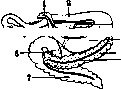
Figure 1. Spiral valve of a ray.
the appearance of nested cones. When a well-developed spiral valve exists, the intestine has a small length but a large diameter. Food passes through such an intestine along a spiral path along the valve. In higher fish and in terrestrial vertebrates, instead of this, an elongation of the intestine itself is observed, which forms more or less significant bends (figure 2). The increase in the absorbing surface of the intestine is also achieved by the formation of a system of smaller folds - transverse ridges, which in the middle intestine have a net-like arrangement. The wall of the intestine then takes on a honeycomb character. The edges of the transverse ridges are often uneven, and on them develop irregular rows of numerous papillae. In birds and mammals these papillae
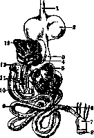
Fig. 2. Intestine of a pigeon: 1-esophagus; 2-crop; 3 and 5-glandular and muscular stomach; 4-spleen; 6-two blind intestines; 7-cloaca; 8-anus; 9-small intestine; 10-12-surface of the intestinal wall; 11-pancreas with its ducts; L-pancreas; 12-liver; giving it a velvety appearance. In mammals, the middle intestine also has a fairly significant system of larger longitudinal and transverse folds of the mucous membrane. Generally, the intestine of mammals reaches considerable length and is clearly differentiated into sections. The initial section of the proper intestine (the "duodenum") also contains special Brunner's glands. The long "thin" intestine forms numerous bends. The posterior intestine is particularly well developed in mammals (compared to other vertebrates), divided into the large intestine with a blind outgrowth at its beginning and into the rectum. There is no blind intestine in many insectivores and in bears. It is also absent in sloths and hippopotamuses, which have a complex stomach. In other herbivores there is usually a long blind intestine, especially in cases where the stomach is simple (horse; fig. 3). Its surface can be further increased by the formation of numerous outpouchings, as in the large intestine (horse, pig), or by the formation of a spiral fold (rabbit). In many mammals of various systematic groups (in some rodents, carnivores, and semi-apes and in anthropoids), the end of the blind intestine forms a thin worm-like appendix [appendix (see)]. In many cases, the large intestine forms lateral rows of regularly arranged outpouchings (haustra), separated by longitudinal muscular bands (taenia; fig. 3). In carnivores and ruminants these formations are absent.
The rectum has considerable musculature but is sharply demarcated from the large intestine. In lower, egg-laying mammals it ends in a cloaca, in viviparous ones - directly with the anal opening.

I. Shmal'gauzen. Development of the Intestine. In an embryo of 3 mm, the intestinal canal is a tube connected in its lower part with the yolk sac via the yolk stalk (ductus vitello-intestinalis, s. omphalo-entericus). Its cranial segment (up to the yolk stalk) gives rise to the pharyngeal intestine (see), stomach, duodenum, and anterior part of the small intestine; the caudal segment gives rise to the remaining part of the small intestine and large intestine. By the beginning of the 2nd month, the intestinal tube forms a loop with the yolk stalk at its apex, which enters the initial part of the formed umbilical cord (skin navel), creating a physiological umbilical hernia that disappears at the beginning of the 3rd month. This loop includes a segment that gives rise to the small intestine; the boundary between it and the duodenum is formed by the flexura duodeno-jejunalis (see Duodenum, Figure 1, and Peritoneum). In the caudal part of the loop, near the apex, an outgrowth appears as a small thickening - the rudiment of the cecum; the segment lying below forms the bend - flexura coli sinistra; thus the boundaries of the future intestinal sections are outlined. Subsequently, the intestine, previously lying in the sagittal plane, undergoes a rotation to the right, as a result of which the segment below the flex. coli sin. moves to the left, and above it - to the right and upward, and thus the rudiment of the large intestine covers the cranial part and duodenum (Fig. 4). By the middle of the 2nd month, the rudiment of the small intestine begins to grow, forming intestinal bends and coils into a ball lying in the umbilical canal. In the large intestine, the descending and transverse parts are outlined first; the ascending part is initially marked by a small bend up to the cecum and develops fully only by the 6th month. The cecum initially represents one large cavity, the free end of which subsequently lags in growth and forms the vermiform appendix. The rectum develops from the cloaca by the formation of a partition, simultaneously with the urogenital sinus. Initially, the large intestine is relatively long; by the end of the 3rd month, the small intestine develops intensively; it becomes significantly wider than the large intestine, especially when it fills with meconium on the 4th month. Only in the 7-8th month does the large intestine begin to grow in thickness. Villi begin to develop in the 4th month in both the small and large intestines; in the latter they disappear in the 9th month, entering the thickness of the mucous membrane, which determines the great length-
Figure 3. Diagram of the large intestine of a horse: 1-colon transversum; 2 and 4-colon dorsale sinistrum et dextrum; 3- ileum; 5- colon ventrale dextrum; 6- taenia coli; 7-caecum; 8- entrance of ileum into caecum; 9-colon ventrale sinistrum of Lieberkühn's glands. Lieberkühn's glands appear later than the villi in the form of
Figure 4. Semi-schematic representation of the rotation of the large intestine around the small intestine: 1- stomach; 2- duodenum; 3- large intestine; 4- small intestine. (According to Kölliker.)
small sacs; circular folds form in the 8th month.
V. Karpov. Anatomical-topographical data. The intestine is divided into two sections: small and large intestines. The small intestines (intestinum tenue) consist of the duodenum, jejunum, and ileum. The small intestines occupy almost the entire lower part of the abdominal cavity, begin at the flexura duodeno-jejunalis at the level of the body of L1 to the left of the spine and end in the right iliac region, where they transition into the cecum (Fig. 5). The diameter of the small intestines gradually decreases from top to bottom: at the flexura duodeno-jejunalis it is about 2.5-3 cm, at the transition point to the cecum - 2.0-2.5 cm. The upper 2/5 of the small intestines are called
Figure 5. Topography of the intestine: 1- ascending
intestine; 2- ileum; 3- sigmoid colon; 4- peritoneum; 5- jejunum; 6- colon transversum. jejunum, the lower 3/5-ileum. There is no sharp difference between them, and this division is arbitrary. The walls of the jejunum are thicker than those of the ileum, richer in blood vessels, and form a large number of folds along their course. In a cadaver, the jejunum is usually empty, the ileum is filled with intestinal contents and distended with gases. Nevertheless, the jejunum differs very little in appearance from the ileum, and a precise boundary between them cannot be drawn. The jejunum is located in the umbilical region and in the left fossa iliaca, the ileum- in the regio hypogastrica, regio umbilicalis, and in the pelvic cavity. - The length of the small intestines varies with age. According to the data of Treves, in individuals aged 20-25 it is 6.75 m; but fluctuations are also possible within the range of 4.5 to 9.5 m. Apparently, individual fluctuations in the length of the intestine depend on a number of external factors and, first of all, on a number of physiological conditions (type of food, degree of habitual filling of the stomach and intestines, etc.). In a newborn, the length of the intestine is 7 times greater than the length of the body, in an adult-3-4 times. Along their course, the small intestines form a series of bends, loops, with the loops of the jejunum lying predominantly transversely, and the ileum - vertically. - On the outside, jejunum and ileum are covered by peritoneum. On the inside, the small intestines are covered by mucous membrane. In the upper part, in the jejunum, the mucous membrane forms folds - plicae, s. valvulae conniventes, circulares Kerkringi. Their number gradually decreases from jejunum to ileum, and in the ileum there are almost none. In addition to the mentioned folds, the mucous membrane also contains thin thread-like villi - villi intestinales. There are more villi in the jejunum than in the ileum. Their total number reaches 4 million. Each papilla (villus) contains inside the so-called central papillary sinus. The latter is a flask-like expansion of the lymphatic vessels of the intestine's mucous membrane, covered with endothelium. Sometimes there are several such sinuses in one papilla. Between the central sinus and the epithelium are numerous vessels of the papilla. In addition, the mucous membrane of the jejunum contains accumulations of lymphatic tissue called solitary follicles (noduli lymphatici solitarii). They reach the size of a pinhead and in places form accumulations (numbering 20-30 or more). These accumulations of solitary follicles are called Peyer's patches [noduli aggregati (Fig. 6)]. Their shape is oval, with the long diameter parallel to the axis of the intestines. In individual cases, the patches reach very large sizes (up to 15-20 cm). In the jejunum, there are few solitary follicles, and Peyer's patches are almost completely absent. The jejunum and ileum are covered with a smooth serous membrane, which transitions into the mesentery (see).
/
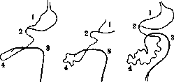
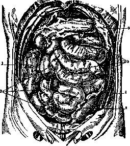

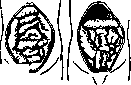
Figure 7. Types of intestinal loops (according to Pavlenko). Loops of the small intestines, when filled and distended with gases, can be palpated through the anterior abdominal wall. The relationship of the small intestines to adjacent organs is difficult to determine precisely: they lie on the side of least resistance and constantly change their position under the influence of movements and changes in body position, respiratory excursions, contractions of the diaphragm, the degree of filling of adjacent organs, the condition of the abdominal press, etc. In the vertical position of the body, the loops of the small intestines descend into the pelvic cavity. In the horizontal position of the body with the pelvis elevated (Trendelenburg position), the intestinal loops are displaced from the pelvic cavity into the upper parts of the abdominal cavity. Despite such great mobility of the small intestines, it can nevertheless be said that certain segments of the jejunum and ileum usually lie in the same places in the abdominal cavity. According to the types of mesenteric position, two types of position of the small intestinal loops are distinguished: in one case—with a narrow lower thoracic aperture and a relatively wide pelvis—the loops lie mostly vertically, in the other—with a wide lower thoracic aperture and a relatively narrow pelvis—the loops lie predominantly horizontally (Figure 7).-At the top, the small intestines are in contact with the mesentery of the colon and with the transverse colon, in front—with the greater omentum and the anterior abdominal wall, to which they are applied on the side opposite to the attachment of the mesentery. On the sides, the small intestines are adjacent to the ascending and descending colons, behind—to the kidneys, spine, ureters, inferior vena cava, abdominal aorta; below they border the pelvic cavity and its contents (urinary bladder, sigmoid colon). In approximately 2% of cases, a remnant of the omphalo-mesenteric duct is encountered (approximately at a distance of 1 foot from the Bauhinian valve); in this case, on the side opposite the site of mesenteric attachment, a blind pouch is observed, the lumen of which is approximately equal to the diameter of the small intestine. This blind pouch is called Meckel's diverticulum or diverticulum ilei. Its length is usually 4-6 cm. Sometimes it is connected by a cord to the navel; more rarely the diverticulum has a communication in the form of a canal with the navel. In these cases, the discharge of feces from the fistulous tract is observed.-The small intestines are attached to the posterior abdominal wall by means of the mesentery. The root of the mesentery (radix mesenterii) has a length of about 13-15 cm, runs obliquely from top to bottom and from left to right. The length of the mesentery is not the same in different parts of the small intestines. It is the longer the farther the segment of the intestine is from its beginning [i.e., it is shortest near the duodeno-jejunal flexure and longest at the terminal ileum (20-25 cm)]. The position and shape of the mesentery are subject to large individual variations. Two types of mesenteric root position should be distinguished: vertical and horizontal (Pavlenko). The first is characteristic of individuals with a narrow lower thoracic aperture and a wide pelvis; the second is found with a wide lower thoracic aperture and a narrow pelvis (Fig. 7). The large intestines are divided into the following parts: the cecum with the vermiform appendix, the ascending, transverse, and descending colons, and the sigmoid colon (Figure 8). In appearance, the large intestines differ sharply from the small ones: their diameter is significantly larger (about 5-8 cm); they have bands of longitudinal muscle, known as taeniae (in number 3); transverse bands or folds (plicae semilunares), corresponding to depressions (sulci transversi); protrusions of the wall of the large intestine (haustra) in the intervals between the transverse bands, and accumulations of fatty tissue on the surface of the intestine in the subserous layer (appendices epiploicae).-The length of the large intestines is approximately 1.3 m. The diameter of the higher parts of the large intestines is greater, that of the lower (caudal) parts is smaller. The walls of the large intestines consist of the same layers as those of the small intestines. The serous membrane does not completely cover the colonic intestines in all parts: in the region of the ascending and descending colon, it is an incomplete covering—their posterior wall is not covered by peritoneum.-The tunica muscularis consists of two layers: an outer longitudinal and an inner circular. The first layer is distributed unevenly in the walls of the large intestine: it forms three flat, sharply delimited bundles, about 10 mm wide and 2 to 3 mm thick. The spaces between them are covered only by a very thin layer of longitudinal muscle. The taeniae are located in the walls of the large intestines in three typical places: one (taenia libera) lies under the serous membrane (shows through it) at the free edge of the intestine, the other two—on the posterior surface of the colon. On the transverse colon, one of the taeniae corresponds to the site of attachment of the mesocolon and is called the taenia mesocolica; another—to the site of attachment of the greater omentum (taenia omentalis).-The circular muscle layer is also distributed unevenly: between the haustra it is somewhat more pronounced. The mucous membrane of the large intestines differs from that of the small intestines in the absence of villi and plicae Kerkringi. In places, the mucous membrane forms clearly noticeable folds—plicae semilunares coli. They consist of mucous, submucous, and muscular tissue (circular fibers) and usually do not encompass the entire circumference of the intestinal lumen, but only one third of it. The number of these folds and the character of their distribution vary greatly. The plicae semilunares and haustra are formed due to the uneven contraction of the longitudinal and circular muscles.-The appendices epiploicae have a flap-like shape and are located at the level of the plicae semilunares in two rows: one on the anterior, the other on the medial side of the colon. The transverse colon has only one row of appendices epiploicae. In obese subjects, the appendices epiploicae reach considerable sizes, arranged in the form of massive polyps on a thin stalk. The ascending colon (colon ascendens) begins in the right iliac region and is the direct continuation of the cecum. It runs from bottom to top, slightly bending
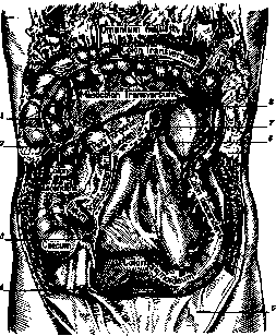
Figure 8. View of the posterior wall of the abdominal cavity: 1-appendices epiploicae; 2-right colic flexure; 3-inferior ileo-cecal recess; 4-ductus deferens; 5-epigastric fold; 6-ascending part of duodenum; 7-duodeno-jejunal flexure; 8-left colic flexure (From Rauber).
to the left side, and at the lower edge of the liver forms a bend called flexura hepatica or flexura coli dextra, after which it passes into the transverse colon (colon transversum). Colon ascendens lies under the liver in front of the descending part of duodenum and the head of the pancreas, and is partially covered in front by loops of the small intestine; in the regio abdominalis lateralis col. asc. lies on the fasciae covering m. quadratus lumborum, m. transversus abd., and on the fascia iliaca and above touches the right kidney and the inferior surface of the liver (impressio colica). The variants observed in this respect are shown in Fig. 9. Col. ascend, is projected on the surface corresponding to the position of the transverse processes of the lumbar vertebrae and the XII rib.- The transverse colon (colon transversum) lies transversely, goes from right to left from the regio hypochondriaca dextra through the regio epigastrica, regio umbilicalis into the regio hypochondriaca sin.; near the lower edge of the spleen it forms a bend called the splenic (flexura lienalis, s. sinistra coli). The angles of coli transversi are attached to the posterior abdominal wall. Usually flexura lienalis lies somewhat higher than flexura hepatica and crosses the left kidney either in the upper or middle, more rarely in the lower third. By means of lig. phrenico-colicum it is attached to the diaphragm. On the anterior abdominal wall col. transv. is projected by means of a line connecting the ends of the cartilages of the X rib. Col. transv. contacts above with the gallbladder, liver and spleen, in front-with the anterior abdominal wall, behind-with duodenum, pancreas, below and behind-with the small intestines. Col. transv. is slightly protruded forward, and both flexures lie more posteriorly.- The descending colon (colon descendens) lies in the left half of the abdominal cavity, goes from top to bottom through reg. hypochondriaca et lumbalis sin. to the left iliac region and passes into colon sigmoideum. Above the boundary of coli descend. corresponds to the XII rib, below-to the left iliac bone. In front it is partially covered by loops of the small intestine, above it contacts the lower pole of the spleen (impressio colica lienis), behind-with the pars costalis of the diaphragm, m. quadratus lumborum, m. transv. abdom. and the left kidney.- The sigmoid colon, col. sigmoideum, forming a bend, passes at the level of S3 into the rectum. The length, shape and position of the entire large intestine are subject to large individual variations. Col. ascend, is sometimes short (up to 8-10 cm), in which cases col. transv. goes in an oblique direction from right to left and from bottom to top, and flexura hepatica is located low. Col. transv. sometimes lies not horizontally, but forms a bend convex downward (U-shaped shape). Relatively more rarely col. descend, forms a convexity directed upward. Its length varies from 30 to 80 cm (on average 50 cm). Col. descend, is also sometimes greatly shortened. The sigmoid colon is the most variable in its position and shape. In some cases it is short, and the bend it forms is insignificant. In other cases, on the contrary, it is long, forms one or several loops, lying either in the cavity of the small pelvis or high (sometimes even under the liver). According to the data of Sozon-Yaroshevich the length of coli sigmoid. on average is 49.36 cm and increases with age: up to 10 years it is 31.55 cm, from 11 to 20 years-36 cm, from 21 g. to 30 years-47.75 cm, from 31 g. to 40 years-51.68 cm, after 40 years-57.27 cm. The relation of col. sigmoid. to adjacent organs [see separate table (t. IV, st. 147-148), Fig. 2] is extremely variable and depends mainly on the state of its filling. In the empty state it hangs into the pelvic cavity and contacts its bottom, with loops of the small intestine, the anterior surface of the rectum and the urinary bladder. In the filled state col. sigmoid. can be displaced high into the abdominal cavity.- Special mention should be made of those anomalies of the position of the intestines, which are explained by their abnormal development. As is known, during embryonic life there occurs torsion of the K. around a. mesenterica sup. in the direction opposite to the clockwise direction. Torsion may deviate in the opposite direction or stop at any point between 90° and 180°. In addition, the differentiation of the peritoneon may stop at the embryonic stage, premature or delayed formation of adhesions, disharmony of growth and delay in the movement of the embryonic intestine. In this way dystopias of the large intestines, sinistro- and dextropositio and others may arise (Shevkunenko). K. is supplied with blood by means of a. mesenterica superior and inferior (Fig. 10). The superior mesenteric artery (a. mesenter. sup.) supplies the entire small intestine and part of the large intestine up to the middle of coli transv.
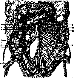
Fig. 10. A mesent sup -9, 2-И duodeno-je-jun,, 2-a. colica med., 3-a. colica dex., 4-a. ileo-colica; 5- ram. colicus; 6-ram. iliac; 7 и *-a. appendic; 10-a. pancreatico-duod. inf.; 11- aa. jejun.; 12-aa. ileae. (By Rauber.)
It originates from the abdominal aorta [see separate table (t. IV, st. 147-148), Fig. 3], lies behind the pancreas and descends downward, going in front of the horizontal part of duodenum in the leaves of the mesentery of the small intestine. A. mesenter. sup. on its way gives off a large number of branches, which anastomose with each other and form a series of arches, arcades arranged in several rows and called the arches of the first, second, third, etc. orders. In its lower part the artery bends somewhat to the right. To the jejunum and ileum a. mesenter. sup. gives about 12-16 branches. Just like the artery itself, these branches are located in the leaves of the mesentery of the small intestine. The lowest branch of a. mesenter. sup. is a. ileo-colica. It departs from the right side of the trunk, goes to the right and down to the ileo-cecal angle, where it divides into two branches: ram. iliacus, going to the left, connecting with the end of a. mesenter. sup. and supplying blood to ileum terminate and ram. colicus, going upward to connect with a. colica dextra and downward-to connect with ram. iliacus. This branch supplies blood to the terminal
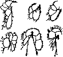
Figure 11. Types of branching of a. mesent. sup. (By Kupriyanov.)
part of the terminal ileum, cecum, vermiform appendix, and ascending colon. From the right side of the superior mesenteric artery, sometimes the right colic artery also branches off. It runs from left to right in a transverse direction toward the middle sections of the ascending colon, and near the latter divides into ascending and descending branches, which connect with neighboring arteries. Very often this artery is merely a branch of the ileocolic artery. From the anterior surface of the superior mesenteric artery, the middle colic artery arises, supplying blood to the transverse colon. It runs from below upward between the layers of the mesocolon, and before reaching the transverse colon, connects by means of arches with the right colic artery and the left colic artery, which belongs to the inferior mesenteric artery system. The arch-like connection between the middle colic artery and the left colic artery is called the arcus Riolani. The inferior mesenteric artery arises from the anterior surface of the abdominal aorta between L1 and L3 and supplies blood to the left half of the transverse colon, descending colon, sigmoid colon, and part of the rectum. It runs from above downward and from right to left, lying in the layers of the peritoneum. The upper branch of the inferior mesenteric artery is the left colic artery, which runs behind the peritoneum from right to left obliquely upward, lying in front of the left kidney, toward the descending colon. It divides into ascending and descending branches and anastomoses above with the middle colic artery, and below with the sigmoid artery. The latter is the second branch of the inferior mesenteric artery. It runs obliquely from right to left toward the sigmoid colon and gives off two branches: an ascending one, anastomosing with the left colic artery, and a descending one, connecting with the superior hemorrhoidal artery, which is the terminal branch of the inferior mesenteric artery. The superior hemorrhoidal artery runs downward and behind the rectum in the thickness of the mesorectum, dividing into two branches that spread along the sides of the rectum. In its lower part, it anastomoses with the inferior hemorrhoidal artery. In the branching pattern of the superior and inferior mesenteric arteries, individual variations are observed, which can be reduced to two types: main and diffuse. The branching types of the superior mesenteric artery are shown in Figure 11. The veins of the Intestine belong to the portal system and consist of two large trunks: the superior mesenteric vein and the inferior mesenteric vein (Fig. 12). The superior mesenteric vein collects blood from the jejunum, ileum, cecum, appendix, ascending colon, and the right half of the transverse colon, lies in the thickness of the mesentery to the right of the artery of the same name, and is composed of the following branches: 1) the jejunal and ileal veins collect blood from the corresponding parts of the small intestine; 2) the ileocolic vein collects blood from the terminal ileum, cecum, appendix, and lower part of the ascending colon; 3) the right colic vein collects blood from the upper parts of the ascending colon; 4) the middle colic vein serves the transverse colon. The area of distribution of the inferior mesenteric vein corresponds to the area of distribution of the artery of the same name and consists of the following branches: the left colic vein and the sigmoid vein. The latter anastomoses with the inferior hemorrhoidal vein. The pattern of vein formation is subject to individual variations. Between the veins of the intestine and the vena cava system, there are anastomoses (so-called portocaval ones), through which ascending thrombi after operations on the Intestine can spread to the venous system of organs supplied by the tributaries of the inferior vena cava (see Cavae venae, Figs. 2, 3, 4, and 7).
Figure 12. Vv. mesent. sup. (7) et inf. (s); 1 - v. pancreat.; 2 - vv. colicae dex.; 3 - intest. ileum; i - a. mesent. inf.; s - vv. intestinal.; e - v. colica sin.; 9 - v. mesent. sup. (After Spalteholz).
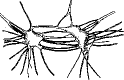
The lymphatic vessels of the Intestine are arranged in two layers: one lies in the submucosa, the other in the subserous layer. The submucosal lymphatic vessels run circularly around the Intestine and are mainly lacteal vessels that absorb products from the Intestine during digestion. The vessels of the subserous layer run along the intestinal wall and are proper lymphatic vessels. However, between these layers there are anastomoses. After leaving the intestinal wall, the lymphatic vessels lie perivascularly. They empty into the mesenteric lymph nodes, located in the thickness of the mesentery (Fig. 13), numbering about 180. The glandular packets sometimes observed at the root of the mesentery are called the pancreas Aselli. The lymphatic vessels of the transverse colon (Figs. 14 and 15) empty into the mesocolic lymph nodes, located at the base and in the thickness of the mesocolon. The efferent vessels of these nodes connect to form the intestinal trunk. The latter, together with the lumbar trunk, empties into the cisterna chyli. The nerves of the Intestine mainly belong to the vagus and sympathetic nerve systems, but contain fibers from the spinal nerves: the intestinal branches, innervating the jejunum, ileum, and the colic branches going to the cecum, ascending colon, and part of the transverse colon. Upon reaching the mesenteric border of the intestine, the nerves form a plexus under the serous membrane and send numerous branches to the ganglionic plexus located between the longitudinal and circular muscle layers of the intestines and called the myenteric plexus or Auerbach's plexus. In the submucosal layer there is a second nerve plexus, which is called the submucosal plexus or Meissner's plexus. It connects with numerous branches to the previous ones. Both plexuses extend along the entire intestinal tract. The aortic plexus also sends branches to some parts of the Intestine, namely: to the descending colon, sigmoid colon, and superior part of the rectum. The vagus nerve sends its branches to the intestines via the celiac branches, which run along the left gastric artery to the celiac plexus, and from there to the Intestine. The quantitative ratio of the branches of the vagus, sympathetic, and spinal nerves in the ganglia varies: in some cases, the branches of the vagus nerve predominate, in others the sympathetic nerve, and in others the spinal nerves. The pattern of branching from the celiac ganglion varies in different individuals and can be represented in 2 types: in one - the number of branches is small, and they depart as trunks that branch weakly; in the other - there are many branches, and they in turn break down into a series of smaller branches (Figs. 16 and 17).
Figure 14.
Figure 15.
F. Walker. Histology of the Intestine. The wall of the Intestine consists of three layers: 1) the mucous membrane (tunica mucosa), which is separated by a layer of smooth muscle (muscularis mucosae) into the proper mucous and submucous (tunica submucosa) layers; 2) the muscular layer (tunica muscularis) and 3) the serous membrane (tunica serosa). Due to the fact that the structure of the mucous and muscular layers in the small and large intestine shows differences, it is more convenient to describe them separately. - Small intestine. The mucous membrane has a thickness of about 0.5 mm; it forms permanent folds running transversely across the intestine for approximately 2/3 of its circumference (plicae circulares Kerkringi), which include the muscularis mucosae. The folds begin in the duodenum at a distance of 2-5 cm from the pylorus as low elevations, reach full development at the duodenal papilla, and continue in this form to the middle of the jejunum, from where they become lower and end in the middle of the ileum. The entire surface of the mucous membrane, including the folds, is covered with villi (villi intestinales), thin projections 0.5-1.0-1.5 mm in length, which give the mucous a velvety appearance (Fig. 18). Their number per 1 cm² in the duodenum and jejunum is 22-40, in the ileum 18-31 (Kolliker). The shape of the villi varies somewhat and can be reduced to two main types: platy or leaf-shaped, and cylindrical or finger-shaped (Sappey); the first predominates in the duodenum, the second in the rest of the Intestine. Between the villi, the openings of Lieberkühn's glands (2 to 7) are visible. - The epithelium covering the mucous membrane and villi is single-layered cylindrical and consists of two types of cells: columnar and goblet cells (Fig. 19). The columnar epithelium (cells 22-30 μ in length, 6-9 μ in width) bears on its free surface a shiny cuticular border, which on thin sections shows transverse striations. Views on its structure have not yet been unified: some authors (Brettauer and Steinach, Prenant) consider it to consist of thin rods or cilia that have lost their ability to move, others (Kölliker, R. Heidenhain, Flemming) see in it a cuticular structure pierced by pores into which thin protoplasmic projections enter (porous epithelium). Acting with 2% NaCl, it is possible to detach the border and pull out processes from it (R. Heidenhain). In the body of the cell, several zones can be distinguished: under the border lies a narrow transparent zone in which the centrosome is described in the form of two grains; behind it is a wider - granular zone; from here to the nucleus is a wide zone containing fibers, tonofibrils (M. Heidenhain), which other authors consider to be mitochondria (Champy); in it is also located the Golgi apparatus, which gives off projections on the sides of the nucleus; further located is the oval nucleus,
Figure 16. Gangl. coeliacum. Concentrated type.

and beneath it the basal zone also contains mitochondria. The striated epithelium serves for absorption; during digestion, the smallest fat droplets appear in the second zone; descending, they merge into larger ones. Goblet cells were formerly considered independent elements (Bizzozero); at present, most authors regard them as mucoid-degenerated striated cells which, after expelling mucus, regain their striation (List, Paneth, Mollendorff).-The base of the mucous membrane (lamina propria), separated from the epithelium by an amorphous basal membrane, consists of reticular tissue, in the loops of which are placed a large number of leukocytes, mainly lymphocytes, fewer eosinophils, plasma cells, which partly undergo degeneration (Scnollenleukocyten), partly emigrate into the cavity of the intestine; a certain number of leukocytes are always found in the epithelium in the form of rounded bodies with nuclei. Villi (fig. 20) as outgrowths of the mucous membrane are constructed in the same way; their peculiarity lies in the arrangement of the vessels and the presence of smooth muscles, which
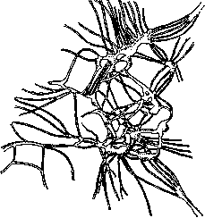
Figure 17. Gangl. coeliacum. Scattered type.
is connected with the function of the villus-absorption. Arterioles, entering the villus, break up into a fine-meshed capillary network located under the epithelium, which then collects into one venule. Along the axis of the villus is located a straight and wide lymph capillary (rarely 2 or 3) 27-36 μ in length, beginning blindly at the top and passing into the deeper-lying lymph network; into it is transported (by an unclear path) the absorbed fat. Between the blood and lymph vessels in the base are located bundles of smooth muscles, rising from the muscularis mucosae to the top of the villus, where they anastomose. At the moment of absorption, the villi fill with blood, swell and rise; the contraction of the smooth muscles shortens and wrinkles them, which facilitates the outflow of fluids.-The Lieberkühn glands (gl. intestinales; fig. 21), **o,

otherwise called crypts, are simple tubular glands, rarely branched, 0.3-0.4 mm long; in humans they are considerably shorter than in carnivores. The cells lining their cavity are of the same character as the mucous membrane, and on sections they are a direct continuation of the villus epithelium; they are only lower (18 μ), and the striation of the deeper-lying cells is thinner. Mitoses are often found in these cells, which is why Bizzozero asserted that the purpose of the crypts is to serve as a site of regeneration for the epithelium of the mucous membrane. But at the bottom of the glands, Paneth described special cells containing in the upper portion
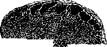
Figure 18. Mucous membrane of the small intestine from the surface at low magnification. Villi and openings of Lieberkühn's glands. (According to Kölliker.) )» *< y/a a distinct protein-like granularity, clearly visible in life and staining with both acidic and basic dyes, which places the glandular nature of the crypts beyond doubt.-In the mucous membrane of the small intestines are also found lymphatic nodules, so-called solitary follicles, 0.5-2 mm in size, in a variable number; developing, they f _\ ,\ / "' \ ;> \ protrude the mucous , 11! l membrane, pushing aside 4!ytf ' \ Г) the villi and glands; they \КШ Щеп М0ГУT can also penetrate /рула П°ДСЛИЗИСТУЮ shell. Small follicles are of round shape; larger follicles often become pear-shaped with a wide base and a pointed apex. Clusters of follicles (usually 20-30 up to 60 and 100), the so-called Peyer's patches (agmina Peyeri), protrude on the mucous membrane of the lower part of the jejunum and ileum in the form of oval plates 2 to 12 cm long (in rare cases more), 1 to 3 cm wide; their total number is 30-40; they are always located on the side opposite to the attachment of the mesentery. On sections, it is seen, Figure 19. Striated and goblet epithelium,
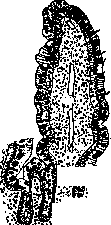

Figure 20.
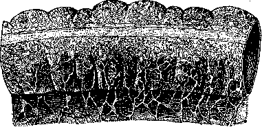
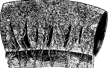
Figure 21. Figure 20. Section of the mucous membrane of the human small intestine. Villi and Lieberkühn's glands. (According to Braus). Figure 21. Section of the bottom of a Lieberkühn gland at high magnification. At the bottom are seen Paneth's granular cells. (According to Braus). that the follicles making up the patches lie deep in the submucous membrane; on their surface the villi are shortened and smoothed.-Muscularis mucosae represents a thin layer of the inner circular and outer longitudinal layers of smooth muscles. Located beneath them is the submucous membrane, consisting of loose fibrous connective tissue and contains a network of blood and lymph vessels and a nerve plexus (plexus entericus, s. Meissneri) (see Vegetative nervous system). The greatest thickness the submucous membrane has in the duodenum, where Brunner's glands are located in it.-The muscular membrane consists of two layers of smooth muscle: the inner-circular, and the outer-longitudinal, developed more weakly; between them lies the nerve plexus (plexus myentericus, s. Auerbachi). The outer membrane is serous, or peritoneum (see). Large intestine. The mucous membrane (fig. 22) differs in the absence of villi
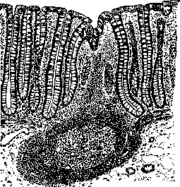
Figure 22. Cross-section of the human large intestine mucous membrane: Lieberkühn's glands, beneath them a solitary follicle with a light center of proliferation. (After Braus.) The surface is covered with a striated epithelium containing goblet cells. Lieberkühn's glands are considerably longer than in the small intestine and reach 5-7 mm in the rectum. The number of goblet cells in them is extremely large, especially in the neck and middle of the gland; at the base are simple cylindrical cells with a thin cuticular covering; among them mitoses are found, and from there apparently the regeneration of the epithelium proceeds. Paneth's cells are absent. The propria tissue of the mucous membrane is infiltrated with leukocytes; scattered in it are solitary follicles of large size (1.5-3 lip) with tubular depressions of the mucous membrane on their summits. The follicles develop particularly strongly in the vermiform appendix, sometimes leading to the complete destruction of Lieberkühn's glands, which after the atrophy of the follicles can apparently grow again (Rudinger). The muscular coat is characterized by the incomplete development of the outer longitudinal layer, which forms three bands (taeniae). The serous coat is absent on the posterior wall of the ascending and descending parts and in the lower part of the rectum. V. Karpov. Anatomical features of the I. in children. Age-related features of the I. concern its dimensions (length and width of the lumen), structure, and topography. From the first days of life to the adult state, the dimensions of the I. continuously increase. But, as established by a number of authors [Beneke, Tar-onetsky, Debele, Anserov, etc.], the rate of growth of the I. at different ages is not the same. Similarly, there is a difference in the ratio of growth of the small and large intestines. Finally, the I. grows in length differently than in width. Even during the first year of life, the I. grows unevenly. According to Frolovsky, the greatest increase in length falls on the 2nd month. Already at this age a disproportion in the growth of the small and large intestines becomes noticeable: the latter grow more vigorously than the former. The relative length of the intestine (compared with the length of the trunk) in newborns is greater than in adults, due to the relatively greater length of the small intestines. Up to 3 years it continues to increase, then gradually falls and by the time of sexual maturity becomes a constant value (Table 1). Table 1. Age Ratio of the length of the spine and intestine of the spine and small intestines of the spine and large intestines 0- 1 yr...... 1- 3 yrs..... 3-10 yrs..... 10-15 yrs..... Adults .... 1:15.3 1:17.4 1:14.0 1:13.4 1:13.2 1:13.0 1:14.6 1:11.5 1:10.8 1:10.7 1:2.3 1:2.9 1:2.5 1:2.6 1:2.6 The indicated change in relative length applies equally to both the small and large intestines, and is explained by fluctuations in the growth not only of the I. itself, but also of the spine, the length of which is taken as the unit of measurement. Hence it is clear that relative length does not give a clear picture of the rate of growth of the I. The latter becomes possible if one takes the percentage increase in length of the I. at each age compared with the previous one (Table 2). Table 2. Age Increase in length (in %) of small intestines large intestines 1- 3 yrs . . . 3-10 yrs . . . 10-15 yrs . . . Adults . . 23.9 8.2 22.0 18.7 36.8 18.6 34.3 18.8 The data in Table 2 indicate that there are two spurts in the growth of the I.: 1) from 1 to 3 years, when the organism transitions from milk, easily digestible food to mixed food, which requires a greater digestive and absorptive surface, and 2) from 10 to 15 years, during the period of sexual maturity, associated with the intensified development of all bodily functions. It is understandable that the I. must satisfy the increased demands made on it; this is expressed in the vigorous growth of it in length. The above applies equally to the small and large intestines. But the latter not only during the first year of life but also later, up to and including the period of sexual maturity, grow at a faster rate. As a result, the difference in length between them gradually smooths out (Table 3). It should be added that in newborns the ascending part of the large intestine (caecum-colon transversum) is as a rule longer than the descending part (Debele, Natiyev), and this ratio Table 3. Ratio of the length of large and small intestines. Age Ratio 0- 1 yr . . . 1:5.6 1- 3 yrs . . . 1:5.0 3-10 yrs . . . 1:4.6 10-15 yrs . . . 1:4.1 Adults . . 1:4.2 according to Debele's observations exists up to 4 years. The width of the lumen, in contrast to the length, increases uniformly.-Most authors recognize that the dimensions of the I. change under the influence of a number of factors. Among the latter, various pathological conditions of a general and local nature occupy a prominent place. Some of them reduce the dimensions of the I. compared to the normal of the same age (e.g. general exhaustion in tuberculosis, peritonitis), others, on the contrary, increase them (for example ascites). Features in the structure of the intestinal tube in its main aspects are as follows. The folds of the mucous membrane of the small intestines in early childhood are less developed than in adults; their length and height are less. The same must be said regarding the size and number of villi. The muscular layer does not reach full development. The mucous membrane, only slightly inferior to it in thickness, is rich in cellular elements as opposed to the weak development of the connective tissue basis, has a relatively more developed network of blood vessels and wide lymphatic vessels (Bagirtsky). The glands of the I. are underdeveloped. This applies predominantly to Brunner's glands; but Lieberkühn's glands also show signs of incomplete development. The lymphoid apparatus has no significant distinctions, if one does not consider certain peculiarities in the structure of Peyer's patches; with age the size of individual patches increases. The absolute number of follicles increases with age, but the relative number in early childhood is 3 times greater (per unit area) than in adults (Gundobin).-One must particularly note certain peculiarities in the structure and topography of the large, especially the colon, intestine. The retarded growth of the large intestines in the embryonic period is manifested in early childhood by the fact that certain parts of this section of the I. retain an embryonic type of structure. The above applies entirely to the cecum, which usually has the shape of a cone, gradually tapering and passing into the vermiform appendix. This same circumstance explains why peculiarities of position have predominant significance only in relation to the large intestines. These peculiarities are mainly conditioned by the nature of the relationships of the large intestines with the peritoneum, essentially different compared to adults. For the cecum, significant mobility is typical, from which depend very considerable variations in its position. The vermiform appendix is located intraperitoneally and as a rule hangs down into the pelvis. The ascending colon very often (according to Natiyev-59.7%) in its initial portion lies intraperitoneally, which explains the ability of the intestine to displacement. The position of coli transv. depends mainly on peculiarities in the size of the liver and stomach. Due to the incompleteness in the development of the hepatic flexure, the position of its right half is particularly variable. The differences in the position of coli descend, are insignificant. For colon sigmoideum, an extraordinary variability of position is characteristic, explained by the great length of the intestine and its high mesentery. Colon sigmoideum is located above the entrance to the pelvis or along the median line or extends to the right side. Due to the underdevelopment of the pelvis and the relatively large liver, colon sigmoideum forms a large number of bends.-These most important anatomotopographic features of the I. in children explain its functional peculiarities and are a predisposing factor for the development of various pathological deviations. The underdevelopment of glands, especially Brunner's, in connection with the rich network of blood vessels and relatively wide lymphatic vessels, finally the insufficient development of musculature-all this leads to the conclusion which was also made by Gundobin, that in early childhood the I. is more adapted to absorption than to digestion of food. The high absorptive capacity explains the easy vulnerability of the child's I. The weak development of the muscular layer with great mobility of the I. causes the frequency of constipation in children. The underdevelopment of the sacrum and coccyx, insufficient fixation and incomplete development of the muscular layer of the rectum cannot but be connected with the frequently occurring prolapse of it in childhood. n. Anserov. Physiology of the I.-see Digestion. P. Methods of research. Among the methods for diagnosing diseases of the I. should be included physical methods of examination (inspection, palpation and percussion of the abdomen-see Abdomen), examination with the aid of duodenal and intestinal tubes, roentgenoscopy, recto-romanoscopy, examination of urine; in addition very important data are obtained in the examination of excreta (see). Functional diagnosis. A. Schmidt and Strasburger founded and refined the technique of examination of intestinal secretions for the functional diagnosis of intestinal diseases. Schmidt proposed a standard diet; the character of the excreta with it does not depend on the accidental composition of the food.
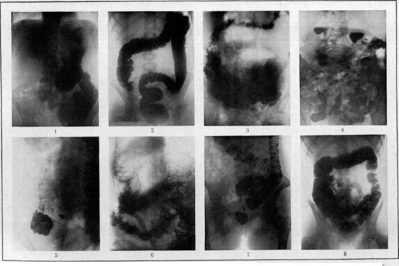
The study of intestinal secretions on this diet provides data characterizing the functional capacity of the INTESTINE. This test diet is as follows: 1st breakfast - 0.5 liters of milk and 50 g of dry bread; 2nd breakfast - 0.5 liters of oatmeal mucilage soup (40 g of oatmeal, 10 g of butter, 200 g of milk, 300 g of water, 1 egg and a little salt); lunch - 125 g of minced meat (raw weight), slightly fried with 20 g of butter, and 250 g of potato puree (190 g of potato, 100 g of milk, 10 g of butter and a little salt); at 5 o'clock - the same as the first breakfast; dinner - the same as the second breakfast. The diet is prescribed to the patient for 3 days, and the intestinal secretions obtained are subjected to thorough macroscopic and microscopic, bacteriological, and chemical examinations. To distinguish the feces to be examined from previous ones, carmine is given. In normal conditions with such a diet, macroscopic examination reveals only a small amount of residues of plant food in the secretions; microscopic examination, in addition to detritus, finds single muscle fibers, individual crystals of fatty acids, residues of oatmeal soup and bread, single residues of potato; the litmus reaction is weakly alkaline or weakly acidic; the fermentation test (see) is either absent or very weak. At present, Schmidt's test diet is not used by all, as many believe that it has not justified the hopes placed in it; thus, an increase in fermentation processes is often noted with a negative result of the fermentation test (acetic acid fermentation), without liquid stools and with an alkaline reaction of the feces to litmus. French authors propose to study the function of the INTESTINE by adding to the ordinary food 75 g of minced meat, raw or slightly fried, and potato. In the Institute of Balneology in Moscow, the test diet consists of 200 g of minced and slightly fried meat, rice porridge, potato puree and grated carrot; in special research of the functional capacity of the pancreas, 100 g of butter is added to this diet. For functional diagnosis, the consistency and form of the feces, the color of the feces, pathological impurities, the degree of utilization of various components of food, the character of the bacterial flora (see Defecation - Constipation, Diarrhea., Intestinal indigestion), and the amount of organic acids and ammonia are taken into account. - The amount of organic acids and ammonia in the feces is determined by the method of Rou and Goiffon (G. Ch. Roux, Goiffon). In a calibrated porcelain mortar with a capacity of up to 70 cm³, 5 g of feces, taken from different parts of the delivered portion, are weighed and triturated with 50 cm³ of distilled water, adding the latter gradually, in small portions. The resulting homogeneous mass of feces, after adding 2-3 drops of a 1% alcoholic solution of phenolphthalein, is treated with a 30% solution of aluminum chloride and potassium hydroxide is added in pinches until a pink color appears. After this, the mixture is allowed to stand for 5 minutes and then 25 cm³ of liquid is filtered. The resulting 25 cm³ of filtrate is brought to a weak red color by adding N/10 solution of HCl and N/10 solution of soda, 5 cm³ of tropaeolin 00 indicator are added (in a flask with a capacity of 500 cm³, 0.1 of tropaeolin is dissolved in 300 cm³ of 96% alcohol and water is added to the mark), and it is poured into a test tube marked 60 cm³. The solution is titrated with N/10 solution of HCl to an orange color; then water is added to the mark and it is titrated again to the color of the previously prepared standard. The standard is prepared as follows: in a test tube marked 60 cm³ (of the same diameter and glass as the first), 1.2 cm³ of N/10 solution of HCl, 5 cm³ of tropaeolin and water are successively poured to the mark. The standard solution is preserved for a long time. The calculation is made by the formula: x = [(y-1.2).100]:25, where y is the number of cubic centimeters of N/10 HCl. The amount of organic acids in the feces in normal conditions is characterized by the figure 14-18. The amount of ammonia is 2-4. An increase in organic acids in the feces is the result of strong fermentation in the intestine, while their decrease indicates that normal fermentation was delayed or by an intensification of putrefactive processes (a large amount of ammonia in the feces), or by acceleration of evacuation, or part of the organic acids was neutralized by the secretion of the INTESTINE. Cases are often encountered where, along with a high content of organic acids and traces of ammonia, the reaction of the feces to litmus is alkaline, which depends on the entry of alkalis into the INTESTINE from the tissues: the acidic reaction of the intestinal contents strongly irritates the intestinal wall, which responds by the secretion of alkalis - the 'protective' reaction of the INTESTINE (Pevzner and Lindenbraten). With an intensification of putrefactive processes, the amount of ammonia in the feces increases with a normal or reduced content of organic acids. Finally, in mixed cases, both the amount of organic acids and ammonia may be increased. The data from fecal examination can, following the example of French authors, be divided into the following 4 groups, characterizing an abnormal function of the digestive apparatus in general and of the INTESTINE in particular. - 1. Insufficient secretion of digestive organs. Insufficient secretion of the stomach is characterized by the presence in the feces of connective tissue and undigested muscle fibers, and often also crystals of calcium oxalate; sometimes there are residues of starch or particles of plant food; putrefactive processes prevail with strong irritation of the intestinal mucosa, although in some cases there is also an intensification of fermentation processes. Insufficient function of the pancreas is characterized by: a) a large amount of feces, pasty or liquid, mostly gray in color (due to the large amount of fat), of a sharp odor, alkaline reaction; b) an abundant amount of drops of neutral fat; c) positive Schmidt's nuclear test; d) the presence of a large amount of residues of starch and cellulose; e) positive reaction for hydrobilirubin. Insufficient secretion of bile into the INTESTINE gives solid or liquid feces of light gray color (absence of hydrobilirubin), acidic reaction; all food substances are well digested, except fats, which are excreted in the form of fatty acid crystals; the test for hydrobilirubin is negative. Insufficient digestion and absorption of milk sugar in the small intestines is characterized by frothy and acidic feces. - 2. Disturbance of the balance of intestinal flora. In the INTESTINE there are two groups of bacteria participating in the processes of food digestion: anaerobic flora, favorable to putrefactive processes, and aerobic flora, saccharolytic, promoting the development of fermentation processes. Depending on the predominance of one or the other group, either a fermentative stool or a putrefactive one is obtained (see Indigestion). When a large amount of Entamoeba coli, Balantidium and other parasites are found in the feces, the putrefactive processes may depend on them. In some cases, both the amount of organic acids and the amount of ammonia are increased in the feces; the reaction of the feces to litmus is amphoteric (so-called mixed forms). - 3. Disturbance of evacuation of the intestine. With accelerated evacuation, the fecal masses are more or less liquid and contain a large amount of poorly digested food residues and iodophilic bacteria. The feces have an admixture of little changed contents of the small intestines, semi-liquid, golden-yellow in color (due to the content of bilirubin), with a very sharp odor; the reaction is alkaline; muscle fibers are poorly digested; much fatty acid and neutral fat, much starch and plant fiber; traces of organic acids and ammonia are present. Feces with an admixture of little changed contents of the cecum - of pasty consistency, yellowish in color, of a fatty odor; muscle fibers and fats are well digested; there is much plant fiber and starch, as well as iodophilic bacteria; bile pigments in the form of stercobilin; organic acids are contained in a normal amount, ammonia in a small amount; the reaction is neutral or weakly acidic. - With delayed evacuation (constipation), the fecal masses are dense, dark in color, do not contain food residues and iodophilic bacteria; the content of ammonia is increased, while that of organic acids is decreased; the reaction is neutral or alkaline. -4. Enterocolitis, the main characteristic signs of which are increased water content in the feces, presence of albumin (from the secretion of the affected intestines), mucus, intensification of putrefactive processes. If there are ulcers in the INTESTINE, the feces, besides mucus, usually (not always) contain blood and a large amount of leukocytes (pus); in case of retention of fecal masses above the point of ulceration, only pathological products are excreted - pus, mucus, blood, while fecal masses are absent. - Symptoms of disturbance of the proper function of the large intestines may be various in Figure 1. Normal picture of filling of the stomach and loops of the small intestines 30 minutes after taking contrast food according to Rieder. Figure 2. Normal picture of the large intestines after contrast enema: the mass fills the rectal ampulla, sigmoid flexure, descending colon, transverse colon and ascending colon. Haustra are well expressed in the upper part of the sigmoid flexure, transverse colon and ascending colon.
The flexura lienalis is much higher than the flexura hepatica. Figure 3. In the area of the small intestine, accumulations of liquid with a horizontal level are visible. Above them, accumulations of gases are visible in the form of a very light shadow (case of stenosis of the small intestine, confirmed by surgery). Figure 4. Multiple diverticula: irregular semicircular-shaped accumulations of contrast mass with gas bubbles underneath are visible (case confirmed by operation). Figure 5. Colica mucosa: in the colon ascendens, there are small point-like accumulations of contrast mass throughout its length. Figure 6. Megasigma: the flexura sigmoidea is elongated and dilated. Interpositio coli: the colon transversum is swollen with gas and is placed above the descended liver and under the right dome of the diaphragm (case confirmed by operation). Figure 7. Carcinoma recti: a filling defect for several centimeters in the area of the ampullae recti (case confirmed by proctoscopy and microscopic examination). Figure 8. Polyposis recti et flexurae sigmoideae: in the area of the rectum, flexura sigmoidea, and colon descendens, round filling defects the size of a small pea are visible (case confirmed by proctoscopy). (For illustration in the article Intestine).
Depending on the localization of the inflammatory process: a) in case of lesion of the cecum and ascending colon, the stool is usually liquid, fermentative, with an admixture of little changed content of the cecum; b) in case of lesion of the left part of the large intestine (including col. sigmoid. and rectum), evacuation is often insufficient with the excretion of non-hemorrhagic feces (see above): so-called false diarrhea is often present; for the diagnosis of this ailment, in addition to X-ray examination, the following method can be used: the patient on an empty stomach is given 0.5 carmine or charcoal half an hour before a meal, and then the first appearance of colored stool is noted; in cases of delayed evacuatory ability of the stomach (motor insufficiency), carmine is introduced through a duodenal tube into the duodenum. The appearance of carmine later than after 24 hours indicates a delay in fecal masses. This method does not give precise results.
M. Pevaner, L. Berlin. Roentgenodiagnosis. Methodology and normal picture. An unprepared K. does not differ upon X-ray examination and in the X-ray from the surrounding parts. Only the accumulation of gas in its cavity, the presence of foreign bodies and fecal masses sometimes give clear images. For systematic study of the K., it is necessary to fill it with a contrast substance that strongly absorbs rays. After taking per os a contrast meal (see Stomach, X-ray examination), already after 15 minutes one can observe partial filling of the loops of the jejunum. Here the food gruel is detained for only a short time; its distribution in a thin layer over the surface of the mucosa promotes rapid digestion and absorption. Correspondingly, a thin peristaltic pattern of the jejunal loops is obtained, on which the delicate folds of Kerckring are visible with rather changeable contour (see separate table, fig. 1) under the influence of contractions of the muscularis mucosae (Forsell). After half an hour of taking the meal, dense shadows of filled ileal loops are visible, and after 3-4 hours the jejunum usually no longer contains contrast substance, which has completely accumulated in the ileum. The latter appears as irregularly arranged, tightly filled loops in the lower half of the abdomen. After 5-7 hours of taking the meal, filling of the cecum and ascending colon up to the hepatic flexure is visible. During the same time interval, it is sometimes possible to obtain an image of the vermiform appendix. The latter is facilitated by thorough emptying of the K. from fecal masses before taking the contrast medium and by palpation of the ileocecal area during X-ray examination. Under normal conditions, both the cecum and the vermiform appendix are easily displaced by the palpating hand. A distinctive feature of the X-ray image of the large intestine is the haustra; their outlines change under the influence of the so-called small movements of the colon. In its initial part, fecal masses are detained the longest: after 24 hours, filling of the transverse colon is still visible; after another 5-6 hours, filling of the descending colon, sigmoid flexure, and rectum. After 30-40 hours, the entire colon is empty. Holzknecht observed under normal conditions very rapid movements of large fecal masses in the direction of the anus. In the ascending colon and the initial part of the transverse colon, antiperistaltic movements also occur under normal conditions. The cecum is usually located at the level of the head of the femur, the hepatic flexure under the lower edge of the liver, the splenic flexure considerably higher, often under the left dome of the diaphragm. The very mobile transverse colon sometimes runs in almost a straight line, more often it sags downward; its upper contour usually lies against the greater curvature of the stomach; both with general ptosis and with limited coloptosis, it can descend to the symphysis. The position of this part of the K. is very variable: it changes even in the same person depending on the tone of the abdominal press, the position of the diaphragm, etc. In general, a more straight course is more often observed with a hypersthenic constitution; a garland sagging downward is observed with an asthenic constitution. The descending colon descends in a straight line downward, the sigmoid flexure presents a series of variants from a slight bend to the formation of a large loop extending to the right and upward. The lumen of the colon gradually decreases from the cecum to the rectum; the ampulla of the rectum appears as a clear dilation. The advantage of the described method lies in its closeness to physiological conditions. For studying the anatomy of the large intestine in the living, filling per rectum (see separate table, fig. 2) - the so-called contrast enema - deserves preference; its approximate composition: a) Barii sulfurici purissimi 150.0, hot water 500.0; b) Decocti Amyli Tritici 30.0:1000.0, mix a and b in hot form, allow to cool to 37-40°. The patient is placed on a trochoscope and the gradual filling of the colon and its emptying is observed on a fluorescent screen (Haenisch). Previously, it is necessary to thoroughly empty the K. with a laxative and an enema. Fischer recommends inflating the colon after partially removing the enema with contrast medium; this gives very clear pictures with images of the mucosal folds. Knothe first introduces a contrast enema and lets it drain, so that a small deposit of contrast medium remains on the wall, on which the mucosa can be studied. With simple inflation of the large intestine, clear images of its outlines are obtained. For studying the small intestine, a thin duodenal tube can also be used, through which a liquid contrast medium is poured, for example, a solution of KI. Pathological picture. Small intestine. (X-ray picture in diseases of the duodenum - see Duodenum.) In fermentative dyspepsia (Lampe) and in some cachectic conditions, the motor function of the jejunum is sharply disturbed; its loops give after taking the contrast medium dense shadows with gas bubbles and horizontal levels. Such gas bubbles and horizontal levels can also be caused by atony and stasis before organic stenosis (see separate table, fig. 3). Since they can be detected even without taking the contrast medium, they serve as a good diagnostic sign in acute obstruction. Individual tightly filled loops, found at a time when the jejunum should already be empty, indicate stenosis and are one of the earliest clinical symptoms of it. - Individual irregular semicircular constant contrast spots with gas bubbles were noted in diverticula (see separate table, fig. 4). A limited bulging near the gastro-intestinal anastomosis indicates peptic ulcer of the jejunum. A prolonged delay in the lower loops of the ileum is often observed in the presence of peritoneal adhesases after pelvic peritonitis, in organic diseases of the cecum, in insufficiency of the Bauhinian valve. For the latter phenomenon, according to some authors, the visible filling of the ileal loops when using a contrast enema is conclusive proof. - Ascarids may appear as light strips in a filled intestinal loop; sometimes an image of the parasite's intestine containing contrast medium is also obtained. Large intestine. Increased tone of the K. leads to narrowing of its lumen; the haustra give a wedge-shaped fine pattern; sometimes with pronounced spasm they are not visible at all, and the filled colon appears as a thin strip. The same pictures are also obtained with irritation of the n. vagi and under the influence of pilocarpine and physostigmine. Atony is characterized by a wide lumen without pronounced segmentation of the haustra; irritation of the n. sympathici, the use of atropine leads to the same change in motor function. In some cases of constipation, a prolonged delay is observed in the ascending colon, in others in the transverse colon or in the sigmoid flexure and ampulla of the rectum. Often a combination of atony of the cecum with spasm in the descending colon is observed. In colica mucosa, light strips of mucus with adherent accumulations of contrast medium and compact "contrast fecal masses" can be seen in the lumen of the intestine (see separate table, fig. 5). Meteorism gives fairly bright pictures of gas accumulation; the distended splenic flexure of the colon pressing against the diaphragm has significance for the so-called gastro-cardial symptom complex; a distended loop of the colon under the right dome of the diaphragm above the liver (colic interposition) is important for the diagnosis of liver ptosis (see separate table, fig. 6). Inflammatory diseases of the colon are characterized by spasmodic narrowing of the lumen and irregularly spotted contrast shadows due to the accumulation of mucus and anatomical lesions of the mucosa. For tuberculosis, an irregularly fine-toothed contour is typical. In tuberculosis of the cecum, a delay in the ileal loops is often noted, the absence of contrast mixture in the cecum, and distinct filling of the transverse colon (Stierlin's symptom). In general, in tumors (see below), both inflammatory and neoplastic, the absence of contrast content at the site of the lesion with normal filling of adjacent parts is noted. In other cases, irregularly contoured filling defects, images of narrow channels with ragged edges are obtained (see separate table, fig. 7). These signs belong to the early stage, when there are as yet no indications of obstruction; but they are conclusive only if they are very constant and especially if the K. has been thoroughly cleansed beforehand, since the simultaneous presence of contrast and non-contrast feces in the intestine can also give a picture of a defect with irregular edges. Clear pictures of polyposis are obtained after removing the contrast enema, when traces of barium salt have settled between individual polyps and give a characteristic filigree pattern (see separate table, fig. 8). In acute obstruction, even without contrast medium, a greatly dilated section of the colon with a horizontal level of fluid and dark strips - the haustra - can be seen. An elongated and gas-distended sigmoid flexure is especially clearly imaged in its volvulus, as well as in congenital megasigma without signs of obstruction. In cases of intussusception, with the help of a contrast enema, an image of the section of intestine that is invaginated into the lumen of another can be obtained in the form of a light strip bordered by dark edges.
For the diagnosis of chronic appendicitis, the uneven filling of the vermiform appendix, its bulbous dilation, and the prolonged retention of contrast medium in it, even after the emptying of the cecum, are of importance. A sign of adhesions is the impaired mobility of the intestinal segment during palpation and the exact correspondence between the site of greatest sensitivity during palpation and the image of the affected part of the intestine. The incorrect position of the intestine under the influence of tumors of the abdominal cavity, in eventrations and hernias, and its pathological fixation by adhesions, are also well determined by X-rays and may be of importance for the diagnosis of the primary disease. - The X-ray picture in diseases of the rectum - see Rectum.
°- Dn- III. Pathological anatomy. Malformations of the I. may manifest as underdevelopment of the I. as a whole or its individual parts, as irregularities of the lumen of the I., as persistence of embryonic formations in the I., or finally as an abnormal position of the intestine. Malformations of the first type include rare cases of complete absence of the I., which is observed only in monsters of the acardius type (see), or underdevelopment of one or another part of the intestine, for example a part of the ileum together with a part of the large intestine, where the free ends of the developed parts of the intestine end blindly. Cases of a too short or too long large intestine may also be observed. In the latter case, one speaks of dolichocolia, and there are observations that such dolichocolia can be hereditary as well as a racial peculiarity, to a large extent conditioned in turn by nutritional conditions; thus, the so-called "long Russian intestine" is included here, which, according to the opinion of Hansemann and others, develops on the basis of a predominantly coarse and abundant vegetarian diet. The question of dolichocolia has practical significance in terms of predisposition to volvulus of the I.; on the other hand, there are indications that there are transitions between dolichocolia and megacolia (or megasigma) and the so-called Hirschsprung's disease; Uspensky speaks of latent forms of Hirschsprung's disease. Congenital irregularities of the lumen of the I. most often manifest as local narrowing, stenosis, or complete closure, i.e., atresia (see) of the lumen of the I. Congenital stenoses and atresias may be observed in various parts of the I., sometimes they are multiple. They are more often encountered in the duodenum, where stenosis or atresia usually occurs in the area of the papilla of Vater, then at the transition point of the ileum into the cecum (atresia ileocaecalis), in various parts of the large intestine, and finally most often in the rectum. With the exception of atresia of the rectum, which is a consequence of maldevelopment of the cloaca, stenoses and atresias of the I. may be the result of pathological processes that occurred during intrauterine life (peritonitis, enteritis), however more often they represent anomalies of development of the I. in the sense of a delay in the formation of the lumen after the phase of its epithelial closure (in a certain period of embryonic life the lumen of the I. is filled with proliferated epithelium). In atresias and severe stenoses, the overlying part of the I. is dilated to one degree or another. Independent congenital dilation of the I. is observed almost exclusively in the large intestine and belongs to the so-called Hirschsprung's disease (see). Limited congenital dilations or protrusions of the lumen are designated as congenital diverticula (see) and most often refer to false diverticula, representing a hernia-like protrusion of the mucous and submucous layers through congenital defects in the muscular layer. - To the embryonic formations persisting in the I. belongs Meckel's diverticulum; to malformations closely related to this belong accessory pancreatic glands in the wall of the I., as well as congenital tumor-like formations of the myoma and adenoma type (see below - tumors of the I.).- Congenital abnormalities of position may affect the entire I., as occurs in situs viscerum inversus, or only a part of it, which most often occurs with respect to the cecum, which may be located too high or, being provided with a mesentery, turns out to be mobile (caecum mobile). Sometimes the entire large intestine has a mesentery common with the mesentery of the small intestine (mesenterium commune type) and is mobile. It rarely happens that the col. ascendens is located on the left side next to the col. descendens. To very rare malformations belongs the doubling of some parts of the small or large intestines. - Congenital hernias, see Hernias. Atrophy of the intestinal wall was formerly considered a very frequent change. Nothnagel and others found atrophy of the intestinal wall in 80% of all corpses. Subsequently, however, this Nothnagelian doctrine of atrophy of the I. was strongly shaken by observations that proved that the pictures which were considered manifestations of atrophy of the intestinal wall are in most cases the result of postmortem changes. Their basis is the stretching of the intestine by gases and the maceration of the superficial layers of the mucous membrane; even in cases where the autopsy is performed immediately after the death of the subject, the bloating of the intestines that occurred during the patient's lifetime can already give such a state of the intestine that simulates atrophy during autopsy. In connection with the above, when judging atrophic changes of the intestinal wall on a corpse, one must be very cautious; the presence of atrophy can be established only in cases where there was no bloating of the intestines and the autopsy is performed soon after death or immediately after death by the introduction of a 20% formalin solution into the abdominal cavity. Research conducted with the above precautions reveals that atrophy of the intestinal wall is observed rarely; it occurs either as a particular manifestation of general atrophy, e.g., in starvation, various cachexias, or as a result of prolonged inflammations of the intestines, e.g., in children as a result of chronic or frequently recurring catarrhs. In these cases, the entire wall of the intestine appears thinner, paler, the mucous membrane is smoothed out, in the small intestine - deprived of its velvetiness. In the small intestine, when straightened in water, with the help of a magnifying glass, one can note a decrease in the number and height of the villi. Under the microscope, the mucous membrane appears lower, the number of Lieberkühn's glands is reduced, they are shortened; the amount of interstitial tissue between the glands is increased; the submucous layer is usually somewhat thinner and denser. The muscular layer often shows no special changes, but sometimes it is also atrophied; sometimes the muscle cells contain a brown pigment. In persons suffering from chronic constipation, Rost as a rule found atrophy of the muscular layer in the col. transv. and col. desc. Sometimes in the col. desc. in the area of haustra, there is atrophy of the muscular layer in limited places with the formation of true diverticula (often multiple), filled with feces (fecal diverticula). From these true diverticula of the large intestine, in the formation of which all layers of the intestinal wall participate, one must distinguish the more frequent false diverticula, which are based on the divergence of muscle bundles and the protrusion only of the mucous membrane with the submucous layer (in the small intestine usually at the point of attachment of the mesentery, in the large intestine - at the points of entry of vessels). Degenerative changes. Among degenerative changes of the I., the greatest significance is amyloidosis. Amyloidosis of the I. in the vast majority of cases is a partial manifestation of general amyloidosis; isolated amyloidosis of the I. is a great rarity (case of Askanazy). The deposition of amyloid can occur throughout the I. However, in the ileum it is most pronounced and begins earlier than in other parts of the intestines. Pathologically, with the naked eye, amyloidosis of the I. can be determined only at a relatively strong degree of it; a weak degree is not accompanied by any special changes in the appearance of the I. More significant deposition of amyloid manifests as pallor of the wall of the I. and a specific greasy appearance of the surface of the mucous membrane: at a very strong degree of amyloidosis, thickening of the wall and its density are noted. As a sign observed in 70% of cases, Lubarsch indicates "hemosiderin pigmentation" in the form of gray and grayish-black spots on the mucous membrane. With a significant degree of amyloidosis, a reaction with iodine and sulfuric acid can be obtained on the mucous membrane. Histologically, homogeneous lumpy masses of amyloid are found, mainly in the walls of blood vessels of the submucous layer and the reticular basis of the mucous membrane; with a significant degree of amyloidosis, deposition is also present in the proper coat of Lieberkühn's glands, as well as in the form of lumps and strands between the bundles of the muscular layer, which can atrophy in this case. The lymphoid follicles of the intestinal wall as a rule do not participate in amyloidosis; only Hayem in amyloidosis in children observed selective deposition of amyloid in the follicles of the I. Very often amyloidosis of the I. is complicated by ulcerative processes; such "amyloid ulcers" apparently arise as a result of narrowing of the lumens of vessels affected by amyloid and anemia of the mucous membrane; their peculiarity is a greasy base that gives a reaction with iodine and sulfuric acid. Among other degenerative-type changes encountered in the I., mention should be made of degenerative obesity of the elements of the intestinal wall and its pigmentation. Judgments about pathological obesity of epithelial cells of the I. must be very cautious, since these cells contain fat physiologically, in the process of absorption. However, in the large intestine, in connection with inflammatory changes, the epithelium of the glands can undergo degenerative obesity.
In consumptives, alcoholics, exhausted old people, as well as during prolonged inflammatory changes in the abdominal cavity, degenerative fatty degeneration of the cells of the muscular layer of the intestine is observed, and sometimes also of the ganglion cells of the nerve plexuses of the intestinal wall.-Of the pigmentations in the intestine, the deposition of hemosiderin (see) is often encountered, and the latter, under the influence of sulfur compounds present in the intestinal contents, appears as an ashen-gray and black color, disappearing upon the addition of HCl solution, which is designated as pseudomelanosis. Lubarsch distinguishes three forms of this pseudomelanosis: villous, nodular, and irregularly patchy. The first affects the ends of the villi and is expressed in a fine-speckled mottling of the small intestine; the second refers to the pigmentation of the lymph follicles (solitary and in Peyer's patches); the third affects certain areas of the mucosa unevenly. Pseudomelanosis is the result of former small hemorrhages and diapedesis of red blood cells; in the villi, it can be the result of absorbing blood present in the intestinal lumen.-True melanosis (melanosis coli), also called ochronosis, is expressed by the appearance of a brownish-black pigment in the wall of the large intestine (in the reticular basis of the mucous membrane and in the submucosal layer), which, in contrast to hemosiderin, does not contain iron and does not dissolve in HCl solution [see separate table (art. 583-584), fig. 6]. The chemical nature of this pigment has not yet been established; it is assumed (Pick) that it comes from aromatic protein decomposition products (indole, skatole), absorbed by the mucous membrane of the large intestine and, in the mucosa itself, converted into pigment through the enzyme tyrosinase.-Besides these pigmentations in the intestine, there is also the deposition of wear pigment, lipofuscin; in small amounts this is observed in atrophic processes of the intestine, but in particularly severe degrees it occurs in a disease called hemochromatosis (see); in the latter case, the pigmentation affects the small intestine, to a much lesser extent the large intestine, and lipofuscin is deposited in the cells of the muscular layer, in the ganglion cells of the nerve plexuses, and partly in the cells of connective tissue. The color of the corresponding parts of the intestine takes on a brownish tint. Syphilitic changes in the intestine can occur in both congenital and acquired syphilis. In congenital syphilis of newborns, involvement of the intestine is observed in 5-8% of cases, with the ileum being the most frequent site of lesion. The change consists in the formation of miliary gummas in the intestinal wall, as well as the development of gummatous infiltrates (see GUMMA), which are sometimes more limited, sometimes more diffuse, often encircling the intestinal wall; in the latter type of cases, multiple, usually circular thickenings of the intestinal wall of grayish-pink, fatty appearance, elevating the mucous membrane, are visibly noticeable. Sometimes they narrow the intestinal lumen, and are often ulcerated. On the serous covering of the intestine, sometimes corresponding to the affected areas, there is a delicate fibrinous deposit containing spirochetes.-In acquired syphilis in adults, clinicians sometimes note acute catarrhal enteritis in the papulous period of syphilis. However, patho-anatomical changes in the intestine in acquired syphilis appear at a later period and are generally rarely observed. The localization of these changes is most often in the large intestine, most frequently in the rectum.-Lesion of the small intestine by syphilis in adults is a great rarity, and in most of the described cases, it concerns changes in the upper part of the small intestine, i.e., the jejunum. The basis of changes in the intestine in acquired syphilis is the development of gummatous infiltrates in the submucosal layer, accompanied by the characteristic syphilitic changes in the blood vessels; these infiltrates elevate the mucous membrane in the form of nodules or encircling ridges; their ulceration leads to the formation of ulcers, often multiple, usually shallow, with dense, roll-like edges and a dense, fatty base. Sometimes these ulcers are sources of bleeding, rarely they undergo perforation. The scarring of infiltrates or ulcers as a rule leads to strictures of the intestine; such strictures in the small intestine are sometimes multiple and lead to phenomena of prolonged, increasing obstruction.--Syphilitic lesion of the large intestine is accompanied by the strong development of dense connective tissue in the surrounding parts, which is especially characteristic for syphilitic lesion of the rectum; here gummatous infiltrates develop not only within the limits of the intestinal wall, but also in the surrounding cellular tissue; subsequent ulceration and scarring give the characteristic syphilitic ulcerative and stenosing proctitis (see) and periproctitis. Actinomycosis of the intestine-see Actinomycosis.-Lymphogranulomatosis of the intestine-see Lymphogranulomatosis.-Anthrax of the intestine-see Anthrax. Changes in the intestine in leukemia-see Leukemia. Intestinal stones, synonym enteroliths (enterolithon), are dense (often of stone-like density) formations encountered in the lumen of the intestine. They are observed most often in the large intestine, e.g., in the caecum, in its vermiform appendix, in the haustra of the large intestine; much more rarely they are found in the small intestine (e.g., in the presence of a diverticulum in it). In their nature, intestinal stones are not always the same. Sometimes a stone is nothing more than a clump of hardened fecal masses; these stones are light, easily broken, on the break have a uniformly granular, more rarely - weakly layered appearance and a dark-brown color. These stones are called fecal stones, coproliths (koprolithon, scybala). Other stones are heavy, very dense, difficult to break; on the break a distinct layering is visible in the form of alternating brownish-gray and dark-brown layers; in the center such a stone reveals a foreign body, in the form of hairs, food residue, a group of epithelial cells, a lump of mucus, a gallstone, etc., and the layers consist of elements of feces with calcium and magnesium salts. Stones of the two aforementioned kinds are usually of the size from a pea to a chicken egg, rarely larger, of round or oval shape. Finally in the intestine small stones can form from a medicinal substance used for a long time, e.g., from salol, from magnesia; in carpenters and polishers due to chronic introduction of lacquer and varnish into the digestive pathways in the stomach and intestines, peculiar, sometimes very large concretions from shellac can form. In addition, gallstones can be present in the intestine, having penetrated here through the bile duct or through the formation of a corresponding fistula, urinary stones in the case of adhesion of the intestine to the urinary pathways and formation of the corresponding fistula. Stones in the intestine can cause inflammatory changes in the mucous membrane or even necrosis, a bedsore in the intestinal wall, resulting in ulceration, and sometimes also perforation of the wall. Sometimes a stone or a group of stones can become impacted in the intestine and cause its obstruction; in particular, stones forming in the lumen of the vermiform appendix, by narrowing the lumen, cause stagnation of its contents and thereby contribute to the occurrence of appendicitis (see).
a. Abricosov. IV. Neoplasms of the Intestine. Pathological anatomy. Tumors of the Intestine are extremely diverse due to the fact that they can originate from the various tissues that make up the intestinal wall. Among benign connective tissue tumors, fibromas and lipomas are found in the Intestine. - Fibromas are very rare, and some of the described cases are not clearly distinguished from inflammatory proliferations of connective tissue (for example, fibromas of the vermiform appendix, Meckel's diverticulum); there are authors (Gosset) who generally doubt the existence of fibromas of the Intestine, considering the majority of described cases to be neurofibromas. - Lipomas are fairly common tumors of the Intestine, most often observed in the large intestine, where they can either project into the lumen of the intestine, raising the mucous membrane in the form of a node or polyp on a stalk (internal lipomas), or protrude outward either in the form of a round node or as a hanging polyp (external lipomas). Lipomas of the latter kind, often originating from appendices epiploicae, can reach enormous sizes (lipomas of the Intestine weighing over 6,000 g have been described); moreover, they are often multiple. It should be kept in mind that lipomas of the Intestine, as is generally characteristic of fatty tumors, do not decrease in volume during general weight loss. - Tumors from smooth muscle tissue, myomas and myofibromas of the Intestine are not particularly rare; they are usually found in the duodenum and small intestine, more often in persons of more advanced age, and can either project into the lumen of the intestine or bulge outward (internal and external myomas). Most often they do not exceed the size of a hazelnut: myomas weighing over 1,000 g are very large rarities. Sometimes myomas are multiple. Myomas are characterized by softening and necrosis of tissue with subsequent petrification; the surface of internal myomas can ulcerate. Internal myomas (which however is also observed in relation to other submucosal tumors, for example fibromas, neurofibromas, angiomas) sometimes pull the wall of the intestine inward into the lumen, which serves as a cause of intussusception; external myomas can pull the wall of the intestine outward and give rise to diverticula. Sometimes a myoma takes on a malignant course (myoma sarcomatodes). Myomas are often difficult to microscopically differentiate from neurofibromas (see below). It is generally believed that myomas of the Intestine belong to hamartomas (see). True tumors of blood vessels of the cavernous or capillary angioma type in the Intestine are great rarities. Much more often here limited telangiectasias of the varicose vein type are observed. They are often multiple, spreading throughout the small intestine in the form of slightly elevating the mucous membrane of dark red nodes of various sizes. Sometimes similar telangiectasias of the skin and internal organs are present simultaneously. The fact that these formations are not tumors but vessel dilatations was established by Müller (P. Müller) by the method of reconstruction. Similarly, from the lacteal vessels of the Intestine, focal dilatations can form, appearing on the mucous membrane in the form of whitish, fine-villous spots; they are more correctly called chylangiectasias, not chylangiomas. True lymphangiomas, limited to the intestinal wall, are extremely rare; in most cases, it is a matter of lymphangiomas of the mesentery, secondarily involving the intestinal tube. - Tumors from nervous tissue, as recent years' work shows, are not so rare in the Intestine; apparently in former times they were misdiagnosed and designated differently (for example, as fibromas, myomas). Of these tumors, neurofibromas are most often found in the wall of the Intestine, having the appearance of small (from a pinhead to the size of a hazelnut) dense nodules; they are most often found in the duodenum and small intestine; in rare cases they are multiple (in Gubermann's case, 250 specimens). Sometimes such neurofibromatosis of the Intestine is as if part of a general neurofibromatosis. Tumors of the neurinoma type and ganglioneuromas containing ganglion cells are less common. A separate place must be given to those neurofibromas that consist of interwoven bundles of nerve fibers and are found in sclerotic changes in the vermiform appendix (similar neurofibromas are sometimes observed in the sclerosed base of a gastric round ulcer, in adhesions after removal of the gallbladder, etc.); these formations are apparently analogous to amputation neurofibromas and do not belong to true tumors. The mentioned neurofibromas of the vermiform appendix develop (according to Masson) from argentophilic cells of Kulchitsky. A malignant tumor from immature connective tissue, sarcoma, is not often found in the Intestine; sarcomas of the Intestine constitute about 3% of all malignant tumors of the intestines, and in general sarcomas of the Intestine constitute 1-2% of all sarcomas of the human body. Sarcoma more often affects the small intestine. The most frequent histological forms are round-cell sarcoma and lymphosarcoma, and in many cases, when the tumor does not show an obvious relation to the lymphatic apparatus of the Intestine, it is impossible to say exactly whether there is a round-cell sarcoma or lymphosarcoma (Oberndorfer). Rarer forms are spindle-cell, polymorphous-cell, alveolar sarcoma, as well as angiosarcoma. - According to external appearance, sarcomas of the Intestine can be divided (Stamm-ler) into nodular and diffuse; the former are either sitting on a broad base with bulging inward or outward of the Intestine or polypoid, and the latter in the form of a limited thickening of the intestinal wall or in the form of diffuse sarcomatosis; in the latter case, the entire wall of the Intestine over a considerable extent is found to be thickened due to infiltration by tumor elements, or the tumor forms a series of flat, sharply limited nodes slightly elevating the mucous membrane. It is precisely to such forms that the manifestation in the intestine of the so-called lymphosarcomatosis of Kundrat belongs, when with lymphosarcomatous lesion of the lymphatic apparatus of the entire body, there is lymphosarcomatous proliferation of follicles and Peyer's plaques in the Intestine, transforming into large, slightly bulging, pale pink, juicy-looking cakes. Primary melanoma of the Intestine can occur only in the region of the anus; multiple metastases of melanoma are very common in the Intestine, appearing in the form of brownish-black nodules. The most common tumors of the Intestine are epithelial neoplasms. To benign proliferations of this type belong polyps and polypoid adenomas of the Intestine; although both formations are essentially different (a polyp is based on hypertrophy, and sometimes simply on prolapse of the mucous membrane, while an adenoma is based on tumor proliferation of the epithelium of the glands), in practice it is very difficult to distinguish them from each other. Polyps and polypoid adenomas occur at any age, but more often in persons under 30 years. Their main site of development is the large intestine, where they are single or multiple; rarely do they exceed the size of a hazelnut. In some cases, with the formation of a huge number of polyps, one can speak of polyposis diffusa [see separate table (art. 255-256), fig. 7]. Many polyps of the mucous membrane of the Intestine have an undeniable connection with chronic inflammation of it, others develop completely independently and sometimes apparently have a congenital origin. True nodular (non-polypoid) adenomas of the Intestine are rare: these include the described cases of adenomas of Brunner's glands of the duodeni. To peculiar benign tumors of the Intestine type belong carcinoids (see). - From true tumors of the epithelium, tumor-like heterotopias (displacements) and other nodular developmental defects of the Intestine must be distinguished. Thus, due to abnormal proliferation of the epithelium of the serous covering of the Intestine, nodes with endometrioid or adenomyoid structure can form. From the side of the mucous membrane, there are heterotopic proliferations of its epithelium, plunging deep into the wall of the Intestine (the so-called enteromas and enterocysts); close to them are glandular proliferations among abnormally located bundles of smooth muscle fibers, occurring in the form of nodes, often multiple, in the small intestine (the so-called adenomyomas, or epitheliomyoses of the Intestine). Finally, in the wall of the small intestine, there can be single or multiple accessory pancreatic glands with ducts opening into the lumen of the intestine. Malignant epithelial tumors, carcinomas, are not uncommon in the Intestine. According to the old statistics of Nothnagel, carcinomas of the Intestine constitute 10% of all carcinomas; according to the latest data: Lubarsch-16.4%, Oberndorfer-20%. According to the combined data of Moscow morgues for 1923-1927 years, out of 2,765 carcinomas, there were 147 (5.3%) carcinomas of the Intestine, of which about 16.9% fall on carcinomas of the small intestines, 51.9%-large (excluding the rectum), 28.8%-rectum and 2.4%-appendix. In terms of frequency of lesion by carcinoma, the rectum is far in first place, followed in descending order: caecum, col. sigmoid., duodenum (Fater's papilla), col. transversum with its splenic and hepatic flexures; carcinomas of the small intestine are encountered least of all. Carcinomas of the vermiform appendix are very rare.
Based on their external properties, cancers of the I. may be: 1) polypoid and fungoid, forming a node protruding into the lumen of the I. on a stalk or broad base, usually with rapid disintegration and ulceration of its surface; 2) diffuse, manifesting as thickening of the I. wall in a limited area, often circumferentially encircling the intestine; in these cases, the mucous membrane may be intact and only edematous and raised in the form of a series of flat elevations; subsequently, ulceration and formation of a crater-like ulcer with raised massive edges occur. In consistency, cancers of the I. may be soft (cerebriform) and dense (scirrhous); not uncommon, especially in the large intestine, are mucous, colloid cancers. The most frequent histological forms of cancer of the I. are malignant adenoma and adenocarcinoma, as well as mucous cancer; solid cancer is less frequently encountered. Squamous cell cancer is sometimes observed in the rectum (from the epithelium of the anal opening) and very rarely in the cecum (as a heterotopic cancer). Primary multiple cancers in the I. are not particularly uncommon (from multiple polyps or from healing tuberculous ulcers).-Cancers cause narrowing of the lumen of the I., and as they grow, they extend to neighboring organs; when the intestinal wall is destroyed, they can cause perforation. Metastasis in cancers of the I. occurs mainly along the serous covering of the abdominal cavity and sometimes (e.g., in colloid cancers) reaches enormous spread. Further, metastases form in the retroperitoneal lymph glands, in the liver; sometimes widespread dissemination to various organs may be observed. Metastases in the I. from malignant tumors of OTHER ORGANS are infrequent. A. Abricosov. Clinic. Benign tumors. Among various types of benign tumors of the I., adenomas in the form of polyps have the greatest practical significance. Symptoms of this condition vary in nature and severity. They manifest as more or less profuse bleeding, discharge of pus, mucus, diarrhea, pain during defecation (especially sharp in cases of incarceration), and tenesmus. The latter occur when polyps are located in the rectum. Polyps often cause narrowing of the intestinal lumen and serve as a reason for the formation of intussusceptions of the I. Multiple polyposis clinically proceeds like ulcerative colitis, is accompanied by discharge of mucus, blood, and pus in the stools, and leads to severe emaciation and anemia of the patient. Fibromas, lipomas, myomas, and other benign tumors give the same clinical manifestations as adenomatous polyps. For a long time, polyps may be asymptomatic. Palpation of the abdominal cavity gives very little indication for the diagnosis of polyps. At present, thanks to radiography and recto-romanoscopy, the recognition of polyps has significantly improved. Polyps located in the rectum can be detected by finger examination. During recto-romanoscopy, it is very important to establish the nature of the tumor by trial excision, since one must always keep in mind the possibility of malignant degeneration of polyps. Some (Schmieden and Westhues) believe that the chances of preventing cancer of the large intestine and especially cancer of the rectum have significantly increased due to the possibility of timely removal of polyps. Postoperative recurrences of cancer were in essence not recurrences, but genuine cancers that formed from unremoved polyps. A great harm to the prevention of cancer diseases is caused by underestimating the danger of polyps, as well as the fact that polyps are diagnosed much less frequently than they actually occur. Often, the practicing physician, in cases of chronic intestinal bleeding, stops at the diagnosis of hemorrhoids, not considering the possibility of the presence of polyps, and does not perform the appropriate examination. The most radical treatment of polyposis is the removal of polyps. Schmiden puts forward the position that 'every polyp should be removed, even if this must be done by means of laparotomy and enterotomy'. Single and multiple polyps located in the rectum and sigmoid colon can easily be removed by galvanoplastic cauterization or by burning with a pacelene through the rectoromano-scope. There is a special indication for the removal of polyps that are dark-colored or provided with a short and wide stalk, since these polyps have the greatest tendency toward malignant degeneration. With a higher location of polyps and with considerable severity of the above-mentioned symptoms, Ozobsov still considered colostomy and appendicostomy with subsequent lavage of the large intestine indicated. Schmiden performs excision of single polyps, in diffuse polyposis-extirpation of the affected part of the large intestine, if necessary-also the entire large intestine in several stages. He considers this the only rational measure, based on taking into account the threatening danger in this disease. In cases of intussusception of the intestines caused by a polyp or myoma, appropriate treatment of the intussusception is necessary. During surgical intervention-unequivocal excision of the polyp or myoma, unless excision of the entire intussusception is required. Clinical manifestations of malignant tumors of the I. differ depending on the location of the tumor.-Cancer of the duodenum-see Duodenum.-Cancer of the small intestine is relatively rare, especially cancer of the jejunum. When the small intestine is affected, signs of intestinal distress, such as phenomena of narrowing (liquid food passes if the intestinal lumen is even partially narrowed), diarrhea, discharge of pus and blood, are in most cases not observed. Initial signs of this condition-intermittent abdominal pains radiating to the back, and constipation. Over time, cancer of the small intestine leads to sudden acute narrowing of the intestine. Before the appearance of narrowing, in many cases, a significantly movable dense tumor can be palpated. Ozobsov described a case where he found a tumor of the ileum in the pelvis (in the posterior Douglas space) by rectal palpation, which several days later he could already determine in the lower abdomen. For early and accurate diagnosis of this condition, it is necessary to resort to X-ray examination, which reveals gas bubbles with a lower horizontal sharply limited plane, formed due to partial stricture. However, one must remember that the same picture-and much more frequently-occurs in tuberculous strictures and in chronic peritonitis.-Much more frequently than cancer, the small intestine is affected by sarcomas. The latter occur at a younger age and predominantly in men (Pagenstecher). Sarcoma is movable and its size varies from that of an egg to a child's head; it never leads to severe narrowing of the intestinal lumen, but still the passage of food masses is difficult; above the location of the tumor, hypertrophy of the musculature develops and visible intestinal peristalsis is observed (Darmsteifung). The small intestine is also affected by lymphosarcomatosis, originating from the retroperitoneal and mesenteric glands. Sarcoma of the small intestine has a rapid course, causes profuse bloody diarrhea, and leads to rapidly developing severe emaciation, anemia, and edema. Cancer of the large intestine proceeds latently for a long time, without giving sharp manifestations. The patient's complaints of emaciation, weakness, loss of appetite, pain in the right iliac fossa, in the right or left hypochondrium, and severe constipation suggest the possibility of cancerous involvement, especially if such phenomena are indicated by elderly persons. In these cases, X-ray examination has exceptional diagnostic value, which reveals beginning narrowing of the large intestine, already present at a time when other clinical symptoms are still little expressed. The best method is examination with the introduction of contrast medium by enema. Great importance is attached to the X-ray study of the 'relief' of the intestines by Berg's method; it often makes it possible to make an early diagnosis of intestinal tumors before such alarming symptoms as partial obstruction and a palpable tumor appear. As narrowing develops, the advancement of intestinal contents in its area is difficult. When contrast medium is given orally, dilatation of the intestine above the tumor is observed. In far-advanced cases, the tumor is outlined, and the lumen of the intestine at the site of the tumor is filled with a smaller amount of contrast medium. A valuable sign is the finding, after emptying the intestine, of residues of contrast medium at the affected site (Rest-schatten). In individual cases, the tumor can be detected without the administration of contrast medium in the presence of a gas bubble, thanks to which the tumor is clearly outlined. With considerable development, the tumor is palpated as a little sensitive, dense, movable formation, located as if isolated from the intestine. Gradually the tumor becomes less movable, and then becomes fixed due to shortening of the mesentery, which is also affected by the neoplasm.-It is very important to differentiate neoplasms of the large intestine from a number of other diseases of the abdominal cavity that have common symptoms. With the help of X-ray examination, it is possible to distinguish tumors of the large intestine from tumors of the kidneys, liver, and gallbladder, retroperitoneal tumors, tuberculosis of glands located in the mesocaecum and mesocolon, etc.
Significant difficulties are presented by the differential diagnosis of cancer and sarcoma of the cecum from tuberculosis of it. The tubercular infiltrate, unlike a neoplasm, gradually decreases toward the ascending intestine (Obraztsov). The large size of the tumor, repeated hemorrhages speak for cancer; diarrhea, elevated temperature and the presence of tubercular bacilli in the feces speak for tuberculosis. The duration of the course of the process does not categorically speak for tuberculosis. Cancer gradually leads to complete narrowing of the intestinal lumen. The course of neoplasms of the large intestine is very diverse: in some cases the symptoms sharply increase and within a year lead to significant exhaustion; in others they develop slowly, over several years. Unoperated neoplasms lead to death due to severe exhaustion, septic intoxications, metastases to other vital organs, etc. The main task of the physician is to establish the presence and nature of the tumor by all available methods of research as early as possible, before the tumor can be well palpated, and to give the appropriate direction to the patient. The only radical treatment for malignant neoplasms is surgical intervention, which gives the better results the earlier it is performed. Unfortunately, a significant part of cancer patients (according to Kuttner's statistics, 40%) are referred for operation when it is already impossible to perform it. The treatment of intestinal tumors with radium and X-rays is still in the study stage. Particular attention deserves the significant mortality in the first weeks after surgery for intestinal cancer, which in a large number of cases can be explained by the fact that patients are delivered late for surgical intervention.
L. Bukhstab, D. Marshakovich. In relation to tumors, the surgeon is interested in questions: 1) about the localization of the tumor, 2) about the nature of the tumor. Tumors located in the middle part of the abdomen and relatively mobile most often belong to the small intestine. Swelling of the small intestine, peristaltic movements in the middle part of the abdomen, phenomena of splashing indicate a tumor of the small intestine, starting from the jejunum and ending with the last loop of the ileum. If the tumor occupies the right iliac region, it most often belongs to the cecum; with localization of the tumor in the left iliac fossa, the tumor obviously belongs to the sigmoid intestine, etc. However, it must be borne in mind that a tumor of the small intestine, due to its weight, can descend into the lower parts of the abdominal cavity. To determine the location of the tumor, one has to take into account other moments, for example, the direction and location of the dilated afferent segment. With a tumor of the cecum, the dilated afferent segment (ileum) goes from the left and below, from the pelvic cavity, into the right iliac fossa. With tumors of the hepatic flexure, with a normally functioning Bauhinian valve, a sac is formed in the right half of the abdomen from the cecum and ascending intestine. This sac during peristalsis has the shape of a flask, the bottom of which is determined in the iliac fossa, and the neck looks under the liver. For a more precise diagnosis of the location of the tumor, if it is suspected along the large intestine, one can first resort to careful inflation of the intestine per anum. The nature of the tumor is determined by the sum of different signs: for example, sarcomas grow quickly and therefore give relatively large smooth tumors. Cancers more often develop in older people, tuberculous lesions simulating a tumor-in younger people. Cancers, tuberculous, and actinomycotic tumor-like foci can be mobile and immobile. All of them are quite dense; cancers are more large-nodular. Actinomycosis is distinguished by woody density. It tends to spread to the coverings of the abdominal wall, in which woody density infiltrates are determined. Cancers of the large intestine are usually accompanied by constipation, which gradually increase and in cancers, especially of the sigmoid intestine, lead to absolute intestinal obstruction. Tuberculous foci, while giving phenomena of obstruction, at the same time cause diarrhea: tuberculous ulcers irritate the intestinal mucosa, cause abundant secretion of mucus, why the intestinal contents even of the large intestine are liquefied. Most often the diagnosis fluctuates between cancer or tuberculosis of the large intestine. For the purposes of differential diagnosis, one can use Fedorov's symptom: in cancers, alimentary glycosuria can usually be easily caused, the type of sugar curve resembles diabetic. In tuberculosis there is hypoglycemia; alimentary glycosuria cannot be caused; the type of blood sugar curve is low. Nevertheless, the diagnosis of the nature of the intestinal tumor is often extremely difficult. From this it is seen that opening the abdominal cavity makes it possible to verify the location of the tumor, establish its nature, although the exact establishment of the diagnosis may require detailed microscopic studies after the operation. An intestinal tumor recognized as malignant (cancer, sarcoma) is subject to excision, provided that the resection is technically feasible. Contraindications to resection are severe exhaustion of patients, metastases of the neoplasm, extensive adhesions of it with surrounding tissues. An intestinal tumor recognized as an actinomycotic lesion is unquestionably subject to resection, if only the resection is feasible. Unfortunately, in actinomycotic processes in the intestines, patients often come under the observation of surgeons when the process has already gone beyond the intestine and spread to the abdominal wall. And in such cases, it is sometimes possible to resect the diseased intestine together with a section of the abdominal wall, but such a resection is difficult, and often impossible (presence of fistulas). The indications for resection in tuberculous tumor-like lesions of the intestines are not so categorical. One must remember that more often intestinal tuberculosis is secondary, that in addition tuberculous ulcers of the intestines after exclusion of the diseased segment have a tendency to scar, and the scarring can lead to complete destruction of the lumen. Results of resections. The distant results of resections of the intestines for cancer can be considered encouraging. Among Fedorov's cases (Punin) there are recoveries traced over a period of 5 years. According to the combined statistics of Korte (cit. by Shaak) out of 266 survivors after resection of the intestine for cancer, 102 people lived from 3 to 20 years, i.e. 38.5%. After resections and exclusions of tuberculous tumor-like lesions of the intestines, recoveries traced over a relatively long period (Tsvetkov, Evoyan) have also been noted.
V. Oppel. V. Disorders of blood circulation. Hemorrhages into the intestine most often occur with various inflammatory and ulcerative processes in the wall of the intestine in cases when the corresponding process produces a violation of the integrity of the blood vessel; these include hemorrhages with typhoid fever, dysentery, tuberculosis, with non-specific follicular and catarrhal ulcerations, with decubital ulcers from intestinal stones, with ulcerating tumors. Hemorrhages into the intestine can also be the result of ruptures of arteriosclerotically changed arteries, varicose dilated veins of the intestinal wall, damage to the wall by a foreign body or some other trauma. Bleeding is also caused by certain intestinal parasites (Ankylostoma duodenale and others). In addition, hemorrhages that permeate to one extent or another the entire wall of the intestine or only its inner layers (the mucous membrane and the submucous layer) are often found in the intestine; more or less extensive hemorrhages of this kind are observed with severe circulatory disorders (embolisms in the mesenteric vessels, thrombosis of them) and are designated as hemorrhagic infarction of the intestine; small, sometimes pinpoint hemorrhages into the mucous membrane occur with severe hyperemia (active and stagnant) of the intestinal wall, with small embolisms (e.g., with ulcerative endocarditis), with various hemorrhagic diatheses (scurvy, leukemia, anemia, thrombocytopenia, etc.), with septic diseases, with typhus in the form of so-called enanthema, with poisonings (arsenic, phosphorus, etc.). Despite the fact that the above-mentioned small hemorrhages occur by diapedesis, they can sometimes give significant effusions of blood into the lumen of the intestine; in particular, there are cases when such hemorrhages from the wall of the intestine (e.g., due to blood stasis in cirrhosis of the liver) are accompanied by fatal loss of blood; at the same time, upon autopsy the mucous membrane is found to be undamaged, and the site of hemorrhage is not found. Hemorrhage into the intestine observed in newborns is designated melaena (see.) neonatorum. Bleeding from hemorrhoidally dilated veins of the rectum-see Hemorrhoids. Blood that has flowed into the lumen of the intestine in case of rapid passage through the underlying segment of the intestine is excreted through the rectum in the form of a bloody, foamy, liquid mass; in case of delay in excretion, the blood in the large intestine thickens, turning into a black, tar-like mass (so-called melaena). It must also be borne in mind that blood can be observed in the lumen of the intestine that has penetrated here from hemorrhages in the stomach, esophagus, from swallowing blood from the oral cavity, nasopharynx and respiratory tract.
A. Abrikosov. The symptomatology of intestinal bleeding consists of general and local signs. Significant intestinal bleeding is usually accompanied by dizziness, ringing in the ears, and fainting; the skin and mucous membranes become markedly pale, the pulse falls; in profuse bleeding, all these phenomena take on an especially threatening character, preceding a fatal outcome. If such a clinical picture is not accompanied by bloody stools or bloody vomiting, it should nevertheless make the physician think of internal bleeding and direct his attention in that direction. With small, but frequently and repeatedly occurring bleedings, symptoms of anemia develop: weakness, rapid fatigue, dizziness, etc. Among local symptoms, patients usually note pulsation, a feeling of heat and tension in the abdomen, and sometimes pronounced painful sensations. If the source of bleeding is located high, for example, in duodenal bleeding, bloody vomiting may often appear. The higher the source of bleeding, the greater changes the blood passing through the Intestine undergoes. The methemoglobin and hematin formed from Hb give intestinal secretions a black color and a tar-like appearance with a shiny lacquer sheen. The lower the source of bleeding, the more the secretions acquire a blood color, and with bleeding from the rectus, bright unchanged blood appears, mixed with normal-colored fecal matter (see). While bleeding from the small intestine or from the upper part of the large intestine may not be accompanied by any painful symptoms, rectal bleeding is often associated with colicky pains, tenesmus, and the discharge of mucus. Particularly characteristic for bleeding from hemorrhoidal piles, fissures, or polyps are normal-colored fecal matter covered on top with strips of bright blood or with a few drops of fresh blood excreted at the end of defecation, while inside the fecal cylinder the feces retain their normal color. With bleeding from higher parts of the rectum or S-Romanum (for example, in carcinoma recti, s. sigmoidei), mucous-bloody secretions are mixed with little-changed fecal matter; if the cancerous tumor is highly ulcerated and is a source of constant irritation of the intestinal wall or narrowing of its lumen, then the appearance of the feces (bloody-purulent or the color of meat slop with the addition of necrotic tissue fragments) and the nature of their discharge (frequent painful urges) may give reason to suspect dysentery or ulcerative colitis, but careful examination of the rectum and S-Romanum with the finger and rectomagnoscope immediately clarifies the truth. Bleeding from higher parts of the Intestine, especially from the small intestine, for example, in typhoid fever, are distinguished by greater profuseness and less frequency. With twists, invaginations, and strangulations, the discharge also has a liquid consistency, but since the source of them are stagnant veins located below the narrowing, they differ for the most part in a very small admixture or complete absence of fecal matter and consist of serous fluid mixed with dark blood. Thus, from the nature of intestinal bleeding, one can with great probability judge the localization of the source of bleeding. To a certain extent, the smell of bloody secretions is also an important diagnostic symptom. Thus, a sharply putrid, cadaverous odor of feces is mainly found in the decay of cancerous tumors. But even tar-like ulcerative feces, with a longer stay in the Intestine, can undergo putrefactive changes and become sharply foul-smelling. In this connection, it is necessary to point out another sign that almost constantly accompanies intestinal bleeding - indicanuria. Strauss and his school attach great importance to this sign and find a pronounced parallelism between gastrointestinal bleeding and the excretion of indican in the urine. Indicanuria in these cases is a consequence of intensified putrefactive processes in the Intestine. With prolonged and severe gastrointestinal bleeding, a sharp foetor ex ore is also very often encountered, which apparently depends on the absorption of products of intestinal putrefaction and their excretion with the exhaled air. From the side of the blood, a picture of secondary anemia is determined. From the side of the heart and vessels - the usual signs of anemia: small and frequent pulse, heart dilation, cardiac and venous murmurs, and phenomena of hydremia. - When intestinal bleeding is suspected, it is necessary to subject the feces to careful examination (see Feces). It is especially necessary to mention the clinical significance of so-called 'hidden' bleedings, i.e., those minimal bleedings that do not cause any visible change in the color of the feces. In a diagnostic respect, the examination of feces for hidden bleeding plays such a prominent role because it is an almost constant or at least very frequent companion of malignant neoplasms and ulcerative processes of the gastrointestinal tract. - When a tuberculous ulcer in the intestines is suspected, a positive reaction to hidden bleeding is also a valuable diagnostic sign. Treatment of intestinal bleeding. The main condition for the treatment of severe intestinal bleeding is absolute rest, both physical and mental. The patient must lie on his back without any movements, if possible not to talk, to use a bedpan and urinal. An ice bag is placed on the abdomen. With signs of excitement or insomnia, pantopon or morphine is given under the skin. By all these measures, an attempt is made to achieve cessation of bleeding and formation of a thrombus. The blood stagnating in the Intestine can at first serve in part as a source of nutrition for the patient, since part of it is digested and absorbed. To a certain extent, this compensates for the complete abstinence from food and drink, which must be carried out with great persistence for at least the first 2-3 days. In recent years, almost completely abandoned nutrient enemas both because their caloric value is extremely small, and because this method of nutrition is associated with a violation of the basic principle of absolute rest for the patient, causing undesirable secretory and motor activity on the part of the gastrointestinal apparatus. Very appropriate are drop enemas of physiological NaCl solution or 6% grape sugar solution as a means of replacing the need for fluid and maintaining heart function. To determine the period of complete rest seems very difficult: it is necessary to carefully watch for the continued discharge of blood - obvious and hidden; if the bleeding is not of gastric or duodenal origin, then nutrition per os can be started somewhat earlier, which at first consists of lukewarm milk 30-50 g per serving, milk or fruit jelly by the dessert spoon several times a day, beaten eggs with sugar and mucous decoctions. Only a slow and gradual expansion of this diet is strongly recommended (see Stomach-ulcer and Duodenum-ulcer). - Of the means directly affecting the cessation of bleeding, intravenous administration of calcium chloride (10-20 g of 5% solution) and hypertonic NaCl solution 1.8-10% is usually recommended; more concentrated NaCl solutions (10-15% in amounts from 20 to 50 cm³) should be administered with great caution in terms of technique, as when this solution gets into the surrounding tissue, it can cause severe necroses. In addition, subcutaneous injection of Merck's gelatin (ampoules of 50 cm³ of 20% solution), ergotin (Ergotini 2.0, Aq. destillat. and Glycerini aa 5.0 - 1 cm³) and horse serum 10-20 g can be recommended. Intravenous administration of 5-20% grape sugar solution from 50.0 to 200.0 is very appropriate. Its action is apparently similar to the action of hypertonic NaCl solution, but at the same time it is a very valuable nutrient. In severe cases, one can also resort to temporary tourniqueting of the limbs with a rubber band. - In threatening collapse, expressed by anemia in a severe degree and falling pulse, in addition to camphor, it is necessary to resort to subcutaneous administration of isotonic NaCl solution (0.9%) or grape sugar (5.5%) with the addition of 1 cm³ of adrenaline (solution 1:1,000). - For hemorrhoidal bleeding, the microclyses recommended by Boas from 5-10% calcium chloride solution (20.0 each) or suppositories of adrenaline are very appropriate. - Surgical treatment of acute intestinal bleeding, in view of the great danger associated with it and the difficulty of finding the bleeding site, is rejected by most surgeons. In recent years, there is some change in views on the surgical treatment of acute bleeding in gastric and duodenal ulcers, since the wider possibility of simultaneous blood transfusion significantly reduces the danger of the operation in severe anemia. In severe bleeding from the rectum, mainly hemorrhoidal, surgical intervention may prove vital. Occlusion of mesenteric vessels, although not often encountered, presents considerable practical interest.
Like the trunk of the mesenteric arteries, their branches can become blocked by an embolus brought from outside or by a thrombus formed in place. Thrombosis of the mesenteric veins occurs very rarely and mostly occurs per continuitatem from a thrombosed portal vein. Arterial occlusion affects almost exclusively the superior mesenteric artery, most often its trunk and less often its branches. Despite the fact that anastomoses exist between both mesenteric arteries and their branches, sufficient collateral circulation rarely develops when an artery is blocked; due to the reverse flow of blood from the veins, a hemorrhagic infarct develops, and the corresponding section of the intestinal wall undergoes necrosis. When the smallest branches of the artery are blocked, small ulcers form on the intestinal mucosa. The picture of the disease changes depending on whether one is dealing with an aseptic or infected embolus. When the trunk of a. mesenter. sup. is blocked, a huge part of the I. undergoes necrosis from the lower part of the duodenum to the descending part of the large intestine. When a. mesenter. inf. is blocked, the I. necrotizes only in some cases. The formation of collateral circulation is more common here. The sources of arterial occlusion are most often endocarditis or atherosclerosis of the abdominal aorta. Thrombosis of veins occurs as a result of infectious-inflammatory processes in the I. The picture of the disease resembles acute obstruction of the I.: sharp acute pain in the abdomen, falling pulse, vomiting, bloating and tenderness of the abdomen, constipation and in individual cases even fecal vomiting. In some cases, pronounced diarrhea is observed. In almost half of all cases, blood appears in the stool, which depending on the location of the source of bleeding is either tar-like-black or completely fresh-red. When paralysis of the I. develops quickly, blood can accumulate inside it and not be excreted. The large loss of blood on the one hand and the sharp pain on the other lead to a state of collapse. The differential diagnosis between internal strangulation, intussusception, volvulus, and occlusion of the mesenteric vessels sometimes presents insurmountable difficulties. It is of course necessary first of all to look for sources of embolism, or-what is no less important-to establish the presence of embolic processes in other areas. The possibility of embolic or thrombotic processes in the mesenteric vessels should be considered in cases where one is dealing with patients of advanced age, in whom, with pronounced changes in the vessels, sudden severe symptoms of intestinal obstruction occur. The prognosis depends of course on the extent of the blocked area, but in all cases it appears doubtful. In lesions of a. mesenter. inf. (blood is excreted in these cases mostly fresh) one can more hopefully expect a favorable outcome due to the formation of collateral circulation. Treatment for non-widespread infarcts can only be surgical and consists in the excision of the affected section of the intestine. In other cases, one has to be limited to symptomatic measures. The so-called abdominal angina also belongs to the area of circulatory disorders of the intestine. In addition to the treatment used for angina pectoris, in abdominal angina special attention must be paid to diet, prohibiting hard-to-digest (fatty and coarse) food, large meals, alcoholic beverages and smoking. The local application of heat in the form of hot water bottles, semi-sitting warm BATHS AND ETC.
I. Bichunsky.
VI. Surgical diseases of the Intestine. Congenital anomalies. These primarily include Hirschsprung's disease (see).-From time to time, surgeons encounter patients with situs viscerum inversus. In itself, such a condition, where the cecum is on the left and the sigmoid colon on the right, is not a disease. But when a patient develops some disease, for example, appendicitis, situs inversus complicates both diagnosis and treatment. Something similar occurs when all the large intestines, due to a halt in embryonic development, end up in the left half of the abdominal cavity. This condition is called congenital distopia of the large intestine. In the literature, only 35 cases of congenital anomalies of the large intestine have been described.-Congenital predispositions to diseases are of three kinds: 1) prolapse of the intestines, 2) abnormal mobility of the intestines, and 3) diverticula of the intestines. The low position of the intestines is called enteroptosis, and the low position of the large intestine is coloptosis. As with enteroptosis and coloptosis, the low position of the intestines itself is not yet a disease. A position that is too low without clinical phenomena is called compensated entero- or coloptosis (Oshdel). The uncompensated state of coloptosis is most often caused by the splenic angle of the large intestine (less often the hepatic angle) being relatively sharply bent, which creates obstacles for the passage of intestinal contents in this area. Therefore, chronic constipation in such patients is occasionally interrupted by abundant repeated stools; in the expelled masses, there is often much mucus and even blood. The condition is complicated by pericolitis: the latter fixes segments of the intestines to each other, hindering their peristalsis. Pericolitis, developing around the hepatic and splenic angles of the intestine, narrows their lumens, which intensifies the symptoms of obstruction.-In the presence of coloptosis, the transverse colon often has its apex dropped into the small pelvis. However, in this case it is mobile, due to which a bend, for example, of the splenic flexure, can be to some extent corrected by the force of peristalsis. When the apex of the colon transv. is fixed by inflammatory adhesions of the greater omentum (colon transv. fixatum), then there can be no question of even partial correction of the bend.-For the treatment of coloptosis, the following operations have been proposed: 1) fixation of the transverse colon, 2) dropping of the splenic (hepatic) angle, 3) exclusion of the angles, proposed by Pair. Exclusions for bends of the hepatic and splenic angles are performed economically. An anastomosis (colo-colostomy) is applied: for a bend of the hepatic angle-between the ascending and right half of the transverse, for bends of the splenic angle-between the left half of the transverse and the descending. Excessive mobility of individual sections of the intestines is a congenital predisposition to disease because it promotes the formation of temporary bends of the intestinal section, the occurrence of temporary overfilling of these sections with subsequent inflammation not only of the intestine itself but also of its mesentery, with scar degeneration of the mesentery and outcome in volvulus of the intestine. Practically, surgeons are mainly interested in two sections of the intestine that often have excessive mobility: the cecum (caecum mobile) and the sigmoid (S-Romanum mobile). The mobility of the cecum is due to the common mesentery for the cecum and ileum. In relation to the mobile and large sigmoid colon, the most radical and reliable method of treatment has to be considered resection of the intestine. The simplest and safest method of resection is Grekov's II resection. Pexia of the sigmoid colon is not justified. If resection is not resorted to, then the operation of Hagen-Torn should be preferred.-The third congenital predisposing factor for diseases turns out to be diverticula, because in them fecal masses and even the smallest foreign bodies (for example, cereal hairs, pieces of enamel tableware, etc.) get stuck, which injure the mucosa and open the gates for infection. The classic diverticulum is the vermiform appendix, then-Meckel's diverticulum, and finally diverticula of the mucosa of the sigmoid colon (Graser). The latter sometimes penetrate through the entire thickness of the intestinal wall and reach its serous covering. Injuries to the intestine are of three kinds: 1) from within, i.e., from the side of the mucous membrane, 2) from the outside without violation of the integrity of the abdominal coverings-so-called subcutaneous injuries, and 3) from the outside with violation of the integrity of the abdominal coverings-so-called penetrating wounds. Injuries from within are caused by sharp objects entering the Intestine through the mouth and stomach. It is interesting that the entry of even sharp objects into the lumen of the intestine does not necessarily lead to injury to the intestine; clinical observations and experiments show that even very sharp objects, such as needles, knives, hooks, can pass through the Intestine and exit per anum without incident. This happens because the sharp object is often enveloped by food masses; in addition, the sharp object causes a reflex spasm of the intestine upon contact with the mucosa: the latter as it were dodges the object. However, sometimes sharp objects get stuck in the intestine, pierce the intestinal wall, and then pierce through it. The piercing proceeds relatively slowly, which is why the signs of irritation of the peritoneum develop relatively slowly. On the path of the piercing foreign body, the peritoneum of surrounding organs creates adhesions and adhesions. Therefore, even such small sharp objects as needles can pierce through the peritoneum and even the abdominal wall without causing noticeable signs of peritonitis. Large objects can pierce through the intestinal wall into the area of formed adhesions and may be found lying inside an infiltrate or abscess. In a number of cases, however, the piercing of the foreign body occurs into the free cavity of the peritoneum, and either a perforating peritonitis occurs or the foreign body (for example, a needle) is encapsulated by parts of the omentum, and the passage itself remains even unnoticed.-The diagnosis of foreign bodies in the Intestine is based primarily on the history. Special attention should be paid to hysterical patients, who sometimes give false data: allegedly they swallow masses of needles and these needles allegedly come out of various parts of the body. That a foreign body has actually passed through the intestines is proven by finding it in the fecal masses; sometimes the body can be found in the rectum. A number of foreign bodies can be confirmed by X-ray examination. The most important moment in diagnosis is determining the impending piercing of the foreign body. The latter can be judged by the signs of irritation of the peritoneum: pain localized in a specific area, local contraction of the abdominal muscles, elevated temperature, infiltrate at the site of the supposed piercing, leukocytosis.--Treatment is initially expectant. To help the body exit, laxatives should not be given under any circumstances; there is no need to give opium either. It is best to leave the Intestine to work physiologically, giving internally coating food: porridge, potatoes, bread. If the body is retained and signs of irritation of the peritoneum appear, laparotomy is necessary. The choice of incision site depends on the localization of the piercing object. If its localization is not established, it is most advantageous to make an incision along the midline. To extract the foreign body, enterotomy is performed. In a number of cases, the abdominal cavity can be completely closed after the operation. Sometimes, with an encapsulated abscess, peritonitis, the wound has to be tamponaded. Sometimes the surgeon, mistakenly taking the foreign body for a tumor, resects the intestine.-According to the statistics of Belfler and Lieblein (Wolfer, Lieblein), one third of all swallowed foreign bodies exit per vias naturales; in 15% of cases, perforation occurs. The latter most often occurs not in the stomach, but in the Intestine (according to Protaiev). Subcutaneous injuries to the intestine. According to data from the Obukhovskaya Men's Hospital, 60% of abdominal contusions are accompanied by injury to internal organs. According to Staroverova-Rudi (Tomsk), among 3,053 stationary patients, subcutaneous injuries to internal organs were noted 9 times. Among subcutaneous injuries of abdominal cavity organs, injury to the intestine ranks first-34.5% according to Podobedova. The thin intestine is most often injured: out of 51 subcutaneous injuries to the intestine, only 3 ruptures of the large intestine occurred (Chistoserdov). The main etiological factor in injury to the intestine is a blow to the abdomen by a horse's hoof, followed by a blow to the abdomen with a fist, a blow to the abdomen with a foot, etc.-'The intestine is injured in three ways: it is torn away from the mesentery (the rarest case), it is crushed, or it bursts. Surgeons are particularly interested in the mechanism of subcutaneous bursting of the intestine. It is easiest to imagine the bursting of the intestine similar to the bursting of a rubber ball if, for example, it is struck with a hammer. The accumulation of gas in the intestine and the difficult exit of it into the efferent and afferent loops of the intestine contribute to the transformation of the intestinal loop into a ball' (Krymov). The most acceptable theory can be considered Gurevich's theory of bursting; in the intestine, gas alternates with semi-liquid contents.'
If the impact occurs, for example, on a section of the intestine filled with gas, the latter cannot at that moment be displaced in the distal 83^ and proximal directions and therefore protrudes and sometimes ruptures the stretched intestinal wall. - Diagnosis of subcutaneous injuries of the I. differs little from the diagnosis of open injury. The only difference is the absence of external injury. However, instead of an injury, there is a precise indication of the site of contusion. Penetrating wounds of the abdominal cavity with injuries to internal organs have their statistics in peacetime. However, gunshot wounds to the intestines acquire special importance in war. It turns out that in 89% there is a combined injury of the I. with other organs (Sharetsky). However, according to Prokin, combined injuries are noted in 38%, isolated injuries in 27.7%. - For diagnostic purposes, the site of application of violence or the location of the entrance wound opening in blunt injuries deserves attention; in through-and-through wounds, the location of both the entrance and exit openings is important. Gridnev, using material from 170 cases of injury to the gastrointestinal tract, thus classifies the frequency of various symptoms in injuries to the small and large intestines: j Small Intestine Large Intestine Symptoms % frequency Symptoms % frequency 1 Abdominal tension 1 Meteorism . . . Abdominal tenderness i Frequent pulse . . 6 39 39 39 Meteorism . . . Vomiting...... Abdominal tenderness .... Disappearance of liver dullness . Slight increase in pulse .... 20 30 In injuries to the large intestine, the symptom of bloody stool is added, which is absent in injuries to the small intestine. The position of the wound helps in diagnosis. Special attention is drawn to the frequency of the pulse; Pavlov-Silvansky says: 'The most reliable sign of damage to internal organs is the pulse, according to the fullness and frequency of which it is sometimes possible to determine whether the I. is damaged or not and whether it is still possible to operate or not'. The same is emphasized by Prokin. - In prognosis, one must mainly be guided by the state of the pulse: the more frequent the pulse (over 120), the worse the prognosis; the less frequent the pulse (down to 100), the better the prognosis. Gridnev insists that it is necessary to diagnose injury to the gastro-intestinal tract before taking up the knife; he refers to 35% mortality in those patients where during laparotomy no injury to internal organs was found. Unfortunately, such diagnostic accuracy, especially in the first moments, is impossible. Statistics show that early laparotomies give the best results. Therefore, the principle of the earliest possible application of laparotomies in subcutaneous and open injuries to the intestine stands firm. The diagnosis of injury or damage to the intestine is equivalent to an indication for surgery. Suspicion of injury or damage to the intestine is also equivalent to an indication for laparotomy. The first act of surgery-laparotomy is the last act of examination. - The conditions of injury (data regarding the site of application of blunt violence, entrance and exit openings of the wound) may necessitate the use of this or that special incision to open the abdominal cavity. In cases of any doubt, the median incision is most advantageous. The question of the final act of surgery can be decided differently: in only beginning signs of peritonitis-blind suture; in signs of peritonitis, especially purulent diffuse or localized-packing. Intestinal, or fecal, fistulas are divided into 2 groups: 1) typical fistulas, which empty part of the intestinal contents, 2) anus praeternaturalis, which expels all intestinal contents outward. According to their origin, intestinal fistulas are again divided into 2 groups: 1) fistulas for therapeutic purposes-external and internal (jejunostomy, ileostomy, colostomy, enteroanastomosis, etc.), 2) pathological fistulas (external and internal). According to Oppel's classification, pathological fistulas are divided into 1) complete fistulas, so-called lip-shaped, when the intestinal mucosa directly transitions into the skin, 2) incomplete fistulas, canal-shaped, when between the fistula opening in the intestine and the fistula opening on the coverings there is a fistula passage, fistula canal. Judging by the pathological process underlying the formation of fistulas, the latter can be divided into 1) uncomplicated and 2) complicated. Complications are caused by suppuration processes (acute and chronic) and neoplasms. Fistulas of the small intestine corrode the coverings surrounding the fistula. For the general condition of the organism, the fistula is all the more dangerous the more it secretes intestinal contents and the higher it is located. Therefore, fistulas of the duodenum and jejunum threaten the patient with exhaustion. Fistulas of the lower part of the ileum, fistulas of the large intestine do not threaten with exhaustion, even if they are of the anus praeternaturalis type. - The origin of fistulas is diverse. They are sometimes formed as a result of an operation to restore the continuity of the intestinal canal, i.e. as a result of intestinal suture. Wounds penetrating into the abdominal cavity and injuring the intestine can lead to the formation of an intestinal fistula. Thus, intestinal fistulas are the result, for example, of gunshot wounds to the abdomen. Furthermore, intestinal fistulas are formed as a result of necrosis of the intestine when it is strangulated. Therefore, they are observed after far advanced strangulated hernias. Finally, a number of pathological processes that destroy the intestinal wall can lead to the formation of intestinal fistulas. These include inflammatory processes, abscesses near the vermiform appendix, ulcers of the intestines, tbc, actinomycosis, cancer. Sometimes fistulas form a long time after intestinal operations. In this regard, fistulas of the cecum, forming a long time after removal of the appendix, gastro-colic fistulas after gastro-enterostomy operation, etc., deserve attention. One must think that in such cases the stump of the appendix does not rupture into the intestine, but opens outward with the formation of an abscess around the cecum, destroying the wall of the cecum. The question of the treatment of fecal fistulas depends to a large extent on the nature of the fistula. As for lip-shaped fistulas, of the anus praeternaturalis type, even Billroth recommended treating such fistulas by resection of the segment of intestine with the fistula and obtained good results. This method of treatment can be called ideal. Recently, Rose also speaks in favor of resection of the intestine with the fistula, however, he uses different techniques in resection of the small and large intestines with the fistula. 'In the first-laparotomy away from the fistula; the resected segment of intestine is everted through the fecal fistula; in the second the fistula is located in the center of the operative field and is removed together with part of the intestine as a tumor' (Chaklin). Thus, Rose in fistulas of the small intestine first completely excludes bilaterally the segment of small intestine with the fistula, excluding it economically, and in the same operation excises the segment of intestine with the fistula, performing evagination of the intestine. But these two moments can also be performed in two sessions. For the treatment of fistulas of the large intestine, Rose does not use resection for fistulas of either the right or left flexure; for the treatment of fistulas in the latter 'for anatomical reasons it will be safer to perform bilateral exclusion' (Chaklin). The surgical treatment of fecal fistulas first of all requires the elimination of fecal discharge; this requirement is absolute. The second requirement is the elimination of the fistula in general. The latter requirement is relative, since in a number of cases (inoperable cancers, actinomycosis, tbc with fistulas) it is unattainable. To fulfill the first requirement, one must either restore the continuity of the intestine at the site of the fistula or completely isolate the intestine with the fistula from contact with feces. Restoration of the intestinal lumen can be achieved 1) by resection of the segment of intestine with the fistula, 2) by suturing the fistula with plastic repair of it, 3) by preliminary elimination of the spur in anus praeternaturalis and subsequent suturing of the intestine. - Complicated canal-shaped fistulas do not lend themselves to such treatment, since a degenerated intestine cannot be sutured. Therefore, for the treatment of complicated fistulas, another method should be used-bilateral complete exclusion of the intestine. Unilateral incomplete exclusion (fig. 23), which Grekov especially recommended, sometimes in narrow canal-shaped fistulas can lead to healing; but this operation does not guarantee the fistula against leakage of feces into it both along the afferent and efferent segments of the intestine. Wide exclusions also do not help, since feces flows in reverse even, for example, from the S-shaped intestine into the cecum (Oppel). For fistulas of the lower part of the ileum, bilateral complete exclusion can be replaced by complete unilateral exclusion with division only of the afferent intestine to the fistula (fig. 24), since the Bauhinian valve turns the lower segment of the ileum after division of the afferent intestine to the fistula into completely bilaterally excluded, with the possibility of emptying the contents into the cecum. --Bilateral complete exclusion (fig. 25) can be applicable to the treatment even of lip-shaped fistulas of the small intestine when they cannot be sutured. This same operation can be applicable in a number of cases for the treatment of lip-shaped fistulas of the large intestine.
When dealing with lip-like fistulas without pathological changes in the intestine itself, after a certain period of time, either resection of the excluded intestine or its demucosation (see) according to Sapozhkov is performed.-The principles of treating internal pathological fecal fistulas are the same as for external ones. However, separating organs and suturing them after destroying the fistula often presents considerable difficulties. To the surgical diseases of the intestines, in addition to those mentioned above, there belongs a whole series of processes. In the first place stands prolonged ulcerative colitis, not yielding to therapeutic treatment. In this case, ulcerative colitis is either of sporadic or epidemic dysenteric origin. The simplest is to resort to the creation of a two-stage appendicostomy (first to sew the appendix into the abdominal wall, and after 2-3 days to cut off the tip of the appendix) and subsequent washing of the large intestine through the appendix. One can resort to colostomy, again two-stage, so as not to infect the operative site. In extreme cases, one can create an anus from ileum. The latter operation is, of course, a last resort. Appendicostomia sometimes helps to such an extent, freeing patients from suffering, that patients, already recovered, refuse to have the fistula closed (Finkel'shtein). The ulcerative process in the intestines can end and sometimes does end with perforation of the intestinal wall with subsequent peritonitis, more often diffuse, sometimes localized. In the latter case, complex cloacas sometimes form in the abdominal cavity, into which both thin and thick intestines open. Perforation of ulcers, caused by the development of either single, simple, or multiple ulcers, requires emergency intervention. It is interesting that timely intervention sometimes allows finding an ulcer that has not yet perforated but is ready to perforate and suture it (Vasilevsky). In such a case, recovery is quite possible. More often one has to intervene when there is perforative peritonitis. The results of such intervention are almost always disheartening. Chronic colitis and chronic enteritis cause a whole series of complications, the latter being divided into three groups: 1) focal inflammatory purulent diseases of the intestinal walls, so-called phlegmons of the intestines; 2) polyposis of the intestine and finally 3) obstruction of both arteries and veins with the outcome in extensive ulcers and even gangrene of extensive areas of the intestine.-Phlegmons of the gastrointestinal tract are rare. Most often the duodenum and the upper part of the small intestine are affected. The causative agent of phlegmon is found to be streptococcus, which implants itself in the mucous membrane thanks to its small wounds. The symptom complex is very unclear. Diagnosis is made either on the operating table or on the autopsy table. If diagnosis is made during operation, the most reliable method of treatment is resection of the affected segment of intestine. At present, only 13 cases of recovery from phlegmon of the gastrointestinal tract are known. Of 18 patients who underwent resection, 8 recovered.-Polyposis of the gastrointestinal tract also belongs to rare diseases. It most often affects the rectum, the sigmoid and generally the large intestine, having a tendency to its curvatures. The symptom complex is similar to that of chronic ulcerative colitis. Aizman recommends a radical method of treatment-resection of the affected intestine. This proposal deserves all the more attention that in 30%, even in 50%, polyposis turns into cancer. The overall percentage of postoperative mortality reaches 30 (Aizman).-Obstruction of vessels most often concerns arteries and occurs from embolism of cardiac origin arteries or from thrombosis on the basis of arteriosclerosis. (Symptom complex of ileus haemostaticus-see Ileus.) In the second place stands thrombosis of large intestinal veins. Since obstruction of arteries and large veins leads to gangrene of the intestine and subsequent peritonitis, the outcome of the disease is fatal. Surgical treatment should be as early as possible. In this case, a cure can be achieved. Often large segments of the intestine have to be resected. Yudin resected about 3 m of small intestine, Nikitin-5.94 m. Both patients underwent the operation successfully. Next, we should stop at an even rarer disease-air cysts of the intestine (pneumatosis intestinorum). In the Russian literature, according to Alferov, only 9 cases are described. Usually diagnosis is made during operation. Treatment-emptying as many cysts as possible by puncture.-Laue at the beginning of the 20th century turned to surgical treatment of constipation (Intestinal-stasis); he either resected the entire large intestine or excluded it, i.e., created an anastomosis between the ileum and the sigmoid colon with transection of the diverting loop of ileum. Exclusion of the entire large intestine cannot be recommended. It is better to resort to resection of the entire large intestine or its segments. However, to this day the question of the applicability of resections of the large intestine for Intestinal-stasis cannot be considered settled for the reason that changes in intestinal activity can be purely functional in nature. Nevertheless, a number of surgeons resort to resection of the large intestine, more often the right half is resected. Foreign statistics note only 4% mortality after colectomy. The data of Russian authors are worse: out of 17 operations, 3 deaths (Samarin). v. opper. VII. Surgical treatment of diseases of the intestine. Operations performed on the intestines can be classified as follows: 1) operations restoring and strengthening the position of the intestines, favorable for the propulsion of their contents; 2) operations emptying the intestine; 3) bypass operations, unloading segments of the intestine; 4) operations of excision of segments of the intestine; 5) operations restoring the lumen of the intestine; 6) operations narrowing and widening the lumen of the intestine; 7) operations for closing fistulas.-Intestinal surgery could begin to develop only after the elaboration by Lembert (1826) of the principle of intestinal suture (see). The first group primarily includes operations of sewing the intestines (enteropexia), fixation, and the matter concerns either fixation of the small intestines by means of fixation of their mesentery (Pavlov) or fixation of different segments of the large intestines-caecopexia, transversopexia, sigmoideopexia). The reason for fixation is too great mobility of segments of the intestine. Caecopexia was first performed by Wilms (1908). It is undertaken for a movable cecum, i.e., such a cecum which has a common mesentery with the ileum (mesenterium ileo-caecale commune) or only its own mesentery.-The technique of Wilms' operation is as follows: after opening the abdominal cavity, after displacing the cecum inward, a semilunar incision is made through the parietal peritoneum behind and under it. The latter is dissected downward, thereby forming a peritoneal pocket. The cecum is lowered into it. The edge of the peritoneal pocket is sewn to the anterior and outer surfaces of the cecum. A number of modifications of the operation have been proposed. The last modification was proposed by Ilyin from the clinic of S.P. Fedorov.-The indication for sigmoideopexia is a long sigmoid colon predisposed to volvulus (see). The operation, according to Pikin, is performed as follows: 'After opening the abdominal cavity-uncurling the intestine and emptying it by squeezing. Then an incision of the peritoneum 25-30 cm long is made along the outer edge of the intestine. Into the space formed by the separation of the peritoneum outward and to the anterior abdominal wall is placed the freely fitting part of the sigmoid colon and over it is thrown the dissected peritoneum, which in turn is sewn with separate stitches to the medial edge of the laid intestine or to the edge itself of the right leaf of the mesentery.'-As can be seen, the principle of sewing the cecum and sigmoid colon is the same. The reliability of the method is not too great, the result is often doubtful. The matter is even worse with fixation of the transverse colon. The indication for this operation is the sagging of the intestine, i.e., coloptosis. It is recommended to shorten the ligamentum gastro-colicum and then sew the transverse colon to the anterior abdominal wall along its entire length. For radical operation on volvuli of the ileocecal segment of the intestines, when a wide mesentery, pouch-like and easily movable cecum are most often observed, Pavlenko in 1922 proposed, after preliminary caecoplication, to sew the mesentery of the ileum starting from the cecum to the plica duodeno-jejunalis at a distance of 3-4 cm from the intestinal edge, thus creating a blind tunnel. The method has been applied clinically and gave, according to the author's observations, excellent results. It is difficult to speak positively about this method for now. In principle, Pavlenko's operation coincides with the operation of Hagen-Torn, undertaken on the mesentery of the sigmoid colon when the latter is large and movable. Hagen-Torn aims with his operation to transform a long, curved flexura sigmoidea with converging knees at the base into a shorter and uniformly arched one. He achieves this by placing a row of stitches on the outer and inner surfaces of the mesentery of the sigmoid colon. Each stitch captures the mesentery at the base and near the apex of the intestine. Thus the apex of the intestine approaches the base of the mesentery, the knees of the intestine at the base diverge and move away from each other.'
Hagen-Torn reports very good results from the operation. All the listed operations aim to create favorable conditions for the advancement of contents. To this category of operations must be added unwinding - detorsio and desinvaginatio (see Volvulus of the intestine). In a number of patients, the main cause of impaired advancement of contents are adhesions that bend or constrict the intestine (for example, pericolitis). Such adhesions are the result of peritonitis; they sometimes form long fibrous strands. After excision of such a strand, the obstacle to the movement of intestinal contents is immediately eliminated. Along the large intestine there are 2 places where even under physiological conditions bends are created: these are the right and left angles of the transverse colon - flexurae hepatica et lienalis coli. Bends of the left angle are particularly frequent. To eliminate the bend, the operation of cutting the lig. phrenico-colici can be performed. The evaluation of the latter operation was given in 1925 by Lindenbaum: 'The simplest and most normal operation for eliminating the bend of the splenic flexure should be considered mobilisatio flexurae lienalis, which consists either in the incision of adhesions, or in the incision of lig. phrenico-colici (Oppel, Raug), or in both manipulations'.
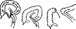
Emptying operations are used when the intestine cannot empty per vias naturales. They are most commonly applied in mechanical and paralytic obstruction of the intestine (see Ileus). They are also used to remove foreign bodies that have become stuck in the intestines (small or large) and do not cause obstruction symptoms. The primary operation in this category is puncture of the intestine. The loop of intestine to be punctured is removed from the abdominal cavity, isolated from the other intestines and the abdominal cavity with towels and drapes. The site of the impending puncture is sutured with a purse-string suture. After this, a trocar is inserted into the intestinal cavity. It is advantageous to use a trocar with a lateral outlet, as a long rubber tube can be预先 placed on it, through which the contents will flow into a vessel placed underneath. While the liquid is flowing out, the abdomen or eviscerated intestines are compressed evenly. After the intestines are emptied, the trocar is removed, the suture is immediately tightened, and the area of the puncture site is thoroughly wiped with physiological solution or rivanol solution. Over the suture, several nodular serosal sutures are placed for strength. After this, the intestinal loop is wiped again, and then it can be returned to the abdominal cavity.
The next emptying operation is opening of the intestinal lumen - enterotomy. The latter ends either with suturing of the intestine (enterorrhaphia) or with the creation of a fistula on it (enterostomia). The indication for enterotomy is foreign bodies in the intestine, even if they do not cause obstruction symptoms. These include: conglomerates of ascarids, gallstones, intestinal stones, dental prostheses, pieces of metal and glass objects, etc. If there are several objects in the intestines and they can be safely moved to one place, then enterotomy is performed at one location. Otherwise, several enterotomies have to be made. During the operation, contamination of the operative field should be avoided in every way; therefore, the intestinal loop to be subjected to enterotomy is isolated from the peritoneal cavity with towels or drapes, the site of enterotomy is separated from the rest of the intestine as much as possible by applying elastic clamps on both the afferent and efferent ends of the intestine. Usually a small incision in the intestine is required. Therefore, the intestinal lumen can be opened with either a transverse or longitudinal incision, but the intestine is sutured exclusively in the transverse direction to avoid possible narrowing of the lumen. It is better to suture the intestine with a two-layer suture (see Intestinal suture).
Enterostomy is more often used to empty the intestine in cases of paralytic obstruction. In peritonitis in general, it is more advantageous to perform enterostomy on the small intestine. Sometimes enterostomy, specifically jejunostomy, is used for feeding the patient as a bypass operation (see below). An emptying enterostomy removes part of the contents from the small intestine; therefore, in such cases, a temporary intestinal fistula is created. - The last operation in the group of emptying operations is also enterostomy, but one that empties all contents of the intestine. Such an operation is called an artificial anus (see Anus praeternaturalis).
The third group of operations on the intestines are bypass operations, i.e., those that unload the intestine. The primary one is jejunostomy in cases of stomach cancer. The purpose of the operation is to bypass the stomach and enable feeding the patient through the small intestine. Technically, the operation differs from the emptying enterostomy in that in a feeding enterostomy not a single drop of contents should leak out of the intestine. Therefore, the technique of the operation is somewhat more complex: the operation is performed according to the type of gastrostomy. - To bypass operations also belongs in part the temporary artificial anus, which precedes resection of the intestines (usually large and rectal). The focus of bypass, unloading operations
lies in the formation of internal intestinal fistulas, enteroanastomoses. The idea and first implementation, albeit unsuccessful, belong to the French surgeon Maisonneuve, who in 1854 reported on the first enteroanastomosis, which ended in the death of the patient. To this day, a simple lateral intestinal anastomosis bears the name of the Maisonneuve operation. Thus, the Maisonneuve anastomosis began the history of intestinal exclusion. At present, surgery has three types of intestinal exclusion: 1) incomplete unilateral - anastomosis of Maisonneuve, 2) complete unilateral - lateral anastomosis + excision of the segment of intestine distal to the anastomosis, 3) complete bilateral - lateral anastomosis + excision of both the segment distal to the anastomosis and the segment proximal to the anastomosis. The indications for their use are obstacles to the advancement of intestinal contents, for various reasons not subject to radical treatment (resection), intestinal fistulas of different origins, inoperable tumors of the intestines, even when they do not cause obstruction symptoms. - One can distinguish between economical and uneconomical types of intestinal exclusion. Under economical exclusion is understood such an exclusion in which the anastomosis is located close to the site being excluded; in uneconomical exclusion, the anastomosis is sometimes located far from the site to be excluded. Generally speaking, it is more advantageous
economical interruptions. Non-economical interruptions create a large interrupted segment of the intestine, which depending on the nature of the interruption either forms a ring-like continuous intestine (incomplete unilateral interruption; fig. 23) or a long blind pouch (complete unilateral interruption; fig. 24). Economical interruption does not have these drawbacks (fig. 26).-The technique of bypass operations on the intestine is first and foremost the technique of creating lateral inter-intestinal anastomoses. It consists of the following: two intestinal loops to be anastomosed are selected. Each of them is grasped parallel to the axis by curved elastic clamps, thanks to which the sections of intestine to be sutured are demarcated from the rest of the intestine. Before applying the clamps, the intestinal loops can be emptied by squeezing the contents between the fingers. When the clamps are applied, the clamped sections of intestine are placed next to each other. A gauze tampon is placed between them at the mesentery. The sections to be anastomosed are isolated from the rest of the abdominal cavity by towels. Then the adjacent sections of intestine are sutured with a continuous sero-serous suture. The length of the suture line is about 3 transverse fingers. When the sero-serous suture is applied, longitudinal incisions are made on both intestines, parallel to the suture line and penetrating through the serous and muscular layers. The incision is located approximately 0.5 cm from the first suture line. The incision may not quite reach the ends of the suture. One should avoid accidentally opening the mucosa at all costs. It is better to work at this moment with a sharp knife but carefully than with a blunt one and with force. Now comes the continuous suture-the second floor, which on each intestine captures the submucosa, muscular layer, and is brought out next to the incision. When the last stitch is finished, two surgical forceps grasp the submucosa first of one intestine, then of the other. Between the forceps, the intestinal lumen is opened, the incision is extended with a knife to the ends of the deep suture. After treatment (see Intestinal suture) of both lumens, the second half of the anastomosis is sutured. Complete unilateral interruption differs from the previous operation only in that the distal end of the intestine from the anastomosis is cut off. For this purpose, the intestine, 5-8 cm away from the anastomosis, is separated from the mesentery over a certain area. A tampon is passed through the hole in the mesentery; after this, a ligature is tightened on the intestine in two places, and the intestine is cut between the ligatures. Each of the stumps is invaginated into the lumen, an purse-string suture is placed and tightened over the invaginated portion. Over the purse-string suture, several nodular sutures or one continuous sero-serous suture can be applied for strength. A rather bulky stump is obtained. To make it thinner, one can proceed in different ways: either make a circular incision on the intestine down to the mucosa, or crush the intestine at the site of ligature application with an enterotome, or finally cut the intestine and then close the intestinal lumens with two layers of continuous suture. The Klapp method (rotation of the intestine around its longitudinal axis) cannot be recommended.-The most advantageous method should be considered the cutting of the intestine with subsequent closure of its lumens: this method gives the most delicate stump and allows for the smallest blind pouch. However, this method is more painstaking. Stumps formed by tightening ligatures should be cut inward into the intestinal lumen. Before cutting, the stump is in some closed space. To be able to invaginate the stump,
Figure 27.
it is necessary to have a section of intestine into which to invaginate. Therefore, on the distal end of the intestine from the anastomosis, a blind pouch is created. In the past, attention was not paid to the length of the blind pouch formed in the proximal segment of the intestine (figure 27 a). The blind pouch was sometimes large, sometimes small. However, it was found that blind pouches, especially at the end leading to the anastomosis, have a tendency to fill and stretch. As they stretch, they bend the anastomosis itself, making it less passable. At the same time, as they stretch, they thin out, become inflamed, and even perforate. Therefore, a rule was introduced (Oppel) to eliminate blind pouches by suturing them if they exist, and even better to create a stump of the cut intestine in such a way that there is no blind pouch. The best conditions for this exist when the intestine is cut close to the anastomosis and the intestinal lumen is closed with two rows of sutures (fig. 27 b). The same can be achieved if the proximal end of the cut intestine is sewn into the wall of the intestine with which the anastomosis is performed (fig. 28).-Complete bilateral interruption differs from complete unilateral interruption only in that in the former, not only the distal segment from the anastomosis is cut off, but also the proximal segment leading to the anastomosis (fig. 25). Initially, complete bilateral interruption was performed by immersing the interrupted segment of intestine into the abdominal cavity. It was found that secretion in the interrupted segment does not cease. Therefore, at present, with complete bilateral interruption, the interrupted segment is necessarily provided with an emptying mucous fistula. Operations of excision of a segment of intestine, resections (resectio intestini). The first question that must be raised regarding resections is the question of permissible sizes of resections.-Laue showed that complete excision of the large intestine (colectomia totalis) is permissible. The question of permissible sizes of resections of the small intestine is somewhat more complex. It was considered permissible to remove one third, or even one half of the small intestine. Nikitin, in the case of gangrene, removed 594 cm of mesenteric small intestine, leaving only 50 cm. Three months after the operation, the patient was found to be completely healthy and able to work ("in jobs of medium severity and in clerical work"). This case shows that in extreme cases, almost the entire small intestine can be excised, however, one should still not remove more than half of the small intestine.-Indications for resections of the intestine are primarily various types of intestinal tumors; next-constrictions, volvuli, incarcerations, gangrene from embolism of arteries or thrombosis of veins, distensions, fistulas, and atonies. As diverse as the indications are, so diverse is the technique of resections. From a technical standpoint, one must distinguish between resections without opening the intestinal lumen (these resections concern sections of the large intestine) and resections with opening of the intestinal lumen. The latter are divided into one-stage and two-stage or one-session and two-session, one-time or double. Usually they are distinguished as one-moment and two-moment. The latter definition is not entirely accurate. Finally, there are three-session intestinal resections: in the first session, an intestinal emptying fistula is applied, in the second session the intestine is resected, in the third session the fistula or anus is eliminated. Resections not accompanied by opening of the intestinal lumen include the operation of S. P. Fedorov on the transverse colon and the operation of Grekov (evaginatio) on the sigmoid colon. Fedorov, excising stomach cancer, separated the transverse colon from the mesentery (1903). Not wishing to complicate the operation, he invaginated the segment of transverse colon separated from the mesentery in the anal direction. The patient recovered. Emelyanov did the same in 1927. Grekov's method, proposed by him in 1911, initially concerned volvuli of the sigmoid colon, but was later expanded. It consists in the sigmoid colon being separated from the mesentery and brought out per anum. One-stage intestinal resection with opening of the lumen consists of three moments: 1) separation of the intestine from the mesentery, 2) cutting of the intestine, and 3) suturing of the intestine, i.e., restoration of its lumen. In resection of the small intestine, it is customary to separate the mesentery near the mesenteric border of the intestine. In resection of the right half of the large intestine, it is advantageous to immediately ligate the trunks of the a. and v. ileo-colicae, i.e., to ligate them at some distance from the intestine. In resection of the sigmoid, one should not go too deep into the mesentery; the same applies to the transverse colon. The small intestine is separated from the mesentery over an area that is to be resected according to the condition of the disease. The situation is different with the right half of the large intestine. Since the removal of one or two dozen cm of the large intestine does not play a role in the economy of the body, it is advantageous to extend the resection to the ascending colon and the hepatic flexure of the transverse colon if it is necessary to resect only the cecum, since this makes it easier to apply the suture and there is less chance of contamination of the operative field (there are many microbes in the cecum, the content is semi-liquid). Moreover, if it is necessary to resect only the hepatic flexure or the right half of the transverse colon, the entire proximal part of the large intestine should be included in the resection for the same reasons. With localization of the pathological process in the splenic flexure, the resection downward should be extended to the sigmoid colon, with which it is easier to anastomose the transverse colon.-One-stage intestinal resection has several variations, the latter being due to the restoration of the intestinal lumens. 1. The lumens of the intestines can be sutured end-to-end.
Figure 28.
2. The lumens of the intestines can be sutured side-to-side. 3. The proximal end of one intestine can be sutured to the side of the other intestine (end-to-side anastomosis). 4. The distal end of one intestine can be sutured to the side of the other intestine (side-to-end anastomosis). 5. The proximal end of one intestine can be sutured to the distal end of the other intestine (end-to-end anastomosis). 6. The proximal end of one intestine can be sutured to the proximal end of the other intestine (end-to-end anastomosis). 7. The distal end of one intestine can be sutured to the distal end of the other intestine (end-to-end anastomosis). 8. The proximal end of one intestine can be sutured to the side of the other intestine (end-to-side anastomosis). 9. The distal end of one intestine can be sutured to the side of the other intestine (side-to-end anastomosis). 10. The proximal end of one intestine can be sutured to the distal end of the other intestine (end-to-end anastomosis). 11. The proximal end of one intestine can be sutured to the proximal end of the other intestine (end-to-end anastomosis). 12. The distal end of one intestine can be sutured to the distal end of the other intestine (end-to-end anastomosis). 13. The proximal end of one intestine can be sutured to the side of the other intestine (end-to-side anastomosis). 14. The distal end of one intestine can be sutured to the side of the other intestine (side-to-end anastomosis). 15. The proximal end of one intestine can be sutured to the distal end of the other intestine (end-to-end anastomosis). 16. The proximal end of one intestine can be sutured to the proximal end of the other intestine (end-to-end anastomosis). 17. The distal end of one intestine can be sutured to the distal end of the other intestine (end-to-end anastomosis). 18. The proximal end of one intestine can be sutured to the side of the other intestine (end-to-side anastomosis). 19. The distal end of one intestine can be sutured to the side of the other intestine (side-to-end anastomosis). 20. The proximal end of one intestine can be sutured to the distal end of the other intestine (end-to-end anastomosis). 21. The proximal end of one intestine can be sutured to the proximal end of the other intestine (end-to-end anastomosis). 22. The distal end of one intestine can be sutured to the distal end of the other intestine (end-to-end anastomosis). 23. The proximal end of one intestine can be sutured to the side of the other intestine (end-to-side anastomosis). 24. The distal end of one intestine can be sutured to the side of the other intestine (side-to-end anastomosis). 25. The proximal end of one intestine can be sutured to the distal end of the other intestine (end-to-end anastomosis). 26. The proximal end of one intestine can be sutured to the proximal end of the other intestine (end-to-end anastomosis). 27. The distal end of one intestine can be sutured to the distal end of the other intestine (end-to-end anastomosis). 28. The proximal end of one intestine can be sutured to the side of the other intestine (end-to-side anastomosis). 29. The distal end of one intestine can be sutured to the side of the other intestine (side-to-end anastomosis). 30. The proximal end of one intestine can be sutured to the distal end of the other intestine (end-to-end anastomosis). 31. The proximal end of one intestine can be sutured to the proximal end of the other intestine (end-to-end anastomosis). 32. The distal end of one intestine can be sutured to the distal end of the other intestine (end-to-end anastomosis). 33. The proximal end of one intestine can be sutured to the side of the other intestine (end-to-side anastomosis). 34. The distal end of one intestine can be sutured to the side of the other intestine (side-to-end anastomosis). 35. The proximal end of one intestine can be sutured to the distal end of the other intestine (end-to-end anastomosis). 36. The proximal end of one intestine can be sutured to the proximal end of the other intestine (end-to-end anastomosis). 37. The distal end of one intestine can be sutured to the distal end of the other intestine (end-to-end anastomosis). 38. The proximal end of one intestine can be sutured to the side of the other intestine (end-to-side anastomosis). 39. The distal end of one intestine can be sutured to the side of the other intestine (side-to-end anastomosis). 40. The proximal end of one intestine can be sutured to the distal end of the other intestine (end-to-end anastomosis). 41. The proximal end of one intestine can be sutured to the proximal end of the other intestine (end-to-end anastomosis). 42. The distal end of one intestine can be sutured to the distal end of the other intestine (end-to-end anastomosis). 43. The proximal end of one intestine can be sutured to the side of the other intestine (end-to-side anastomosis). 44. The distal end of one intestine can be sutured to the side of the other intestine (side-to-end anastomosis). 45. The proximal end of one intestine can be sutured to the distal end of the other intestine (end-to-end anastomosis). 46. The proximal end of one intestine can be sutured to the proximal end of the other intestine (end-to-end anastomosis). 47. The distal end of one intestine can be sutured to the distal end of the other intestine (end-to-end anastomosis). 48. The proximal end of one intestine can be sutured to the side of the other intestine (end-to-side anastomosis). 49. The distal end of one intestine can be sutured to the side of the other intestine (side-to-end anastomosis). 50. The proximal end of one intestine can be sutured to the distal end of the other intestine (end-to-end anastomosis). 51. The proximal end of one intestine can be sutured to the proximal end of the other intestine (end-to-end anastomosis). 52. The distal end of one intestine can be sutured to the distal end of the other intestine (end-to-end anastomosis). 53. The proximal end of one intestine can be sutured to the side of the other intestine (end-to-side anastomosis). 54. The distal end of one intestine can be sutured to the side of the other intestine (side-to-end anastomosis). 55. The proximal end of one intestine can be sutured to the distal end of the other intestine (end-to-end anastomosis). 56. The proximal end of one intestine can be sutured to the proximal end of the other intestine (end-to-end anastomosis). 57. The distal end of one intestine can be sutured to the distal end of the other intestine (end-to-end anastomosis). 58. The proximal end of one intestine can be sutured to the side of the other intestine (end-to-side anastomosis). 59. The distal end of one intestine can be sutured to the side of the other intestine (side-to-end anastomosis). 60. The proximal end of one intestine can be sutured to the distal end of the other intestine (end-to-end anastomosis). 61. The proximal end of one intestine can be sutured to the proximal end of the other intestine (end-to-end anastomosis). 62. The distal end of one intestine can be sutured to the distal end of the other intestine (end-to-end anastomosis). 63. The proximal end of one intestine can be sutured to the side of the other intestine (end-to-side anastomosis). 64. The distal end of one intestine can be sutured to the side of the other intestine (side-to-end anastomosis). 65. The proximal end of one intestine can be sutured to the distal end of the other intestine (end-to-end anastomosis). 66. The proximal end of one intestine can be sutured to the proximal end of the other intestine (end-to-end anastomosis). 67. The distal end of one intestine can be sutured to the distal end of the other intestine (end-to-end anastomosis). 68. The proximal end of one intestine can be sutured to the side of the other intestine (end-to-side anastomosis). 69. The distal end of one intestine can be sutured to the side of the other intestine (side-to-end anastomosis). 70. The proximal end of one intestine can be sutured to the distal end of the other intestine (end-to-end anastomosis). 71. The proximal end of one intestine can be sutured to the proximal end of the other intestine (end-to-end anastomosis). 72. The distal end of one intestine can be sutured to the distal end of the other intestine (end-to-end anastomosis). 73. The proximal end of one intestine can be sutured to the side of the other intestine (end-to-side anastomosis). 74. The distal end of one intestine can be sutured to the side of the other intestine (side-to-end anastomosis). 75. The proximal end of one intestine can be sutured to the distal end of the other intestine (end-to-end anastomosis). 76. The proximal end of one intestine can be sutured to the proximal end of the other intestine (end-to-end anastomosis). 77. The distal end of one intestine can be sutured to the distal end of the other intestine (end-to-end anastomosis). 78. The proximal end of one intestine can be sutured to the side of the other intestine (end-to-side anastomosis). 79. The distal end of one intestine can be sutured to the side of the other intestine (side-to-end anastomosis). 80. The proximal end of one intestine can be sutured to the distal end of the other intestine (end-to-end anastomosis). 81. The proximal end of one intestine can be sutured to the proximal end of the other intestine (end-to-end anastomosis). 82. The distal end of one intestine can be sutured to the distal end of the other intestine (end-to-end anastomosis). 83. The proximal end of one intestine can be sutured to the side of the other intestine (end-to-side anastomosis). 84. The distal end of one intestine can be sutured to the side of the other intestine (side-to-end anastomosis). 85. The proximal end of one intestine can be sutured to the distal end of the other intestine (end-to-end anastomosis). 86. The proximal end of one intestine can be sutured to the proximal end of the other intestine (end-to-end anastomosis). 87. The distal end of one intestine can be sutured to the distal end of the other intestine (end-to-end anastomosis). 88. The proximal end of one intestine can be sutured to the side of the other intestine (end-to-side anastomosis). 89. The distal end of one intestine can be sutured to the side of the other intestine (side-to-end anastomosis). 90. The proximal end of one intestine can be sutured to the distal end of the other intestine (end-to-end anastomosis). 91. The proximal end of one intestine can be sutured to the proximal end of the other intestine (end-to-end anastomosis). 92. The distal end of one intestine can be sutured to the distal end of the other intestine (end-to-end anastomosis). 93. The proximal end of one intestine can be sutured to the side of the other intestine (end-to-side anastomosis). 94. The distal end of one intestine can be sutured to the side of the other intestine (side-to-end anastomosis). 95. The proximal end of one intestine can be sutured to the distal end of the other intestine (end-to-end anastomosis). 96. The proximal end of one intestine can be sutured to the proximal end of the other intestine (end-to-end anastomosis). 97. The distal end of one intestine can be sutured to the distal end of the other intestine (end-to-end anastomosis). 98. The proximal end of one intestine can be sutured to the side of the other intestine (end-to-side anastomosis). 99. The distal end of one intestine can be sutured to the side of the other intestine (side-to-end anastomosis). 100. The proximal end of one intestine can be sutured to the distal end of the other intestine (end-to-end anastomosis).


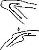
This method is particularly recommended by us by S. P. Fedorov. In this case, a two-layer suture of Cherny is usually applied. Special difficulties are presented by the suture on the mesenteric border of the intestines. It is precisely here that it reveals the greatest dangers of its insufficiency. To avoid the latter, some surgeons use an invaginating suture: the leading end of the intestine is invaginated for a certain distance into the diverting end. The closure of the lumens occurs well. 2. Clinic of Spasokukotsky prefers the insertion of the opening of the leading segment of the intestine into the side of the diverting intestine. In this case, the end of the diverting intestine a
is tightly sutured. 3. The
simplest suture consists in the formation of a lateral anastomosis between the ends of the resected intestine. This suture is the most reliable and can be performed without special technical skill. In this case, it is rather indifferent
but, whether to arrange the connected bends of the intestine isoperistaltically (Fig. 29 b) or antiperistaltically (Fig. 29 a), since with both arrangements isoperistalsis is obtained. The disadvantage of this method is that in addition the ends of the severed intestine must be sutured, which creates the danger of forming blind pouches. Blind pouches are eliminated in exactly the same way as in exclusions. All the above-mentioned methods immediately restore the lumen of the intestines. To complete such a resection, the openings between the mesenteries must be eliminated. Intestinal loops can become incarcerated in them after operations. The openings between the ends and edges of the mesenteries are sutured. When the intestines are sutured end to end, difficulties can and do arise, since the afferent segment may turn out to be narrower or wider than the efferent one. It is more often narrower than the efferent one when the small intestine is sutured to the large one, e.g., the ileum to the transverse; the afferent segment may turn out to be wider than the efferent one when the lumen, e.g., of an dilated small intestine is sutured to the lumen of a collapsed small intestine below the site of obstruction. To equalize the lumens, one has to suture the efferent or afferent end, or to obliquely cut the afferent and efferent ends. In a word, one has to find ways to bring the width of the lumens to the same diameter.-One-session resection of the intestines is a relatively difficult and dangerous operation. Therefore, Mikulicz long ago proposed a 'two-moment', in essence two-session, resection of the large intestines. The latter is applicable mainly to the large intestines, in extreme cases to the lower part of the small intestines. Mikulicz's method is as follows: 'The loop of intestine subject to resection together with the tumor and other diseased parts (glands) is freed from connections with the mesentery etc. and is placed in front of the abdominal wound; only after the abdominal cavity is completely closed and the skin is sutured up to the protruding intestine, the latter is cut off. The protruding intestine can be separated either immediately, as is customary at present, or after 12-24 hours. The thus formed anus praeternaturalis is closed in such a way that first the spur is crushed with a crusher, then the fistula is closed' (Mikulicz). From what has been said, it is evident that the resection itself in recent times has been performed by Mikulicz in one session. But still the entire operation turned out to be two-session.-The meaning of the two-session operation lies in the attempt to prevent infection of the peritoneum as much as possible. However, an anus can be applied only in resection of the large intestine, in extreme cases of the lower loops of the ileum, because with an anus on the jejunum the patient will become exhausted.-In recent times Grekov introduces into the principle two-session resection with exclusion. This operation has been named 'Grekov II'. Its technique is as follows: 'Median guiding incision; after finding the tumor, a wide incision of the posterior parietal peritoneum outward from the corresponding intestine with division of the lig. phrenico-coli-cum'. Resection of the descending intestine according to Grekov is performed as follows: 'The loop is bluntly dissected together with the vessels and glands nourishing it up to the transverse colon upward and to the S-Romanum downward. An extensive free loop is obtained, which can be easily brought out, especially through a new incision on the left. The entire diseased mesentery is usually resected, and the loop is brought out with the calculation that both legs of the loop remaining in connection with the mesentery will be on the outside, of sufficient length so that after removal of the loop both its stumps can be invaginated and sutured at a distance of several cm from the edges of the lateral wound. After fitting between the two bends of the loop, a wide anastomosis is applied (usually transversosigmoideostomia), entirely or for the most part remaining inside the abdominal cavity. After 2-3-4 days, without anesthesia, under morphine and if needed by spraying the mesentery with novocaine, the second stage is performed, i.e., resection of the protruded loop with a blind three-layer suture of its stumps. The intestinal stumps are abundantly moistened with sterile vaseline oil and covered with a dry dressing' (Grekov).-Thus Mikulicz's resection in respect to removal of the intestine is one-session, while Grekov's operation is two-session. This is its weak side, since the intestine separated from the mesentery must necrose. In Grekov's operation it is necessary to prevent the outflow of contents during the first days, which can infect the wound, which can be prevented by simple ligation of the intestinal stumps protruding from the wound. If Grekov's operation is performed as the author himself proposes, it is three-session: 1) dissection of the intestine and its protrusion with unilateral exclusion, 2) cutting off the intestine, 3) closure of the intestinal fistulas. To three-session resections also belongs the operation of Schloffer. The latter however begins with the application of an anus praeternaturalis on the afferent segment of the intestinech In the second session, resection of the intestine is performed, in the third-closure of the anus. And this operation under suitable conditions (phenomena of obstruction, fairly strongly expressed) is acceptable.-Of the above-mentioned methods of intestinal resection, many surgeons prefer one-session resection and obtain satisfactory results. The two-session method of Mikulicz's resection requires closure of the anus. The two-session method with formation of fistulas, exclusion of the intestines with formation of mucous fistulas often requires elimination of the latter. A doubly excluded intestine can be resected under certain conditions (e.g. in cancer of the intestine). Long ago surgeons have sought to simplify the matter by forming artificial constrictions of the intestinal lumen. In 1897 Mosetig-Moorhof began to apply the formation of intestinal valves: around the intestine through the mesentery a thick silk ligature is passed, which is tightened. Over the ligature a nodal or continuous sero-serous suture is applied. The same operation independently of Mosetig-Moorhof was performed by Chernyakhovsky. It turned out that the thread tightened on the intestine is cut through and the intestinal lumen is restored. The most advantageous method of forming a valve, if it is needed for a long time, is Bogolyubov's method, which Razumovsky called biological. The intestine is tied with a wide strip of aponeurosis. 'A wide strip narrows the intestine over a more extensive area and forms a tubular stricture of the intestine with a narrow long channel, difficult to pass for intestinal contents' (Bogolyubov).-To operations narrowing the lumen of the intestine can also be attributed the operation of Rozanov-correction of the Bauhinian valve. It is performed as follows: after clarifying the question of insufficiency of the valve, 'the ileo-caecal fold is incised, and thus the angle of entry is corrected; the ileum at the site of its entry into the large intestine, above, where the superior ileo-caecal recess is located, is fixed with one, two, three catgut sutures to the ascending intestine, in order to thus secure the corrected angle of entry. By this technical maneuver we lower the upper lip of the valve downward and in addition create an artificial semilunar fold'.-On the intestine, operations expanding its lumen are rarely applied. Even if there is a scar stricture in the intestine, the latter is either excised or excluded. In recent times expanding operations find application on the Bauhinian valve. Grekov, and then Rozanov, apply them in bauginospasm (see.). v. Onpel. VIII. Neuroses of the Intestine - diseases of the Intestine of a purely functional nature, having no organic substrate in the form of pathological, morphologically determinable changes of it. This definition does not fully correspond to modern concepts both of neuroses in general and of pathological functional disorders of the Intestine and requires certain limitations. Thus e.g. intestinal dyspepsias (see.) can for a long time give functional disorders without being at the same time neuroses of the intestine; this also applies to functional disorders of endocrine nature. Furthermore, a number of functional diseases of the intestine are known as a result not of anatomical changes but of pathophysiological reactions to irritation of the type of allergy or anaphylaxis; these too cannot be considered pure neuroses of the intestine. Finally even functional disorders of nervous origin, e.g. disorders of defecation in organic diseases of the spinal cord, must be considered as nervous diseases of the intestine, but not as its neuroses. On the other hand sometimes organic processes, e.g. an anatomically barely visible ulcer of the duodenum, adhesions of the intestine, a fissure of the anus etc., give a series of functional disorders superimposed to a large extent on symptoms of organic origin, and the picture of the disease takes on the character of a 'pure' neurosis of the intestine. The latest methods of investigation of the intestine, functional diagnosis of its diseases, rectomanoscopy and especially roentgenodiagnosis (the relief method, Bergmann's technique etc.) often reveal an organic basis for diseases, for a long time considered pure neuroses of the intestine. That is why the doctrine of neuroses, which formerly occupied such an important place in the pathology of the intestine, has considerably narrowed in recent times, and in the latest major manuals (Bergmann u. Staehelin, Kraus and.'
Brugsch) find no separate description of intestinal neuroses as a special nosological unit at all. The rich autonomous nervous system of the intestinal wall, the close connection with the higher vegetative centers and nerve trunks, and finally with the central nervous system—both the spinal cord and the brain—explain why on the one hand organic and functional diseases of the Intestine sharply affect the psychoneurotic system of a person, and on the other hand why not only organic but also fine pathophysiological processes arising in the central nervous system, processes of a purely psychic nature, find expression in disorders of the function of the Intestine. That psychic affections have a great influence on the function of the intestines has been known for a long time, and already Trousseau described clinical observations on emotional diarrhea; later a whole series of clinicians described numerous motor, secretory, and sensory nervous disorders of the Intestine that were causally connected with psychic reactions and were cured by psychotherapy. Experimentally, the influence of psychic affections on the functions of the Intestine was established much later by roentgenoscopy (Cannon) and by the method of the celluloid window in the abdominal cavity (Katsch). Most often based on disturbances in the dynamics of psychic processes, intestinal neuroses are a manifestation of the pathological psychic reaction of the organism to external irritation. Hence the obvious closest mutual connection between functional disorders of the central nervous system and the activity of the Intestine. The reason why the Intestine so often reflects psychic affections and conflicts, some authors (v. Bergmann and his school) tend to see in the fact that the unconditional reflex process of defecation is inhibited in civilized man by a mass of conditioned reflexes developed by upbringing and conditioning his behavior in the final analysis (see Defecation); the disinhibition of these conditioned reflexes by psychic affections is one of the causes of intestinal neuroses; the formation of new conditioned reflexes under the influence of deep psychic affections lies at the basis of another series of neuroses. The prerequisite for the development of neuroses is either constitutional peculiarities, defects of the psycho-physical personality (hereditary neuropathy) or a restructuring of the personality as a result of the most diverse psychic traumas experienced by this patient. Etiologically, in the development of these psychic conflicts, there is often fear, e.g., before the disease due to a misinterpretation of a somatic symptom (fear of developing a duodenal ulcer, cancer of the rectum, etc.); a poorly understood interpretation of the disease given by the physician ('prolapse of the internal organs', 'catarrh of the intestines', appendicitis, colitis). In these cases, one can speak of the so-called iatrogenic disease. Often, sexual conflicts also lie at the basis of intestinal neuroses: sexual dissatisfaction as well as physical and especially psychic, and all kinds of abnormalities of sexual life (coitus interruptus, condomatus), fear of venereal disease, pregnancy, physical aversion in married life, shame, etc. Small organic lesions, e.g., bleeding erosions of the rectum, slight strangulation of hemorrhoidal nodes, or functional disorders, e.g., meteorism due to intestinal dyspepsia in neurotically predisposed persons, are also the cause of the development of severe and persistent forms of intestinal neuroses. Often the cause of intestinal neuroses is the living conditions. Thus, disorders of defecation may appear due to external conditions unfavorable for proper intestinal functions (for example, cold, distant toilets, the need to stand in long queues with an insufficient number of toilets in overcrowded apartments, etc.). In a number of cases, the appearance of intestinal neuroses is observed on the basis of an already existing viscero-visceral reflex on the Intestine, e.g., in diseases of the sexual sphere in women, the urogenital apparatus in men (gonorrhea and its complications), or other organs of the abdominal cavity (diseases of the bile ducts), and finally in organic diseases of the nervous system (radiculitis, tabes dorsalis, etc.). The basis of these neuroses is frequent abnormal sensations, mainly pains in the abdomen, which are painfully reflected in the psyche of the patient and lead to a restructuring of his psychic personality, fixing the patient's attention on the physiological processes of digestion. This constitutes what the old doctors called hypochondria intestinalis. The etiology and pathogenesis of intestinal neuroses are very diverse, and if they most often occur in constitutionally burdened neurotics, in hysteria, neurasthenia, and in asthenics, nevertheless intestinal neurosis is also often observed in people who are otherwise balanced, under the influence of a psychic trauma or a deep conflict, often not realized by the patient himself or long forgotten by him. Attention is also drawn to the appearance of intestinal neuroses as a result of sharp changes in external living conditions, e.g., in peasants and workers when they give up their customary physical labor for intensive mental work. Corresponding to such a complex pathogenesis of intestinal neuroses and their clinical pictures are extremely diverse and very changeable. Based on pathophysiological prerequisites, intestinal neuroses are conventionally divided into motor, sensory, and secretory, guided by the most important functional disorder of the Intestine that occurs in a given neurosis. Motor neuroses in turn are divided into forms expressing a weakening of the motor function of the Intestine (atony, paralysis of the intestines, habitual nervous constipation) and forms appearing as a result of strengthening of the motor function of the intestines (hypertonia); here belong the numerous spastic phenomena of the Intestine with strengthening of its peristalsis, as a result of which in some cases spastic constipation, in others—diarrhea as a result of hyperperistalsis of the intestine may appear. To motor neuroses is also attributed the so-called peristaltic restlessness of the intestines (tormina nervosa intestinorum). The disease is expressed by an unpleasant sensation by the patient of the peristalsis of the intestines, accompanied by loud sound phenomena in the abdominal cavity, gurglings in the Intestine. These sound phenomena suddenly appear and suddenly disappear, attract the attention of those around the patient and make his stay in society burdensome. It is a matter of sudden attacks of spastic contraction of the Intestine, which can be seen if one observes the patient during the very attack, who has flabby and thin abdominal coverings. According to Boas, the picture of the abdomen in such patients resembles a bag filled with potatoes. If the peristaltic restlessness of the Intestine is not the result of an organic process (e.g., adhesions), the prognosis in this neurosis is entirely favorable. To sensory neuroses are attributed a) hyperesthesia of the Intestine, expressed by indefinite pains, sensations of burning, stabbing pains appearing in the abdomen without visible causes or under the influence of psychic irritations; b) intestinal enteralgia, expressed by attacks of severe spastic pains occurring periodically without any visible cause, and c) neuralgias in the region of the anus—attacks of severe pains during defecation and without it without any visible cause. To secretory neuroses belongs the excretion in the Intestine of significant quantities of liquid, 86» causing violent, completely unmotivated diarrhea, rapidly appearing and rapidly disappearing, which however on detailed examination can be connected with a psychoneurotic trauma; here some authors also attribute myxoneurosis intestinalis membranacea, seu enteritis membranacea, a disease most often known under the name colitis mucosa. This is a peculiar disease, having undoubtedly great practical importance, which belongs rather to the vegetative disorders of the Intestine; however, it is often confused with an intestinal neurosis based on a restructuring of the entire personality of the patient as a result of severe psychoneurotic conflicts. The classification presented above has in mind the presence of one dominant symptom of intestinal neurosis. Meanwhile, the monosymptomaticity of neuroses of the stomach and intestines, if we are dealing with actual psychoneurotic diseases of them, and not with reflex nervous disorders, is rightly questioned, why a strict classification based on the disorder of individual functions of the Intestine should be considered artificial, since in practice we are dealing with complex clinical pictures with a whole series of functional disorders, both motor, sensory, and secretory at the same time. In a whole series of cases, it is not a matter of any one complaint of the patient, but of a whole bundle of very acute and indefinite subjective sensations, changing in their combination every day and united by a very pessimistic mood of the patient. The attention of the patient is entirely occupied by the study of the state of his digestion, and the completely normal activity of the Intestine becomes the subject of numerous complaints of the patient. These are the patients who carefully and daily follow their stool, its quantity and color, the content of undigested food in it, and they suffer equally from an increase as well as from a decrease in the number and quantity of evacuations and conscientiously keep journals of their observations for years. Here less than in other cases can one speak of the monosymptomatic type of intestinal neurosis.
This hypochondria intestinalis of older authors represents one of the most severe forms of intestinal neuroses, and indeed the iatrogenic origin of the patient's complaints and subjective sensations often has a basis in the overly detailed examination of the patient by numerous doctors, to whom he constantly applies. Not the least role here is played by research documents (stool analyses, X-ray protocols, proctoscopic examinations), arbitrarily interpreted by patients unfamiliar with terminology, most often people of mental labor, often standing high in their intellectual development. The diagnosis of intestinal neurosis is by no means an easy task, even when it comes to people with a clearly expressed neuropathic constitution or in the presence of a definite psychological trauma in the anamnesis. First of all, it is necessary to exclude the presence of organic disease or alimentary intestinal dyspepsia by the most thorough examination of the intestine using methods of functional diagnosis. It is necessary to keep in mind diseases of other organs of the abdominal cavity and especially of the sexual sphere as the most frequent source of viscero-visceral reflexes on the intestine, as a result of which spasms of the intestines, constipation, diarrhea and constant pains appear. Finally, it is not always possible to establish endocrine causes of intestinal disorders (e.g., thyrotoxicosis, hypofunction of the adrenal glands) as the cause, for example, of diarrhea and (which is even less taken into account) allergic diseases of the intestine, significantly more frequent than is generally thought, and causing unmotivated diarrhea and pains of 'nervous' origin. The differential diagnosis between organic reflex nervous diseases of the intestine and its frequent neuroses presents in each individual case features that cannot be accounted for in general. Typical are the lability of the clinical picture, the absence of connection between the onset of the patient's attacks and physical exertions or dietary errors, and conversely, the dependence of the attack on the state of the psychoneural sphere of the patient, the sudden onset and sudden end of the attacks, and finally the general psychoneural physiognomy of the patient, which imposes a certain color on the entire clinical picture of the disease. A careful study of the anamnesis, living and working conditions of the patient, detailed acquaintance with the inner life, experiences of the patient, especially in his sexual sphere, point to the right path in the diagnosis of intestinal neurosis and determine the therapy. Prevention of intestinal neuroses represents a great and grateful task for the physician: regulation of labor, especially mental, proper alternation with physical culture and sports, struggle with sexual perversions and excesses, prohibition of excessive smoking, systematic physical exercise, especially during the period of sexual maturity, organization of reasonable rest, especially collective summer excursions - such in general are the foundations of prevention of these frequent diseases. Very useful are daily physical labor activities for persons detached from their usual physical work and engaged, for example, in study or household and public activities with excessive mental strain; on the other hand, attention should be paid to the careless prescription of diet by doctors and to psychological traumas to persons predisposed to intestinal neuroses from the side of medical and paramedical personnel (negative psychotherapy).-Treatment of intestinal neuroses is in close connection with their pathogenesis in each individual case, therefore more than in other diseases of the intestine, here a deep individualization of each case and a deep study of the patient's personality is required, and less success can be expected from general prescriptions and orders. For example: if one patient can be allowed any kind of food and categorically reject a 'strict' diet, to which he has most often put himself, then in another such a decision to consume any food will undermine the authority of the doctor or lead to the idea of the incurability of the intestinal disease. There are no general rules here and by their very nature cannot be. The basis of treatment should be a broadly understood psychotherapeutic effect on the patient with an attempt to study the immediate or distant causes of the psychological conflict causing the intestinal neurosis. If this succeeds, then often years of suffering are quickly cured. The methods of psychotherapeutic effect on the patient are very different, ranging from explanation and direct medical suggestion to psychoanalysis and hypnosis, requiring the intervention of a specialist. -In addition to psychotherapeutic treatment and regulation of the patient's lifestyle (especially in the sexual sphere), some benefit is provided by a change of surroundings, travels, even short ones; much less benefit can be expected from treatment at specialized resorts, e.g., at Caucasian mineral waters, rather contraindicated in these cases; physical methods of therapy, especially warm general procedures (aromatic, carbonic baths, sometimes general electrotherapy), can be used, but local procedures on the abdomen and in the rectum are rather contraindicated. From medicinal substances, one should avoid medications used for organic diseases of the digestive tract (hydrochloric acid, pepsin, alkalis, bismuth, salol, etc.), as they reinforce in the patient the idea of intestinal disease, and one should try to apply atropine and its derivatives, sometimes cocaine (3 times a day 10 drops of 3% solution before meals for diarrhea), caffeine for constipation, quinine, strychnine, etc. Sometimes great benefit is also provided by appropriate organotherapeutic preparations (ovarin, spermin, antithyreoidin, etc.). r. luria. IX. Tuberculosis of the Intestine. Pathological Anatomy. Tbc of the intestine can be observed in the form of primary tuberculous lesion and in the form of secondary tbc. The primary tbc complex, developing at the site of primary introduction of the tubercle bacillus into the body, due to the fact that the digestive tract comparatively rarely plays the role of the portal of entry for tbc, is observed in the intestine rarely. As described by Ghon, Pototschnis and Siegmund, it, just like the primary tbc complex in the lung, represents a combination of a limited tuberculous lesion at the site of infection, i.e., in this case in the mucous membrane of the intestine [analogous to Ghon's focus (see) in the lung], tuberculous lymphangitis and tuberculous lesion of the regional lymph gland (in the mesentery). Most often the primary focus is in the small (ileal) intestine, less often in the large. In the mucous membrane the change can be expressed in the form of a small ulcer, or a fresh one with a curdled bottom and nodules at the edges or already healing; in other cases it is limited to the formation of a curdled nodule in the submucosal layer, while the mucous membrane remains intact. Tuberculous lymphangitis in the fresh period is detected in the form of a series of nodules extending from the focus in the mucous membrane to the mesenteric lymph gland; later there may remain only induration along the course of the indicated lymphatic vessels. The change in the mesenteric lymph gland is usually very pronounced and manifests as a solid curdled lymphadenitis with subsequent liquefaction of the curdled mass. In view of the fact that the change in the intestinal mucous membrane can be easily overlooked, e.g., when healing a small tuberculous ulcer or with submucosal localization of a small tuberculous nodule, sometimes it seems that there is a lesion of the lymph gland without a lesion at the site of the portal of infection, i.e., in the wall of the intestine. It is self-evident that often in connection with the spread of the process, not one but several mesenteric lymph glands are affected. Secondary tbc of the intestine, i.e., the lesion of the intestine by a tuberculous process from an existing tuberculous focus in another organ, is observed very often; at the same time, such secondary tbc of the intestine most often occurs in patients with pulmonary tbc (about 85% of all patients dying from pulmonary tbc have tuberculous changes in the intestine) due to the introduction from the lumen of the intestine into the mucous membrane of tubercle bacilli that enter the intestine with swallowed sputum. The hematogenous route of infection of the intestine, assuming retrograde transport of tubercle bacilli from affected lymph glands of the mesentery into the wall of the intestine, cannot be considered proven. Pathologically-anatomically, secondary tbc of the intestine, complicating pulmonary tbc, most often affects the ileum; however, it should be noted that any part of both the small and large intestine can be the site of development of the tuberculous process. Initially, the appearance of nodules in the area of solitary follicles or, more often, follicles that are part of Peyer's patches is usually noticed; visually this is noticeable as the appearance of yellowish-gray specks in some of these follicles. Nodules can also arise outside the lymphatic apparatus and in this case appear under the mucous membrane. Subsequently, the nodules ulcerate and small lentil-like ulcers appear; in Peyer's patches usually several such ulcers are noticeable; later they merge, as a result of which larger ulcers with uneven, as if gnawed and undermined edges are formed. The bottom of the ulcers is marked, caseous; in it nodules can be noticed, just as in the edges of the ulcers [see separate plate].
(pp. 583-584), fig. 7]. The outline of ulcers is extremely irregular; however, ulcers that arise in Peyer's patches may later occupy the entire patch and repeat its shape. Usually, the tuberculous process from the site of the initial lesion of the intestinal wall begins to spread along the lymphatic pathways that ring the intestinal tube; this results in the spread of the tuberculous ulcerative process in the transverse direction along the length of the intestine and the formation of encircling, circular ulcers, which are very characteristic of tuberculosis. The involvement of lymphatic pathways is also manifested by the appearance of tubercles on the serous covering of the intestine and the nearest parts of the mesentery, and these tubercles are arranged in characteristic rows resembling beads or strings of pearls. In the large intestine, tuberculous ulcers can have an extremely irregular shape, but they also often show a tendency to encircling spread; in some cases of very long-standing tuberculous ulcers in the large intestine, hypertrophic thickening of the mucous membrane at the edges of the ulcers and in the intervals between them is observed, sometimes with the formation of multiple polyps (hypertrophic form of tuberculous ulcers). In the lower part of the large intestine, the tuberculous process can cause almost continuous ulceration with the preservation of only small islands of mucous membrane; such forms, which give a patho-anatomical picture very similar to that of chronic ulcerative dysentery, Aschoff calls 'tuberculous dysentery'. The course of the tuberculous process in the intestine can vary and in most cases coincides with the course of the process observed in the same subject in the lungs. Predominantly exudative forms of pulmonary tuberculosis are usually accompanied by the formation of tuberculous ulcers in the intestine that spread rapidly, often giving hemorrhages due to the destruction of blood vessels, sometimes leading to perforation of the intestinal wall. On the contrary, in productive and cirrhotic forms of the pulmonary process, intestinal tuberculosis shows an extremely slow course, with a noticeable alternation in the ulcers between the formation of new tubercles and scarring. Sometimes tuberculous ulcers of the intestine show a very great tendency to heal, forming scars that contract the tissue, causing narrowing of the intestinal lumen with all its consequences. The healing of ulcers is often accompanied by atypical proliferation of the epithelium of the regenerating mucous membrane, and sometimes in chronic healing tuberculous ulcers, cancers arise (cases have been described of cancer developing immediately in several tuberculous ulcers). Tuberculosis of the rectum - SEE Rectum.
A. Abricosov. Clinic. During life, the diagnosis of intestinal tuberculosis is very difficult, and the frequency of clinical diagnosis varies sharply depending on the thoroughness of clinical observation and the methodology of research. Isolated tumor-like foci, ileocecal infiltrative processes accessible to methodical palpation by Gausmann or Obraztsov, can be recognized by physical examination, taking into account the entire complex of symptoms. To some extent, strictures of the small intestine due to the scarring of circular ulcers can also be recognized. Of course, in those cases where there are signs of obstruction, the diagnosis is already quite late. Scattered intestinal ulcers may not cause any symptoms until death. Therefore, it is especially important to carefully monitor all aspects of the disruption of the normal function of the intestine and the general changes in the patient's condition that may suggest intestinal tuberculosis. This group of diagnostic signs should include: 1) Pain (when the process spreads to the peritoneum and affects the terminal branches of the intestinal nerves, in strictures and adhesions). The pain is either diffuse or localized in the ileocecal area. Pain points have been described along the edge of the rectus muscles at the level of the second lumbar vertebra and inward from McBurney's point, which authors explain as being due to lesions of the regional mesenteric glands (periadenitis) - an inconstant sign. 2) Diarrhea alternating with constipation; profuse diarrhea in consumptives with foul-smelling stools and meteorism very often accompany intestinal tuberculosis. On the other hand, they are not pathognomonic for tuberculous lesions of the intestine, as they are often caused by improper dietary regimen (overfeeding, excess fats), poor condition of the teeth, and general intoxication. Therefore, in the presence of these disorders, it is first necessary to regulate the patient's dietary regimen. Persistent complaints of such diarrhea in the anamnesis should be taken into account very carefully. 3) Laboratory data: a) Tubercle bacilli in the stool - only the fact of repeated detection of large quantities of tubercle bacilli is significant, especially on the surface of fecal masses and when the patient is strictly disciplined, guaranteeing against swallowing sputum; b) The presence of hidden blood in the stool, indicating capillary hemorrhages from ulcers (Weber's test or the more sensitive Gregersen's test, see). 4) X-ray examination (with a special cone of the Buchi system; films only, powerful apparatus) 6, 7, 8, 9, 24 hours after the administration of contrast medium. Characteristic findings are: increased peristalsis of the entire intestine or of individual segments, filling defects (ulcers!), segmentation or dilatation of individual segments. The symptom of non-filling of the ascending part of the cecus, described by Stierlin, is rejected by a number of authors. 5) Symptoms of general intoxication - paleness with a subicteric tint, anemia, weakness, poor general condition, poor appetite, tachycardia - should be considered as indicating intestinal tuberculosis in the absence of other localizations that could explain the complex of symptoms. 6) Of diagnostic value are partly the negative tuberculin reaction, high erythrocyte sedimentation rates, and the blood count. Prevention of enterogenous intestinal tuberculosis in consumptives is largely possible and consists first of all in teaching patients to spit out sputum without swallowing it; the rational organization of the patient's diet, excluding all excesses, and strict observation of the regularity of bowel movements are also of very serious importance. - Therapy. In the foreground is the general hygienic-dietary regimen (a sparing diet); great success has been achieved with ultraviolet rays; radiotherapy, according to many observations, does not give a satisfactory effect. Tuberculin therapy is contraindicated in most cases due to negative anergy. In isolated tumor-like lesions, ileocecal infiltrates, strictures - surgical intervention. Postoperative mortality is very high (20-25%), and in a number of cases - generalization of tuberculosis. The prognosis is very bad, as intestinal lesions in most cases are accompanied by generalized amyloidosis of the organs. If it is possible to localize and arrest the pulmonary process and with prolonged systematic treatment, significant and lasting improvement and even healing of tuberculous ulcers is possible.
B. Holzmann. X. Intestinal flora. The intestinal flora of a healthy person consists of a relatively small number of bacterial species capable of breaking down carbohydrates with the formation of acids. The intestinal tract of an infant begins to be colonized by bacteria in the first hours of life. According to the research of Tissier, during the first three days of a child's life, there is a gradual colonization of the intestinal tract, initially by different bacteria to a large extent of a random nature. By the end of the 3rd day, in the case where the child is breastfed, a permanent microflora is established, in which Bacillus bifidus Tissier predominates (fig. 30). In smears from the child's stool, this microorganism is seen almost exclusively, but in cultures, enterococcus (Enterococcus), Bact. coli com., and Bacillus lactis aerogenes can always be found. All these microorganisms should be classified as part of the normal or basic microflora of the child's intestinal tract. In artificially fed children, the same bacteria are present in their intestines, but the predominance of Bacillus bifidus is usually absent. Quite often, to the basic microflora are added Bacillus acidophilus, Bacillus exilis, Staphylococcus albus, etc.-In the intestine of an adult, according to the research of Vasina (1929), the basic microflora includes enterococcus. It is present in all healthy people without exception, and is distributed quite evenly along the entire intestinal tract, starting from the duodenum and ending at the rectum. Also almost constantly in the intestinal contents is Bact. coli com. Much less frequently in a healthy person can Bacillus lactis aerogenes and Bacillus acidophilus be found. Characteristics of the microbes of the basic microflora of the intestines. 1. Bacillus bifidus Tissier (1905) in smears prepared from faeces 1 of a child, appears as a straight rod, 3-4 μ long, 0.5-0.6 μ thick. In cultures on solid nutrient media, the form of this bacterium is more diverse: it varies not only in length, but bacteria with thickened, bifurcated ends or branching ones are encountered. Bacillus bifidus stains positively by Gram, is immotile, does not form spores, and has no capsule. It

Figure 30.
belongs to the anaerobic bacteria. When sown on agar containing glucose or any other sugar, Bifidus bacillus grows in its mass in the form of colonies of two types: rather large lentil-shaped and small oval ones. Broth with glucose it clouds and simultaneously settles to the bottom in the form of a voluminous precipitate. Does not coagulate milk. Ferments grape and milk sugar with the formation predominantly of lactic acid. Does not release gas during fermentation. Does not form indole. -2. Bifidobacterium acidophilus Moro (fig. 31) (1900) is very close to the previous species of lactic acid bacteria, but can be unmistakably differentiated from them by its ability to grow in the presence of air. Bifidobacterium acidophilus is known in two variants: the first forms delicate small disks with a smooth surface and even edge (Merezshkovsky's type). These smooth colonies should be classified according to English terminology as type S (smooth). The second type, described by Moro, grows on the surface of glucose agar in the form of small colonies having a rough surface and edges scattered in strands. According to the doctrine of microbial dissociation (see), Bifidobacterium acidophilus Moro should be considered the rough variant, that is, type R (rough) of the same species as the S variant described by Merezshkovsky. In colonies of the smooth type, the bacteria have the form of straight rods 2-3 μ in length with a thickness of 0.5-0.6 μ. Type R consists of straight, slightly curved or wavy rods varying in length from 2 to 10 or more μ, with a thickness of 0.5-0.8 μ. Bifidobacterium acidophilus stains positively by Gram, is immobile, does not form spores or capsules. It splits glucose and lactose with the formation of lactic acid. Does not form gas. Milk coagulates slowly.-3. Enterococcus (fig. 32) (Streptococcus faecalis
Figure 33.


Figure 34. by English authors) has the form of elongated oval or lanceolate, and sometimes round cocci, arranged in pairs or chains. When growing in glucose broth, the chains can reach considerable length, and they are characterized by uneven size of cocci and variability of their shape from cocci to short rods. On carbohydrate agar, enterococcus grows in the form of small colonies, transparent (S), turbid (O) or slightly rough (D). It breaks down glucose and lactose with the formation of lactic acid. Fermentation occurs without gas evolution. Mannitol is generally not split. Milk is curdled without whey separation. These 3 Gram-positive bacterial species belong to the genus of lactic acid bacteria (Genus Lactobacillus).- 4. Bact. coli commune (see) (fig. 33 and t. II, p. 720).- 5. Bac. lactis aerogenes has the form of a coccobacillus, not staining by Gram, immobile, not forming spores. On the surface of solid nutrient media, Bac. lact. aerog. grows in the form of large mucous, juicy, milk-turbid, hemispherical colonies (fig. 34). The rough colony type of this bacillus does not form mucus; it ferments glucose, lactose and mannitol with the formation of acids and gas. Milk is curdled. Indole is not formed. Apparently this microorganism is identical to Bact. acidi lactici (Hilppe), often found in sour milk.-Bac. lactis aerog. is not constantly present in the intestine of a healthy person. Bact. coli and Bac. lactis aerog. belong to the group of bacteria that, in the breakdown of carbohydrates, besides lactic acid, produce acetic, formic, propionic acids, then alcohol, hydrogen and carbon dioxide. All the microorganisms of the normal intestinal flora described above possess one common property: the ability to ferment carbohydrates with the formation of lactic and some other acids. It is precisely this function, according to Metchnikoff, that determines their beneficial effect on humans. They sour in the I. food pulp just as is done in industry and food technology when preserving such food products as sour milk, kefir, koumiss, sauerkraut, etc. The acidic reaction of the food pulp prevents the development of putrefactive processes in it, which are harmful and sometimes dangerous to human health. The most useful microorganisms for a breast-fed child should be considered Bac. bifidus and enterococcus. It is they that are constantly present in the I. of a healthy child. In an adult, enterococcus comes to the fore, performing the main work in souring the food pulp. There can hardly be any doubt that enterococcus is identical to the salivary streptococcus (Streptococcus salivarius). Consequently, the process of infection of food masses with lactic acid bacteria begins already in the oral cavity, but systematically it occurs in the duodenum and small intestine.-Of all five main species of bacteria of the normal intestinal microflora, only Bact. coli is capable of forming indole and phenol in the breakdown of protein digestion products. On this basis, Metchnikoff classifies Bact. coli among the microbes harmful to humans, contributing to chronic poisoning of intestinal origin. But such a statement cannot be accepted as definitively proven, since the I. of perfectly healthy and long-lived people also contains Bact. coli. Metchnikoff put forward a hypothesis about the connection between longevity, the structure of digestive organs and intestinal microflora. According to his assumption, the greatest longevity is possessed by those animals that have a relatively short I. and often empty it, for example birds or reptiles. The large intestine is necessary for mammals as a reservoir for food, freeing them from the need to frequently empty the I. In the large intestines, residues of digested food stagnate, and conditions are created favorable for the multiplication of putrefactive bacteria. The products of protein decomposition formed during putrefaction, as well as the toxic substances of the bacteria themselves, chronically poison the bodies of animals with well-developed large intestines. Intestinal intoxication is intensified with constipation. As a result of intestinal self-poisoning, premature aging sets in. Metchnikoff proposed to eliminate the processes of putrefaction in the intestines by introducing lactic acid bacteria with food. He especially recommended dairy products containing Bulgarian or Caucasian bacillus, close to Bac. acidophilus. It should be emphasized that putrefactive processes in the intestines arise with intestinal infections, improper nutrition, decreased internal and external secretion. Poorly digested food pulp is a mass that easily undergoes putrefaction.-The question of whether animal life is possible without intestinal microbes was studied by a number of authors. They conducted experiments on growing guinea pigs, chicks, tadpoles in sterile conditions. These experiments proved that life without bacteria is possible, that animals with a sterile I. do not even die under unfavorable living conditions; but they still develop significantly worse than the controls. According to the experiments of Cohendy, contamination of food with enterococcus is sufficient for the chick to develop normally. Consequently, the experiment also provided material for proving the benefits of intestinal lactic acid bacteria. In the I. of humans there are also butyric acid or putrefactive bacteria. In a healthy person, they are few. With digestive disorders, they can multiply and become the predominant bacteria. One can always find: 1. Bac. perfringens Veillon and Zuber. This microbe is called perfringens because in cases of hernia incarceration, it is the first to perforate the intestinal wall and appears in the exudate of the abdominal cavity (perfringo-I pierce). 2. Bac. sporogenes Metchnikoff is identical to Bac. putrificus in biochemical and cultural properties, but is coarser in morphological characteristics and grows on agar with rough colonies. Possibly, it is the rough (D) variant of Bac. putrificus (see t. VII, p. 448, fig. 1-4) (see Putrefaction).-In pathological processes accompanied by violation of the integrity of the intestinal mucosa, putrefactive bacteria capable of destroying proteins or peptone appear in the feces. The indicative aerobic proteolytic bacteria include: 1. Proteus vulgaris Hauser, having the form of a small, motile, Gram-negative rod. Its smooth variety (S) grows on the surface of agar as a thin, slightly turbid film, turbidifies broth, rapidly liquefies gelatin. The genus Proteus includes the second species-Bact. dahlem Gilde-meistern Baerthlein(1911), differing from Proteus vulg. in the inability to destroy proteins, weak motility and growth on agar in the form of round transparent colonies; The biochemical properties of both bacteria are identical. In English literature, Bact. dahlem is described as a species of paratyphoid bacteria (Bact. paratyphi Morgan), which is hardly true. 2. Flavobacterium aromaticum Stutzer (see Putrefaction). It energetically destroys proteins. On agar it grows in the form of round juicy colonies of yellow color, aromatic in smell. Very often in ulcerative processes in the intestinal contents, staphylococci appear, more often golden and less often white. In putrefactive processes arising from constipation, overfeeding or irrational nutrition, bacteria of the polysaprobic type multiply in the intestinal contents. Of these, the most characteristic can be considered Bac. alcali-genes, Sarcina flava, Bac. pseudodiphtheriae, Micrococcus candidus. - According to Finkelstein (1907), childhood diarrhea occurs from improper feeding due to poisoning by products of insufficiently digested food. Bacterial processes in this case play a secondary role in his opinion. Another point of view was expressed by Metchnikoff (1908), who considers it proven that Proteus vulgaris causes infantile cholera. According to Tissier, in pathological processes in the I. of children, Bac. perfringens, Bac. proteus, etc. multiply. Nicoll (M. Nicoll) assumes that non-pathogenic intestinal bacteria under conditions weakening the body's resistance (overheating, improper nutrition, etc.) can become pathogenic (microbes de sortie). Such bacteria include Bact. coli, enterococcus, Bac. alcaligenes, etc. The role of overheating in the origin of diarrhea has been experimentally proven by Zdrodovsky (1928).-In various intestinal diseases, numerous varieties of Bact. paracoli (see Bact. coli commune) appear in the intestinal contents, which are not present in the normal I. This microorganism can be classified among the indicators of the presence of pathological processes in the I. Sometimes Bact. paracoli turns out to be the predominant microorganism. Its appearance in the intestinal contents is apparently associated with the presence of a bacteriophage. With the help of a bacteriophage, Breinl and Hoder experimentally obtained various variants of Bact. paracoli from Bact. coli. Just like Bact. coli, para-intestinal bacteria can participate in pathological processes of the intestinal mucosa as microbes de sortie.-The distribution of microflora along the intestinal tract is quite uneven. In the duodenum and jejunum there are few of them: enterococcus is always present here and often Bact. coli. In the ileum, the number of the same bacteria becomes larger.
Sometimes Bacillus acidophilus, Bacillus lactis aerogenes are added. In the large intestines, anaerobic Bacillus perfringens, Bacillus putrificus, Bacillus sporogenes are found in greater quantities than in the small intestines. In the rectum, bacteria are present in the greatest quantity. None of them predominates (Tissier). In a breast-fed infant, Bacillus bifidus and enterococci are quite evenly distributed throughout the entire intestinal tract.
M. Stutzer. XI. Parasites of the Intestine. Parasites of the Intestine belong to the protozoa and worms; as false parasites, various arthropods (mites and insect larvae) can live in the Intestine. The Intestine is either the final habitat of the parasites or serves only as a stage in their biological cycle. In the second case, the parasites are not discovered during diagnostic examination. Parasites inhabiting the Intestine itself and reaching maturity there are the following. Among protozoa, the parasites are amebae (see)-dysentery (E. histolytica), intestinal (E. coli), etc. Non-pathogenic amebae remain in the cavity of the intestine, while the dysentery ameba ulcerates the intestinal wall and localizes in the submucosal tissue. Parasites of the Intestine are also various flagellates (see)-Giardia intestinalis (see), Trichomonas (syn. Cercomonas) intestinalis, Chilomonas Mesnili and the infusoria Balantidium coli (see), which also penetrate the thickness of the intestinal wall. Coccidia (see) described as human parasites, Eimeria oxyspora, E. Wenyoni, E. Snijdersi, are actually parasites of fish and enter the human Intestine with food. Blastocystis hominis, classified as a flagellate, is apparently a saprophytic fungus.-Among parasitic worms, the majority inhabit the Intestine (see Human helminthiases, as well as individual helminthic diseases). Additionally, various rhabditids and Rhabditides are found in the Intestine, whose parasitic role is unclear.-Under the abdominal covering of the intestine, there may be larvae of Porocephalus armillatus (see Anguatulidae). In the intestine itself, larvae of various flies (see Myiasis), some beetles (see), such as larvae of various species of Coleoptera, and some centipedes, such as Geophilus (see), Chete-chaelyne, Stigmatogaster, Himantarium, Scutigera, Julus (all false parasites), can live. The frequent finding of Tyroglyphidae mites in feces has been noted, which are swallowed with food. Furthermore, the Intestine serves as the starting point from which the parasite begins its life cycle. In the intestine, multiplication of trichinae occurs, the larvae of which then pass into the bloodstream. Here also the eggs of ascarids are opened and they are bored into the intestinal wall for subsequent migration.-The fact of finding any organism in feces does not yet indicate that it actually lived in the Intestine. There is a fairly significant number of coprophilic protozoa, whose cysts pass unchanged through the digestive tract; they are opened in already deposited feces. Additionally, various worms (e.g., flukes parasitizing in the liver of cattle, sheep, goats; free-living nematodes or nematodes inhabiting plant objects with vegetables) can be swallowed with food, and their eggs can be found in feces and lead to a false conclusion about the presence of supposed parasites in the Intestine (supposed alimentary trematodiases, Heterodera radicicola of vegetables, whose eggs were described under the name Oxynris incognita, etc.). Finally, animal organisms can enter the feces from the outside and also mislead the physician (various protozoa, rhabditids, mites, etc.). E. Pavlovsky.
Related articles
Cite this page
“INTESTINE.” Soviet Medical Encyclopedia. English translation of Bolshaya Meditsinskaya Entsiklopediya, 1st ed. (Moscow, 1928–1936), ed. N. A. Semashko. https://sovietmedicalencyclopedia.pages.dev/article/intestine/