Coccidia
Historical document, translated for reference. It reflects medical knowledge of the 1920s–30s and is not medical advice.
Summary
Coccidia are a large group of parasitic protozoa belonging to the Sporozoa class, characterized by both sexual and asexual reproduction with complex life cycles involving one or more hosts. They infect various animal species including humans, causing diseases in intestinal, hepatic, and blood tissues.
Encyclopedia article (1928–1936)
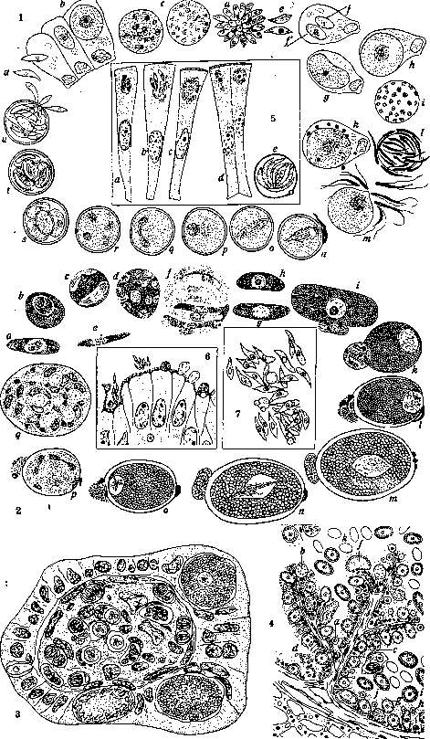
Coccidia (Coccidiomorpha), an extensive group of parasitic protozoa of the class Sporozoa, which consists of two subclasses—gregarines (see) and coccidia, or coccidiamorphs. Coccidia are intracellular parasites for most of their life cycle, residing in the intestinal epithelium, bile ducts, hepatic parenchyma, blood cells, and reticuloendothelial cells. C. parasitize among all classes of the animal world: in invertebrates and vertebrates—cold-blooded and warm-blooded, including humans. C. reproduce sexually and asexually, i.e., they are characterized by alternation of generations, which in some cases is associated with a change of hosts (figs. 1 and 2). Morphology and development history. A classic example of C.—Eimeria Stiedae Lindemann, parasitizing in the liver and intestines of rabbits. Merozoites, single-nucleated, pointed at both ends, contractile and motile formations, actively penetrate healthy epithelial cells, where they round off and become schizonts. The schizonts grow, their nucleus divides, and correspondingly their body also divides; finally, when the affected cell is destroyed, the resulting schizonts or merozoites fall into the cavity of the organ, intestines, or bile duct, penetrate fresh cells and begin a new asexual cycle of schizogony. With each subsequent schizogony cycle, the number of merozoites formed decreases, and they themselves become larger; finally when the schizonts begin to produce only 4 merozoites, the latter turn out to be equipped with flagella and, having penetrated the epithelium again, grow and transform into gametocytes, giving rise to the sexual cycle. (The decrease in the number of merozoites and the formation of flagellated forms, which are essentially precursors of gametocytes, is a feature of the described species and does not have universal distribution among C.) Gametocytes—male and female—undergo reduction divisions and accordingly transform into female macrogametes and motile male microgametes. Each macrogamete is fertilized by a microgamete, after which a layer of mucus is secreted around it and a firm two-contoured shell of yellowish color with an opening (micropyle), closed by a mucous plug, is formed. Inside the thus formed oocysts (20-50 μ × 16-30 μ), the protoplasmic body (sporont) divides into 4 spherical sporoblasts, around which in turn a shell, the sporocyst, with its own micropyle, is formed. Inside each sporoblast, two sporozoites are formed. The entire process of development of the sporont into sporozoites takes place after the oocyst is released outside and continues for about three days. When swallowed by a fresh rabbit, the sporocyst reaches the duodenum unchanged, where under the influence of trypsin the micropyle opens, the sporozoites come out and penetrate the intestinal epithelium, etc. Methods of spread. The spread of infection can occur directly between individuals of the same species, by contamination, i.e., ingestion of cysts. This is how C. spread among mammals, where both sexual and asexual cycles occur in the body of one host. In other cases, the relationships are complicated by the participation of an intermediate host, which plays the role of a passive carrier, however not affecting the parasite's development cycle. Thus, the coccidia parasitizing in the intestines of the centipede Lithobius forficatus undergo both sexual and asexual cycles of its development in the walls of its intestines and are released outside in the form of sporocysts. The latter are swallowed by water slaters (Asellus aquaticus), in whose intestines they do not change. Centipedes feed on slaters and thus become infected with the sporocysts contained in them. Further complication is observed in the case of the coccidia Aggregata Eberthi, parasitizing in the intestines of cephalopod mollusks, cuttlefish. In the latter, C. undergo the sexual cycle and are released outside in the form of sporocysts along with its excreta. Having fallen to the bottom silt, the sporocysts are absorbed by the crab Portunus, in whose intestines they open; the sporozoites freed from them penetrate the intestinal epithelium of the crab and begin the asexual cycle of schizogony, at the end of which the formation of gametocytes begins, which however do not receive further development in the crab. Crabs serve as food for cuttlefish, which become infected by eating infected crabs. Thus, having entered the intestine of the cuttlefish, the gametocytes of Aggregata begin a new sexual cycle.—Further complication of these relationships consists in the transition from the contaminative method of infection to the inoculative method, in which infection is carried out by the bite of a vertebrate host by an arthropod or worm carrier; in the most developed cases, this change of hosts is associated with a change of generations, when the sexual cycle takes place in the body of the carrier, as for example in the case of transmission of the malarial plasmodium (Coccidiida, Haemosporidiidea, Plasmodiidae). However, in a number of cases, the inoculative method of transmission is in no way associated with a change of generations, and the entire development process, both in the sexual and asexual parts, takes place in the main host, and in these cases there is a kind of preparation for the transfer of the sexual cycle to the body of the carrier. This is expressed in the fact that the process of schizogony, as well as the formation of macrogametes, occurs in the intestinal epithelium, while the formation of microgametes, fertilization and development of sporozoites occur, although in the same organism, however are transferred from the entodermal organ to the mesoderm, namely to the subepithelial connective tissue layer. This is observed, for example, in the development of the coccidia Schellackia bolivari, parasitizing in the lizard Acanthodactylus vulgaris and transmitted by the tick Lyponyssus saurarum. Infection of lizards occurs by eating infected ticks. In the body of the lizard, the entire development process of the coccidia takes place. Sporozoites developed in the subepithelial tissue penetrate the vessels and settle in the erythrocytes. When the lizard is bitten by a tick, infected erythrocytes enter the intestine of the latter, where they are phagocytosed by its epithelial cells. In the latter they do not die, but also do not develop. Their further development occurs in the intestine of the lizard, to which they get in the case that it swallows an infected tick. Systematics. Subclass Coccidiomorpha (Doll.; 1901). Predominantly intracellular parasites. Reproduction sexual and asexual; in many, change of hosts. Systematics is built mainly on differences in the properties of zygotes, i.e., their motility or immobility, their ability or inability to grow during development, on the degree of rigidity of their covering shell (sporocyst), on whether sporocysts are formed inside oocysts or not, on the number of the latter and on the number of sporozoites formed in them.-I. Order Coccidiida is characterized by the fact that male and female gametocytes have the same size and develop separately; male gametocytes form a large number of microgametes.-1. Suborder Eimeriidea. Microgametes are formed from the superficial layer of multinucleated microgametocytes. The immotile zygote is covered with a hard shell and does not grow during development. All development takes place in one host. Infection is contaminative by eating sporocysts.-1. Family Selenococcidiidae. Sporozoites develop in the cavity of the intestine to the 8-nucleus stage, at which they penetrate the epithelial cells. Gametocytes also initially develop extracellularly. The theoretical significance of this group lies in the fact that it connects C. with gregarines. Representatives of this family parasitize in invertebrates (fig. 11).-2. Fam. Cryptosporidiidae—the entire development cycle occurs on the surface of intestinal epithelial cells. Genera Cryptosporidium muris and Cryptosp. parvum parasitize in mice: the first—in the gastric glands, the second—in the small intestines (fig. 6).-3. Fam. Eimeriidae—the most typical coccidia; development intracellular. Differ in forms that do not form sporocysts in oocysts (Asporocystida), forming 2, or 4, or many sporocysts (Di-, Tetra-, Polysporocystida). In sporocysts, 1-2-4 or many sporozoites are formed (Mono-, Di-, Tetra-, Polyzoa). 1. Subfamily Cyclosporinae forms 2 sporoblasts, 2 sporocysts and 2 sporozoites. Cyclospora caryolytica parasitizes in the intestinal epithelium of the mole, causing severe enteritis. Similar forms are found in snakes (adders and vipers). 2. Subfamily Isosporinae forms 2 sporoblasts, 2 sporocysts and 4 sporozoites. Isospora bigeraina (Stiles; 1891) parasitizes in cats, dogs and ferrets, in which it is the cause of chronic enteritis, affecting the epithelium of all small intestines. Two varieties are described, differing in the size of oocysts and sporocysts. In the large one—the size of oocysts is 18-20×14-16 μ, and of sporocysts—13.5-15.5×9-10 μ; in the small one—10-16×7.5-10 μ and 7.5-10×5-8 μ. Isospora Rivolta (Grassi; 1879) is found in cats and dogs. The size of oocysts is 20-24×15-20 μ, of sporocysts—12-15×9-10 μ. Isospora felis (Wenyon; 1923)—in cats and dogs; size of oocysts 39-48×26-37 μ, of sporocysts—20-27×18-21 μ (figs. 3 and 8). Isospora hominis (Railliet & Lucet; 1901) is found in humans. Histologically, only one case of infection of the villi of the small intestine in humans has been described to date (Virchow;)
(1860). Described in feces by several authors. Size of sporocysts 16 x 10.5 µ. Isospora belli (Wenyon; 1923) occurs in human feces. Size of elongated oocysts 25-33x12.5-16 µ. A case of laboratory infection in humans has been described. The form is comparatively very rare. Isospora Lieberkuhni (Labbe; 1894) parasitizes in the kidneys of frogs. Other species of Isospora also occur in birds. 3. Subfamily Eimeriinae. In the oocyst, 4 sporoblasts, 4 sporocysts and 2 sporozoites are formed. Widely distributed among vertebrates—humans, horses, small ruminants, dogs, cats, mice, rats, birds, reptiles, amphibians, fish and invertebrates. The typical species E. Stiedae (Lindemann; 1865) parasitizes in rabbits in the intestinal epithelium and in the bile ducts (fig. 4). 5 cases in humans have been described. Size of oocysts 20-40x16-25 µ, 28-44x21-30 µ, 33-43 x 18-30 µ. Eimeria perforans (Leuckart; 1879) parasitizes in rabbits; size of oocysts 16-23x12-16 µ, 15-30x11-18 µ. Eimeria Zurnii (Rivolta; 1878) differs from Eimeria Stiedae in its round and smaller (12-25 µ) cysts (fig. 9). Parasitizes in the intestines of ruminants. The causative agent of bloody diarrhea mainly in young individuals. Carriage in about 22% (Berlin statistics). In severe infection, mortality up to 5%. Eimeria clupearum (Thelohan; 1892) parasitizes in the liver of herring, sprats and mackerel. Round sporocysts are covered with a thick shell and have a diameter of about 20 µ. E. sardinae (Thelohan; 1890) parasitizes in the testes of sardines, herring and sprats. Oocysts with a diameter from 33.6 to 50.6 µ, on average 42.5 µ; elongated sporocysts 25-30x6-7 µ. The last two forms are of particular interest in that for a long time they were considered parasites of humans because the corresponding oocysts were found in the feces of patients with enteritis. As such, they were described under the names E. oxyspora, E. Snijdersi, E. Wenyoni, until recent research (Thomson & Robertson; 1926) showed that the first two forms are identical to E. sardinae, and the third to E. clupearum, that they enter the human body with food and have no pathological significance (fig. 10). 4-7. Subfamilies Barrouxinae, Caryosporinae (fig. 10), Pfeifferellinae, Caryotrophidae parasitize in cold-blooded animals and invertebrates. 4. Fam. Aggregatidae parasitize in cephalopod mollusks (cuttlefish). Intermediate host—crab. 5. Fam. Lankesterellidae. 1. Subfamily Schellackinae. 2. Subfamily Lankesterellidae. Parasitize in cold-blooded animals, transmitted by the former—ticks, by the latter—leeches.
coccidia
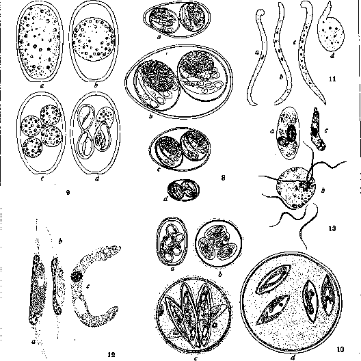
Figure 1. Eimeria Schubergi-development cycle in the centipede Lithobius forficatus: a-sporozoite; b-penetration of the sporozoite into the intestinal epithelial cell, growth of the schizont (c) and formation of merozoites (d,e); f, h, i-formation of microgametes; f',g,h-formation of macrogametes; k-fertilization; l-formation of oocyst; m-s-formation of sporoblasts; n-mature oocyst with 4 sporocysts, each with 2 sporozoites; o-emergence of sporozoites from sporocysts and oocysts, swallowed by another centipede. Figure 2. Adelea ovata-development cycle in the centipede Lithobius forficatus: a-e-schizogony in the intestinal epithelium of the centipede; f-formation of large merozoites; g-young female gametocyte; h-young male gametocyte; i-union of female and male gametocytes; k-male gametocyte divides and forms 4 microgametes; l-one of the 4 microgametes has penetrated the macrogamete, which is enveloped by an oocyst; m,n-fertilization; o-formation of zygote; p-division of nuclei in the zygote; q-mature oocyst contains many sporocysts, each with 2 sporozoites. Figure 3. Section through an intestinal villus of a cat with developmental stages of Isospora felis in the epithelium and I. bigemina in the subepithelial tissue. Figure 4. Section through the bile duct of a rabbit liver, infected with Eimeria Stiedae. Hypertrophy of the epithelium and developmental stages of coccidia: a-young forms, from which both schizonts and gametocytes can develop; b-adult multinucleate schizonts; c-merozoites inside the cell, resulting from schizogony; d-release of merozoites from the cell; e-mature microgametocyte with microgametes on the surface; f-section through the central part of the microgametocyte; g-cross section through a macrogametocyte lying in a cell; h-same-longitudinal section; i-section through a freely lying oocyst; k-unstained oocysts; l-merozoites arranged in the cell in a barrel-shaped group. Figure 5. Caryospora simplex from the intestine of a viper: a, b-schizogony; c-formation of microgametes; d-formation of macrogametocytes; e-formation of 8 sporozoites inside the oocyst. Figure 6. Cryptosporidium parvum from the intestine of a mouse. Developmental stages are located on the surface of epithelial cells. Figure 7. Klossiella cobayae in the kidney of a guinea pig; formation of gametocytes inside the renal tubule. Figure 8. Oocysts of different Isospora species: a-I. belli from the human intestine; b-I. felis from the intestines of cats and dogs; c-I. Rivolta from the intestines of cats and dogs; d-I. bigemina (=I. hominis?) from the intestines of cats, dogs (and humans). Figure 9. Eimeria Stiedae: a-d-development of the oocyst in the rabbit liver. Figure 10. Oocysts: a-Eimeria perforans of the rabbit; b-E. Wenyoni (=clupearum) from humans; c-E. oxyspora (=E. sardinae) from humans; d-E. Snijdersi (=E. sardinae) from humans. Figure 11. Selenococcidium intermedium: a-c-free worm-like schizonts; d-shedding of the latter. Figure 12. Leucocytozoon Ziemanni from the owl Athene noctuae: a-macrogametocyte; b-microgametocyte; c-ookinete. Figure 13. Haemoproteus from the kestrel: a-female gametocyte; b-formation of microgametes; c-ookinete. ____________ 2. Suborder Haemosporidiidea. Microgametes are formed from microgametocytes in the process of so-called flagellation, accompanied by intense protoplasmic currents. The fertilized zygote forms a motile ookinete, which becomes an oocyst and grows during development. Representatives parasitize in the erythrocytes of vertebrates, warm-blooded and cold-blooded, where their asexual cycle takes place; they are transmitted by insects, in which the sexual stages occur. The entire development cycle proceeds as in typical coccidia.-1. Fam. Haemoproteidae (Doil; 1916). Parasites of endothelial cells and erythrocytes of vertebrates (asexual reproduction period) and tissues of the invertebrate vector (sexual development period) (fig. 13).-Genus Haemoproteidae (see).-Genus Leucocytozoon (Danilewski; 1890). Parasites of birds. During development in erythrocytes, pigment is not formed. In peripheral blood, they are found in internal organs (fig. 12). Found in geese, ducks, birds of prey.-2. Fam. Plasmodiidae (Mesnil; 1903). Representatives parasitize in the erythrocytes of vertebrates, where they undergo the asexual cycle. The sexual cycle takes place in the body of the invertebrate vector. During development in erythrocytes, pigment is formed.-Genus Plasmodium. Representatives of this genus parasitize in various vertebrates, both warm-blooded and cold-blooded. In humans, they are the causative agents of malaria. In all studied cases, the vectors are mosquitoes. 3. Suborder Piroplasmidea a. Parasites of blood corpuscles of vertebrates-cattle and small ruminants, pigs, horses, dogs, mice, monkeys. The development cycle has not been studied in detail. The asexual cycle takes place in the erythrocytes of the vertebrate; the sexual cycle, insufficiently studied-probably in the insect vector.--1. Fam. Babesiidae (Poche; 1913). Non-pigment-forming parasites of erythrocytes of mammals.-Genus Babesia bigemina-causative agent of babesiosis, or piroplasmosis-hemoglobinuria of cattle (see Babesia).-2. Fam. Theileriidae. Non-pigment-forming parasites of erythrocytes of mammals. Schizogony takes place in the endothelium of capillaries of internal organs. As a result, peripheral blood is not infectious.-Genus Theileria. Species Th. parva (Theiler; 1904). Causative agent of theileriosis of cattle, occurring without hemoglobinuria, jaundice, and anemia, which characterize piroplasmosis. High temperature, hypertrophy of peripheral lymph glands. Mortality up to 90%. Vector-the tick Rhipicephalus appendiculatus. Found in cattle and small ruminants. II. Order Adeleida (Leger; 1911). The zygote is immobile, enclosed in an oocyst. The change of generations takes place in the same host. Infection is contaminative.-1. Suborder Adeleidea. The zygote is immobile, the oocyst does not grow. Of the 4 families (Dobelliidae, Legerellidae, Adeleidae and Klossiellidae), differing from each other in the number and method of formation of sporozoites-with or without preliminary formation of sporocysts-the most significant is the latter (Klossiellidae), since its representatives are found not only in invertebrate hosts like the first three families, but also in vertebrates as parasites of laboratory animals.-Fam. Klossiellidae. In the oocyst, several sporocysts with a large number of sporozoites are formed. The macrogametocyte forms only two microgametes.-Genus Klossiella (Smith & Johnson; 1902). K. cobayae (1914) parasitizes in the renal tubules and in the endothelium of blood vessels of the guinea pig. K. muris (Smith & Johnson; 1902) is a common parasite in the kidneys of white mice (fig. 7).-2. Suborder Haemogregarinidea. Forms a zygote having the characteristic of a motile ookinete, which increases during development. The change of generations is associated with a change of hosts, with the sexual cycle taking place in the vector.-1. Fam. Haemogregarinidae (Neveu-Lemaire; 1901). In the oocyst, sporozoites are formed, bypassing the sporocyst stage. The asexual cycle takes place in erythrocytes or other cells in vertebrates. The sexual cycle takes place in the intestine of leeches. The number of microgametes formed is 2-4.-The only genus-Haemogregarina (Danilewski; 1885). Species H. Stepanowi (Danilewski; 1885) parasitizes in the European tortoise. Vector-the leech Placobdella catenigera.-*2. Fam. Hepatozoidae. In the oocysts, a large number of sporocysts and sporozoites are formed. Asexual reproduction-in the internal organs of vertebrates, sexual-in the intestine of invertebrates (ticks). Contains one genus Hepatozoon (Miller; 1918). Species H. muris (Balfour; 1905)(synonym H. perniciosum). Parasitizes in black, red, and white rats. Schizogony occurs in liver cells. Formation of gametocytes occurs in leukocytes, with which they enter the intestine of the tick. In the intestine of the latter, sporogony occurs. Infection of rats occurs by eating infected ticks Laelaps echidninus. H. canis (James; 1905). Vector-the tick Rhipicephalus sanguineus. Ticks become infected by sucking the blood of infected dogs; dogs become infected by eating infected ticks.-3. Fam. Karyolysidae. Species Karyolysus lacertarum (Danilewski; 1886). Parasitizes in Lacerta muralis; vector-Lyponyssus saurarum.
_) Effect on the organism. Infection with coccidia and their development in the host's organs manifest as a complex of diseases known as coccidiosis. Depending on the affiliation of the pathogen to one or another group of Coccidiomorpha, a distinction is made between coccidiosis in the narrow sense, caused by representatives of the order Eimeriidea (order Coccidiida), as well as representatives of the order Adeleida, and haemosporidioses, caused by representatives of the suborder Haemosporidiidea (order Coccidiida); this group includes plasmodioses caused by representatives of the family Plasmodiidae (human malaria - plasmodiosis), and finally piroplasmoses (babesioses and theilerioses), caused by representatives of the suborder Piroplas-midea (order Coccidiida). An example of coccidiosis in the narrow sense: rabbit coccidiosis. - Infection and immunity. The severity of the disease depends to a certain extent on the amount of infectious material, i.e., on the number of sporocysts that penetrate during a single infection. Pathological destruction caused by C. is associated with their schizogony and the involvement of new cells in the process. But since schizogony has a tendency to transition to sporogony, the infectious agent is naturally removed from the body, and the active process ceases. However, it is very probable that with the onset of sporogony, part of the schizonts may temporarily pass into an inactive state, in order to later return to active schizogony and serve as a source of clinical relapse (analogy with relapses in malaria). Under ordinary conditions, the matter is not limited to a single infection, and an already infected rabbit continues to become infected with oocysts swallowed from the outside. But apparently as a result of the developing relative immunity, reinfection (resp. superinfection) proceeds more easily, in most cases not affecting the intestine, being mainly concentrated in the liver. - Pathological anatomy. In the affected organs, destruction of the epithelium is observed, as well as inflammatory scar changes in the submucosal layer of different parts of the intestine. For liver changes, nodules originating from the affected epithelium of the bile ducts are characteristic, which expand, proliferate, and are surrounded by connective tissue. In connection with this, a very strong hypertrophy of the liver may develop. - Diseases with coccidiosis among warm-blooded animals occur in rabbits (Eimeria Stiedae and E. perforans), in cattle (E. Zurnii), in sheep, goats, pigs, in predatory animals, as well as in birds (E. avium), being the source of deadly epidemics of enterocolitis and hepatitis among domestic poultry (external symptom - blackening of the comb, "black head"). Coccidioses caused by Isospora affect cats and dogs (Isospora bigemina, Isospora felis). - C O C C I D I O S I S O F M A N. In the literature, 5 cases of coccidiosis caused by E. Stiedae are known. In 2 cases, the diseases were fatal and proceeded with symptoms of enteritis, anemia, and fever. Autopsy revealed hypertrophy of the liver and numerous foci of caseous decay. The latter are also described in the spleen and in the mucous membrane of the small intestines. Coccidioses caused by Isospora hominis and belli are apparently very mild diseases. In the case of laboratory infection, diarrhea appeared after six days, lasted for 1-2-3 months, and ended with recovery. On false findings of C. in man, see above. - D i a g n o s i s of coccidiosis is established on the basis of examination of excreta, and ordinary helminthological enrichment methods can be used, in particular emulsification of feces in a saturated salt solution. - Treatment of coccidiosis in man due to its rarity has not been developed. In cattle, enemas of a 1/2% solution of tannin are used; orally, Creolin Pearson 20.0 is administered; T-ra Rhei aqu., T-ra Calami aa 4.0 pro die. - P r o p h y l a x i s. Avoid contaminated products.
Related articles
Mentioned in
Cite this page
“Coccidia.” Soviet Medical Encyclopedia. English translation of Bolshaya Meditsinskaya Entsiklopediya, 1st ed. (Moscow, 1928–1936), ed. N. A. Semashko. https://sovietmedicalencyclopedia.pages.dev/article/coccidia/