Cnidosporea
Historical document, translated for reference. It reflects medical knowledge of the 1920s–30s and is not medical advice.
Summary
Cnidosporea are a large group of parasitic protozoa that infect tissues and cavities of cold-blooded invertebrates and vertebrates. They are characterized by the formation of spores equipped with one or more polar capsules throughout their life cycle.
Encyclopedia article (1928–1936)
CNIDOSPORIDIA, an extensive group of protozoa parasitic in the tissues and cavities of cold-blooded invertebrates and vertebrates. Some forms are described as parasites in warm-blooded animals—the rabbit and man, for example, Encephalitozoon cuniculi and E. rabiei (the supposed causative agent of rabies), but this question has not yet been resolved (see below). The parasitological interest of the group C. lies in its comparative pathological significance, since most of its representatives possess specific organotropism, including toward the nervous system. C. are characterized by the formation of spores, which, unlike the spores of typical sporozoans (Sporozoa), are formed in C. not at the end of the vegetative period, but throughout their entire life, and are provided with one or more polar capsules. Based on the ability of Cnidosporidia to form spores at any moment of their existence, Schaudinn (1900) opposed them as Neosporidia to the other sporozoans (Coccidiida, Gregarinida), which form spores only at the end of their vegetative life (Telosporidia), assigning both of these groups to the class Sporozoa. However, the differences between C. and typical sporozoans are so profound that other authors have separated them into a special class C., characterized by the peculiarities of the structure of their endogenous spores and their development, which is so complex that it has given some researchers occasion to express the thought that C. are not even Protozoa, but a prototype of Metazoa (Emery, 1909; Dunkerly, 1925). - On the basis of morphological peculiarities in the structure of the body and spores in the group C., 3 orders are distinguished: Myxosporidiida, Actinomyxidiida, Microsporidiida, which however all develop according to a general scheme. Developmental history. In the intestine of the host, the spore opens; from it emerges a uninucleate amoeboid, which penetrates into the tissue or into the body cavity, where it grows and transforms through a series of successive divisions into a multinucleate plasmodium. The latter can multiply by division into 2, schizogony, or budding. Simultaneously with this, spores are formed. The initiation of the latter occurs through the endogenous isolation in the body of the plasmodia of uninucleate areas of protoplasm, so-called propagative cells (fig. 1a), from which the spores are subsequently formed. Each propagative cell divides into one larger gametoblast, from which later a new amoeboid is formed, and one smaller sporoblast, forming the common spore covering (fig. 1b). Two such pairs unite in a sporocyst or so-called pansporoblast, containing two gametoblastic nuclei (fig. 1c). Thus the pansporoblast consists of 2 sporoblasts, each of which gives rise to one spore. The development of both spores occurs in the pansporoblast simultaneously; in this process each of the gametoblastic nuclei forms by division 6 daughter nuclei, of which two become nuclei of gametes, two enter into the composition of the polar capsules, and the remaining two participate in the formation of the shell valves of each of the spores (fig. 1d-k). Both gamete nuclei of each spore fuse (fig. 1g-h). Thus the mature spores contain uninucleate amoeboids. The question of when the actual sexual process occurs has not been finally resolved, with some authors referring it to the moment of union of the gametoblasts, and others to the moment of fusion of the gamete nuclei. A third group admit the existence in some C. of the process of autogamy. In different C. the described processes may differ in minor details. Nutrition occurs osmotically. There are harmless and pathogenic forms. 'Systematics. I. Order Myxosporidiida Bütschli. This includes mostly large multinucleate amoeboid forms; large spores are formed in pansporoblasts, mostly in pairs, contain 2 or 4 polar capsules and one amoeboid embryo and are surrounded by a bivalve shell (fig. 1h). Myxosporidia parasitize intercellularly in tissues (muscles) and in parenchymatous or cavity organs (kidneys, gallbladder and urinary bladder) of fish, amphibians, and reptiles. Some are dangerous pests of fish. The most significant are: 1. Myxobolus Pfeifferi (Thelohan; 1894)- the causative agent of an epidemic disease in barbel (Barbus fluviatilis). It affects the skin, gills, musculature, parenchymatous organs, producing severe destruction of tissues in the form of diffuse infiltrations or in the form of localized, encapsulated tumors from 1-2 cm to the size of a hen's egg. Size of spores 10-12 μ (figs. 1, 2a, 2, 7, 12 and 14). 2. Myxobolus neurobius Schuberg-Schredner parasitizes in the nerve trunks and in the spinal cord of trout, in the former being located between the Schwann and myelin sheaths (fig. 11). 3. Leptospora cerebralis Hofer - a pathogenic parasite of salmonid fish; it selectively affects the cartilaginous skeleton and perichondrium, causing various deformities and curvatures of the skeleton; when the lesion involves the semicircular canals, the fish develops a peculiar disease expressed in circular movements ('whirling'). Infection occurs through eating other infected individuals. II. Order Actinomyxidiida is characterized by the formation in the pansporoblast of 8 spores (fig. 4), having a three-rayed structure, covered with a single shell and containing 3 polar capsules each (fig. 5) and numerous uninucleate amoeboids or one multinucleate. They parasitize exclusively in the body cavity and intestine of marine oligochaetes (Oligochaeta). III. Order Microsporidiida (Balbiani). Mostly small forms; they form small spores, which in most forms are formed in pansporoblasts singly, in pairs, in groups of 4, 8, or in larger numbers. They are covered with a single shell, have a simplified polar capsule in the form of a vacuole containing a very long polar filament, and contain 1 amoeboid.
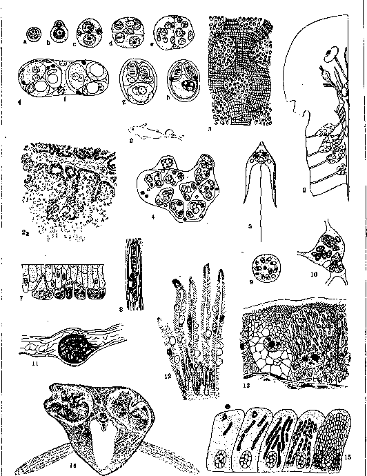
Figure 1. Formation of spores in Myxobolus Pfeifferi. Figure 2. Rudd with a tumor near the caudal fin, caused by Myxobolus piriformis. Figure 2a. Section through this tumor, which in its structure resembles a papillary adenocystoma. Figure 3. Plistophora typicalis in the muscles of the fish Cottus scorpius. Figure 4. Initiation of 8 sporoblasts in the pansporoblast of Tetractinomyxon. Figure 5. Spore of Synactinomyxon tubificis. Figure 6. Part of the central nervous system of Lophius piscatorius with nerve tumors caused by Nosema lophii. Figure 7. Myxidium Lieberkuhni on the surface of the epithelium of the pike's urinary bladder. Figure 8. Nosema apis in the muscles of the silkworm larva. Figure 9. Section through a ganglion cell of Lophius containing spores of Nosema lophii. Figure 10. Connective tissue cell of the worm Chaetogaster, affected by spores of Thelohania. Figure 11. Longitudinal section through a nerve fiber with a cyst containing Myxobolus neurobius. Figure 12. Cysts of Myxobolus minutus on section through the gill fillets of perch. Figure 13. Intestinal epithelium of the silkworm, affected by Nosema bombycis. Figure 14. Giant cysts in the musculature of barbel, caused by Myxobolus Pfeifferi. Figure 15. Nosema apis
in the intestinal epithelium of the bee. They parasitize almost exclusively intracellularly in worms, bryozoans, but mainly in arthropods, less frequently in fish and amphibians, rarely in the body of other parasitic protozoa (gregarines). The most important are: 1. Genus Nosema. Does not form pansporoblasts; thus spores are formed directly from uninuclear elements, a) Nosema bombycis (Naegeli) - the causative agent of a fatal epidemic disease of the silkworm ('pebrine'). Infection occurs by eating spores, from which amoeboid forms emerge; the latter penetrate into the cells of the intestinal epithelium and are carried by the blood to all organs, where they multiply by schizogony, as well as by division into two, forming characteristic chains, and then proceed to spore formation. Penetrating into eggs, they cause germinal infection of offspring (fig. 8 and 13). b) Nosema apis (Zander) is described as the causative agent of epidemic diarrhea in bees; but it is possible that the combination with other infectious factors is required for the epidemic to occur; since this parasite is also found in small quantities in healthy bees. This form is also found in wasps, horseflies; it has been possible to infect butterflies and other dipterans with it (fig. 15). c) Nosema pulicis (Noller) parasitizes in the epithelium of the intestine and Malpighian vessels, in the fat body and salivary glands of the dog flea (Ctenocephalus canis). d) Nosema adiei (Shortt, Swellengrebel) (India) is described in the same organs and in the ovary of the bedbug (Cimex lectularius) and was mistakenly considered a developmental stage of Leishmania donovani. e) Nosema anophelis (Kudo) - in the intestinal epithelium and in the fat body of larvae of Anopheles quadrimaculatus. f) Nosema culicis (Bresslau) - in larvae of Culex pipiens. g) Nosema stegomyiae (Lutz-Splendore) - in the intestine of adult Stegomyia fasciata. h) Nosema lophii (Doflein) - in the nervous tissue of the fish Lophius piscatorius ('sea devil'), forming grape-like tumors of nerves (fig. 6 and 9).-2. Genus Glugea (Thelohan) is characterized by the formation of 2 spores in the pansporoblast; numerous species of this genus parasitize in fish, amphibians, and reptiles.-3. Genus Thelohania Henneguy, 8 spores are formed in the pansporoblast. Representatives of this genus are found in arthropods (crustaceans and insects) (fig. 10). a) Thelohania legeri Hesse. b) Th. obesa Kudo, c) Th. piriformis Kudo - in the fat body of Anopheles larvae. d) Thelohania opacita Kudo, e) Th. rotunda Kudo. f) Th. minuta Kudo - in larvae of Culex. This infection is often the cause of death of larvae; with a weaker degree of infection, it can also pass to adult mosquitoes. g) Th. corethrae - in the body cavity of Corethra larvae. h) Th. tipulae - in the fat body of Tipula lateralis larvae. The last two forms cause hypertrophy of fat cells. 4. Genus Plistophora Gurley; an indefinite number of spores, up to 16, are formed in the pansporoblasts. Parasitizes in fish and arthropods, a) Plistoph. typicalis - in the trunk muscles of various freshwater fish (fig. 3). b) Plistoph. periplanetae - in the Malpighian vessels of cockroaches (non-pathogenic).- Some authors also include among the Microsporidia the causative agent of rabbit encephalitis Encephalitozoon cuniculi (see). Others (Levaditi) believe that the causative agent of rabies also belongs to this group (Glugea lyssae = Encephalitozoon rabiei). However, doubts are expressed about the correctness of assigning these forms to Microsporidia, especially since in mammals representatives of this group are not known at all, except for the mentioned forms.
G. Epstein. CNICUS BENEDICTUS L., blessed thistle, an annual herb of the composite family (Compositae), grows wild in Southern Europe, Transcaucasia, Uzbekistan, and is cultivated in some places. It is collected shortly before flowering or during flowering. The stem reaches 60 cm in height and branches in the upper part. The leaves are alternate, reddish and, like the stem, are covered with hairy or sticky hairs. The pharmaceutical preparation is commonly called Herba Cardui benedicti. Components: cnicin (or centaurein), C42H56O15 (0.2%), a crystalline bitter substance, essential oil (0.3%), tannins and malic acid magnesium. In powders (0.5-1.5), infusion and decoction (5.0-10.0:150.0), the herb is used as a bitter that stimulates appetite; large doses cause vomiting.
Related articles
Cite this page
“Cnidosporea.” Soviet Medical Encyclopedia. English translation of Bolshaya Meditsinskaya Entsiklopediya, 1st ed. (Moscow, 1928–1936), ed. N. A. Semashko. https://sovietmedicalencyclopedia.pages.dev/article/cnidosporea/