Clubfoot
Historical document, translated for reference. It reflects medical knowledge of the 1920s–30s and is not medical advice.
Summary
Clubfoot is a deformity of the foot with inward rotation and plantar flexion. This article discusses the congenital and acquired forms, their genetic basis, associated anomalies, and various etiological factors.
Encyclopedia article (1928–1936)
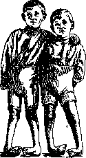
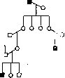
CLUBFOOT (pes varus, pes varo-equi-nus, talipes varus, talipes equino-varus according to English authors), a deformity of the foot with inward rotation and plantar flexion. The deformity in C. usually consists of three main components: adduction, supination (talipes varus according to English authors), and plantar flexion (talipes equinus according to English authors). Depending on which element predominates, the deformity is called: pes varus, equino-varus, etc. Opposite deformities of the foot are called pes valgus - deviation of the foot outward, pes calcaneus - pathological position of dorsal flexion (heel foot); a combination of both types of deformity is called pes calcaneo-valgus. These latter types of deformity, in contrast to pes equino-varus, are more often of paralytic origin. - C. in the vast majority of cases is congenital; according to Bessel-Hagen, 73.8% of all cases of C. are congenital, and 26.2% are acquired C. According to Lannelongue, one case of congenital C. occurs in 2,000 births; according to Bessel-Hagen, one case in every 1,100 births. Genetics of C. Congenital C. is in the overwhelming majority of cases a genotypic disease. Not only the anomaly itself but also its degree is usually inherited (Fig. 1). In most cases, C. is inherited as an autosomal recessive trait (Fig. 2), and, as in other similar cases, relatively frequent consanguineous marriages (inbreeding) between parents of patients are observed. However, some cases (Kochs, Müller, etc.) must be interpreted rather as a dominant form of inheritance; finally, individual genealogies by Fetscher indicate the possibility of recessive, sex-linked inheritance (Fig. 3). From the foregoing, it follows that C. is a heterogeneous anomaly, combining clinically similar but biologically (genotypically) completely different cases. The fact mentioned above of clinical differences in the manifestation of C. in different families (with similarity within one family) also speaks in favor of this. In boys, C. occurs twice as often as in girls (Bessel-Hagen, Fetscher). The reason for the more frequent occurrence in the male sex has not been finally clarified. In some cases, this is explained by the probable fact of recessive, sex-linked inheritance; the remaining cases must apparently be interpreted as partial restriction to the male sex. It is extremely interesting that among the sisters and brothers of recessive homozygotes, the male sex predominates: according to Fetscher's data, for 386 brothers there are only 267 sisters. A satisfactory explanation for this phenomenon does not yet exist. As one hypothesis, it can be suggested that there is partial lethality of the C. gene in the homozygous state (assuming that some cases of C. are inherited as a recessive, sex-linked trait). At least this allows interpretation of the data provided by Fetscher. The large number of carefully verified clinical facts forces recognition of the existence of congenital paratypic C. In the not too distant past, the opinion prevailed that this form of C. is predominant, and that primary C. should be assigned a very modest place. Representatives of another group (J. Wolff) energetically objected to this. As exogenous intrauterine factors, pathological position of the fetus, anomalies of the uterus and amnion (uterus bicornis, narrowing of the uterine cavity, deficiency of amniotic fluid, adhesion of the amnion to the surface of the fetus, amniotic constrictions), bone defects, multiple pregnancy, mismatch between the size of the uterus and fetus (Martini, Malgaigne, Liicke, etc.) are cited. All these factors probably play only the role of contributing factors. - C. is often combined with other congenital malformations: spina bifida occulta, torticollis, club hand, syndactyly, harelip, cleft palate, as well as with deaf-mutism, nocturnal enuresis, etc. In his material, Fetscher among 184 patients with C. found mental deficiency in 12% of cases (idiocy in 4.67%); among 460 investigated idiots, C. was present in 20 cases (4.4%), among 407 mentally ill patients - in 4 cases. Such cases must be interpreted as pleiotropic action of the C. gene. As for the question of the mechanism of action of the clubfoot gene, according to B. Ashner (V. Ashner), the area of its manifestation is the corresponding affected limbs; Fetscher, on the contrary, assumes that a lesion of the central nervous system is transmitted, which in turn leads to the development of CLUBFOOT.
I. Rybkina. Congenital C. is more often bilateral (about 60%); when one side is affected, the right side predominates (Whitman, Kirmisson). Acquired C. has several etiological factors. There are forms of static C. due to pathological stress. Such is, for example, C. in pronounced genu valgum, in partial acquired defects of the tibiae (osteomyelitis, extensive resection), after various types of fractures within the ankle joint with partial dislocation of the talus (traumatic C.). Acquired C. with such etiology has very atypical forms - from simple positioning in the va-r position to severely expressed forms. On the basis of scar, dermo-desmogenic and tendogenic contractures after inflammatory processes, C. often develops, Fig. 3. Hereditary
I. Rybkina. Congenital C. is more often bilateral (about 60%); when one side is affected, the right side predominates (Whitman, Kirmisson). Acquired C. has several etiological factors. There are forms of static C. due to pathological stress. Such is, for example, C. in pronounced genu valgum, in partial acquired defects of the tibiae (osteomyelitis, extensive resection), after various types of fractures within the ankle joint with partial dislocation of the talus (traumatic C.). Acquired C. with such etiology has very atypical forms - from simple positioning in the va-r position to severely expressed forms. On the basis of scar, dermo-desmogenic and tendogenic contractures after inflammatory processes, C. often develops, Fig. 3. Hereditary
I. Rybkina. Congenital C. is more often bilateral (about 60%); when one side is affected, the right side predominates (Whitman, Kirmisson). Acquired C. has several etiological factors. There are forms of static C. due to pathological stress. Such is, for example, C. in pronounced genu valgum, in partial acquired defects of the tibiae (osteomyelitis, extensive resection), after various types of fractures within the ankle joint with partial dislocation of the talus (traumatic C.). Acquired C. with such etiology has very atypical forms - from simple positioning in the va-r position to severely expressed forms. On the basis of scar, dermo-desmogenic and tendogenic contractures after inflammatory processes, C. often develops, Fig. 3. Hereditary
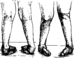
which develops gradually. na(poKFecharPu°)STY There are also rheumatic, ischemic, and myogenic forms of clubfoot. Finally, among the various types of acquired clubfoot, the paralytic forms constitute a huge group, arising mainly after infantile paralysis. Regardless of the etiological factor, a very typical deformation occurs in clubfoot, and rather complex pathological-anatomical relationships develop between the bones of the foot. Pathological anatomy. The deformation present in clubfoot occurs due to excessive rotation of the foot around its three main axes. Movement around the transverse axis in the direction of plantar flexion is so pronounced that the surface of the trochlea of the talus protrudes forward over a considerable distance. The neck of the talus is turned inward and downward. Normally, it is turned inward in the embryo at 35.7°, in the adult at 12.3°, in clubfoot at 50.7°. The anterior part of the talus in such a deformation is not covered by the articular surface of the navicular bone. The calcaneus is in a position of plantar flexion and rotation around its longitudinal axis, turned around the vertical axis so that its anterior end is positioned medially, and in addition there is a twisting of the calcaneus medially in the posterior-anterior direction. Its upper surface faces forward and inward. The articular surface for the cuboid bone faces significantly inward and downward. All the bones of the foot are underdeveloped. Due to the adduction and rotation of the anterior part of the foot, the navicular bone is turned inward, backward, and upward, so that it articulates with the posterior part of the head of the talus and may come into contact with the medial malleolus. The relationship between the cuboid bone and the calcaneus is also changed: the upper surface of the former faces outward and downward. The malleoli are usually not very changed in their development and mutual position. In long-standing cases, twisting of the bones of the leg around their longitudinal axis is also noted. This explains the circumstance that even after correction of the foot, the latter appears turned inward in relation to the knee joint. Anatomical changes also occur in the ligaments, fasciae, and muscles. The entire ligamentous apparatus on the medial and posterior surfaces of the foot and sole is shortened. The tendons of the muscles assume a pathological direction depending on the displacement of their attachment points. The tendon of the tibialis anterior muscle lies directly at the medial malleolus, and in severe cases even on its medial side. The tendons of all the anterior muscles are correspondingly displaced medially. The tibialis posterior muscle, instead of the normal forward turn behind the medial malleolus, is directed downward, and since the navicular bone approaches the medial malleolus significantly, the part of the tendon below the malleolus is barely distinguishable. The tendons of the flexor digitorum longus and flexor pollicis longus are also displaced medially depending on the degree of foot deformation. The path of the tendon of the peroneus muscle is lengthened, the muscles are overstretched and have lost their tone. The described pathological changes increase when the patient begins to walk. Usually, the support is not the sole, but the dorsal or, more accurately, the anterolateral part of the foot, with the cuboid bone and the anterolateral part of the calcaneus bearing the greatest weight. Here a significant callus-like thickening of the skin forms, under which a bursa is located. The talus loses its articular cartilage on the protruding anterior surface of the trochlea, the neck elongates and is directed downward and medially. Due to the load during walking, coarse bone changes occur, especially in the talar, cuboid, and calcaneal bones, which in advanced cases sometimes serves as an almost insurmountable obstacle to the complete correction of the clubfoot during redression. The clinical picture of clubfoot is extremely characteristic. Besides the described deformation involving the foot, some atrophy of the leg muscles is noticeable, a known disproportion in the development of the limbs and trunk, and a characteristic gait with support on the dorsum of the foot. Due to the persistent position of varus-equinus, the dynamics of the muscles are sharply disrupted. The peroneal group is overstretched and inactive; the tibialis anterior and triceps surae muscles, due to the extreme shortening of their path, although they contract actively, do not possess normal contractile ability. The same can be said about other groups of foot muscles. In essence, clubfoot represents a contracture of certain muscle groups with subsequent remodeling of the skeleton (Figure 4). There is a significant difference between congenital clubfoot and pes cavus that developed later and is always associated with myelodysplasia, with dysplasias of the lower end of the spine (spina bifida occulta) or with infantile paralysis. Congenital clubfoot is more often bilateral, and all components of the deformation are equally sharply expressed; in bilateral clubfoot, the varus element may be less pronounced on one side than on the other. In paralytic or myelodysplastic pes cavus, the cavus element sharply predominates over other deformities. Often the deformation is well expressed on one leg, while only traces of it are present on the other; the deformation of the feet develops gradually in childhood or adolescence during the period of most intensive skeletal growth. It should be noted that persistently recurring clubfoot, not yielding to systematic treatment, often finds explanation upon careful examination of the lumbar spine and nervous system. Clubfoot associated with strangulating constrictions of amniotic origin on the leg is also characterized by extreme rigidity of soft tissues and stubbornness in treatment. Such clubfoot greatly resembles ischemic contracture of the hand. Clubfoot arising from bone changes that occurred in utero, after trauma, or osteomyelitis is easily recognized radiologically and clinically. Timely classification of clubfoot into one of the indicated groups is important from a therapeutic point of view, as depending on the etiological factor and pathological-anatomical changes, a treatment plan is outlined. The treatment of clubfoot depends on the period in which the patient comes to the doctor. Treatment in the early period, starting from the first month to 1 year, although disputed by few authors, is still recognized as quite appropriate by most major orthopedists. The main obstacle to treatment in the first months of the child's life are technical difficulties, the need for painstaking manipulations, as well as the complexity of subsequent treatment and the tendency to relapses. But the technical difficulties are quite surmountable, and there is no need to wait until 9 months to a year or until the child begins to stand. It is beneficial to achieve early elimination of the deformation by teaching parents redressing manipulations followed by application of a bandage according to Fink-Oettingen. There is a completely false opinion that in mildly expressed forms the deformation disappears as soon as the child begins to walk. In reality, the deformation increases and will progress with the load of the body. Haglund, being an advocate of early therapy, still believes that in some cases correction has to be postponed. Early therapy can be begun at the end of the first month of the child's life. It consists of manual redression ('closed' redression) and application of a bandage. Manual redress
Figure 4.
Figure 5.
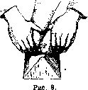
Figure 6.
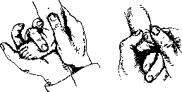
such correction at this age can be performed without anesthesia, by manipulating delicately. The general principle of correction in C. remains the same here: first, the adduction and supination of the foot are corrected, and only after the complete elimination of these two components of C. is the 'equinus' corrected. Manual correction for clubfoot is performed as follows (figs. 5 and 6): with one hand, the leg is grasped around the ankle joint. The anterior part of the foot is grasped with the other hand in such a way that the foot rests on the palmar surface of the hand; the fingers grasp the outer edge of the foot, and the thumb grasps the inner edge. With slow, elastic movements, adduction and supination are eliminated, and at the same time, the hand holding the ankle joint presses with its palm against the calcaneus, creating a firm point of support. The final phase is the correction of the equinus element, and the heel should be brought completely downward and its inward rotation eliminated so that the foot is set in a slight valgus position and the calcaneus. Before eliminating adduction and supination, the equinus position serves as a good lever for all manipulations, and it would be a great mistake to attempt to correct the equinus immediately, let alone tenotomize the Achilles tendon. To correct the equinus in small children, manual corrective movements in the dorsal direction are usually used, while slightly valgizing the foot. The Lorenz technique is also good, in which one hand pulls the foot in the plantar direction with a loop thrown over the dorsal surface of the foot, while the other hand corrects the foot in the dorsal direction. It is necessary to hypercorrect the entire foot along with the heel, not just the anterior part. In persistent cases and in older children, it is necessary to resort to correction with a hinged board or with the support of a regular board against the chest of the physician performing the reduction. With this technique, considerable force can be developed (fig. 7). Correction in a child can be considered complete if a light touch of the finger to the sole creates the correct position of the foot with a tendency toward valgus. In mild cases, correction can be achieved in one or two sessions through manual manipulation without causing significant pain to the child. In severe cases, corrective manipulations have to be repeated several times with intervals. Immediately following correction, a bandage should be applied to maintain the achieved position. The general requirement for the bandage: it should not correct, but only fix the position achieved by manual correction. Among the many bandages proposed for early childhood, the Fink bandage deserves attention, which is also called the Ettingen-Fink bandage and is applied as follows: after manual reduction, the skin is smeared with kleiol from the lower third of the thigh to the toes. The foot to the ankles is covered with cotton, which is tightly bandaged with gauze bandage. A special aluminum sole is applied to the foot (for better correction of the foot as a whole), after which bandaging with a flannel bandage 2a-4 cm wide is begun. The bandage is applied spirally from the thigh around the calf to the inner ankle, then along the sole to the outer ankle, covers the arch, goes along the sole and returns to the outer ankle, from where it is led upward to the thigh, where the bandage is looped by the assistant and pulled tightly. With further spiral turns, the bandage covers the calf, lies over the first loop, pulling it to the calf and thereby abducting the foot (cf. the heel). This is the first loop for pronation of the heel.
Fig 7 bandage-similar to those described, but bandaging can be conveniently combined with frequent repeated corrective manipulations. All this should be done by the physician himself or a trained assistant until the parents, if they are sufficiently cultured, master the manual manipulations and bandaging perfectly themselves. A bandage of adhesive tape, although it holds the position well, irritates the skin, especially in small children, and is practically less suitable. Tregubov, after reduction and hypercorrection of the foot, proposes to maintain the achieved position with a flannel strip that only covers the foot and attaches to the flannel covering the thigh above the knee. Separate ties on the thigh and foot are tightened, with periodic correction performed. A plaster cast, while holding the position perfectly, is less convenient for infants due to frequent dampening and contamination, but with good care and supervision, it can be used even at the very earliest age. The first cast after reliable correction is usually applied for 4-6 weeks. Then manual correction is repeated again to achieve hypercorrection, obtaining pes abductus and a mild pes valgus. A cast is applied again for 4-6 weeks. Usually it has to be changed 4-5 times, sometimes more. In small children, treatment should be continued until the beginning of walking.
After the period of fixation with a cast, there should follow a period of fixation in an apparatus
in which the child must walk for at least a year. The apparatus must be able to maintain the position of abduction and pronation of the foot well. During this period, systematic massage is also necessary. Treatment can be considered complete when the child can actively produce pronation and abduction of the foot with muscular strength. If the child comes to the doctor at the age of 1 year, manual redressation is more difficult; when the child has been walking for several years, manual correction presents serious difficulties, but it should always be attempted before resorting to other types of conservative therapy or surgical intervention. The treatment plan for C. after 1 year of life remains the same as stated above: first complete correction, fixation with a bandage, then an apparatus. After one year, redressation must be performed under anesthesia, and during vigorous manipulations, deep anesthesia is required. Here, not all components of C. can be easily eliminated manually, and one has to resort to a wedge, on which it is convenient to correct adduction and supination (figure 8).-For the purposes of unsafe mechanical redressation of more persistent cases of clubfoot, there are various devices. Such are, for example, 'Thomas's wrench', Gocht's forceps, Phelps-Gocht's, Zwinge's and many others. Their purpose is to replace manual force with mechanical levers, which can be manipulated more vigorously. In this respect, the osteoclast of Schulze (Schulze's table) represents a significant improvement, which, although complex in design, combines all the elements of a redressing apparatus: precise fixation, compression and traction. With the help of the Schulze osteoclast, almost all types of C. can be corrected conservatively, even the most advanced. Correction with the Schulze osteoclast is usually done in stages: first, adduction of the foot is corrected with a loop, then, by applying direct pressure on the talus and combining it with simultaneous correction of supination, it is possible to partially reduce the talus. In one or two sessions, the foot can be placed in a valgus position. It is necessary to ensure that there is no excessive bending of the anterior part of the foot. Then, on the board, the equinus position is corrected, and in persistent cases, open achiloplasty should be resorted to. Redressation in childhood and adolescence usually has to be repeated several times before the necessary hypercorrection is achieved. After each redressation, a bandage is applied to fix the corrected position. For the specified age, the plaster bandage is also irreplaceable, and when applying it, the following conditions must be observed: do not make the cotton padding too thick; areas subjected to the greatest pressure should be well protected, preferably with thin felt plates over the cotton; ensure that the protective layers do not slip, for which the skin in these areas should be smeared with kleol. The following places need to be 'protected' particularly carefully in C., as they are subjected to the greatest pressure: the inner side of the anterior part of the foot and heel, the outer part in the region of the Chopart joint and the outer ankle. Good cotton padding is necessary between the toes, the toes should be spread out, free, and not compressed by the plaster bandage. The soft bandage over the cotton padding should lie smoothly, without folds, and not cutting into the areas of bending. It is now necessary, before applying the plaster bandages, to give the foot its final position so that the plaster bandage does not perform further correction: the plaster bandage should lie freely, not tightening the foot, going from inside to outside, from the dorsal to the plantar side, thus turning the foot, abducting and valgizing it. In children, it is better to apply the plaster bandage slightly above the knee joint, giving it a slightly bent position. This prevents the bandage from slipping down. For this purpose, modeling of the bandage over bony prominences is also necessary. In the first 24 hours, the foot is given an elevated position. If there is suspicious loss of sensation in the toes, the entire front of the bandage together with the soft bandage must be cut and slightly spread apart. The correct application of a plaster bandage in C. is no less important than skillful redressation. Such a bandage should be changed in 3-4 weeks. If, despite vigorous redressation, the deformation cannot be corrected or there is a constant tendency to relapse, then in such cases one has to resort to minor palliative operations, such as excochleatio tali according to Ogston, division of the plantar fascia, lengthening of the Achilles tendon. The latter intervention, however, should be performed only after the adduction and supination of the foot have been eliminated. In particularly persistent cases where complete redressation is not achieved, Erlacher suggests freezing the tibial nerve openly in the popliteal fossa. Treatment cannot be considered complete after correction and even hypercorrection have been achieved. Immediately after the period of correction and fixation with a plaster bandage, there follows a long period of wearing an apparatus with simultaneous massage and active exercises to achieve active pronation. From the moment when well-expressed active pronation appears, there is less risk of relapse. It is extremely important at this time that the heel is slightly valgized. In individual cases, despite complete correction of all components of C., the foot still occupies an incorrect position in relation to the knee joint. This is explained by the twisting of the bones of the leg around the longitudinal axis. In such cases, one has to resort to the extreme but only effective measure-osteotomy of the bones of the leg with rotation of the distal part according to the normal axis of the limb. The treatment of old cases of C. represents the most difficult task. Manual redressation on a wedge and even with the help of the proposed auxiliary redressators (Thomas's wrench) and others does not give a complete effect due to the remodeling of the foot skeleton. After a series of redressing manipulations, sometimes only correction of the anterior part of the foot can be achieved, but this is an illusion of correction, as the rotation of the talus and calcaneus remains uncorrected. The Schulze osteoclast is a powerful means for correcting even advanced cases of C., but due to its high cost, it is available only to a few orthopedic institutions. And its application in inexperienced hands is unsafe. For most old cases of C. in adults, surgical treatment is indicated. Among a number of bloody interventions, some serve only as an additional factor to correcting manipulations. Such are, for example, excochleation of the talus, achillotenotomy. Other operations of a more radical type aim to eliminate the deformation completely by surgical means. Surgical intervention is indicated where closed methods of redressation have not led to the desired results or where it can be assumed a priori that bloodless measures will not be effective. For a long time, the Phelps operation has been used to correct C., consisting in the open division of all soft tissues from the inner side of the foot. Later, osteotomy was added to this. The Phelps operation does not give favorable results and has been abandoned by most orthopedists. Division of the tensing soft tissues from the inner side is sometimes done subcutaneously as a preliminary step before redressation, but no significant effect is obtained from this operation. Of other operations on soft tissues, various types of tenotomies are widely used. Many surgeons consider that achillotenotomy should be done openly and, in severe cases, combine it with division of the posterior ligament (lig. talo-tibiale post.), which is a serious obstacle to the complete elimination of equinus. Individual authors (Broadhurst) recommend division of the tendon of m. tibialis poster., which also represents a serious obstacle to correction. More rarely, one has to resort to division of m. flexoris digitorum longi and tibialis ant.-One intervention on soft tissues rarely leads to complete elimination of the deformation in adults. For this purpose, operations on the skeleton are used. Excochleatio tali is performed through a small incision in front; spongy substance is scooped out from the head, neck and body of the talus so that the cartilaginous membrane remains at least 1/2 cm thick. Thanks to such scooping out, the talus becomes pliable, and correction is easily achieved. This type of intervention is possible in childhood up to 6-8 years. In adults with very significant deformation, complete extirpation of the talus is used, which is fully developed by Whitman and is called astragalectomy. It was first performed in 1892 (Lund). With a literal arc-shaped incision, the articulatio talo-cruralis and talo-calcaneo-navicularis are exposed, the ligamentum talo-naviculare and the outer lateral ligament are divided, after which the talus, grasped with bone forceps, is removed with strong adduction and supination of the foot. If after removal of the talus the correction is hindered by the anterior part of the calcaneus, it should also be resected so that the os cuboideum can be freely placed in hypercorrection of the foot.
Astra-gallectomy is a simple and highly effective operation for severe forms of Clubfoot; it can be performed simultaneously on both feet without first severing the Achilles tendon. Good results are achieved with wedge resection of the tarsus. With a transverse incision through the most prominent bony projection, the tarsal bones are exposed. From the lateral side, a wedge is excised from the calcaneus, talus, and cuboideum so that the wide base of the wedge is turned outward and the apex includes the navicular bone. With a sufficiently large wedge, a fully satisfactory correction is achieved. In stubborn cases with unfavorable position of the calcanei, it is necessary to resort to its osteotomy according to Hohmann. The clinic of Vreden has developed (Kuslik) a method of crescent-shaped resection, which spares the skeleton and eliminates all components of the deformity. After bone operations, a subsequent immobilizing bandage is also required, followed by wearing an apparatus for 6-7 months. Mention should be made of the Wullstein operation, which combines redression and tenotomy with muscle transplantation. After incision of the plantar aponeurosis and detachment from the calcaneus of the mm. abductor hallucis longus, hallucis brevis, they are severed from the navicular bone by the m. tibialis posterior; redression not by wedge. Through a medial incision on the leg above the ankle, the Achilles tendon is lengthened, and the tendon of the m. tibialis post. is also drawn here, which is then passed through the interosseous membrane to the anterior surface of the leg by a special incision and secured subperiosteally to the IV and V metatarsal bones. From the incision on the outer side of the foot, the tendon of the m. peronaei brevis is severed and in severe pronation is sutured to the strong tendon of the abductor digiti V. Plaster in the corrected position for 4 weeks. The Wullstein operation, although based on active preservation of abduction and pronation of the foot, cannot give a significant effect in severe bone deformity. Ludloff, also attaching importance to the t. tibialis poster., transplants it to the peronaeus brevis after preliminary correction. The various operative methods for treating Clubfoot require a critical approach and appropriate indications. They do not exclude subsequent orthopedic treatment. In the presence of a bone defect (more often absence of the inner ankle) or partial defect of the tibia, conservative therapy does not give good results. Even if it is possible to correct the deformity in childhood, a recurrence occurs. In such cases, after correction of the deformity, an attempt should be made to plasticly restore the bone defect or, after correction, to wear an apparatus for a long time. In Clubfoot that arose on the basis of former osteomyelitis, scars on the inner side of the leg and foot are a serious cause of recurrence. In such cases, along with intervention on the skeleton, skin plastic surgery is indicated. Metatarsus varus and pes adductus represent only one component of Clubfoot—adduction—and are easily treated. In adults, functional adaptation usually occurs so well that they do not notice their defect. In childhood, bloodless redression is possible, followed by prolonged wearing of an orthopedic apparatus until the skeleton is remodeled in the tarsometatarsal joint. There are many transitional deformities of the foot (see), which have a specific etiology and a peculiar patho-anatomical picture and require special orthopedic treatment. The methods of conservative and operative treatment of Clubfoot presented, with appropriate selection and systematic application, give reliable and fully satisfactory results. The earlier treatment of congenital Clubfoot is started, the easier it is and the better the results.
Related articles
Mentioned in
Cite this page
“Clubfoot.” Soviet Medical Encyclopedia. English translation of Bolshaya Meditsinskaya Entsiklopediya, 1st ed. (Moscow, 1928–1936), ed. N. A. Semashko. https://sovietmedicalencyclopedia.pages.dev/article/clubfoot/