Flatfoot
Historical document, translated for reference. It reflects medical knowledge of the 1920s–30s and is not medical advice.
Summary
Flatfoot is a deformity characterized by lowering or complete disappearance of the foot arch. This article distinguishes between congenital and acquired flatfoot, with static flatfoot being the most common form, caused by factors like excessive load and weakness of the muscle-ligamentous apparatus.
Encyclopedia article (1928–1936)
FLATFOOT, pes planus, pes valgus, pes plano-valgo-abductus, represents a deformation characterized by lowering, and sometimes complete disappearance, of the arch of the foot. A distinction is made between congenital and acquired flatfoot. Congenital flatfoot is observed very rarely. It arises from primary malformations of the rudiment, from narrowness of the amniotic cavity, underdevelopment of the fibula (in the so-called Volkmann's deformation), and some other pathological factors. Acquired flatfoot, on the contrary, is encountered extremely frequently. Three forms of flatfoot are distinguished: static, traumatic, and paralytic. Static flatfoot is the most common and is one of the most frequent orthopedic deformations. The deformation is almost always bilateral. In its severely expressed form, static flatfoot represents a reformation of the foot, in which the arch is completely absent and the foot touches the ground with all points of its plantar surface. In these cases, the foot resembles a bear's paw, increasing in length and as if spreading in width. The imprint left by such a foot lacks the characteristic inner depression of the human foot. The bones of the foot forming the arch, as it were, slide from their usual places, the articular surfaces become flattened, enlarged, atrophied, completely disappear, and appear in new places. The mutual position of the bones changes. New processes, tuberosities appear on the bones, the attachment points of tendons and ligaments proliferate and enlarge, some bones become rarefied, completely disappear, and sometimes new small bones appear. The ligaments become loose in places, lengthen in places, become dense and shortened, and atrophy. Some muscles relax, stretch, and lengthen, while others contract and pass into a state of chronic contracture, which is reflected in changes in their histological structure. The picture of flatfoot described does not appear immediately; on the contrary, flatfoot develops extremely slowly. Along with cases of severely expressed flatfoot, we distinguish a large number of initial and intermediate stages. There are certain age, racial, sexual, and individual differences in the shape of the foot, differences in the height of the arch, length and width of the foot, differences in the stability of ligaments, development of muscles, etc., which, in themselves representing no pathology, can under appropriate professional or domestic conditions predispose to the development of this deformation. Flatfoot develops when there is a mismatch between the load on the foot and the endurance of its ligamentous-muscular and bony (rickets) apparatus. Age differences of the foot should also be remembered. Thus, the child's foot up to 2 years of age appears completely flat, devoid of an arch. However, this is apparent flatfoot, depending not on deformation of the foot arch, but on the development of the plantar fat layer that fills the entire foot arch and makes it flat. With time, the fat pad partially disappears, the foot assumes its usual contours, and the depression on the plantar surface of the foot increases more and more. The foot begins to grow particularly strongly during the transitional age. If one judges flatfoot by the ratio of the length and height measurements of the foot, then due to the comparatively faster increase in length, there will be a relative decrease in the height of the arch, which however will not yet be indicative of flatfoot. During the transitional age, during the period of foot growth, it is particularly sensitive to load. The etiology of static flatfoot consists of the following main moments: body weight, excessive and prolonged load, and weakness of the muscle-ligamentous apparatus supporting the foot arch. Factors contributing to the development of flatfoot include carrying heavy loads, standing professions, and sedentary occupations. Literary data and works by a number of authors (Smirnov, Klimenkova, Golyanitsky, Kalistov, Khesin, Gorynevskaya, Shtriter, etc.) allow us to conclude that in the pathogenesis of static flat-valgus foot, the decisive role belongs to the conditions of professional labor. E. V. Smirnov found among 848 Astrakhan dockworkers 103 flattened feet, i.e. 121 per 1,000. In contrast to Hoffa, who saw the highest percentage of flat feet in the 16-20 year age group, according to Smirnov's data, the highest percentage of flat feet occurs in the 56-60 year age group (20%) compared to 7.5% in the 26-30 year age group. In the work of a dockworker, the size of the load is of particular importance; the percentage of flatfooted individuals (according to Smirnov) increases in parallel with the increase in the weight of carried loads. Mechanization of loading and unloading labor is of great importance from the point of view of mass prevention of flatfoot. In the origin of flatfoot, fatigue of the muscles of the lower extremities is of particular importance. Flatfoot is noted particularly frequently among weavers-170 per 1,000, and among spinners with considerable seniority-550 per 1,000 (Klimenkova). These data far exceed the frequency of flatfoot in other professions (9 per 1,000 among women and 24 per 1,000 among men in a study of 30,948 workers-data from Golyanitsky). Klimenkova notes the undeniable influence of the duration of work seniority on the frequency of flatfoot. Dynamic studies of the feet of factory school students and adolescents (Kalistov, Gorynevskaya) show that professional overload noticeably affects the foot in terms of its flattening. In addition to professional factors, the development of flatfoot is influenced by weakening of general nutrition (anemia), disturbance of local nutrition of the lower extremities (varicose veins), irrational footwear, hard pavements that deprive the modern human foot of natural support from the ground (Blencke), and rickets. Several theories have been proposed to explain the mechanism of development of flatfoot. Some consider the arch as a system of five arches corresponding to the five metatarsal bones forming them (Riedinger, Fick, Meyer, Haglund), others see in the arch a system of a two-armed lever (Krukenberg, Semeleder), and finally still others see a system of two arches. The last theory, exhaustively developed by the Viennese orthopedist A. Lorenz, enjoys the greatest recognition. He distinguishes between the outer and inner arches of the foot. The outer arch rests on the ground at three points: behind-the calcaneal tuberosity, and in front-the distal heads of the last two metatarsal bones; the apex is represented by the cuboid bone. The inner arch rests in its posterior part on the calcaneus, supported on it by the talus, and in front rests on the ground at the distal heads of the first three metatarsal bones; the apex of the inner arch is represented by the navicular bone and the head of the talus (articulatio talo-navicularis). The passive force strengthening the arch is lig. et fascia plantaris, the active force-mm. tibialis ant. et post., peroneus longus, and the short flexors of the foot. The arch of the foot represents an important organ, the elastic oscillations of which protect the body from rough jolts and shocks when walking and jumping. The violation of the normal elastic properties of the arch begins in connection with functional overload and fatigue of both tibial muscles. Due to their physiological weakness, the short flexors cannot for long replace the powerful long muscles. Deprived of the normal counteraction of the supinators (mm. tibialis ant. et post.), both pronators-mm. peroneus longus et brevis-turn the foot inward, creating a pes valgus position. The navicular bone sinks more and more, stretching the talo-navicular ligament, rich in endings of sensory nerves. In doing so, shifting forward, the navicular bone deprives the talus of its support, whereby the head of the latter slides down and becomes wedged between the navicular and calcaneus bones. In the extreme degree of flatfoot, the navicular bone and the head of the talus reach the
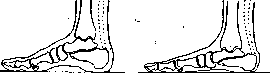
Figure 2.
of the floor (compare fig. 1-normal with fig. 2-severe flatfoot), the foot is pronated and its anterior part is turned outward-pes plano-valgo-abduetus. Flatfoot is often accompanied by hallux valgus and a lowering of the transverse arch of the foot, formed in front by the heads of the I-V metatarsal bones. Clinically, flatfoot manifests with painful symptoms and a disruption of the normal contours of the foot. The faster flatfoot develops, the more severe the pain, and vice versa. Flatfoot develops particularly quickly and painfully between the ages of 16 and 20 years in young people who, having poorly developed musculature, engage in work involving intense loading of the lower extremities-pes-valgus adolescentium, as well as in those who have not fully recovered from a severe illness if they too quickly resume physical work. A similar clinical picture can develop in obese people who, accustomed to a sedentary lifestyle, suddenly switch to a profession involving prolonged standing. In all these cases, a painful reflex-spastic contraction of the peroneal muscles may develop, so that the tendons of these muscles stand out sharply under the skin behind the lateral malleolus with a strongly pronated foot-pes planus contractus. In slowly developing static flatfoot, weakly expressed pains, aching, and rapid fatigue are observed. Pain sensations are least pronounced in the rachitic form of flatfoot in children. This form is characterized by a sharp settling of the arch when standing and its rapid restoration when transitioning to a sitting or lying position. In rachitic flatfoot, a significant role is played by the decreased elasticity of the foot skeleton itself due to its decreased calcification. Pain in all forms increases with walking and standing, especially by the end of the day; after rest, the condition always improves. Pain can be localized in areas corresponding to the overstretched muscles and ligaments of flatfoot and the compressed nerves, or it can radiate upward, as far as the buttock and lower back. Most often, pain is noted on the sole, in the area of the navicular bone or on the dorsum of the foot in the same area, at the inner edge of the heel, under the inner and outer malleolus, between the heads of the metatarsal bones, and in the calf muscles. In the latter, specifically in the substance of the tibial muscles, especially the posterior one, fairly painful nodules of induration can sometimes be palpated upon deep palpation.-As for the external form of the foot, it appears relatively elongated, widened in the middle part, the heel part is also somewhat widened, the longitudinal arch of the foot is lowered (fig. 3 and 4), the entire foot is valgus, the navicular bone is clearly outlined through the skin; the vertical axis of the heel forms an open angle outward with the line of the Achilles tendon, which can be easily verified when examining the patient from behind in a standing position. Flatfoot is characterized by an awkward gait, they excessively turn the toes outward, cannot run quickly, and jumps are often painful. Active and passive movements are limited, especially supination.
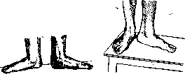
Figure 3.
Figure 4. The diagnosis of flatfoot in severe cases is very easy, but in early, initial forms, when the deformation is not yet visible to the eye and when treatment is especially effective, recognition, on the contrary, is often very difficult. In such cases, special research methods are used: plantography, radiography, or podometry. Plantography consists of obtaining an imprint from the plantar surface of the foot. For this purpose, it is most convenient to first coat the entire sole with a 10% aqueous solution of tannin, then have the patient stand on a sheet of clean paper and immediately after this spray the paper with a 10% aqueous solution of ferric chloride. The size of the uncolored part of the depression in the middle of the foot reflects the degree of depth of the arch. (Figures 5 and 6 give an idea of the plantographic prints of a normal and flat foot.) For comparing the obtained mass footprints, a number of methods for analyzing the obtained prints have been proposed (Perthes' method, Chizhin's method, the method proposed by the Leningrad Institute for the Study of Occupational Diseases). All these methods are based on measuring the ratio of the dark and light parts of the obtained print in its middle part. The radiography method reveals all
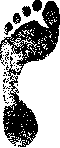
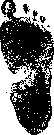
Figure 5.
quite clearly various changes in the skeleton, on the basis of which it is possible to determine fairly early stages of flatfoot. The method of podometry, developed by Fridland, seems practical and convenient. It can be performed using a compass and a measuring tape. The essence of the method consists in determining the percentage ratio of the height of the foot's arch to its length. First, by spreading the legs of the compass, determine the height
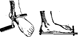
Figure 7.
Fig. 8. The height of the foot from the floor to the upper surface of the navicular bone, which is palpated approximately one finger forward from the ankle joint. The magnitude of the divergence of the compass legs is noted on the measuring tape. Then, with the same instrument, the length of the foot is measured from the tip of the big toe to the posterior curvature of the heel, marking these points beforehand with a pencil on paper. Having both measurements in millimeters, multiply the height of the foot by 100 and divide the product by the length of the foot. Thus, the desired podometric index is obtained. For greater convenience of measurement, the author of the method has proposed two models of a special instrument—the 'podometer.' Figs. 7 and 8 show measurements of the height and length of the foot using one of these instruments. An index within 29-27 is found in most people; an index from 27 to 25 indicates flatfoot; an index below 25 indicates severe flatfoot. Thus, we have two main methods for studying and characterizing the foot. The first method—characterization of the supporting surface of the foot—was widely used by Chizhin, the Leningrad Institute of Occupational Diseases (Shtriter), Gorinevskaya, and Kalistov. The second method—direct measurements of the dimensions of the foot (length, width, and height) and their interrelationship (proposed by Fridland). As shown by the work of Gorinevskaya, both methods correlate with each other, and therefore in mass examinations, one can use whichever is more convenient and simpler. These methods are of great service in medical examination and military conscription commissions, in professional selection, in mass examinations, and in dispensaries. Flatfoot must be differentiated from gout, Achilles bursitis, calcaneal bursitis, calcaneal spur, chronic muscular rheumatism, Köhler's disease, and sometimes even from tuberculosis of the foot, sciatica, and some other diseases. Mass examinations show that the norm of the foot arch cannot be expressed by any single number; it is necessary to note certain limits of variation typical for a particular age, sex, and professional group. When evaluating the feet of different groups, we can speak of arches belonging to the typical group, above the typical and below the typical, low, very low, and finally the group of flat feet (flatfoot), which is already a pathology. From the point of view of mass prevention, we should be interested not only in persons suffering from flatfoot, but also in groups with a low foot arch, groups with the initial stages of flatfoot. In cases of pronounced flatfoot, serious therapeutic measures, change of profession, etc., are necessary, while in cases of initial stages of flatfoot, with a low foot arch, preventive measures are necessary. Early detection of such initial flattening of the foot is possible only through widespread population dispensarization; it is especially indicated in youth, among adolescents and students in factory-apprentice schools. Preventive measures include: selection of an appropriate profession not involving prolonged standing or carrying heavy loads; special physical exercises aimed at strengthening the muscles of the foot and leg; general physical exercises that strengthen the entire muscular system of the adolescent, develop correct gait, and provide proper work skills. If a change of profession and lifestyle is not possible, timely use of supinators can stop the beginning flattening of the foot. Individual prevention of flatfoot should begin in childhood with the cultivation of correct gait. When standing and walking, the toes should point almost straight forward and in any case not diverge in a lateral direction by more than 5-8°. Persons in professions requiring prolonged standing are advised to rest during the day several times for a few minutes on the outer edges of supinated feet, and at the end of the workday to take warm foot baths up to the knees, followed by massage of the foot arch and leg along the front and inner side. Among physical exercises, walking barefoot, walking on tiptoes, running, jumping, playing basketball, volleyball, and football, and swimming are especially indicated. The correct construction of footwear is of extremely great importance. It must exactly correspond to the dimensions of the foot, the medial edge of the shoe must be straight, in no way turning the big toe outward, the height of the heel should be within 3-4 cm, and the arch support (insole under the arch) should be made of sufficiently elastic material. In terms of the prevention of flatfoot, great benefit can be provided by physicians working in professional selection and physicians in charge of medical supervision of athletes. Treatment. In the early stages of flatfoot, warm foot baths, massage, and corrective gymnastics have a significant effect: squatting on supinated and adducted toes, climbing a pole, and simply vigorous supination movements of the feet in a lying position. In severe pain—bed rest, baths, and injections of a 0.5% solution of novocaine (up to 30.0) into the area of painful points. Some recommend for pes planus contractus to freeze the peroneal nerves with chloroethyl (Stracker) or even crush the nerves with fingers. In both cases, the nerve is exposed by an incision at the head of the fibula. In already developing but not fixed deformity, and especially in rachitic flatfoot, good results are obtained by redressation. The operation is performed under deep anesthesia. First, the adduction of the anterior part of the foot along the line of the Chopart joint is eliminated, then the arch is bent upward, and finally the foot is brought into a varus position, for which the Achilles tendon is first subcutaneously severed; a plaster cast is applied for 3-4 weeks, and after the specified time, the patient is prescribed supinators to wear constantly during the day. They can be purchased ready-made, but it is much better to make them from an individual plaster model. The bare foot is wrapped in plaster bandages, which, after hardening, are cut with a knife along a previously laid string on the dorsum of the foot. The resulting negative is exactly folded along the cut line and filled with liquid plaster paste. After half an hour, the latter hardens, and then the negative is removed from the positive. The positive is smoothed with the blunt side of a knife; accordingly, the plaster (by a few millimeters) is scraped out in the area of the arch, the entire plaster is rubbed with glass paper, powdered with talc, and the model is ready. On it, with a pencil, the outline of the future supinator is drawn: its leather part, metal lining, and steel spring plate. In this form, the model goes to a prosthetics master, who prepares the apparatus itself. Fig. 9,
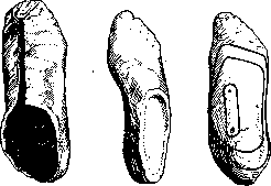
Fig. 9.
Figs. 10 and 11 illustrate the described stages of preparing the negative, positive, and model, and Fig. 12—the supinator in the form in which it performs its function in the shoe under the foot arch. Some surgeons, to enhance the effect of redressation, add a muscle-plastic operation to it—transplantation of half of the Achilles tendon to the posterior tibial muscle (Nicoladoni)—or shorten the posterior tibial muscle (Hoffa), or transplant the entire Achilles tendon to the medial side of the heel (Anzoletti, Gocht). Fridland transplants the long fibular muscle to the inner edge of the foot into a split in the periosteum near the navicular bone.—In chronic flatfoot, when there are persistent and coarse changes in the bones, more complex interventions have been proposed. The best results are obtained by a combination of me
M_,-----' on 2 toes ni
Fig 12 shows the ankle and ending at the distal head of the first metatarsal bone. The long abductor of the great toe is pulled downward. The periosteum of the navicular bone is incised vertically, and a wedge-shaped piece is excised from it, with the base up to 2 cm, directed medially and downward. The bone wedge is temporarily kept in a gauze tampon. An arcuate incision is made below the outer ankle, extending to the heel bone. At a distance of 1 finger's breadth from the calcaneocuboid joint, a vertical osteotomy of the heel bone is performed. The foot is then forcefully corrected (adduction, inversion, supination). As a result, the gap in the navicular bone closes, while that in the heel bone widens, creating space for the implantation of the bone wedge taken from the navicular bone (figures 13 and 14). The periosteum over the latter is sutured. After this, the second part of the operation is performed: the tendon of the anterior tibial muscle is incised lengthwise, its anterior half is detached from the bone, passed under the intact half to the attachment site of the posterior tibial muscle, and here fixed with silk transplantation. The rapa is sutured. A plaster cast of high density is applied in slightly hypercorrected position for 4 weeks. Then, for several weeks, warm foot baths and gymnastics are prescribed. Traumatic flatfoot (pes planus traumaticus) is rare, usually occurring as a unilateral deformity after a fracture of the outer ankle, but it can occur after more complex injuries in the ankle joint area and after various destructions of muscles controlling the foot. Deformation can be prevented by proper immobilization of the foot in the very early stages of injury and subsequent proper functional treatment of the main condition. Paralytic flatfoot (pes planus paralyticus) is second only to static flatfoot in frequency. It arises from anterior poliomyelitis and only exceptionally after other types of nervous system lesions. It can be unilateral (figures 15 and 16) and bilateral. In paralytic flatfoot, both tibial muscles and triceps surae are most severely affected. On the contrary, the long extensor of the great toe is mostly spared. Clinically, emaciation of the limb, cyanosis, and decreased skin temperature are striking, so the affected foot and leg feel colder than on the healthy leg; the tendon of the long extensor of the great toe is tense and causes hammer-like curvature of the great toe. Movements in the foot joints, in contrast to static flatfoot, are not only not decreased during passive manipulation, but even increased, while active mobility is sharply reduced. Prevention of paralytic flatfoot consists of physiological fixation of the foot at a right angle to the leg and in a slight
varus position from the very first moments of detecting paralysis. Splints and rubber traction serve as fixing devices. The latter can be adapted to the boot. At the same time, regeneration of the nervous system and strengthening of the muscles are achieved through warm baths, massage, and electrification, preferably of the supinating muscle groups. - Treatment depends on the degree of paralysis. In mild cases, an orthopedic boot with elastic traction is sufficient, while in severe cases, correction combined with muscle plastic surgery is necessary. The most recommended procedure is the transplantation of the peroneus longus muscle to the medial edge of the foot, to the area of the navicular bone. The transplanted muscle is passed under the skin and sutured under the periosteum with sufficient tension. To this is added the shortening of the posterior tibial muscle. In severe valgus of the heel, Anzolotti-Gocht's suture is added (see above). Finally, a plaster cast is applied. After a month, it is removed, warm foot baths, massage, and light elastic traction (supinating and slightly adducting the foot) are prescribed for several weeks.
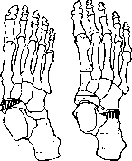
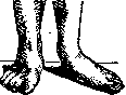
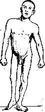
Figure 15.
Figure 16. varus position from the very first moments of detecting paralysis. Splints and rubber traction serve as fixing devices. The latter can be adapted to the boot. At the same time, regeneration of the nervous system and strengthening of the muscles are achieved through warm baths, massage, and electrification, preferably of the supinating muscle groups. - Treatment depends on the degree of paralysis. In mild cases, an orthopedic boot with elastic traction is sufficient, while in severe cases, correction combined with muscle plastic surgery is necessary. The most recommended procedure is the transplantation of the peroneus longus muscle to the medial edge of the foot, to the area of the navicular bone. The transplanted muscle is passed under the skin and sutured under the periosteum with sufficient tension. To this is added the shortening of the posterior tibial muscle. In severe valgus of the heel, Anzolotti-Gocht's suture is added (see above). Finally, a plaster cast is applied. After a month, it is removed, warm foot baths, massage, and light elastic traction (supinating and slightly adducting the foot) are prescribed for several weeks.
M. Fridland. From the point of view of military service, flatfoot is not an obstacle to service in the army. When assessing fitness for military service of persons with flatfoot, as with other defects, one should primarily consider the degree of functional disorders caused. Since flatfoot in the vast majority of cases does not cause any functional disorders, it as a rule is not a reason for exemption from military training. Obstacles to military training in the Red Army can be the relatively few accompanying severe deformities of the foot shape, such as toe adduction, their deviation to the side, etc., but these defects are provided for by special articles of the schedule of diseases. Similarly, cases of acute uncompensated flatfoot limit acceptance for military service, at least in regular units. Persons with complete flattening of the plantar arch are formally considered fit for non-combat service in the Red Army. However, the bodies determining fitness for military service and army physicians are obliged, when assessing fitness for service (in the Red Army in general or in particular branches of the armed forces) of persons having various defects, including flatfoot, to apply the method of individual assessment taking into account a number of factors [character of work before service, degree of functional impairment (according to case history and medical observations), character of work at present time, etc.].
Related articles
Mentioned in
Cite this page
“Flatfoot.” Soviet Medical Encyclopedia. English translation of Bolshaya Meditsinskaya Entsiklopediya, 1st ed. (Moscow, 1928–1936), ed. N. A. Semashko. https://sovietmedicalencyclopedia.pages.dev/article/flatfoot/