Malformations
Historical document, translated for reference. It reflects medical knowledge of the 1920s–30s and is not medical advice.
Summary
Malformations are persistent congenital abnormalities in the relationships between parts of an organism that occur during individual development. This article discusses classification, frequency, experimental reproduction, and correlations between different malformations.
Encyclopedia article (1928–1936)
Malformations, persistent congenital abnormalities in the relationships between individual parts of an organism, arising during individual development and going beyond the variations of these relationships in the given species. Malformations may affect the structure of the organism as a whole or its major anatomical regions, or be limited to abnormalities in the structure of individual systems, organs, parts of organs, and even groups of cells (for example, birthmarks). Depending on the extent and degree of such developmental defects, 'severe malformations' (Monstrum, Monstrosity) and anomalies are distinguished; in reality, there is no sharp boundary between these, just as there is no sharp boundary between an anomaly and a variation. If an anomaly occurs frequently in a race, it is sometimes extremely difficult to distinguish between it and a variation. Thus, from physiological structure to malformations there is a chain of variations and anomalies connecting these two extreme links. Examples of such transitions are monozygotic conjoined twins—symmetrical double malformations (thoracopagi, xiphopagi, etc.)—asymmetrical double malformations. A further continuation of this teratological chain is inclusio fetalis, teratomas (see), mixed tumors, ordinary tumors from embryonic rudiments. Thus, malformations belong to the so-called dysontogenies. This term, proposed by Schwalbe (E. Schwalbe), combines all violations of ontogenesis that manifest in one form or another during fetal or post-fetal development (for example, tumors from dysontogenetic tumor rudiments). Corresponding to the phases of normal ontogenesis, the moment of origin of malformations (the so-called teratogenetic terminal point, see Teratology) may be in the change of genotype, or in abnormal blastogenesis, or in abnormal organogenesis. In this group of malformations, those changes that arose as a result of reactive processes (inflammation, regeneration during embryogenesis) are also considered, although some researchers (V. Fischer) exclude them from the group of malformations. Arising as a developmental defect, malformations are mostly accompanied by growth disturbances. Nevertheless, there may not be complete parallelism between the violation of development and the violation of growth, and malformations sometimes even show a marked dissociation between growth and development (for example, eunuchoidism, dwarfism, precocious puberty, etc., in which there is a halt in development with continued growth or vice versa).

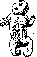
Distribution of malformations. Malformations occur at all levels of the animal scale. Experimental reproduction of many of them that occur spontaneously in some animals (insects, amphibians, reptiles, birds) is one of the most essential methods for studying the mechanism of their occurrence. Statistics on human malformations do not exist due to the poor clinical definition of this concept and the absence of sharp boundaries between malformations, anomalies, and variations. Elements of disturbed development in one form or another (sometimes microscopic) are found in almost every person (birthmarks, skin horns, patent foramen ovale, lobulation of kidneys, lobulation of lungs, accessory spleen, accessory pancreas, kidney cysts, Meckel's diverticulum of the intestine, etc.). Schwalbe finds such changes in at least 50% of all autopsies. Severe malformations occur, according to imprecise data (G. Gruber), in 1-3% of all corpses. The frequency of malformations of one or another organ or system in humans is proportional to the complexity of their embryonic development pathways. Malformations most often occur in the urogenital apparatus, brain, heart, face, and skull. The rule is the multiplicity of malformations in the same individual, depending on the correlative or relative connection of normal, as well as pathological development between different organs or systems. Such a connection has been experimentally demonstrated by Dürken (Darken), who obtained a combination of developmental defects of the central nervous system and hind limbs when damaging the embryonic rudiments of one of the corresponding anatomical regions. The presence of such a connection is also found in spontaneous human malformations ('syngenetic malformations') (Fig. 1 and 2). Malformations of many organs and systems show a certain connection with sex. Thus, for example, according to Lorenz (Lorenz), congenital hernias, 'Figure 1. Amniogenic malformation. Partial exencephaly; remains of amnion, fused with the skull.'
Figure 2. Combined (syngenetic) malformation: arhinencephaly, underdevelopment of shoulders, synostosis of the right elbow joint, flat foot, etc. Coxa vara occurs in the vast majority of girls (671 girls to 82 boys), urethral cleft is also much more common in girls than in boys (Motzfeld); most anencephalics are also girls. Double M. (see below) are especially common in girls. On the other hand, congenital heart defects, diverticula of the bladder are more common in boys. Pathogenesis of M. The mechanism of the occurrence and development of M. lies in a deviation from normal development, mostly during embryogenesis. These deviations consist either 1) in inhibition of development (hypoplasia), absence (agenesis) or underdevelopment of an organ or system ("monstra per defee-tum"); 2) in excessive development or duplication of an organ or the entire organism ("monstra per excessum"); 3) in splitting of an organ either due to non-fusion of paired parts of the organ ("primary splits", for example cleft palate, hare lip), or splitting of an already formed organ by an amniotic constriction ("secondary split"); 4) in a completely unique, sharply distorted embryogenesis ("monstra alienantia"). Often in the formation of M. various mechanisms participate, either in simultaneous combination or following one another. Thus, for example, as experiments by Barfurth, Tornier, Przibrarn and others have shown, superregeneration after a preceding defect may have significant importance. This is the probable mechanism of the occurrence of certain forms of polymelia and polydactyly in humans. Furthermore, as experiments by Przibrarn and others show, the occurrence of M. may be the result of heteromorphosis, i.e., due to the development of another organ "during regeneration in place of a lost one. The occurrence of M. may also be the result of displacement and transplantation of tissues with a certain prospective potency into other parts of the embryo. In this way various M. have been obtained in lower animals (true twins, parasitic twins, sometimes similar to human teratomas and teratoid tumors, etc.) (Born, Harrison, Spemann, Mangold, Schaxel, Braus and others). The causes that lead to the occurrence of M. consist either in properties of the embryo itself (internal teratogenic factors) or in properties of the environment (external teratogenic factors). Internal teratogenic factors consist of hereditary or non-hereditary changes in germ cells, which are the cause of the development of M. both during the intrauterine period and in extrauterine life. Genotypic factors can be realized either by direct transmission of a gene for a specific malformation or through genotypically conditioned defects of the amnion, placenta; this can be the cause of the occurrence of although different M. in members of the same family, but nevertheless genotypically conditioned. Thus, the cause of the occurrence of this group of M. must be sought in genotypic changes, although they can be realized in later periods of development. To reveal the genotypic factor in the occurrence of M., a thorough genetic analysis is necessary in each individual case, since the presence of malformations in several siblings may be associated with pathological changes in the mucous membrane of the uterus in the mother or with other changes in the environment and by no means yet indicates in favor of heredity of this M. Genetic analysis gave some grounds for the conclusions that a number of M. (dwarfism, gigantism, double M.) are not hereditary and their occurrence is associated either with violations of the conditions of pregnancy or with non-hereditary changes in germ cells (polyploidy of spermatozoa, etc.). On the other hand, the following M. with dominant and recessive heredity are known: M. more or less completely dominantly inherited - congenital dislocation of the hip, syndactyly, polydactyly, brachydactyly, hyperphalangia of the thumbs, prognathism, hare lip and cleft palate, ectopia of the lens, partial albinism, etc. Recessively inherited are: M. of the inner ear with deaf-mutism, some forms of chondrodystrophy and flatfoot, complete albinism, etc. The given list concerns only hereditary M. in the narrow sense of this concept, not touching the large group of hereditary diseases. But even with respect to M. it is undoubtedly incomplete due to the little study of this question. It is necessary to note that one and the same M. can be the result of both internal and external factors. External factors in the occurrence of M. These are various physical, chemical, infectious-toxic and neuropsychic effects on the developing embryo. Assumptions that teratogenic changes can arise under the influence of external factors in germ cells even before fertilization, causing either stable, congenital, ugly non-hereditary modifications or even being the cause of the occurrence of mutations, have also proved quite plausible. The most important of the external factors are the following: 1. Physical factors: 1) mechanical: single or prolonged (for example pressure) trauma. Experimentally in anamniotes, various mechanical traumas of the embryo (shaking, pressure, partial detachment of cells of the first cleavage, punctures, incisions) caused the appearance of various M. In anamniotes with an egg protected by a shell and in amniotes, the significance of such mechanical effects from the outside for the occurrence of spontaneous M. is significantly reduced due to protection by the shell or amniotic fluid. Apparently these factors have even less significance for humans. 2) Sensitivity of the embryo of some animals to gravity with changed position. M. occur in this case apparently as a result of redistribution of substances of different specific gravity, as a result of which normal development is disrupted. 3) Abnormal temperature. The amplitude of temperature fluctuations compatible with normal development varies for different embryos; for example, trout develops at temperature 0-15°, chick 25-42°. Transition beyond these boundaries experimentally caused M. According to Stockard, the action of lowered temperature is carried out through a decrease in oxidation. The significance of the temperature factor (increase in temperature during fever) in human teratology is problematic; nevertheless, from this point of view, the obtaining of double M. of tritons at increased temperature is of interest. 4) Radiant energy or absence of light. Absence of light completely stops the development of frog eggs (experiments of Jung, 1878). The teratogenic effect of radium and X-ray rays on selachians, reptiles, birds has been established. Experimentally the influence of X-ray rays on the formation of abnormal germ cells (O. Hertwig, 1911) and on the occurrence of mutations (Muller, 1928) has been shown. The teratogenic influence of these rays on the human embryo and germ cell is not excluded. 5) Abnormal osmotic pressure, which, as experiments show, affects differently in different periods of development. Hertwig obtained in frogs and axolotls various M. of the nervous system, associated with inhibition of development (anencephaly, spina bifida, etc.) with an increase in the concentration of NaCl in the water. For humans the significance of osmotic pressure is not proven, but can be assumed in various inflammatory processes of the mucous membrane of the uterus and changes in the composition of maternal blood. 6) Changes in the amnion occupy a completely special place in human teratology among other factors of a physical-mechanical nature (so-called amniogenic M.). Amniotic threads (see Amniotic threads, 40S, constrictions, adhesions), forming either secondarily due to adhesions of the embryo with the amnion or primarily as a remnant of the supposed primary multicamerality of the amnion (Grosser, 1925), are often the cause of the most diverse M. (spontaneous amputations with subsequent, sometimes excessive regeneration, formation of splits, prolapses of internal organs, adhesions and scars of the skin, malformations of the skull and brain, oblique facial cleft, agenesis of organs, etc.) (fig. 3). Narrowness of the amnion and oligohydramnios, in which the amnion loses the ability to protect the embryo from unilateral pressure and in turn presses on it, primarily on the protruding parts of the embryo (head, extremities), can also lead to M. The adhesions forming between the amnion and the fetus with movements of the fetus or increase in fluid are

drawn into the above-mentioned
Figure 3. Combined (syngenetic) amniogenic malformation of the skull and
Diagnosis of similarity of M. requires great skill.
caution; only experience shows that the above-mentioned Malformations are usually caused by anomalies of the amnion. Meanwhile, insufficient closure of the medullary ring, persistence of the embryonic facial fissure, etc., Malformations may be the result of either amniotic defects or weakness in the growth of the edges of these openings due to internal (or external, but of a different nature, e.g., chemical) causes. Therefore, Schwalbe (1906) sets two requirements for diagnosing the amniogenic origin of Malformations: 1) the presence of amniotic adhesions or threads at the site of the Malformation; 2) the mechanism of the Malformation must be understood purely mechanically by means of threads or adhesions. However, these conditions are not always met even when the amniogenic origin of a particular Malformation has been empirically established beyond doubt, since the adhesions that caused the Malformation may later rupture or be resorbed. Therefore, the amniogenic origin of certain Malformations, e.g., cyclopia (Fischel), is hypothetical. 2. Chemical factors. These include lack of oxygen, which experimentally caused Malformations in insects and some lower vertebrates (Dewitz). Stockard explains the effect of this factor as follows: under normal conditions, the rate of development of different parts of the body varies; during temporary asphyxia, it is temporarily reduced to zero, and the ability of different parts to outpace others is temporarily eliminated. When development resumes, other parts develop abnormally, creating abnormal, malformed relationships. For humans, the influence of this factor is possible either due to local changes in the uterine mucosa or due to general asphyxia of the mother. The importance of the chemical composition of the medium is inseparable from osmotic pressure. A number of Malformations (e.g., cyclopia) have been obtained experimentally in lower vertebrates by adding magnesium chloride, alcohol, chloroform, ether, etc., to water (Stockard, McClendon, Werber). Dürken believes that in teratology, chemical factors are underestimated compared to mechanical ones, all the more so because the effect of a chemical factor on the embryo is territorially less limited than that of a mechanical one, although it can be limited by the different affinities, resp. different sensitivities, of different cells to the same substance. Chemical factors can cause not only single but also double and multiple Malformations. The introduction of a cytotoxic serum with respect to the lens into the blood of a chicken caused prolonged somatic induction in the form of blindness in several generations. In humans, the teratogenic significance of altered maternal blood (nephritis, alcohol, morphine, various medications, etc.) can be assumed. 3. Infectious-toxic influences on the fetus occupy a prominent place in teratological literature, although according to the latest data, changes caused by infection in the egg membranes are probably of particular importance. The effect of the infectious agent on the fetus is largely shielded by the placental barrier, and in case it crosses this barrier, it is unlikely to have practical teratological significance due to the death of the fetus. From the point of view of Bromann, infectious diseases of the egg membranes are of much greater importance. Mall (Mall, 1910) found inflammations of the chorion in aborted eggs with a malformed fetus and explained the occurrence of Malformations by the change in the chemical composition of the medium during inflammation, which is also confirmed experimentally. Mall also explains the frequency of defective embryos in tubal pregnancy by abnormal environmental conditions in the tube. Nevertheless, the occurrence of tubal pregnancy due to a primary defect of the egg is not excluded. In addition, inflammations of the membranes and their adhesions may contribute to the occurrence of amniotic defects. 4. Nervous-psychic influences, as directly causing Malformations, are denied. However, the teratogenic significance of this factor, mediated through uterine contractions, circulatory disorders, changes in the hormonal composition of the mother's blood, etc., is not excluded. It is necessary to emphasize that the same Malformation can arise as a result of both external and internal factors, just as different external factors often activate the same teratogenetic mechanism with the same outcome; e.g., duplicitas anterior has been obtained by shaking (Wilson, 1893), pressure (Fischel, 1898), severing a blastomere (Spemann, 1903, 1904), decrease in osmotic pressure (Loeb, 1895), increase in osmotic pressure (Bataillon, 1901), increase in temperature (Driesch, 1893). At the same time, the same cause can lead to different Malformations. Classification of Malformations. All Malformations are divided into single and double. The old attempt at a pathogenetic systematization of Malformations created the following groups: 1) monstra per defectum; 2) monstra per excessum; 3) monstra per fabricam alienam. Despite the imperfection of such a classification, it has persisted to the present day. Přibram (1920) retained it, simplifying only the names: 1) defects; 2) excesses; 3) aliena. As Gruber points out, when assigning Malformations to one group or another, caution is required, since a defect can be the result of inhibited development or agenesis and atrophy of an already formed organ; doubling can be the result of inhibited development ('defect', e.g., bicornuate uterus) and excessive regenerative development ('excess', e.g., certain forms of polydactyly); at the same time, excess development can be the result of preservation of the embryonic state (e.g., persistent thymus). Course of Malformations. Most Malformations develop during the fetal period and by the time of birth of the fetus are stable, having a more or less constant character. However, some forms of Malformations exist for a long period in a latent form and are realized in later age periods (eunuchoidism, dwarfism, gigantism, Malformations of the inner ear with deaf-mutism, congenital dislocation of the hip, etc.). Many Malformations do not undergo significant changes during life (hare lip, polydactyly, bicornuate uterus, etc.), while others may progress, increasing disproportionately to the growth of the entire organism, sometimes being a source of severe lesions (e.g., birthmark-melanoma). The clinical significance of Malformations depends on the severity of the functional disturbances they cause. Some of them are not associated with any functional disturbances (e.g., menstruation and ability to conceive in a bicornuate uterus, etc.), while others cause severe lesions of the corresponding systems, sometimes incompatible with life (clinical significance of Malformations-see separate organs). According to viability, the following groups are distinguished: 1) death in the early embryonic period. In such cases, death usually occurs within the first two intrauterine months without apparent causes. Experimental genetics indicates the lethal effect of certain genes. 2) Viability in utero, but non-viability extrauterine (some heart defects, Malformations of the central nervous system, respiratory organs, digestive organs, excretory organs, some double asymmetric Malformations, etc.). 3) Viability in the extrauterine period (Malformations of the limbs, small parasitic forms, cleft palate, hare lip, etc.). Some Malformations are compatible with life in themselves but promote rapid infection of the corresponding areas (defects of the skull, spine, abdominal wall-meningitis, defects of the abdominal wall-peritonitis, etc.). In addition, many Malformations create a slight vulnerability of the organ and predispose to various diseases (e.g., horseshoe kidney, creating prerequisites for urinary stasis and stone formation). It is also necessary to note that developmental defects of some organs (adrenal glands, thyroid gland) are accompanied by severe general constitutional disorders. The development of some Malformations often causes a severe course of pregnancy and labor, sometimes fatal for the mother (double Malformations).
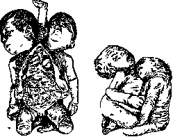
Double and multiple Malformations represent a special group of Malformations that develop due to incomplete separation of twins (Fig. 4). Double Malformations (so-called duplicitus-duplicity) are most commonly encountered. In this case, the distinction is made between symmetrical and asymmetrical dicephalus, tribrachius, tetrapus, thoracopagi, rhamphus. pa - meningitis, defects of the abdominal wall-peritonitis, etc.). In addition, many Malformations create a slight vulnerability of the organ and predispose to various diseases (e.g., horseshoe kidney, creating prerequisites for urinary stasis and stone formation). It is also necessary to note that developmental defects of some organs (adrenal glands, thyroid gland) are accompanied by severe general constitutional disorders. The development of some Malformations often causes a severe course of pregnancy and labor, sometimes fatal for the mother (double Malformations).
Figure 4. Anterior duplication: ris. 5. dissymmetrical dicephalus, tribrachius, tet
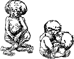
Figure 6. Double Malformation: symmetrical, Figure 7. Thoracopagus cephalothoracopagi.
Figure 6. Double Malformation: symmetrical, Figure 7. Thoracopagus cephalothoracopagi.
parasiticus. They distinguish two main types of this M.: 1) dupl. symmetros (gemini aequales), in which both fetuses are developed symmetrically and uniformly, and 2) dupl. asymmetros (gemini inaequales), in which one fetus develops correctly ('autosit') and is the carrier of another ('parasite'), lagging in development and attached to the autosit on its surface or inside its cavities. As experiments show, the parasite lags in development from the very earliest phases of embryogenesis. In symmetrical double M., three types of connections are distinguished: 1) vertical, 2) horizontal, 3) medial. Vertical in turn is divided into ventral and dorsal (see Thoracopagus, Pygopagus); horizontal into cranial and caudal (see Craniopagus, Ischiopagus); finally, in medial connection, both individual parts have a common axis in one part of the body, while in the other part the axes diverge-dupl. anterior, media, posterior (the latter is extremely rare in humans). As for dupl. asymmetros, depending on the place of attachment of the parasite to the autosit, they speak of parasitic craniopagus, ischiopagus, thoracopagus, pygopagus, etc. Double and multiple M. always develop from one egg, are of the same sex, have a common placenta, common chorion, and often a common amnion. As for the mechanism of formation of double and multiple M., as experimental research (Born, Spemann and his school) has shown, both the connection of initially free twins and their incomplete separation are possible here. Spemann's experiments establish* the teratogenetic termination period for double M. only until the end of gastrulation; for humans this period lasts until the formation of the embryonic disc and the amniotic cavity. In humans, double M. apparently always form by incomplete constriction of the embryonic rudiments, since with a double amnion-twins are always free. Spemann succeeded in obtaining various forms of double M. by transplanting rudiments of one embryo to another. Similar M. could also be obtained by various physical and chemical influences (Loeb, Driesch, Wilson, etc.). M. of individual organs- see these organs and Fetus, malformations of the fetus.
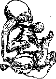
Figure 8. Ischiopagus parasiticus.
mental research (Born, Spemann and his school), here possible are both the connection of initially free twins and their incomplete separation. Spemann's experiments establish* the teratogenetic termination period of double M. only until the end of gastrulation; for humans this period lasts until the formation of the embryonic disc and the amniotic cavity. In humans, double M. apparently always form by incomplete constriction of the embryonic rudiments, since with a double amnion-twins are always free. Spemann succeeded in obtaining various forms of double M. by transplanting rudiments of one embryo to another. Various physical and chemical influences also made it possible to obtain similar M. (Loeb, Driesch, Wilson, etc.). M. of individual organs- see these organs and Fetus, malformations of the fetus.
Related articles
Mentioned in
Cite this page
“Malformations.” Soviet Medical Encyclopedia. English translation of Bolshaya Meditsinskaya Entsiklopediya, 1st ed. (Moscow, 1928–1936), ed. N. A. Semashko. https://sovietmedicalencyclopedia.pages.dev/article/malformations/