Malaria
Historical document, translated for reference. It reflects medical knowledge of the 1920s–30s and is not medical advice.
Summary
Malaria is a group of closely related diseases characterized by periodic fever attacks. The article covers its history, discovery of the parasite by Laveran, classification of different species, and the life cycle of the Plasmodium parasite.
Encyclopedia article (1928–1936)
MALARIA, from the Italian malaria- bad air, intermittent, remittent, marsh fever (malaria, febris intermittens, French paludism). Under this name is united a group of closely related diseases, characterized by a specific clinical course, in particular the strict periodicity of fever attacks. I. History. Clinically, M. has been known for a long time, and its exact description was given by physicians of ancient Greece (Empedocles and Hippocrates), as well as a number of physicians of ancient Rome. Galen denied the need to isolate M. from the general number of febrile diseases, and this view held until the 16-17 centuries, when the question of M. as an independent disease was raised again (Fracastor, Sydenham). Turning points in the history of M. are: for therapy - 1640 - the import of cinchona bark from America to Europe, and for the study of the disease - November 6, 1880, when Laveran in Constantine (Algeria) discovered the causative agent of malaria. In the autumn of 1885, Golgi began his famous work on establishing the relationship between the development of parasites and the clinical manifestations of M. In 1891, Romanovsky proposed his method for staining blood parasites, which made it possible to study the structure of protozoa. In 1884, Laveran expressed the hypothesis about the role of mosquitoes as transmitters of the disease. In 1895, R. Ross experimentally proved the transmission of M. by mosquitoes in birds and in 1898 gave a description of the development of protozoa in the mosquito's body. In 1898, Bignami for the first time described a case of experimental infection of a healthy person with M. through the bite of an infected mosquito. A large number of subsequent works on the study of the biology of the parasite and mosquito have significantly clarified the epidemiology of M. and made it possible at the present time to carry out a rational fight against M., the enormous economic significance of which in the life of entire peoples was realized and proven only recently. II. Parasitology. Historical data. In the 60-70s of the 19th century, the causative agent of M. was considered to be various plant cells, algae. In 1879, Klebs described Bacillus malariae, the role of which in the etiology of M. was generally recognized for a number of years, and therefore the discovery in 1880 by Laveran of the blood parasite Plasmodium M., attributed by him to the type Protozoa, was at first met with distrust and even hostility by most researchers. However, further works established the role of Plasmodium firmly. The place of this parasite among protozoa remained unclear for a long time. Mechnikov attributed it to the class Sporozoa. Many authors still adhere to this view. Hartmann (1907-1912) attributes it to the class Mastigophora, subclass Flagellata, order Binucleata, family Plasmodidae. A feature of parasites belonging to the order Binucleata is their inherent multi-stage development cycle, which reduces to asexual (schizogony) and sexual (gamogony) development with adaptation to different types of animals. Morphology and biology of the parasite. A detailed study of the morphological features of Plasmodium, as well as protozoa in general, became possible only after the discovery by Romanovsky (1891) of the method of selective staining of protoplasm and nucleus with a mixture of methylene blue and eosin (this method was first applied by Gentsinsky in Odessa in 1888). This method, as well as all its later modifications (Laveran, Ziemann, Leishman, Giemsa, etc.), gives a bright blue color to the protoplasm and an intense red color to the nucleus. When studying the details of the parasite's structure, a thin blood smear is stained, but for diagnostic purposes, the method of staining an unfixed thick drop of blood according to Ross is currently used; Hb of erythrocytes is leached out during staining; Plasmodia, usually somewhat changed in shape, stand out sharply against the light, unstained background. The body of Plasmodium consists of protoplasm, nucleus and pigment. The latter is a product of Hb breakdown and is deposited in the protoplasm in the form of the smallest grains, located peripherally or clustering in one place. When observing a living Plasmodium, one can see the active movement of the pigment. Both Laveran and a number of other researchers accepted the existence of one species of malaria Plasmodium, believing that the different clinical course of three-day, four-day and tropical M. is due to the adaptation of the parasite to the clinical, racial and individual characteristics of the host. At present, the majority, in contrast to this unitarist view, adheres to the theory of the multiplicity of causative species - pluralism. The data of comparative morphology and development of the parasite, culture data and the absence of microscopic research data confirming the transition of one species of Plasmodium to another speak for this. The presence of at least three species is established: Plasmodium vivax (Grassi et Feletti, 1890), the causative agent of three-day M., Plasmodium malariae (Laveran, 1881), the causative agent of four-day M. and PL immaculatum (Schaudinn, 1902), the causative agent of tropical M. Experimental infection of humans with M. for the treatment of progressive paralysis showed that each of the three species of human M. strictly retains its original character and does not pass into another species. Thus, Mühlens and Kirschbaum (1921) made 20 passages in humans with PI. vivax, 4 with PL immaculatum and 3 with PL malariae; the parasite always retained its species. To the present time, there are already many hundreds of passages of PL vivax and PL malariae through humans, and the above position on the independence of all three species of human M. remains unshakable. The most common names of the causative agents of malaria: three-day - Plasmodium vivax (Grassi et Feletti, 1890), Haemamoeba vivax (Grassi et Feletti, 1890), PL malariae tertianum (Labbe, 1889), Haemosporidium tertianum (Lewkowicz, 1887); four-day - Plasmodium malariae (Laveran, 1881), Oscillaria malariae (Laveran, 1881), Haemamoeba malariae (Grassi et Feletti, 1890), Haemosporidium quartanae (Lewkowicz, 1897), PL malariae quartanum (Labbe, 1899), Laverania malariae (Jancso, 1905); tropical - PL immaculatum (Schaudinn, 1902), Plasmodium praecox (Doflein, 1901), Haemamoeba malariae (Laveran, 1890), Laverania malariae (Grassi et Feletti, 1890), Haemamoeba praecox (Grassi et Feletti, 1890) v H. Laverani (Labbe, 1894), H. immaculata (Grassi et Feletti, 1891), PL falciparum (Blanchard, 1905), Haematozoon falciparum (Welch, 1897). The position in the zoological system of other Plasmodium species is still far from clear. Ziemann describes Laverania perniciosa, a form very close to PL immaculatum and differing from it only in minor morphological and biological features: small pigment, fewer merozoites, fewer half-moons, etc. Close to this species are PL tenue (Stephens, 1914) and PL caucasicum (Marzinowski, 1916). Emin (1914) described as a separate form PL vivax, varietas minuta - a parasite in the early stages of its development approaching PL immaculatum, in later stages - PL vivax. Mixed infection - the simultaneous presence in the blood of parasites of different species - is often observed: up to 10% and higher. The presence of two and even three specimens of Plasmodia in one erythrocyte is most often observed when infected with PL. immaculatum. A distinctive feature of the causative agent of M. is the double development cycle. Asexual development (schizogony), described by Golgi (1885), occurs exclusively in erythrocytes. The formation of sexual forms also occurs here. The sexual cycle takes place in the mosquito's body (R. Ross, Grassi, Bignami, Bastianelli). Schizogony (Fig. 1) proceeds in general terms similarly in all three species of Plasmodium. All forms resulting from schizogony are called schizonts. When a person is bitten by a mosquito infected with Plasmodia, a sporozoite - the final product of the parasite's development in the mosquito's body - enters the blood. It consists of protoplasm and a small nucleus and has the appearance of an elongated body pointed at both ends (:/). Having penetrated into an erythrocyte, it turns into a roundish clot consisting of transparent protoplasm with a small vacuole and a dot-like chromatin nucleus, and takes the form of a ring (2) - a signet ring, in which the stone is the nucleus (when stained according to Romanovsky - red color), and the rim is protoplasm (blue color). The opening in the ring is formed by the vacuole. The Plasmodium grows, the protoplasm increases in size, acquires the ability of amoeboid movement and can take the most diverse shapes. The amount of chromatin increases, the vacuole grows. The Hb of the erythrocyte is absorbed by the Plasmodium and turns into clumps of pigment - the smallest yellow-brown grains. A semi-mature form (3) is obtained. With further growth, the parasite reaches half development, a mature schizont (4) is formed, after which the division process begins. The nucleus divides into 2, 4, 8, 16, 32 parts by simple constriction, depending on the species of the parasite. At the same time, the dividing protoplasm surrounds each nucleus. The pigment in the form of a clot accumulates in the center of the remaining undivided protoplasm.
The erythrocyte disintegrates, the pigment and the preserved part of the protoplasm remain as a dead residue (5), young plasmodia-merozoites enter the bloodstream and penetrate the erythrocyte (6). The cycle is complete and begins again (7-11). During schizogony, along with asexual forms (agamonts), sexual forms (gamonts, gametocytes, gametes) are formed. Young and semi-mature forms (1a-3a and 1b-3b) of gametes differ from schizonts in the absence of vacuoles and amoeboid movement. Mature forms (4a, 5a, 4b) are characterized by the presence of a large nucleus and rounded protoplasm with coarse pigment in the form of rods embedded in large quantities. Female forms (macrogametocytes) (1a-4a) have a small nucleus and much protoplasm rich in nutrients, which is intensely colored dark blue. In males (microgametocytes) (1b-4b) the nucleus is large, the protoplasm is poor in nutrients and is colored weakly. For gametocytes of Plasmodium immaculatum, half-moon forms sharply different from other species are characteristic (22). In the human bloodstream, even outside erythrocytes, sexual forms can circulate unchanged for a long time (months). In individual cases, schizogony of gametes is observed, i.e., asexual reproduction of an unfertilized macrogamete (fig. 1, 5a-7a). This process was described by Schaudin under the name of retrogressive development-schizogony of macrogametes (parthenogenesis by other authors). Outside the bloodstream, apparently under the influence of changes in physical conditions (temperature, properties of serum, change in surface tension of erythrocytes, etc.), a metamorphosis of sexual forms is observed. Under natural conditions, this process occurs only in the stomach of a mosquito, but it can also be observed in artificial conditions, for example, when examining a thick drop.
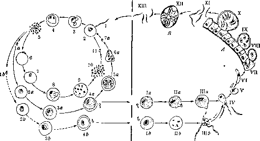
Sporogony (fig. 1, I-XIII). Entering the mosquito's stomach along with the blood of a malaria patient, mature gametocytes, leaving the erythrocyte (1a and 1b), undergo a series of transformations. In the male cell, division of the nucleus (1b) occurs, formation of 4-6 flagella-whips (IIIb) takes place, which separate from the cell and possess lively movement (spermatozoa); in the female cell, the so-called reduction division occurs. Part of the chromatin substance of the nucleus-the reduction body-is rejected, moves to the periphery of the cell and disappears (IIIa). The significance of this process is not clear. The cell becomes oval, the protoplasm protrudes, a bump-fertilization hillock, hillock forms.
Figure 1. Scheme of asexual and sexual development of the malaria parasite.
Figure 1. Blood in malaria tertiana: 1 and 2-rings; 3-half-grown schizont; 4-mature schizont; 5-beginning of division; 6-later stage of division; 7-merulation; *-male gamete; 9-female gamete. Anisocytosis; poikilocytosis; polychromatophilia. Figure 2. Blood in malaria tropica: 1-rings; 2-beginning of division; 3-merulation; 4-male gamete; 5-female gamete with remnants of Maurer's spots; 6-female gamete, free from Maurer's spots. Figure 3. Blood in malaria quartana: 1-ring; 2-transition of ring to band; 3-band; 4 and 5-schizonts; 6-merulation; 7-male gamete; 8-female gamete; 9-polychromatophil. Figure 4. Plasmodium praecox (Proteosoma) in the blood of a chaffinch: 1-merozoite; 2-schizont; 3 and 4-merulation; 5-female gamete; 6-female gamete in plasma. Figure 5. Malaria parasites in the blood of monkeys: 1, 2 and 3-Plasmodium Reichenowi in chimpanzee; 4, 5, 6 and 7-Plasmodium malariae in chimpanzee; 8, 9 and 10-Plasmodium Kochii. (According to Gander-Berenberg-Gossler.) Figure 6. Malaria parasites in the blood of Vespertilio Daubentoni: 1-young merozoite; 2, 3 and 4-schizonts; 5-merulation; 6-female gamete. (According to Shingareva.) Figure 7. Brain in comatose malaria; capillaries filled with parasites look like dark lines. Figure 8. Liver in malaria; abundance of pigment in reticuloendothelial cells. Fig. 9. Brain in daalaria. Durck's granuloma^ with hemorrhages. Infected erythrocytes fill the capillaries.
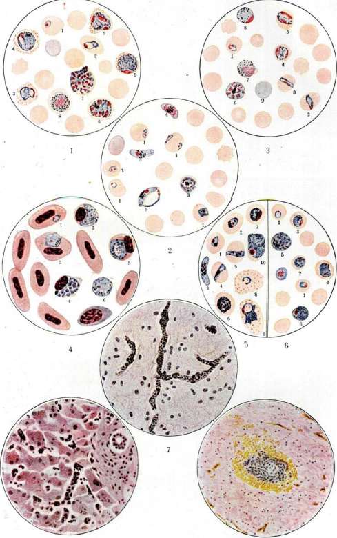
(For illustration to the article Malaria.) To the article Malaria.
of the epithelial cell (Ilia). To it a sporozoite is directed and drawn into it. The nuclei of both cells fuse, and thus copulation (IV) occurs. The fertilized cell is surrounded by a mucous membrane preventing further penetration of sporozoites into it. After a day a zygote or ookinete—a motile egg cell (V-VII)—is formed. It possesses active movement. It penetrates the wall of the mosquito's stomach (A) as far as the serous covering. Here it transforms into a spherical or oval pigmented oocyst (VIII). The process of ookinete penetration into the stomach wall ends in 48 hours. As a result of continuous nuclear division, the oocyst increases in size, and daughter cysts—sporoblasts (IX)—are formed. They are filled with sickle-shaped bodies arranged like a rosette (X). When mature, the sporoblast bursts, and its contents enter the body cavity of the mosquito (XI). Sporozoites are found in all organs of the mosquito, but mainly concentrate in the salivary glands (B), in whose cells they are located with pointed ends directed into the lumen of the excretory canal (XII). Together with saliva (XIII), they enter the human blood (1) during a bite, causing infection. In oocysts, curved dark bodies—Rossi bodies, black spores—are sometimes encountered. Ruge and others considered them as involutive forms of sporozoites, Noe—as developmental stages of Nosema, Kerr—as a special type of protozoa (parasite of the mosquito)? Their nature has not been finally clarified. Main (W. Main), based on the fact that they always give a reaction to chitin and are almost always found as thickenings at the ends of tracheal tubes, considers them an anomaly of the tracheal system. The duration of sporogony is from 10 to 20 days depending on the species of parasite and external (temperature) conditions. The number of oocysts can be very large (up to several hundred). The number of sporozoites in the salivary glands reaches 10,000. The question of the possibility of sporozoits overwintering in the mosquito's body has not been finally resolved. The possibility of transmitting the parasite to the mosquito's offspring through the egg is probably excluded. The process of sporogony, like schizogony, is in general outline the same for all three species of malaria parasites. Mitzmain observed that a large number of gametes of P1. immaculatum pass through the intestine of Anopheles maculipennis and are expelled with the mosquito's excrement, since the number of oocysts developing on the stomach wall of Anopheles is not proportional to the number ingested. This question is of great importance for the epidemiology of M., as it explains the percentage of Anopheles infected with malaria. In experiments with feeding Anopheles on people with gametes in the blood, it was not always possible to infect Anopheles with M. Also important is the number of gametes in 1 mm3 of blood of the patient, since with 150-200 gametes in 1 mm3 of blood, a person does not infect even 50% of Anopheles that fed on his blood. The ability of different species of Anopheles to be infected with different species of human M. is not the same. Thus, the work of Dutch authors (Swellengrebel and others) experimentally proved that Anopheles Ludlowi is infected with P1. immacul. in 100%, P1. vivax already in 80%, and P1. malariae only in 4.7%, whereas in Anopheles Kochii P1. immaculatum does not develop at all, just like P1. malariae, and P1. vivax develops in 16.7%. Species of parasites (see separate table, fig. 1-3). Morphological features of the most common species P1. vivax (from Latin vivax—living, motile) are described in the description of the parasite's development cycle in the human body. The duration of schizogony is 48 hours. For this parasite, the following changes in the morphological picture of blood are characteristic: erythrocytes are enlarged, pale; when stained according to Romanovsky, Schüffner's granularity is often observed (see separate table, fig. 1-3 and 9). Delicate, light-red granules are evenly distributed throughout the erythrocyte, apparently formed as a result of changes occurring in the composition of the erythrocyte protoplasm under the influence of parasite penetration into them. Young forms (rings) of Plasmodium malariae (fig. 2, II) differ little from Plasmodium vivax, but are smaller. Amoeboid motility is weakly expressed, even half-schizonts have a round shape. Very characteristic is the formation of the so-called bands—the plasmodium is thrown from one edge of the erythrocyte to the other in the form of a strip of protoplasm with parallel edges (fig. 2). There are usually no more than 8 merozoites; they are arranged in a rosette (fig. 2). Sexual forms are significantly smaller than in P1. vivax (fig. 2). The size of erythrocytes does not change. Development cycle 72 hours. From schizonts of P1. immaculatum (formerly called P1. praecox), only ring forms (fig. 2, III) are usually observed in the peripheral blood. They are very small, have little protoplasm, the ring border is very thin. The presence of two nuclei in one ring is often observed (see separate table, fig. 2,1). Schizogony occurs in internal organs (capillaries of the brain, bone marrow, spleen). The number of merozoites is most often 16-20 (fig. 2). Development cycle 24-40 hours. The external appearance of sexual forms is characteristic. As they grow, they elongate, taking the shape of a half-moon. The pigment is located in the middle of the plasmodium (fig. 2). The erythrocyte is not enlarged. When stained, Maurer's dotting—large, coarse red grains, usually grouped in one place—can be detected. The scheme of the main distinguishing features is given in table 1. The cultivation of the causative agent of M. with transfers, obtaining a series of generations in the sense as observed in bacteria, has not yet been resolved. To date, it has not been possible to inoculate human M. parasites into any experimental animal, despite the enormous effort spent by a number of authors. For example, Base (1922) unsuccessfully inoculated human M. into horses, dogs, foxes, monkeys, rabbits, mice, guinea pigs, hedgehogs, bats, wolves, cats, domestic and wild pigeons, magpies, owls, turtles, frogs, and lizards. Not a single animal became infected with M., although in some guinea pigs and sea Tabl. 1. Scheme of the main distinguishing features of the causative agents of M. Plasmodia (forms) and erythrocytes Young forms...... Half-adult forms..... Adult forms....... Division ......... Development cycle........ Erythrocytes affected by parasites ....... Adult forms....... P1. vivax P1. malariae Asexual forms - agamonts Ring. V*-Vs diam. of erythrocyte Large ring with pigment. Amoeboid forms. Vi-*/« diam. of erythrocyte Oval or round. Pigment—one or several clumps. Size UP TO 11/2 ERYTHROCYTES 18-24 merozoites. More often 16. Shape of mulberry berry 48 hours. Pale, enlarged. Schüffner's granularity Same Round or bands. Pigment often golden-yellow. Much pigment Same, but size smaller than erythrocyte 6-12. More often ft. Daisy Normal Thin, very small ring. V,-V4 diam. of erythrocyte Ring. Up to V« diam. of erythrocyte. Often 2 nuclei Rarely in peripheral blood. Round. Up to V2-3U diam. of erythrocyte. Delicate pigment 8-24. More often 16-20. Morula. Small Not enlarged. Maurer's dotting Sexual forms - gamonts Larger than erythrocyte. Irregular disc. Pigment brown, fine-grained Smaller than erythrocyte. Irregular disc. Pigment dark brown, coarse-grained Straight or curved half-moons Cultivation of plasmodia. All attempts to cultivate plasmodium on nutrient media before 1912 ended in failure. Sakharov (Tiflis) managed to maintain the viability of plasmodium in leeches that had drunk blood of M. patients with a large number of parasites for 7 days. Tsiman—only one day. Morphological features of plasmodium did not change, but further development was not observed. In 1912, Bass (Bass), and then a whole series of researchers (Tsiman, Gurko and Hamburger, Müllens, Perecropov, etc.) succeeded in obtaining cultures and subcultures of the second and even third generation when sown on human blood. Bass maintained the development of plasmodium in culture for up to 7 weeks. Method of Bass (1914): blood taken from a patient's vein is immediately defibrinated and to each 10 cm3 of blood, 0.1 cm3 of a 50% solution of dextrose is added. Row (Row; 1917) and Sinton (Sinton; 1922) described a method that made it possible to obtain the growth of one generation of parasites even in a few drops of blood. Zhukov (Moscow) and Perecropov (Kazan) managed to obtain sporozoites in cultures according to Bass. Nevertheless, the question of the possibility of guinea pigs in the blood and remnants of malaria parasites circulated for 24 hours. Yoshino (Yoshino; 1926) claims that he succeeded in inoculating P1. vivax into young guinea pigs, but the illustrations accompanying his work are unconvincing. Zia and Faust (Zia S. H., Faust E.; 1928) also unsuccessfully attempted to infect young hamsters (Crice-tulus griseus).
P. Muffel. III. Epidemiology. The epidemiology of M. is determined by four basic factors: the reservoir of the virus, the carrier, the susceptible organism, and the external environment. The reservoir of the virus in M. is formed by an infected person when their blood contains the sexual forms of the plasmodium—gametes (resp. gametocytes). Gametocyte carriers are the only source for infecting the carrier that spreads the infection; this determines their exceptional epidemiological significance. Infection of the carrier is possible only when the carrier's peripheral blood contains a sufficient number of mature male and female gametes. All other conditions being equal, the carrier becomes infected more strongly the more gametes are in the carrier's blood. Hence the particularly important significance of cases of M. with abundant parasite content in the blood. In endemic areas, the main reservoir of the M. virus is children; the epidemiological significance of immigrant masses after their infection can be equivalent; in both cases, the parasites are especially abundant in the blood. However, the adult population of malarial areas also often contains parasites in the blood, although usually in limited quantities. Gametocyte carriers may be among patients with malaria; they are also found in latent and clinically mild forms of infection. In primary infection, gametes usually appear in the blood some time after the onset of the disease, on average in the tropical form after 8-15 days, in three- and four-day forms after 7-10 days 3. This circumstance undoubtedly has great significance for the epidemic spread of tropical M. In three-day M. the number of gametes is smaller, but it is characterized by frequent relapses with the appearance of gametes. In four-day M. the number of gametes is the smallest, but the parasites persist in the blood for a long time. According to observations by some authors, seasonal fluctuations in the appearance of gametes are observed in the same individuals. Thus, according to Sinton (1926) in India, during systematic examination of 2,000 people, gametes of P. immaculatum (praecox) were detected from 1% to 4% from July to October and reached 15% in November, falling to 0% in February. The sexual composition of gametes changes in different phases of infection: in the first days of appearance of gametes, male ones predominate, later—female ones; later male ones disappear and only female ones remain. Thus, according to Simons XI/1917-11/1918=0.5%, III-VI/1918=0.3%, VII-X/1918 = 1.5%, XI/1918= = 0.2%. If one takes into account that oocysts can mature at a corresponding increase in temperature, the possibility of overwintering of the parasites seems reliable. The quantity of the carrier has only relative importance and is by no means always proportional to the actual spread of M. in a given area; in particular, despite the very limited number of Anopheles, M. can be very widespread. The decisive importance is the presence of infected mosquitoes, and it must be taken into account that the same infected mosquito can relatively long serve as a source of mass diseases (attack over a series of days and multiple bites during the same night). On the other hand, the mosquito factor has significance only in the general complex of other factors. In the absence of a virus reservoir or necessary external temperature conditions, despite the huge quantity of the carrier, the area may be free of M. In particular, the diversion of the carrier from humans by animals (especially large ones) has great practical importance. Anopheles very readily (apparently even preferentially) attacks animals that provide it with blood feeding under the most accessible conditions (large surface).
With appropriate quantitative ratios between mosquitoes, animals, and humans, a special equilibrium can be established, under which humans are freed from mosquito attacks. Such ratios can acquire significant importance for the epidemiology of M., limiting the spread of M. despite the large number of vectors. Some authors, especially Roubaud, believe that as a result of prolonged animal feeding, 'zoophilic races of Anopheles' that do not attack humans can form; these races also differ in morphological features of the maxillary armature (14-15 teeth on maxilla). Roubaud attempts to explain this 'zoophilia' as the spontaneous disappearance of M. in some places (e.g., in France) despite the preservation of a huge number of Anopheles ('anophelesism without paludism'). However, according to many authors, there is no basis for recognizing special 'zoophilic' races; the entire phenomenon is explained by simple ratios of labile equilibrium (see Biocoenosis, Zoophilia). Susceptible organism. The causative agent of M. is capable of infecting humans regardless of their age, sex, and race; but a number of factors can influence to varying degrees, either predisposing to the disease or, conversely, protecting from it. Thus, children easily contract M., as they are particularly accessible to the vector (tender skin, lack of self-protection, low resistance), while strong individuals may better resist the disease compared to exhausted individuals, etc. All factors that lower the body's resistance in any way contribute to its infection with M. and the severity of the infection. These include inadequate nutrition, overwork, lack of adaptation to climate among immigrants, occupational factors predisposing to infection (agricultural workers in rural areas, the army), various states of pathological aggravation (alcoholism in particular), moral depression, and others. The accumulation of unfavorable factors can lead to catastrophic consequences, especially among immigrants arriving in malarial areas; under these conditions, the most severe epidemics of M. typically develop. An illustration of this is the epidemic of M. experienced by the army in Azerbaijan in 1920, when military units were subjected to almost universal infection (from June to December, 21,920 malaria cases were registered in the hospitals of Baku, of which 646 died). Social catastrophes under certain conditions (massive settlement of malaria patients, lack of quinine, etc.) can lead to pandemic spread of M. throughout entire states. A striking example in this regard is also the most severe pandemic of M. that arose in the USSR during the civil war (see below - statistics). The possibility cannot be excluded that when fresh masses are affected, there is an increase in the virulence of the plasmodium due to its massive passage. The question of congenital M. has not yet been finally resolved. Most Western authors deny its possibility. However, a number of observations, particularly by Russian authors, establish with sufficient obviousness that under certain conditions placental infection in M. can occur. In this regard, particularly indicative are findings of plasmodia in the blood and organs of premature fetuses. For example, in one case (Leger; 1918), P. vivax was found in the blood of a 7-month-old fetus born to a mother suffering from three-day M. In the case of Zdrodovsky (1929), half-moons of P. falciparum were discovered in the spleen of a dead fetus 6-7 months old, born to a mother in a state of malarial coma. In this same case, the barrier role of the placenta was clearly revealed (a large number of plasmodia in the mother's blood and placenta while their content in the fetus's organs was negligible). According to Tsiman, the normal placenta apparently represents an impassable barrier to plasmodia. It is possible that cases of placental infection occur only in the presence of pathological changes in it (e.g., infarcts). The relative rarity of congenital M. speaks in favor of the plausibility of this assumption. The question of M. among the permanent population of endemic areas is of special interest; it is connected with the general problem of immunity relations in malaria. Immunity. Innate immunity to M. is absent and if observed in some individuals (Sechenov), it is only as a very rare exception. Racial immunity is also absent, although there have been repeated indications of the relative resistance of negroes (Laveran and others); in the latter case, the question obviously concerns only acquired tolerance to the infection (see below). In full analogy with most protozoan infections, M. is incapable of causing sterilizing immunity: a past illness does not protect against subsequent infections. This became particularly clear in experiments with artificial inoculation of M. Only in some cases of artificial infection with blood containing P. vivax, a more or less prolonged (6-20 months) immunity to subsequent infection with virulent blood of the same species is noted; however, even here susceptibility to infection through mosquito bites remains. Sterilizing immunity to M. does not develop among the population of endemic areas. At one time R. Koch (1899-1900) attempted to justify the existence of such immunity based on comparative age-related examination of populations in severely malarial tropical areas (Java and New Guinea). According to his observations, in such places only younger children (up to 5-10 years) contain parasites in the blood at the maximum of infection (80-100%) within up to 2 years; semi-adult and adult population, on the contrary, is free from parasites due to immunity acquired through infection in childhood. However, later numerous observations in various countries refuted Koch's position, showing that the adult population very often contains plasmodia in the blood even in the most severely malarial areas despite universal infection in childhood. Thus, for the adult population of tropical Africa, the following figures for parasite findings in the blood were established in mass examinations: Plehn (1906) - 50%; Tsiman (1909-1910) - up to 60%; Reichenow (1917) - 50-54%; Leger and Nogue (1923) - 47%; similarly according to Bass (1917) in the Mississippi area - 19.2%; according to Christophers (1925) in India - 50%, etc. To illustrate the immunity relations in M., the following data from mass examinations of the local population in Azerbaijan of various ages can serve (see table 2). Table 2. Thus, in endemic areas, the child population is affected by M. to a particularly severe degree, but the adult population, even in very old age (examination of 30 elderly people aged 71 and above), can show infection with plasmodia in a large percentage. Sterilizing immunity does not develop in M. This lack of immunity, however, is compensated for by relative tolerance to the infection, which M.-affected populations gradually develop as a result of continuous infections; but this tolerance is actually associated with the presence of latent infection. In other words, the immunity relations in malaria are formed according to the general principle of labile infections (see Immunity, immunity in protozoan infections). However, the acquired tolerance is relative (possibility of relapses when equilibrium is disturbed and only relative resistance to superinfection). Later observations by Christophers (1925) in India provide particularly illustrative examples of the dynamics of gradually developing resistance of the M.-affected population to M. According to Christophers' research, children under 2 years with a parasitic index of 100% contained in their blood more than 10,000 plasmodia per 1 mm3 and all showed a temperature reaction; at ages 2-5 years with 100% infection (parasitic index 100%), the number of plasmodia in the blood was 1,200, and the temperature reaction was not constantly observed; at ages 6-10 years with the same percentage of infection, the number of parasites in the blood fell below 1,000, and fever a) Childhood age (Bogoyavlensky; 1924-29). Age (years)....... Spleen index (in %) Plasmodia (in %)...... 40.7 2 47.3 50.8 57.1 .52.1 4 54.9 57.4 5 64.8 53.0 6 63.1 44.9 7 66.6 35.4 74.3 35.8 75.2 34.0 10 78.7 26.3 On average 63.1 42.9 b) All ages. Winter examination after a sharply epidemic season (Zdrodovsky and Lindtrop; 1922-23). Age (years)...... Spleen index (in %) Plasmodia (in %)..... 1-5 6-10 11-15 16-20 100.0 100.0 97.8 87.5 83.4 77.0 60.0 40.6 21-30 77.2 45.6 31-40 79.1 40.9 41-50 51-70 62.1
47.8 29.0 | 30.4 On average 82.0 50.7 was absent; in adults, parasites were found in 50%, there was no temperature reaction at all, and only in 10% were there enlarged spleens. External environment. In the complex of factors of the external environment, climate and specifically temperature relations have special importance. The epidemiological role of temperature is twofold: it decisively influences sporogony and at the same time has a significant influence on the biology of the carrier. The importance of temperature for sporogony has already been considered. As for the biology of the carrier, temperature primarily affects the aquatic metamorphosis—his. All other conditions being equal, this metamorphosis occurs the faster the higher the temperature. Thus, according to Martini, at 24-27° the metamorphosis of Anopheles ends within 147 days; at 16-19°—within 307 days. According to observations by Brenn and Voskresensky in Azerbaijan (1927), the aquatic metamorphosis of Anopheles maculipennis in natural conditions in different months had the following durations: VII—average temperature = 24.5°, VIII—average temperature = 26.9°, IX—average temperature = 18.9°; corresponding duration of metamorphosis: 17-19 days, 17 days, 30 days. Thus, the longer and warmer the summer season, the more generations of the carrier are born; simultaneously go the conditions ensuring sporogony. As a result, countries with a long hot season show the greatest predisposition to M.; hence the special predisposition of subtropical and tropical countries. Temperature relations change depending on the altitude of the terrain; hence the significant importance of the orographic factor for the spread of M. Lowlands are especially predisposed to M., while altitudes, on the contrary, are less favorable and beyond a certain limit become immune. These limits, however, vary greatly in different places. Sometimes foci of M. are found at significant altitudes; e.g. at altitudes up to 1,000 m in Macedonia, 1,600 m in German East Africa, 2,000 m in the Himalayas, 2,500 m in Peru (Müller; 1921). An example of the dependence of the spread of M. on altitude is provided by the experience of surveying the Gandzhinsky district in Azerbaijan: in the lowlands (below 250 m) in 30 villages among 829 children the splenic index was 71% and the parasitic 34%; in the same district in 9 villages of the foothills and mountains (from 750-800 m to 1,000-2,000 m) among 417 children the splenic index was 11.3% and the parasitic—1.4% (Chergeshtov; 1925).—Of other climatic factors, some authors (Bentley and others) point to the importance of relative humidity.—The importance of winds, 'bringing' mosquitoes to leeward places, occurs only in limited cases (Rudnik notes the importance of winds in some areas of Macedonia). The water factor, along with the temperature factor, has decisive importance for the epidemiology of M., since it is connected with the breeding places of Anopheles; the latter can only be characterized schematically. In typical cases, these are non-turbid, stagnant or slowly flowing water bodies with relatively clean and not too saline water and developed submerged or floating flora. This characterization, however, is conditional and only corresponds to the optimal type. Thus, breeding places of Anopheles can also be found in heavily polluted water bodies (e.g. with oxidizability by Kubel up to 545 mg—Voskresensky and Brenn); the salt content in them can sometimes be very high: 19.5‰ (Anopheles maculipennis—Corsica), 25-30‰ (Anopheles Rossi—India), 40‰ (An. multicolor—Sahara); according to observations by Zdrodovsky in Transcaucasia, the limiting concentration is 10‰. The optimal pH for the development of Anopheles larvae is 7.2-7.6 (observations on peat quarries by Smorodintsev, Odova and Sebensov). On the other hand, while the presence of submerged or floating flora (e.g. 'water cotton' in Transcaucasia) is especially characteristic, at the same time breeding places can also be in the absence of flora or with the presence of only emergent vegetation (observations by Zdrodovsky and Voskresensky in Transcaucasia). In some species of Anopheles, breeding places are extremely peculiar (e.g. water collections in hollows—Anopheles nigripes). There is also selective colonization of certain types of water bodies by specific species of Anopheles (e.g. stagnant water bodies in the channels of mountain rivers in Azerbaijan are selectively colonized by Anopheles superpictus). Water bodies can be places of abundant breeding of Anopheles regardless of their size (sometimes small water bodies acquire special importance); but water bodies must have a certain duration of existence ensuring the minimum period of Anopheles metamorphosis. Hence e.g. in the southern outskirts of the USSR, water bodies with a duration of less than 2 weeks have no epidemiological significance. Evaporation, infiltration and surface runoff are the main types of water circulation; any difficulty in this natural circulation can lead to the formation of water stagnation and breeding places. According to their origin, stagnant water bodies can be classified according to the following, most practically widespread types: a) Stagnant flooding of spontaneous and natural origin: 'flood' flooding along river systems due to spring river floods; coastal stagnation in rivers, as well as collections of stagnant water in the beds of rivers (e.g. in the beds of mountain rivers); stagnation of water in the mouths of rivers flowing into the sea, with dune blockages; swamps of the coastal strip, formed due to difficulty in runoff of drainage waters by dune deposits ('lagoon type'); spring waterlogging; waterlogging from exposed groundwater of high standing; infiltration waterlogging of depressions from neighboring water systems; lake type of water bodies; surface collections of atmospheric water and others. b) Stagnant flooding of artificial origin: stagnation of water from irrigation systems—inside canals (stagnation from overgrowths, coastal stagnation, stagnation from destruction of banks) and around canals (waterlogging from water breaks and infiltration waterlogging of natural or artificial depressions); waterlogging from drainage systems of irrigation; waterlogging from irrational irrigation (irrigation by 'flooding'); waterlogging of rice plantations with continuous irrigation with slowly flowing water or flooding; stagnation of water in incorrectly arranged open drains; waterlogging from discharge of excess or used water; subsoil and infiltration flooding of artificial excavations (quarries along railway lines, peat quarries, etc.); artificial ponds, basins, farm reservoirs; gushing water sources; drinking wells, cisterns and others. Epidemiological characteristics of a malarial area. Geographical position, orography, climate, hydrography, condition of the carrier, distribution of the population according to orographic zones, socio-economic conditions and others determine the general predisposition of a given area with respect to M. Statistics of actual population morbidity from M. and mortality from it, accounting for the movement of primary and recurrent diseases during the year and analysis of their comparative parasitology give objective elements for the general characterization of a malarial area; especially valuable are long-term statistical observations. Detailed data on malarial morbidity of populated areas (or separate population groups) are compiled by determining on a mass basis the so-called malarial indices. Among the latter, the following are distinguished. 1) Splenic index, or the percentage ratio of persons with enlarged spleens to the total number of those examined. The index is relatively stable and gives a very valuable characterization of the spread or extent of M. in a given area. 2) The Ross index is an indicator of the average degree of enlargement of spleens among those examined; characterizes the intensity of malarial morbidity of the population; is calculated by the formula: a+3b+6c+9d / a+b+c+d where a—number of non-enlarged spleens (the volume of a normal spleen is taken as unity), b—number of small spleens, protruding from under the ribs by 1-2 transverse fingers (enlargement 3 times), c—number of medium spleens, protruding by 3-5 fingers (enlargement 6 times), d—number of large spleens, i.e. reaching the navel or more (enlargement 9 times); the Ross index in practical conditions gives a quite satisfactory characterization of intensity; an increase of this index to 5 and above indicates especially heavy morbidity. 3) Parasitic index, or the percentage ratio of persons with plasmodia in the blood to the total number of those examined; the index is extremely valuable, as it gives a characterization of the virus reservoir (especially when accounting for gametes), but is very variable and therefore not very accurate; depends in particular on the research methodology; at present it is determined as a rule with the help of a thick drop. 4) Endemic index, or the total percentage of persons in whom objective signs of M. were found.
(enlargement of the spleen and plasmodia, in combinations or separately), the index can give higher values compared to the splenic and parasitic, since in the absence of spleen enlargement there can be plasmodia in the blood and vice versa. 5) Cystic and sporozoite index, or the percentage of finding infected mosquitoes; the index is especially valuable in epidemiological terms, but extremely conditional, as it depends on a whole series of factors (see above). The sporozoite index has preferential value, since the finding of cysts does not yet indicate the actual ability of Anopheles to infect people (e.g., immature cysts in hibernating mosquitoes).-Malaria indices, determined in humans, including the splenic index, in countries with seasonal M. undergo changes depending on the season (higher values during malaria time and lower outside of it). For examination according to malaria indices, children (usually up to 10 years old) are preferable as the most sensitive and at the same time most sedentary element. The presence of a malaria focus in a given locality is most accurately determined by two signs: the presence of Anopheles and the occurrence of clearly primary diseases (e.g., among children born after the end of the malaria season of the previous year). On the basis of examination of populated localities (all or typical) with determination of splenic indices, a malaria map of the given locality is compiled, on which the malaria of individual localities is depicted graphically.
p,
Zdrodovskin. IV. Statistics and Geographical Distribution. 1. World distribution. Malaria is one of the most widespread diseases on the globe both in terms of the number of people suffering from it and the extent of areas where it is observed endemically. The largest foci of it are concentrated in countries with hot and humid climate, in particular in tropical and subtropical countries; but its distribution extends far beyond these countries and extends in the northern hemisphere to the northern borders of the temperate zone. Its distribution in individual parts of the world is as follows. In Asia, it is a great scourge for British India, where its distribution reaches enormous proportions. In the last 10 years, over 1 million people were treated for M. in hospitals in India and over 2,000 died. In some provinces, more than 50% of all children die from M. In Bengal for 1925/26 year, about V2 million people died from M., which constitutes 66% of total mortality. In individual cities of India, mortality from M. in recent years has reached 250 per 100,000 population (see below). In Ceylon (pop. 4,500 thousand) for 1922-26 years, about 1 million M. patients were treated annually in dispensaries, and about 30,000 in hospitals. In the Dutch Indies, according to data from 171 hospitals for 1924, M. patients constitute about 10% of all patients. In the Federation of Malaya (pop. in 1928 1,534 thousand), deaths from M. were registered in 1926 - 16,531, in 1927 - 20,736 people. M. constitutes over 30% of all causes of death. On the island of Cyprus in 1926, M. constituted 75.8% of all infectious diseases. M. is widely spread in Asia Minor, in Southern China and on the Philippine Islands. Table 3 shows mortality rates from M. in recent years in some cities in Asia. Table 3. Mortality from M. per 100,000 population. Cities Bombay . . Calcutta Madras . . Manila . . Rangoon . . Singapore . Tehran . . 1926 1927 1928 19.0 1929 14.3 42.7 25.5 149.3 119.7 110.2 78.9 258.6 249.9 - 126.8 16.1 10.4 10.8 13.0 61.6 59.2 64.7 45.7 396.0 259.3 240.3 201.3 155.2 98.4 93.2 30.4 Africa is a classical country of M., which is the main enemy of the colonization of the country (Nocht). Information on the distribution of M. is extremely scarce. It is widely spread in Egypt. In Kenya in 1926, 42,972 people were treated in hospitals (2,565 thousand pop); on the island of Mauritius (pop. 388 thousand), about 2,000 people died from M. annually in 1925-27. In Nyasaland (pop. 1,306 thousand) for the same years, from 3 to 4 thousand people died from M. annually. In Tanganyika, mortality from M. constitutes 21.5% of total mortality. In Uganda (pop. 3,242 thousand), about 30,000 cases of M. are registered annually.-Malaria is widely
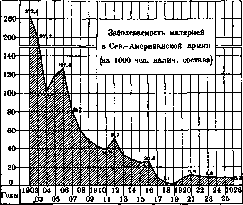
Figure 3.
spread in America. In the USA, about 160,000 cases of M. are registered annually in recent years. It was extremely widespread there at the beginning of the 20th century, but at present it has decreased significantly. The indicator of the success of the fight against M. in America is the decrease in morbidity from it in the North American army: morbidity from M. among it was 272.3 per 1,000 personnel in 1902, in 1919 it decreased to 2.3, in subsequent years it, as in many other countries, gave an increase in order to then begin to decrease again (Figure 3). In Mexico for 1925-28, from 8 to 9.5 thousand deaths from M. were registered annually. In the West Indies in Haiti (pop. 2,500 thousand) about 1,000 people die from M. annually; in Puerto Rico, mortality from M. in 1911 was 190 per 100,000 population; in 1921 it dropped to 114; in 1925 it was 105 per 100,000 population; on the island of Jamaica, in the city of Kingston, mortality from M. was 40 in 1924, 80 in 1925, 31 in 1926 per 100,000 population.-Large foci of M. are concentrated in Central America. The construction of the Panama Canal went down in the history of M.: the first period of construction (1881
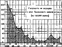
Figure 4. 1889), when a French company was in charge, was accompanied by such cruel mortality of workers from M. and yellow fever (annual mortality up to 240 per 1,000), that it was necessary to completely stop the work. In the second period of construction, carried out by Americans (1905-12), M. initially also gave a high increase, but soon thanks to systematically conducted anti-malarial measures it sharply decreased and dropped to very small figures. Morbidity and mortality in the Panama Canal zone for 1906-1921 are given in Table 4 and in Figure 4. Table 4. Morbidity and mortality from M. in the Panama Canal zone, for 1906-1921 (per 1000 population). Years Morbidity Mortality 1906 , 1907 , 1908 . 1909 . 1910 , 1911 , 1912 , 1913 . 821' 424 282 215 187 184 110 76 7.45 3.51 1.37 0.85 0.81 0.84 0.31 0.30 Years Morbidity Mortality , 82 0.14 0.23 1916 . 1 16 0.06 1917 . 0.09 1918 . 0.08 1919 . 0.08 1020 . 0.15 1921 . In recent years (1925-28) in the Panama Canal zone, from 1,000 to 1,500 cases and from 20 to 48 deaths from M. are registered annually (population 127 thousand).- In South America, M. is especially widespread in the valleys of the Orinoco and Amazon rivers, in particular in Argentina and Brazil. For some South American cities, mortality from M. per 100,000 population for recent years is expressed in such indicators (Table 5): Table 5. Cities Belém..... Callao ..... Lima...... Manaus..... Mexico .... Niterói . . . Rio de Janeiro Salvador . . . Santos ..... 1926 1927 1928 271.2 248.8 233.8 77.4 39.1 41.3 68.9 45.7 50.4 562.8 697.2 781.2 8.2 5.7 4.1 26.6 22.8 28.5 25.2 25.4 21.5 175.3 137.9 143.7 36.4 42.5 44.0 The construction of railways in malarious areas of America met the greatest 1856-60. On the height of M. at the end of the 19th century, Table 6 gives an idea, where mortality from M. per 100,000 population, on average per year for 1891-1900, in some countries of Western Europe is given. Table 6. States |Mortality | from M. States
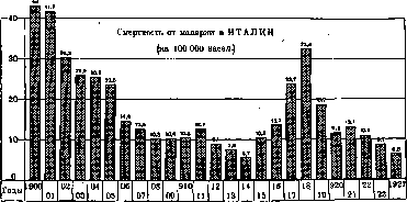
Figure 5.
difficulties due to M.-In Australia, in the last 10 years, from 50 (1916) to 35 (1926) people die from M. annually per 100,000 population. The most affected state is Queensland. In Western Europe, M. is most widespread in Mediterranean countries. Since ancient times, extensive areas where Greek and Roman culture once flourished have suffered severely from it. Many researchers believe that the decline of ancient culture was facilitated by the rapidly spreading M. in these areas. 'Where in ancient times beautiful cities flourished, now there are only weak, exhausted by malaria faces of very sparse pastoral and peasant population' (Nocht). In the 19th century, several major epidemics of M. were observed in Europe. The first of them fell on 1806-12 (Napoleonic wars), the next ones on 1824-27, 1846-49, 1856-60, 1866-72, and the last one, covering Holland and northwestern Germany, on 1899-1903. The highest, pandemic-like, were the increases of M. in 1806-12 and Mortality from M. Italy.....
46.2
23.0
Scotland ...
1.7
Of individual countries in Western Europe, M. is most widespread and most studied in Italy. Thanks to systematically conducted anti-malarial campaigns, it is gradually decreasing. In 1887, 71 people died from M. per 100,000 population; in 1900, 49 people; in 1926, mortality decreased to 7 per 100,000. During the world war, a significant increase in malaria was observed (Figure 5). Malaria is unevenly distributed across individual provinces. Table 7 (see article 603) indicates mortality from malaria in individual provinces of Italy per 100,000 population (according to data for 1891-1900). Spain is severely affected by M. At the beginning of the 20th century, 4 to 5 thousand people died from M. annually in it; in recent years, mortality from M. has fallen below 1 thousand (in 1928, 736 people; Fig. 6). Pittaluga calculates the number of M. patients in Spain at 300,000 people. The province most affected by M. is Caceres, where mortality from M. for 1922-1924 amounted to from 41.3 to 63.4 per 100,000 inhabitants (on average in Spain for the same years - from 5.5 to 7.1). M. is widely prevalent in Greece, in the Balkan states, in Hungary, Galicia, Bosnia, Dalmatia.
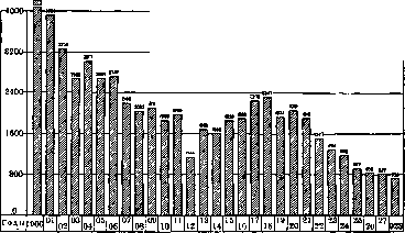
Figure 6.
Mortality from Malaria in SPAIN (absolute figures)
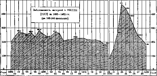
Table 7. Provinces Mortality from M. Provinces Mortality from M. Sardinia . . Basilicata Apulia . . . Calabria . . Sicily . . . Lazio . . . Abruzzi . . 255.2 186.6 116.6 109.5 101.0 74.9 59.0 Tuscany ..... Lombardy . . . Liguria .... 45.4 11.3 7.5 7.1 6.7 1.7
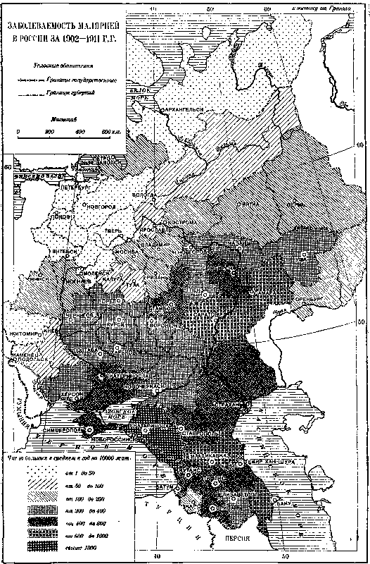
2. Malaria in the USSR. Before the world war, about 3.5 million cases of M. were registered annually in Russia, or about 215 per every 10,000 population. Among other infectious diseases, M. occupied the highest place (see Infectious diseases, Table 9). The largest number of diseases was registered in southern regions, the smallest in northern regions (see below). Remaining in the same region for many years at approximately the same level, M. from time to time experienced sharp increases in morbidity in these areas; it acquired the character of an epidemic, which after a certain period subsided, and M. again passed for several years to its usual endemic level for that region. From time to time, M. went far beyond its usual range, covered vast areas, and acquired the character of a severe pandemic. Table 8 provides figures for malaria morbidity in Russia and the USSR for the period from 1892 to 1929. Figures 7 and 8 show the curves of M. morbidity in Russia and the USSR from 1892 to 1929 and in 3 provinces: Samara, Saratov, and Simbirsk (Ulyanovsk). These curves give an idea of the periodic increases in M.-of its 'epidemics'. In Russia as a whole before the war, these increases for the indicated period were in 1894-95, in 1897,
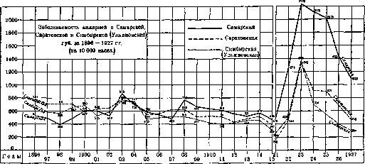
Figure 7.
they increase as one moves from north to south: in northern districts they constituted relatively small values (in Northern, Lake and Belarusian regions - see Table 10. Provinces most affected by malaria in 1911-14 (morbidity per 10,000 population). Provinces and regions 1 1911 1912 I 1913 1914 Black Sea . . 1448.8 1278.2 1461.4 1169.8 Kuban . . . . 1145.3 1204.2 1253.4 1067.0 Baku . . . . 826.4 1151.7 1028.9 868.6 Kutais . . . . 653.8 730.7 711.8 642.6 Terek ...... 630.3 678.7 674.8 673.9 Tiflis . . . . 614.4 656.4 603.6 509.2 Dagestan . . . 558.7 646.3 508.9 438.9 Yerevan . . . . 541.0 640.2 638.4 537.8 I Stavropol . ! 585.9 633.5 634.2 386.2 Samara . . . . 1 610.0 553.5 521.0 566.0 Saratov . . . 911.2 430.2 479.7 Astrakhan . . . 343.4 450.4 452.0 468.7 Voronezh . . . ! 430.1 398.4 415.0 337.9 Tambov . . . . 401.9 343.9 362.0 363.1 Simbirsk . . . . 399.5 420.6 458.0 451.2 ! significantly elevated. Apparently the lack of medical assistance and deficiencies in registration explain the relatively low morbidity rates in Central Asia (about 2% of the population), where the actual morbidity was probably significantly higher. The World War, and especially the civil war, created the conditions for an unprecedented rise in M., which by the end of the civil war had taken on the character of a sharply expressed pandemic and a dire national disaster. The morbidity rate for M. in the entire country according to official data rose from 215 per 10,000 population in pre-war years to 450-475 in 1923-24. M. went far beyond its usual area of distribution; it covered the entire country and in particular advanced into northern districts. Severe forms of tropical malaria, observed before the war exclusively in notoriously malarious districts - in the Caucasus, in the Lower Volga and in Central Asia, «07 spread throughout the republic. Malaria during these years ran in very severe forms, with high mortality. The causes of this unprecedented development of M. are still a subject of study for epidemiologists [both Russian and foreign (Wolter; 1930)]. Individual factors contributing to the development of the pandemic are as follows: 1) unprecedented movement of masses of people and the introduction of M. from unfavorable areas to favorable ones; 2) changes in climatic conditions; a sharp rise in temperature in 1921-22, which favored the multiplication of Anopheles; 3) the going wild of the soil due to the cessation of its cultivation in many places; 4) the marshification of vast territories due to the destruction of sanitary-technical structures and water supply systems, for example in Central Asia and the Caucasus; throughout the USSR according to official data it was 100 per 10,000 inhabitants - a figure that clearly did not correspond to reality. In 1921 it rose to 140 for the entire country. But already in this year an unprecedented rise in the rate was noted in individual districts. Thus, in the Northern district with a morbidity rate of 19-27 per 10,000 in pre-war times (see table 9) it rose to 119 per 10,000 in this year, i.e. increased approximately 5 times; in Arkhangelsk province, where the morbidity of M. before the war was about 10 per 10,000 population, the rate in 1921 rose to 176, i.e. increased 17 times; in North Dvina province in the same year it rose to 278, in Kotelny district the morbidity in 1921 reached 72.5% of the population. Such are some figures relating to the extreme north. M. rose sharply in places
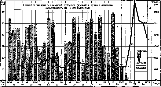
Figure 9. 5) a sharp decrease in the number of livestock, which diverts Anopheles from humans; 6) the complete absence of quinine during the civil war due to the blockade of the country and the accumulation of a huge number of gametocyte carriers; 7) finally, hunger played a major role in the susceptibility to M., sharply weakening the population's immunity to M. It was also the cause of the development in famine-stricken areas of a large number of severe forms of M. (mass diseases with coma-like forms with high mortality). Only approximate numerical expressions of this pandemic can be given. With the beginning of the World War, the number of annually registered cases of M. sharply falls, which must be explained exclusively by the deterioration of their registration. If in 1911-13 on average per year in all Russia 215 cases of M. were registered per 10,000 inhabitants, then in 1914 the morbidity rate drops to 208, in 1915-to 185, in 1916-to 190. For 1917-19 the information is so incomplete that it does not give an idea of the morbidity of M. In 1920, the morbidity rate for
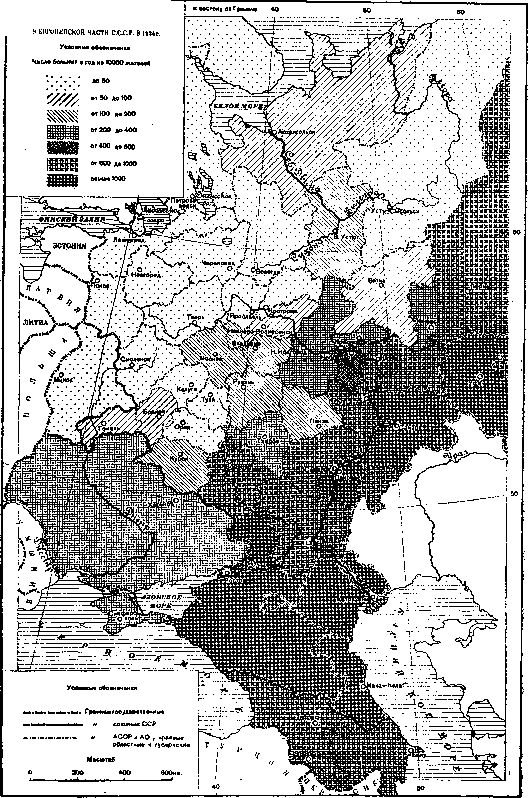
its usual area of distribution. Thus, in the Volga German Republic the morbidity of M. in 1921 according to official data was 25% of the population. This year should therefore be considered the first year of the pandemic spread of M. The following years give a further rise of it. Overall in the USSR in 1922, 268 cases of M. were registered per 10,000 inhabitants; in Arkhangelsk province the morbidity rate for this year was 274 per 10,000 (27 times higher than pre-war). 1923 gives the highest morbidity rate for the entire USSR: 474 per 10,000, i.e. out of every 100 people 4.7 were registered as malaria patients. In Arkhangelsk province for this year the morbidity rate was 409 per 10,000 (in individual districts-over 8%); in North Dvina-338 (in individual districts-up to 34%), in the Volga German Republic-5,084, in Saratov province-1208, in Samara-2,347, in Astrakhan-2,542 (in 1913-452), in Moscow-537 (before the war 120 per 10,000) etc. 1924 gave for the Union 445.7 cases
Figure 10.
Malaria morbidity
Figure 11. Malaria morbidity AND THE NETWORK OF ANTIMALARIAL INSTITUTIONS OF THE EUROPEAN PART OF THE USSR. IN 1828 designations shHHH Tropical Institutes Protozoan Departments of San. Institutes Malaria Stations Number of patients B year per 10000 inhabitants . - . ' . Less than 50 /////y/// from 50 to 100 'Ж'T'Я from 100 to 200 from 200 to 400 1 Leningrad 2 Bogorod 3 Shatura 4 Mikhnovo 8 Kudinovo v El. st. im Kloi 7 Bolshovo 6 Orekhovo-Zue 9 Vladimir 10 Izhevsk 11 Yoshkar-Ola 12 Kazan 13 Cheboksary 14 Kaneino 15 N.Novgorod 16 Ryazan 17 Tula 18 Skopin 19 Ranenburg 20 Lipetsk 21 Ramon 22 Pavlen 23 Ostrog 24 Voronezh 25 Tambov 26 Ulyanov 27 Syzran 28 Penza 29 Voronezh 30 Kamyshin 31 Balashov 32 Saratov 33 Sverdlovsk 34 Perm 35 Chelyabinsk 36 Ufa 37 Buguruslan 38 Buzyuluks 39 Samara 40 Dergachi 41 Pugachev 42 Pravol 43 Orenburg 44 Uralsk 45 Novo-Uzensk 46 Yalta give antimalarial 47 Kherson 48 Nikolaev 49 Kryvyi Rih 50 Mogilev 51 Kharkov 52 Kuren 53 Sumy 54 Poltava 55 Kremenchug 56 Dnepropetrovsk 57 Konstantinov- 58 Izium 59 Artemovsk 60 Luhansk 61 Stalino 62 Mariupol 63 Starobelsk 64 Kamensk 65 Sochi 66 Tuapse 67 Tiraspol 68 Gelendzhik 69 G. Klyuch 93 Grozny 70 Maikop 95 Mozdok 71 Novorossiysk 96 Georgievsk 72 Anapa 97 Armavir 73 Krasnodar 98 Nalchik 74 Slavyansk 75 Armavir 76 Kropotkin 77 Umansk 78 Yeysk 79 Rostov 80 Novocherkassk 'O* Poti 98 Nalchik 99 Ochamchiri 100 Zugdidi 101 Gali 102 Senaki 103 Ozurgeti 81 Labinsk 82 Stalingrad 83 Enotaevsk 84 Krasny Yar 85 Astrakhan 86 Dolban 87 Sleptsovsk 88 Khasavyurt 89 Derbent 90 Buynaksk 91 Vladikavkaz 92 Nazran 105 Kobuleti 106 Batumi 107 Karayaz 108 Kutais 109 Gori 110 Echmiadzin 111 Telavi 112 Idjevan 113 Namarli 114 Delijan 115 Lizakh 116 Ganja OT.Ani-Abad, 118 Nukha 119 Kuban 120 Agdash 121 Geokchai 122 Agdalyan 123 Jebrail 124 Kalagin 125 Salyan 126 Megri 127 Nakhichevan 128 Davalu 129 Yerevan 130 Tiflis .131 Baku 182 Makhach-Kala 183 Odessa '*4 Moscow 185 Kim 1.36 Ivanovo-Voznesensk 137 Sukhumi
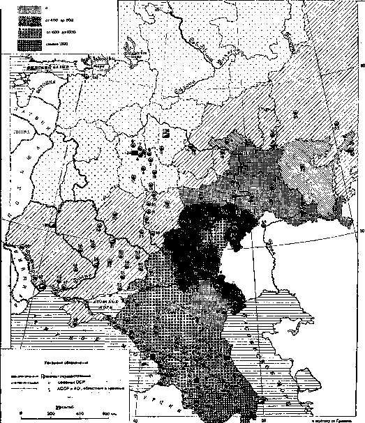
Figure 12. 20 «15 per 10,000 population. Malaria morbidity by individual provinces for 1924 is given in Fig. 11, and for the most affected provinces for 1923-27 in Table 11. Table 11. Malaria morbidity in the most affected provinces (per 10,000 pop.). Provinces and 1911-1923 1924 1925 1926 1927 regions 13 years. German Volga Republic...... - 5,084 3,407 3,843 2,474 1,792 Samara prov. . . . 2,779 2,109 2,020 Astrakhan prov. . 2,554 2,547 2,232 Ulyanovsk prov. | 426 Saratov prov. ! 607 1,342 Bashkir Republic . 1,214 Tatar Republic . . ! 289 441 . Chuvash Republic . . ! - Stalingrad prov. | - 1,115 Kalmyk region . . 1,272 Moscow prov. . . 1,124 Voronezh prov. . . . Mari region . . . - Vladimir prov. . ! 132 Tambov prov. . . t 373 Nizhny Novgorod prov. . . Arkhangelsk prov. . . Regions and autonomous republics Official morbidity figures for Malaria during the pandemic years are significantly below the actual figures due to the weakness of the medical network during these years and poor regt a b l_ 13.istration of patients. According to local health workers, these figures for individual regions should be increased from 2 to 5 times or more. If we take an average correction coefficient of 2.5 for these years, the total number of cases would be over 14,000,000 for each of 1923 and 1924. In subsequent years, malaria morbidity gradually decreases to pre-war levels. This decrease is observed steadily year after year despite significant improvement in disease registration due to the strengthening of the general medical network each year and the opening of a large number of malaria stations, which contributed to the increased influx of malaria patients. Corresponding figures for the entire USSR are given in Table 8 and in Fig. 7. Morbidity by individual union republics and individual regions Table 12. Malaria morbidity by individual republics of the USSR from 1924 to 1929 (per 10,000 pop.). Among the individual republics of the USSR, the Transcaucasian republics continue to be the most unfavorable for malaria, in which the morbidity rate for malaria, until recent years, amounts to 10% or more of the total population. The endemic rate of malaria in these republics shows no tendency to decrease. The annual increase in malaria morbidity in some of these republics may be explained to some extent by the expanding each year network of anti-malaria institutions and improving registration of patients.-For the RSFSR, the malaria morbidity rate in 1929 decreased almost 3 times compared to 1924, in Ukraine - approximately 6 times. The endemic rate of malaria in Belarus remains at a low level for all years. In the Central Asian republics, it gradually increases. Malaria morbidity by regions of the RSFSR (per 10,000 pop.). North-eastern region (Northern Territory) Leningrad region......... Karelian ASSR.......... Western region.............. Central-industrial region...... Ivanovo industrial region ..... Moscow
624,0 Turkmenistan .... I - Uzbekistan......: - 1925 г. 382,5 229,4 29,1 866,5 866,8 608,2 232,6 291,7 1926 Г. 319,7 160,4 10,4 1001,1 947,5 802,8 271,0 285,3 1927 Г. 243,7 130,7 18,4 946,9 994,4 779,6 401,4 232>6 1928 Г. 1929 Г. стям РСФСР за 1924-29 гг. приводится в, табл. 12 и 13. Заболеваемость по отдельным1 губерниям в 1929 г. приводится на рис. 12. шается, что должно также в известной степени объяснить улучшением регистрации больных.-Из отдельных областей РСФСР наиболее неблагополучными по M. продолжают оставаться Dagestan, North Caucasus, Lower Volga region, Kirgiz Republic and Middle Volga region. Наиболее низко регистрируется она в северных областях. 3. O смертности от M. за последние годы дает представление табл. 14, где приведены показатели смертности от ---------------- M. за 1926-28 гг. по нек-рым городам CCCP (по данным ЦСУ). B табл.приведены лишь те города, где смертность от M. в среднем за 3 года выше 10 на 100 000 нас. 195,8 92,5 27,7 996,0 912,6 830,5 461,2 255,5 163,1 58,0 11,3 1386,1 1 080,4 781,6 540,5 341,5 Табл. 14. Смертность от M. в нек-рых городах CCCP за 1926-28 гг. (на 100 000 нас). Города 1926 Г. | 1927 Г. \ 1928 Г. 11926-28 гг.; 1 I'; Maikop . . Grozny . . Stalingrad Astrakhan . Orenburg . Pyatigorsk . Samara. . . Baku . . Syzran . . Ulyanovsk . Tambov . . . Barnaul . . Ufa .... .Vladikavkaz Novosibirsk Kazan . . . . Chelyabinsk . . Novorossiysk Armavir . . . 102,2 75,8 99,4 90,7 68,2 27,8 34,8 30,3 26,4 25,1 16,2 27,9 18,1 21,9 15,8 13,6 16,9 15,2 65,2 66,0 54,6 57,5 37,3 47,1 16,2 21,5 17,6 16,4 16,9 18,8 20,0 39,6 25,8 22,1 26,9 19,5 13,4 20,6 9,5 12,0 12,7 15,0 7,8 17,7 13,7 10,9 - 16,5 12,3 11,6 14,2 5,7 9,6 11,8 3,8 62,5 60,5 59,9 56,8 44.1 32,1 21,5 19,1 18,3 18,2 16,9 16,5 14,9 13,2 10,4 10,3 Bo многих из этих городов смертность от M. выше, чем смертность от брюшного тифа и др. инфекционных б-ней. Города эти расположены гл. обр. на Северном Кавказе, в Нижнем и Среднем Поволжье и в Закавказьи, но нек-рые из них находятся в ЦЧО (Тамбов), на Урале (Уфа, Челябинск) и в Сибири (Барнаул). B Москве в среднем за 1926-28 гг. смертность от M. составляла 0,5, в Ленинграде-0,2 на 100 000 населения. 4. Малярия в городах и сельских местностях. По данным официальной регистрации в городах регистрируется около 20% всех заболеваний M., в сельских местностях-около 80%. По отношению к населению в городах заболеваемость M. регистрируется выше, чем в Сельских местностях. B;табл. 15 приводятся соответствующие показатели за 1924-27 гг. по РСФСР. табл. 15. B проц. отношении Ha 10 000 населения ""o-ды Города Сельские местности Города Сельские местности 1924 1925 1926 . 1927 19.7 80,3 590,1 20,8 ! 79,2 i 466,5 19,2 ! 80,8 ! 453,0 24,5 S 75,5 | 345,5 ! ! 451,0 343,7 308,0 221,4 Такое же соотношение заболеваемости отмечается и по большинству наиболее пораженных M. районов. B табл. 16 приведена заболеваемость в 1927 г. на 10 000 соотв. населения в городах и сельских местностях отдельных районов РСФСР. Более высокая заболеваемость M. в городах вероятно в значительной степени объясняется более полной регистрацией б-ных в них. 5. 3 аб o л ев аемо сть и см ертность от M. по возрасту. B эндемических районах M. особенно сильно -поражает детей. R. Koch в Новой Гвинее нашел маленьких детей до 5 лет сильно инфицированными,-среди же более взрослых-редких пар a-зитоносителей. Panze в Африке путем исследования крови нашел' зараженных среди детей до 1 года-48%, от 1 до 3 лет- 87%, от 4 до 7 лет-65%, среди старших детей-39%, среди взрослых-15%. Обследо- Области и автономные республики Казанская ACCP . . Киргизская ACCP . Дагестанская ACCP Cp.-Волж. район . . Башкирская ACCP . Ншкне-Волш. район Сев.-Кавк. край . . Сибирский край . . Вятский район . . . Уральская обл. . . Табл. 16. Заболеваемость малярией на 10 000 нас. Адми-иистр. центры 110,9 785,2 579,7 394,1 944,8 847,6 807,6 403,4 340,3 279,7 139,7 Прочие города Сельские местности 812,4 564,7 809,3 275,6 68S.0 325,2 608,0 132,2 95,6 212,0 334,7 176,0 234,3 482,9 362,5 267,5 629,3 789,0 124,9 56,5 197,1 87,2 вания в CCCP также показали высокую по-раженность M. детей. Bo время пандемии дети оказались сильно пораженными M. в разных районах CCCP: в Орехово-Зуеве (Московской губ.) в 1923 г. среди детей яслей оказалось б-ными M. 21,9%, в детских домах-69%, в школах I ступени-47% (результаты обследования на паразитарный и селезеночный индексы). B Москве детские учреждения в том же году дали от 10% до 21,6% б-ных M., в Брянской губ.-31,5%, в Туркестане-от 60% до 100%, на Ташкентской жел. дороге-от 75% до 100%. Высокая пораженность M. детей в эндемических районах наблюдается и в неэпидемические годы. B Поволжьи например дети до 10-летнего возраста болеют M. чаще, чем другие возрасты; после 10 лет число заболевших резко уменьшается (Кушев).-Смертность от M. регистрируется гл. обр. среди детей. B 1926 и 1927 гг. по городам Европейской части CCCP c населением свыше 50 000 чел. (без Москвы и Ленинграда) зарегистрировано умерших от M. на 100 000 чел. соотв. возраста (табл. 17): Возраст Ta б л. 17 Возраст 1926 г. 1927 г. 1926 г. 1927 Г До 1 г. . . 1- 4л... 5-14 » . . 15-29 » . . 170,0 37,2 7,0 3,1 92,9 32,3 3,5 2,1 30-59 Л. . . 60 Л. И CT. По всем возрастам. 5,4 17,7 12,6 4,4 12,9 8,8 6. Летальность при M. колеблется в зависимости от формы M., состояния питания б-ного, своевременности лечения и пр. За 1905-09 гг. она составляла на 100 пользованных б-ных в австро-венгерской армии 0,08%, во франц. армии в Африке-0,65%. B б-цах Германии за период c 1889 по 1897 гг. она в среднем составляла 0,9% (Prinzing), в госпиталях Индии за 1916-25 гг. около 0,2% (Hoffman). B б-цах России до мировой войны летальность при M. составляла около 0,8%. Bo время пандемии она резко повысилась. B 1923 г. она по данным стационаров составляла в Ярославле-2,0%, в Астрахани-до 2,3%, по лечебн. учреждениям Даг-рыбы (Дагестан)-от 1,5% до 3,5%, в Тифлисе-до 5%. По отношению к общему числу зарегистрированных б-ных летальность при M. за этот год колеблется от 0,5% до 0,8% в центральных губерниях и на севере, до 2,4% и выше в нек-рых районах Нижнего Поволжья и Кавказа. Высокая летальность наблюдалась преимущественно y б-ных c коматозными формами M. За период c 1923 по 1928 гг. летальность при M. в б-цах Астраханской губ. составляла: в 1923 г.-от 0,1% (май) до 2,3% (сентябрь); в 1924 г.-1,3% (февраль-апрель 3,5%); в 1925 г.-0,6% (август 1,4%); в 1926 г.-0,2%; в 1927 г.- 0,7%; в 1928 г.-0,3%. 7. Сезонность заболеваемости M. Помесячное распределение M. неодинаково в отдельных районах. B Европейской части РСФСР, в Белоруссии, Украине и Сибири максимум заболеваний наблюдается весной (в апреле - мае), в Закавказьи и среднеазиатских республиках-в конце лета или в начале осени (в августе-сентябре). Ha рис. 13 приводятся помесячные кривые 'л / ч 5sp s* -' ?*1и I24\ 110// ,& \ i / / Y $» Js^s / "^ ^ As Z% ^г- jy/ ^ Vs "&S тгт Щ^- .>ъ -*ч h&>3 Европ. Россия Азиатская Россия 1911 г 1911 г. ------------1912 г, ----------_ 1912 г. Рисунок 13. Помесячное распределение заболеваний M.в1911 и1912гг.в Европейской и Азиатской России (в последнюю входят Кавказ, Сибирь и Средняя Азия); на рис. 14 и 15 - соответствующие кривые
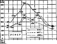
i и m iv v vi vii viii ix x xi хн 1 1___1___I___1___L-I-----1-----1-----1-----!-----1 Рисунок 14. Помесячное распределение заболеваний малярией в РСФСР (абс. цифры). по РСФСР и Закавказью за 1926-28 гг. Неодинаковое помесячное распределение заболеваний M. зависит от преобладания отдельных форм ee: там, где преобладающей формой является трехдневная M. (в большинстве районов РСФСР, в Белоруссии и пр.), наблюдается весенний подъем; наоборот, там, где большое распространение имеет тропи- ческая форма M., отмечается летне-осенний подъем (см. ниже). B нек-рых районах из года в год отмечаются два подъема: весен
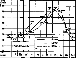
Рисунок 15. Помесячное распределение заболеваний малярией в Закавказьи (абс. цифры).
and, depending on the increase of the three-day form, and summer-autumn, due to the development of the tropical form of M. (separate districts of the Lower Volga region, Transcaucasia and Central Asia). This constancy of the monthly curve of M. is also observed in those foreign countries where M. is widespread: in Italy, for example, the maximum incidence falls every year on August (see Figure 16). During the pandemic period, the character of the monthly curve in the USSR changed sharply: in the European part of the USSR, where the maximum of diseases previously fell every year on May, it moved to September in 1922; in 1923 it was observed in June, and only in the following years the curve again assumed its usual character with a maximum in May (Figs. 17 and 18). This distortion of the monthly curve must be explained by the spread of the tropical form of M. throughout the country during the pandemic years. 8. Ratio of individual forms of M. Systematic studies of the blood of malaria patients in laboratories
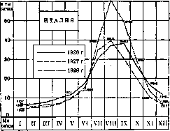
Figure 16. Monthly distribution of malaria cases in Italy (absolute numbers).
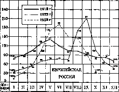
Figs. 17. Monthly distribution of M. cases in the European part of Russia (in relation to the average daily number of cases = 100).
ke) are given for comparison the results for several years. The tropical form of M. (Plasmodium immaculatum) is widespread throughout the USSR. It is noted in individual cases in Siberian stations (Omsk, Tomsk), but in Barnaul it already occurs in large numbers. It is observed relatively rarely in the central provinces. In the Volga region, in the North Caucasus and in Transcaucasia, it occupies a very high place. Four-day M. (Pl. malariae), very rarely
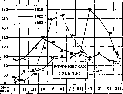
Figure 18. Monthly distribution of malaria cases in the Voronezh province (in relation to the average number of cases = 100).
occurring in Siberia and central areas, is widespread in the North Caucasus and in the Volga region, especially on the left bank of the Volga (Samara, Buguruslan, 23 Buzuluk), and it gives the impression that "in recent years it is growing at the expense of tropical M., which accordingly decreases. The latter also decreases at the expense of the three-day M. (Pl. vivax). Mass laboratory diagnosis of M. made it possible to establish also the monthly movement of its individual forms. Regardless of the district where observations were made, it was established,
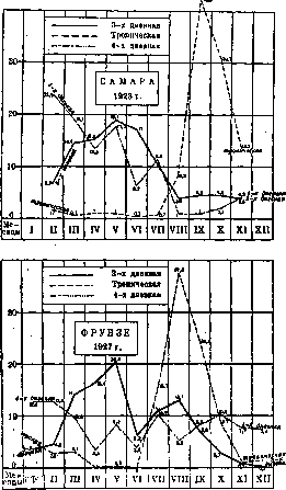
hampers the development of some industries, where it is observed as a professional disease among workers; in places of endemic spread it is the biggest brake to the development of industry; in some places there is extinction of the population due to malaria. 1. Wars and "M. Wars of the 19th century were often accompanied by the strongest outbreaks of M. 1 1 1 1 1 1 --------------- Tropical <v / \ \ 2jt)H 1923 / / \ i / ".4 / / (2.S \ \ \ / м \ \ \ o is "-Q- / " *лз тропа Ktcttan ' "1 II 1 1 /' N? \ь, / / / V \ 1927 г. h i \ /v \ i i t \ \ \ \ \ \ i \ 2.» +^** 1 1 1 \ ,«sV. пическ a.4.'- J°*s- 1 t--2i* _Jo_ ^-. Figure 19. Monthly distribution of individual"" forms of malaria (in percentages of the annual total of each .form). that everywhere the three-day M. (Pl. vivax) gives its maximum in May, the tropical form (Pl. immaculatum) in August or September, the four-day form (Pl. malariae) is distributed more evenly throughout the year (Fig. 19). The combination of all forms gives the monthly malaria curve of a given area. V. Malaria as a social disease. M. has great social significance. Public calamities (war, famine, etc.) cause an increase in it and worsen its course. Incorrectly organized large-scale work, carried out without sufficient regard for sanitary requirements, lead to mass morbidity and mortality of workers (the digging of the Panama Canal, the laying of railways in malarious areas, etc.). On the other hand, malaria epidemics severely disrupt the national economy; M. in the troops. Thus, in the American Civil War (1861-65), among white troops, on average per year, 544.7 contracted M. and 5.0 died, while among colored troops, 870.6 contracted and 18.8 died per every 1,000 men present. During the entire war period, among white troops, 1,163,814 people contracted M. and 8,140 died, while among colored troops, 15*2,141 contracted and 1,923 died. In some departments located along the Mississippi, among the troops, there was universal incidence of M. During the Japanese military expedition to Formosa (1874), out of 5,990 Japanese soldiers, 4,673 contracted M. During the Russo-Turkish war (1877-78), both the Russian Danube and Caucasian armies suffered severely from malaria. In individual units of the latter, M. constituted up to 7/8 of all diseases. In total, during this war, 610,591 people contracted M. in the Russian army (247 per 6Й5 - up to 142.0 (i.e., 14.2% of the entire composition contracted), in 1924, 1,000) and 1,293 died (0.5 per 1,000). The incidence rate strongly increased to 33.3, in ! 1922.-up to 106.5, in 1923.-de in Turkmenistan (1880-81): out of 12,000 military personnel, 8,411 contracted M. and 65 died. During the Spanish-American war (1898), M. claimed many victims among the Spanish and American armies: Spanish troops in Cuba in 1897 lost up to 7,000 dead from M., among American troops in 5 months (May-September) there were 38,833 cases and 97 died from M. with an average strength of 167,000 men. The largest outbreaks of M. were observed during the campaigns of European troops in tropical and subtropical countries, for example, during the French campaign in Senegal (1878), the English campaign in Afghanistan (1878-1879), the French campaign in Tonkin (1884-86) and in Madagascar (1884-85 and 1895), the Italians in Abyssinia (1895-96), and others. The morbidity rate among armies of various countries per 1,000 men of average strength before World War I (1901-10) was expressed in such
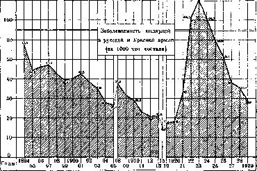
indicators (Table 19; according to Vasilevsky):
59.8
|
34.1
33.0
;
29.3
;
! 1.6 »
|
| 0.2 Branches of labor World War I gave a significant increase in M. in different armies. Above (see World distribution of M.) this increase was already noted for individual countries (Italy, Spain, SASU). In the German army, the incidence of M. per 100,000 men of average strength in individual years of the war was: 1st year of war-35; 2nd year of war-132; third year of war-620; 4th year of war-1,370. On the Balkan front, this incidence reached 9,260 in the third year of war, and 23,240 in the fourth per 100,000; on the Turkish front in the third year of war - up to 65,120, in the 4th - up to 18,370 per 100,000 men. The morbidity in the Russian and Red armies is shown in Fig. 20. In 1888-92 it averaged 106.5 per ;------------------ 1,000 men of strength per year in the Russian army, in 1900-02 it dropped to 40, in 1913 - to 20.8 per 1,000. In* 1921 go Figure 20. a decrease in morbidity to 104.3 per 1,000 is noted; subsequently M. quickly decreases to 28.9 per 1000 in 1929 (still: higher than in 1911-13). 2. M. among the industrial proletariat. According to the data of the Central Administration of social insurance in 1925, out of 100 all" cases of illness M. constituted among workers in mining and processing industry-4.3%, and among railway transport workers-9.2%. In Table 20, the number of cases of illness M. and days of illness from it per every 100 insured in the USSR in 1925 is indicated. Giving a high percentage for all branches of labor, M. for some of them still in 1925 constituted the most frequent disease: among workers in the extraction and processing of oil, for example, M. constituted about 20% of all cases^ of illness and causes of loss of days due to illness; in the basic chemical industry - about 10%. The average duration of 1 case of M. constituted a 100 Table 20. Number of cases of insured days . 1925 I -ni o t M. in the USSR. Cases of illness Days of illness male. Agriculture ... Mining industry . . Incl. coal mining » » » peat . . » » » oil . . Metal processing . . . » wood . . . . Processing of fiber substances » mineral » » food » » animal products Chem. industries .... Incl. basic chem industry....... Incl. oil processing Rail transport . 10.4 8.5 7.0 7.3 23.8 .5.8 10.2 2.4 7.5 5.5 6.5 8.7 12.4 30.5 7.1 female total male 2.2 8.6 84.9 6.5 8.3 82.8 6.4 6.9 75.8 10.8 8.6 67.5 - 23.8 220.7 4.7 5.7 55.9 8.5 9.9 92.9 2.3 2.4 23.7 5.4 6.9 65.6 10.4 7.0 42.7 4.7 6.2 47.9 3.1 6.8 77.4 17.0 12.8 134. S - 30.9 280.7 6.3 7.8 61.6 19.0 70.4 67.7 81.2 72.5 75.5 93.4 76.7 - 220.9 47.7 55.0 104.2 94.7 27.1 25.8 57.1 63.2 72.4 38.6 46.0 33.4 208.0 57.3 287.7 61.2 vlya for the entire USSR for men 9.3, and for women 10.0 days. Corresponding data on M. in the 7
MALARIA
in the Moscow province for 1925-28 are given in table 21 (per 100 insured). Groups of transport workers (water workers) and agricultural workers (on rice and cotton plantations). Table 21. Branch of labor Metal processing . . . Wood . . . . . Textiles . . . . . Leather goods . . . . . Minerals . . . . . Food . . . . . Animal products. All industry . . . Cases of disease Days of disease 1925 1926 1927 1928 1925 1926 1927 1928 1.7 2.1 1 2.1 | 1.4 20.2 24.6 1 20.1 13.8 1.2 1.3 1.3 26.4 14.6 | 15.6 12.3 3.4 3.8 2.8 2.4 43.6 44.2 | 28.6 22.5 4.4 2.6 2.1 4.1 59.8 28.9 23.1 56.2 2.2 1.4 1.1 1.3 24.8 16.0 11.6 12.0 1.7 1.4 1.4 1.7 24.2 12.6 20.3 12.5 2.8 3.0 2.3 2.0 35.4 35.2 23.6 18.9 M. was recorded at the highest level during these years among textile workers and workers in the processing of minerals.-The average Table 23. duration of a case of loss of working capacity due to M. decreases each year. It was for the entire industry of the Moscow province for both sexes: in 1925 - 12.8, in 1926 - 11.6, in 1927 - 10.3, in 1928 - 9.6 days. If in the Moscow province, which is relatively fortunate in terms of malaria, the latter occupies a high place (for the years mentioned, M. in terms of days of loss of working capacity among infectious diseases ranks only after influenza and tbc), then for some groups of seasonal workers it continues to remain one of the most common diseases. It continues to be recorded at particularly high levels among peat workers, for whom malaria is a professional disease. For the 1927 season (May-November), the incidence of malaria among peat workers is characterized by the following indicators (table 22): 3. The economic damage from M. is extremely great. In 1923, in the year of greatest M. development, the population of the USSR theoretically lost 75-112 million working days due to malaria, the insured - about 12,000,000 working days. During the pandemic, the most important branches of the national economy were disrupted due to M.: besides the peat industry, mentioned above, the fishing industry suffered most severely from M. (in Dagestan - Incidence on peat bogs of the Moscow province (per 100 workers). Peat bogs Kupavinskaya .......... Nikol'skaya........... Aksininskaya ........ Electroperevodka ........ Shaturskaya ......... All peat bogs of the Moscow province ... ......... 1922 1923 1924 1925 ! 1926 84.0 35.7 9.5 13.0 11.9 63.2 49.7 29.4 28.4 32.5 54.5 29.5 13.0 23.4 19.2 51.6 30.0 8.7 6.6 8.0 26.3 31.6 15.2 20.4 30.3 50.0 31.2 16.7 15.4 19.9 11927 11.5 23.4 7.3 7.3 12.8 14.3 Table 22. Groups of seasonal workers Peat workers.....| 28, Bricklayers.....j 2, Construction workers. Permanent industrial workers Per 100 insured % M. to total cases} days cases days 28.8 172.8 21.8 20.0 6.0 2.9 21.0 2.3 1.9 7.2 1.6 17.4 1.9 2.3 10.8 0.9 2.9 1.1 1.1 9.9 M. is recorded about 30 times more often among seasonal peat workers than among permanent industrial workers, and despite the fact that the duration of the disease is shorter among them than among other seasonal and permanent workers, still 20% of all cases of disease and loss of working capacity are due to M. The high incidence of M. among peat workers, which was a strong brake on the development of this industry in 1922-23, prompted health authorities and economic bodies to create a special system of medical-sanitary service for them (see below). As a result, a gradual decrease in M. among them is observed. For illustration, we can present the course of malaria among peat workers on some peat bogs of the Moscow province for 1922-27 (table 23). M. is a very common disease in some other branches of industry: in fishing, in some fishing grounds in the Caucasus, on average 65% of workers had the disease, on individual grounds - up to 100%), textile industry, oil, coal and others. The incidence of workers in timber rafting reached 72.5% (Kotlas). The operation of some railways and shipping was disrupted due to the mass illness of workers. In many areas, fields remained unsown and unharvested due to the mass illness of the rural population. M. causes no less trouble in places of its constant spread: in Turkestan and Transcaucasia, the extinction of a number of villages due to M. has been noted. All this places the fight against M. as one of the major social problems.
I.
Dobreitser. VI. Pathological anatomy. At the center of pathological-anatomical changes in M. lie changes in the blood, namely - of erythrocytes and blood pigment, which fully corresponds to the general characterization of M. as a blood infection. In this connection, anemia of one degree or another is always observed at autopsy (especially in chronic forms). Malaria plasmodia can also be found in the corpse; this mainly concerns tropical forms, in which abundant parasites are found in smears from the brain, spleen, bone marrow; however, Bystrov in cases of death from tertian malaria found parasites in the blood of the corpse (even 4 days after death). There are indications that in the blood of the corpse, multiplication of the parasite resembling the maturation of merozoites from schizonts in vitro on defibrinated blood with dextrose is possible; however, the ability to inoculate new erythrocytes in the blood of the corpse is denied.-As a consequence of destructive processes in erythrocytes, the so-called melanemia and melanosis (or hemomelanosis) of organs are noted. Under melanosis MALARIA
#29 imply deposits of a special black-brown pigment (hemomelanin) in various organs and primarily where the reticulo-endothelial apparatus is located: in Kupffer's cells of the liver, in the endothelium of the spleen sinuses, lymph glands, and in the bone marrow. However, outside this apparatus, along the stroma of all organs, significant pigment deposits may also be noted. The amount of the latter can be so great that macroscopically the organs acquire an ashen-gray, smoky, or almost black coloration. The largest amount of pigment is usually found in the spleen, liver, and pancreas. Sometimes pigmentation is extremely uneven: for example, a complete absence of pigment is observed in the spleen while there is an abundance of it in the liver and pancreas. It should be kept in mind that in chronic malaria, as well as with significant intervals between attacks, or in cases where attacks occurred in the distant past, the malarial pigment may almost completely disappear. Under otherwise equal conditions, the amount of pigment is also subject to strong individual fluctuations, apparently depending on the adsorptive capacity of the reticulo-endothelial apparatus itself, the rate of pigment absorption, and possibly also on the physicochemical properties of the latter. The formation of the pigment occurs in the process of intraparasitic changes of the blood hemoglobin; the pigment is therefore hemoglobinogenic, and although it does not typically give the usual reactions to iron, iron is nevertheless contained in it. Some authors, based on findings of iron and a number of other properties of hemomelanin, consider it close to, though not identical with, hematine. Many propose not to use the term 'melanin' in relation to malaria at all, leaving it exclusively for proteidogenous pigments (for example, pigments of the skin, hair), which have no direct relation to hemoglobin and do not contain iron (but contain sulfur), and to call this pigment simply 'malarial pigment.' Malarial hemomelanin has anisotropic properties, is soluble in weak alcoholic-alkaline solutions; the latter circumstance somewhat brings this pigment closer to the so-called formalin pigment. Along with hemomelanin, another pigment that gives the usual reactions to iron and apparently represents hemosiderin is often found in the same organs in malaria. - The periodic and prolonged load on the reticulo-endothelial system with pigment and other colloidal particles formed in connection with the massive breakdown of erythrocytes, and finally the very fact of intravascular parasitism of plasmodia—all this makes understandable the significant participation of the reticulo-endothelial system in the morphological picture of malaria. This participation is expressed in desquamative and proliferative processes, which sometimes lead to very significant changes in the volume and structure of the corresponding organs (hepatomegaly, splenomegaly, cirrhotic changes); an echo of this participation is also the so characteristic for malaria monocytosis in the peripheral blood. Progressive anemia, the overload ('blockade') of the reticulo-endothelial system with pigment, atrophic, degenerative, and sometimes sclerotic changes in organs (see below) often lead to general exhaustion, cachexia, and death. Sometimes, as a severe complication reflecting irreparable disorders in protein abundance, amyloidosis of internal organs, especially the kidneys, spleen, and adrenal glands, joins in. Finally, relatively often death in malaria patients occurs in the form of malarial coma. In terms of changes in individual organs, the spleen is of the greatest importance. In acute attacks of the disease, it significantly swells, and its capsule is sometimes strongly stretched; in some cases, for example, in artificial infection of progressive paralytics with malaria, tears and ruptures of the spleen with fatal hemorrhage are observed. Sometimes, on the contrary, the spleen is flaccid, mushy, with abundant scraping of pulp. In the malarial spleen, foci of gray and dark-brown necrosis (infarcts) are often observed. If the foci are numerous, a special appearance of marble spleen (Shirokogorov) results. Follicles are either sharply hyperplastic or barely noticeable. On microscopic examination of pulp smears, an abundant amount of parasites in all stages of development, clumps of pigment, lymphocytes, plasma cells, myelocytes, as well as free cells of various sizes of the reticulo-endothelium containing pigment (macrophages) are found. Chronic, especially tropical, malaria gives the most enlarged and most dense spleen (up to 6 kg; Shirokogorov), so that sometimes one speaks of malarial splenomegaly. Such a spleen often cannot be held by its ligamentous apparatus and becomes highly mobile, unless a perisplenitis with adhesions develops, which can wrap around the organ. The development of adhesions is associated either with diffuse perisplenitis or with the development of infarcts, which leave behind scars of various sizes, sometimes sharply distorting the contours of the spleen. Parasites in the pulp from chronic cases are often absent; pigmentation is noted to varying degrees; sometimes there is no pigment at all, and such spleens do not look dark-brown but red. The number of free pulp cells in these cases is sharply reduced, fibrous tissue develops, and the sinuses are sometimes sharply dilated, which gives the microscopic picture of the organ an angiomatous appearance. The liver is most often significantly enlarged (mainly in chronic cases); sometimes one can speak of hepatomegaly; in other (also chronic) cases, on the contrary, the liver is reduced and resembles the picture of atrophic cirrhosis. The surface of the organ is usually smooth, although sometimes phenomena of perihpatitis and the development of adhesions with the diaphragm are noted. Pigmentation is usually significant, and the liver looks ashen-gray or chocolate-colored on the cut [see separate table (art. 583, 584), fig. 8]. The malarial pigment lies mainly in the cells of the reticulo-endothelium, often freely in the circulatory system; there is none in the liver cells, but accumulations of hemosiderin are often found in them. It should be kept in mind that in terms of size and color, the liver, especially in acute cases, can be completely normal. In acute cases, phenomena of parenchymatous and fatty degeneration are often observed, sometimes phenomena of parenchymal dissociation, as well as necrobiotic changes affecting both groups of cells and entire lobules or groups of them. These changes in the parenchyma and probably lie at the basis of the atypical structure of the parenchyma, as well as the cirrhotic phenomena observed in chronic cases. - Lymph glands are enlarged, especially in chronic cases; the enlargement falls mainly on the glands of the abdominal cavity (area of the liver gates, spleen, periaortic). The color of the glands is either light gray or brown. Microscopically—desquamative and proliferative changes from the side of the sinus endothelium and reticulum cells. - In the bone marrow, usually large amounts of hemomelanin; the color of the marrow instead of red becomes brownish. In chronic cases complicated by cachexia, mucoid degeneration of the tissue is noted. The bone marrow is probably the only organ in which sexual forms of plasmodia—gametes—can be found. - The pancreas is often noted for its sharp pigmentation. Cases of necrosis of fatty tissue and parenchymal cells are observed, and in chronic cases, cirrhotic changes as well. - The peritoneum is also often intensely pigmented. In chronic cases, ascites is observed as a manifestation of marantic edema. - From the side of the intestine, a sharp melanosis of the mucosa is usually noted. Inflammatory and ulcerative changes of the small and large intestines (malarial enterocolitis) are relatively common. In the large intestines, dysenteria-like pictures (malaria dysenterica) are also observed. In the vessels of the affected areas, an abundant amount of parasites is found. The very mechanism of the development of intestinal changes should be represented as a toxicosis of the mucosa on the one hand and as a consequence of severe circulatory disorders on the other. - In the stomach, phenomena resembling those in the intestine are possible. Hemorrhages, as well as ulcerative-gangrenous changes from the oral cavity, gums, and pharynx probably belong to the complication of scurvy. Necrotic changes in the salivary glands are also described by Shirokogorov. - The lungs, besides their diffuse pigmentation to a steel color, usually do not show changes; sometimes edema, pneumonic foci, infarcts are encountered. Shirokogorov points to the significant resistance of malaria patients to tuberculous reinfection. - From the side of the heart in acute cases and in coma, ecchymoses of the epicardium, parenchymatous degeneration of the myocardium are found; from the side of the muscle fibers—sometimes loss of cross-striation, swelling and vacuolization of nuclei, more rarely—perivascular inflammatory infiltrates. In chronic cases, sclerotic changes of the myocardium of a focal and diffuse nature are possible. - Arteries do not show specific changes.
Shirokorodov observed, however, proliferation of endothelial and adventitial elements in vasa vasorum, with which he sometimes associates the sclerotic changes in the intima even observed in young malaria patients. The skeletal muscles are usually dark red in color, resembling smoked ham; necrobiotic changes of Zenker's degeneration type may occur. The kidneys often show glomerular pigmentation. In acute cases, there are pictures of hyaline-drop and granular degeneration; in chronic cases, amyloid degeneration is often observed. Nephritis develops mainly in tropical M. and in m. quartana. In rare cases, phenomena of so-called acute hemolytic M. are observed (see Hemoglobinuric fever). The adrenal glands of those who died in coma from tropical malaria often show degenerative-destructive changes in the cortex; in the medulla, the formation of miliary granulomas is described. The lipid content in the adrenal cortex in acute M. usually remains high (Zhgenti), which distinguishes M. from other acute infections. Changes in the testes in chronic forms are often observed and consist in the death of the generative epithelium and proliferation of the interstitial connective tissue. A characteristic picture is presented by the changes in the brain in tropical M., less frequently (M. Alekseev) in other forms. At autopsy, the smoky discoloration of the brain is striking [see separate table (pp. 487-488), Fig. 1], especially of the gray matter; upon microscopic examination, universal development of blood stasis is found, and in the swollen pale erythrocytes, plasmodia with pigment are found everywhere [see separate table (pp. 583-584), Fig. 7]. These pictures are pathognomonic for so-called malaria coma (see below), and their basis lies not only in the abundance of parasites but also in the changes in the physical properties of the erythrocytes themselves. The process is often accompanied by small hemorrhages, and in the extravasated erythrocytes, parasites are as a rule not found. This indicates the stickiness and phenomena of conglutination of the changed erythrocytes in the vessel lumen, which makes them incapable of diapedesis. In cases of death from uncomplicated acute three-day malaria, Byystrov found a peculiar swelling of the brain without pigmentation of the gray matter and without signs of edema and hyperemia. In the brain substance, pictures of focal encephalitis can also be found, expressed in perivascular and periganglionic proliferations of glia, in the formation of so-called malaria granulomas of Durck (Dllrck) [see separate table (pp. 583-584), Fig. 9]. The development of granulomas is often combined with hemorrhages and necroses, with the zone nearest to the vessel being necrotic, then the zone of hemorrhage, and behind it the granuloma proper. The radial arrangement of glial cells gives the granulomas a characteristic daisy-like figure (Ganseblumchen-figur of German authors). The most granulomas are found in the white matter and in the central ganglia. In the cortex, there are few or no granulomas despite the abundance of parasites precisely in the cortical capillaries; this circumstance makes it unlikely to explain the causal genesis of granulomas by the direct action of the parasites themselves on the brain tissue. There is an assumption that on the basis of successive degenerative changes of nerve fibers, mainly their myelin sheaths, pictures of disseminated sclerosis may then arise. However, even at their origin, granulomas have considerable similarity with fresh foci of disseminated sclerosis (disappearance of myelin sheaths, preservation of axis cylinders, proliferation of glia). The specificity of Durck's granulomas for malaria should, however, be considered not proven. On the basis of necrotic changes in the brain substance, foci of petrification have also been observed. From the side of the meninges, inflammatory changes in the form of diffuse lymphocytic infiltrates with subsequent sclerosis and clouding of the membranes are sometimes also observed. These same infiltrates can penetrate along the vessels into the brain substance. Exanthema in M. is rare, mainly in comatose forms; it appears either as petechiae or as blue-purple spots and erythemas, especially on tense areas of the skin. Histologically - extravasates, intravascular parasitism, deposits of pigment. On the lips, herpes sometimes appears. It is necessary to distinguish the pathology of M. from the pathology of malaria coma as the final act of the disease, especially frequent in tropical M. Among the phenomena characteristic of coma, one must first of all include massive peripheral stases of blood with universal parasitism of plasmodia in markedly swollen pale erythrocytes; it must be thought that these phenomena themselves create the most severe conditions for blood circulation. To the manifestations of coma should also be attributed hemorrhages, pulmonary edema, as well as a peculiar yellowish discoloration of the coverings, indicating the onset of hemolytic phenomena. According to data from Moscow morgues for 1923-27, out of 37,899 autopsies, only 46 cases of death from M. as the main disease were noted; 7 of these cases fall on tropical M. In general, these figures show that malaria mortality in the climate of the middle and northern zones is very small, and mortality is also insignificant, since malaria must still be considered a very widespread suffering. I. Davydovsky, I. Shirokorodov. VII. Pathogenesis. The pathogenesis of M. is determined by the relationship between the state of the human organism and the vital activity of the plasmodium as a parasite of its erythrocytes. Plasmodia in the form of sporozoites, entering the bloodstream of the bitten person along with the mosquito's saliva during a bite, penetrate into erythrocytes and grow here, feeding on Hb. Upon reaching maturity, the plasmodia divide asexually (schizogony) into daughter cells - merozoites. The latter, destroying the erythrocyte, enter the blood plasma and strive to find shelter in new erythrocytes. At the same time, a significant part of the merozoites dies under the influence of the parasiticidal properties of blood plasma. Numerous observations have established that the most typical expression of the malarial disease - the febrile attack - occurs at the moment of simultaneous division of a large number of blood parasites. The very cause of the febrile reaction of the organism to the indicated moment in the life cycle of the plasmodium has not yet been finally established. Marchoux attributes the destruction of erythrocytes to the action of a hemolytic toxin formed and released by the plasmodium at the height of its maturity. The onset of the febrile attack was associated with the action of a special pyrogenic toxin, the existence of which, however, has not been proven by direct experiments. Another hypothesis explains the onset of the temperature reaction by the entry into the blood of protein bodies formed from the death of part of the blood parasites and the breakdown of erythrocytes. Varazzi caused a typical temperature curve of three-day fever by injecting a rabbit every 48 hours with a solution of egg white. Abrami and Senevet consider the malarial attack as a clinical expression of a sudden violation of the colloidal equilibrium of the plasma (hemoclastic shock), occurring at the moment of division of the plasmodium. Brown put forward the assumption that the attack is caused by the release of malarial pigment (its injection into rabbits causes an increase in temperature). Whatever the mechanism of the malarial paroxysm, the vital activity of the plasmodium lying at its basis causes primarily the destruction of a large number of erythrocytes and Hb. In the periods free from febrile attacks, these losses of peripheral blood are more or less quickly replenished, and at the same time, apparently, the physico-chemical state of the blood changes, as a result of which the number of circulating schizonts in it gradually decreases. These immunobiological changes are confirmed by the fact of spontaneous cessation of attacks with a decrease in blood parasites in the peripheral blood to a minimum not detectable by the methods available to us. Abrami, Senevet and Carnot proved the presence of schizontolysins in the blood serum of malaria patients. The instability of these lysins explains the rapid exhaustion of immunity and the occurrence of early relapses (Ruckfalle, rechute) of febrile paroxysms. The mechanism itself of both these early and later seasonal relapses (recurrences) of the disease has not yet been finally clarified. The hypotheses proposed to explain them can be divided into two main groups: according to one, the recurrence is a consequence of continuing, only quantitatively increasing schizogony (Ross, Thomson), others assume that schizogony of recurrence is preceded by the maturation and fertilization of gametes (Rowley, Biedl, Grassi) or parthenogenesis of gametes (Schaudin) in the blood of the patient. Predisposing causes and provoking moments for recurrence are extremely diverse: these include colds, dietary errors, overwork, trauma, etc. However, none of these moments can certainly provoke a recurrence. The reasons for the mass occurrence of seasonal recurrences in general are still not sufficiently clear.
They may lie in seasonal fluctuations of the physicochemical properties of the patient's blood, as well as in the biology of the parasite itself. Relapses of schizogony of the same form of parasite in the peripheral blood with clinical manifestations of a malarial attack must be distinguished from reinfection—a new infection with the same form of parasite after the primary disease has been cured or during its course. The initial infection with each new form of parasite is determined by the term 'fresh infection.' VIII. Clinic. The clinic of malaria includes a number of very diverse pathological manifestations that are difficult to fit into a specific classification. In the most general terms, M. can be characterized as a chronic disease that at times manifests as acute febrile attacks. In accordance with the three varieties of the causative agent, clinically M. proceeds in the form of a three-day form (malaria tertiana—causative agent Plasm. vivax), a four-day form (malaria quartana—causative agent Plasm. malariae, s. Laverani), and a tropical form (malaria tropica—causative agent Plasm. immaculatum). In the last pandemic, cases of infection with two or (more rarely) all three species of plasmodium were often observed. The incubation period of M. varies within rather wide limits depending on the type of parasite, the time of infection, and the state of susceptibility of the infected. Ziman considers the incubation in M. to be on average 10-14 days; it is shortest in the tropical form, somewhat longer in the three-day form, and longest in the four-day form. Accumulated observations of cases of the primary manifestation of M. in the early spring months, when infection through a mosquito bite was still impossible, have led to the introduction into the clinic of M. of the concept of prolonged incubation. Some malariologists prefer to define this period as a primarily latent infection. Prodromal phenomena are not always sufficiently clearly expressed; often the first febrile paroxysm occurs suddenly; sometimes it is preceded by sensations of indefinite malaise, fatigue, aching in the limbs, and headache. More often and more sharply these sensations preceding the febrile attack are noted by patients during relapses of the disease. The characteristic of acute malarial disease the alternation of febrile attacks (febris intermittens) is not established from the very beginning of the disease. Often an initial fever of a continuous or remittent type is observed (Fig. 21), which then passes into a proper intermittent type. According to the observations of Wethmar, the initial fever more often occurs with a short incubation period. |[temperature-- DAYS OF ILLNESS batatura 2 3 4 5 6 7 8 9 1 -x~i Щ------q :£ " -X Z3 ___ 0 *° j " o 3&^ _ id_ хв : II т X* : ± 3S ~ ' L ± Hl_ ji ,:I/l 3 ,1-___Zl 1 : :: :_ :___ I _:_:__:_ Figure 21. Initial fever in mal. tertiana. A typical malarial paroxysm is characterized by the fairly rapid succession of brief periods of chill, heat, and sweat, standing in close connection with the cycle of asexual development of the malarial parasite in the patient's blood. The chill, with which the attack begins, can be of varying intensity—from barely noticeable shivering and the sensation of running ants along the spine to a chill that shakes the entire body of the patient and causes uncontrollable chattering of the teeth. The patient cannot overcome the sensation of cold, even by putting on all available clothing. The state of chill lasts 20-30 minutes, sometimes much longer, and, gradually subsiding, gives way to the sensation of heat, which is accompanied by headache and thirst. The period of heat lasts from 1 to 12 hours, after which t° begins to fall more or less rapidly. First the forehead and upper part of the chest, and then the entire skin of the trunk and extremities become moist, the period of sweat sets in, often taking the character of profuse sweating. With the fall of t° and the appearance of sweat, the distressing sensations disappear fairly quickly, the patient usually calms down and falls asleep. During the subsequent period of apyrexia, coinciding with the growth and maturation of schizonts in the erythrocytes, the patient feels only some weakness, which disappears by the beginning of a new attack. With the onset of chill, the skin becomes dry and rough (goose skin), the extremities grow cold, the lips and fingertips become cyanotic. With the end of the chill, the patient's face reddens, the skin becomes hot and dry to the touch and remains so until the appearance of sweat. During the period of apyrexia, the skin takes on a normal appearance or sometimes remains moist and pale. In individual cases, the appearance of various types of rashes (see below) is possible. The tongue during the febrile period is often slightly coated, sometimes swells and has indentations—impressions of teeth—on its lateral surfaces. Breathing shortens and quickens already from the beginning of the chill and returns to normal only with the passing of the paroxysm. The pulse quickens and becomes tense from the beginning of the attack, and at the height of the attack, dicrotia is sometimes felt. With the fall of t°, a significant slowing of the pulse and improvement in its quality are usually observed. Blood pressure in the stage of chill is considerably elevated, gradually returning to normal or even falling below normal in the stage of heat and sweat, due to «37 weakening of the vascular tone (Shabashov, Molchanov and Egorov, Mikeladze).—A malarial attack is very often accompanied by vomiting, in severe cases with an admixture of bile in the vomitus. Occasionally, especially in childhood, diarrhea accompanies the attack.—The characteristic for M. enlargement of the spleen, caused by hyperemia of the organ and hyperplasia of its cells, begins with the onset of febrile attacks, and sometimes even in the prodromal period. In fresh cases, after the attack has passed, the spleen fairly quickly contracts to its normal size. However, it should be borne in mind that neither the fact of enlargement of the spleen nor its size and density can serve as a measure of the severity of M. During the attacks, the liver also enlarges and becomes painful, functioning intensively due to the need to process a mass of erythrocytes destroyed by blood parasites. The filling of the bile capillaries and the entry of bile into the blood sometimes causes a yellowish discoloration of the skin and sclera. From the side of the nervous system, during an acute malarial attack, signs of general diffuse damage, characteristic of febrile states in general and manifesting as general malaise, headaches, sometimes delirium, and semi-consciousness, are noted above all. However, from the very first attacks, manifestations of a hyperkinetic character in the form of convulsions of individual muscle groups or paretic states, and in children also general convulsions, are possible. In accordance with the pathogenesis of the disease, significant changes are observed in the erythrocytes. A certain percentage of them (according to Marsh—well noticeable with proper staining of the smear). Maurer described similar changes in erythrocytes in tropical M.; Maurer's speckling is sparser, coarser, and has irregular contours. With vital staining with basic aniline dyes, the number of erythrocytes showing granularity (substantia reticulo-filamentosa) is significantly increased compared to normal (Sarkisyan). The decrease in the total number of erythrocytes and their Hb content has already been mentioned above. A malarial attack also causes quite significant changes in the quantity and composition of leukocytes. Two hours before the onset of chill, leukopenia g is noted (Abrami and Seneve), which by the time of the chill is replaced by a slight leukocytosis, gradually increasing at the beginning of the rise in t° and quickly disappearing by the time it subsides. Together with fluctuations in the number of leukocytes, the ratio of their individual types also changes: neutrophilia, characterizing the beginning of the attack, is replaced at its end by neutropenia, mainly due to segmented forms, relative lymphocytosis, lymphocytosis. Eosinophils at the height of the attack decrease in number or even disappear and reappear in apyrexia. As in other acute infections, Turk's cells often appear in the blood during the acute period. Braylovsky, having made* during the height of an epidemic over 1,500 blood studies in various forms of M., found that the range and character of quantitative fluctuations of leukocytes are not connected with the type of parasite-causative agent, but in the qualitative composition of the white blood such a connection is noticeable: three-day M. gives a shift to younger forms, the number of eosinophils is reduced,.
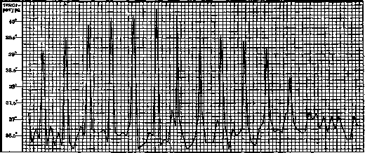
Figure 22. Therapeutic mal. quartana. Spontaneous subsidence of fever. Transition of paroxysms
from evening hours to morning hours. up to 12% in severe cases) contains plasmodia in one or another stage of development depending on the moment of examination. Erythrocytes containing plasmodia are paler due to their impoverishment of Hb. In three-day fever, the introduction of Plasmodium vivax leads to an increase in the size of erythrocytes and causes the appearance in its protoplasm of fine, so-called Schüffner's granules, and they are rarely absent. In tropical M., the shift of neutrophils reaches myelocytes, is more sharply expressed, eosinophils often completely disappear, monocytes are encountered more frequently than in three-day M.; usually the presence of degenerative forms is observed. Four-day M. more closely resembles three-day M. in blood changes, but causes greater lymphocytosis. The number of blood platelets decreases. The excretion of urine during the stage of chills can be somewhat increased, then it sharply decreases, and during the entire period of heat and sweat, concentrated urine is excreted in small amounts. The specific gravity of urine is elevated due to the content of a large amount of protein breakdown products; the breakdown of erythrocytes also causes an increase in the excretion of iron and urobilin; the amount of chlorides is either unchanged or somewhat elevated. The above-mentioned symptoms are more or less common to acute attacks of all three forms of malaria. The further course of the disease has peculiarities in each individual form. Four-day M. (fig. 22) is distinguished by the most regular course. Fever attacks in this form recur after two days of apyrexia at the same hours. The premature onset of an attack (m. anteponens) or its delay (m. postponens) is observed less frequently than in other forms. In the presence of two generations of the parasite, after two days of fever, one day of apyrexia occurs (m. duplicata), with three generations (m. triplicata) attacks occur daily. Usually with double or triple m. quartana, one of the generations predominates and gives the temperature curve a distinct 4-day type (fig. 23). The asynchronous maturation of schizonts of P. malariae gives this form more noticeably expressed prodromal phenomena in the form of fatigue and headache; the stage of chills is more prolonged, and the rise in temperature occurs less violently than in other forms. Sweat appears quickly and is abundant. Vomiting
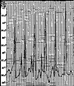
The three-day M. (m. tertiana), which is most common in our latitudes, has a less regular course. The attack of three-day M. occurs every other day, and in the presence of two generations-daily (m. tertiana duplex) (fig. 24). In t. tertiana, constant or remittent initial fever is especially frequently observed. The chill in a typical attack of three-day malaria is very sharply expressed but not prolonged. The warming of the body occurs quickly, the temperature reaches high figures and then slowly subsides. Sweating does not set in immediately and sometimes lasts up to 8 hours. Attacks of m. tertianae are often accompanied by vomiting and intestinal disorders; all clinical phenomena in this form are more sharply expressed than in four-day malaria; in the period of apyrexia, painful sensations do not leave the patient. But m. tertiana is more prone to spontaneous cessation of attacks and more readily responds to quinine treatment. Tropical M. (figs. 25 and 26) is distinguished by the most severe and irregular course. Its temperature curve rarely retains the three-day character, taking on, especially during relapses, an extremely irregular course (fig. 27). The superposition of attacks gives the temperature a remittent or constant character. The other features of the febrile attack are also as irregular and variable in m. tropica: the chill can be very brief and mild, or sometimes completely absent. The rise in temperature in fresh cases occurs very quickly, usually in morning or daytime hours. The fever lasts very long, giving a decrease only on the following day, and often the temperature does not have time to fall to normal before the beginning of the next attack. In later stages, on the contrary, brief isolated febrile paroxysms separated by long intervals of apyrexia are possible. The stage of sweating may be completely absent or weakly expressed. The clinical symptoms of an attack of tropical M. are similar to those of three-day malaria but are distinguished by particular severity: vomiting often takes on a bilious character, nervous phenomena reach their most pronounced development; the sleep that follows the attack is interrupted by nightmares. The brevity or almost complete absence of apyrexia does not allow for rest, and the strength of the patient quickly declines. Tropical M., more than the other two forms, sometimes takes on a malignant course. Described are: a) typhoid, or hyperthermic form with constant high temperature (fig. 28) and symptoms of severe intoxication; b) cholera-like, or algid form, running with all the symptoms of cholera; c) dysenteric form; d) pneumonic form with pneumonic foci and pulmonary hemorrhages; e) hemorrhagic form with rashes and hemorrhages. Great caution is necessary in establishing the diagnosis of such malignant forms; it is necessary to exclude the possibility of coincidence of M. with typhoid, cholera, dysentery, etc. Special mention is deserved by the cerebral, or comatose M., which has spread widely, especially during the last pandemic. The occurrence of this form is explained by the influence of a particularly virulent hypothetical toxin (toxic theory) or more often by the blockage of brain capillaries by disintegrating infected erythrocytes (mechanical theory). Comatose state in M. occurs in individuals weakened by undernutrition or previous diseases
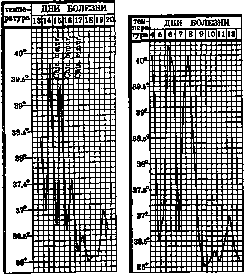
or pregnancy (Kitaev), or in those completely without immunity to M. In the latter case, coma can arise from the very first clinical attacks of M. The temperature in the comatose form is high, of constant ty
Fig. 28. Tropical malaria, typhoid form. Relapse.
Fig. 26. Tropical malaria. Fig. 27. Tropical malaria, fresh disease! Long paroxysms.
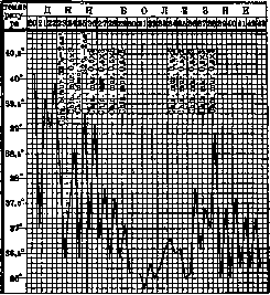
Fig. 26. Tropical malaria. Fig. 27. Tropical malaria, fresh disease! Long paroxysms.
The temperature may sometimes, conversely, be normal (in 20% of all cases of Kitaev); the patient is 'immobile, in a sleepy, stuporous, or completely unconscious comatose state. The pupils are dilated, do not react, the pulse is tense, the patient urinates on himself; often there is continuous sweating. The spleen may not be enlarged at all or only slightly enlarged. In the peripheral blood, a large number of schizonts is usually found; at the same time, the appearance of mature schizonts and forms of division (Kitaev) is characteristic, which are not observed in the peripheral blood in ordinary cases of tropical M. Sometimes, however, the number of parasites in a thick smear is remarkably small and does not correspond at all to the severity of the disease. Comatose tropical M. in most cases ends in death despite the most energetic treatment. - More rarely, M. is complicated by hemoglobinuria. The acute febrile* stage of any form of M. may end even without any treatment. After a greater or smaller number of febrile attacks, a prolonged apyrexia sets in. A kind of equilibrium is established between the patient's organism and the life activity of the plasmodium. The patient feels quite well, but upon examination of the blood, both sexual forms and schizonts may be found. The duration of such an interparoxysmal period for three-day M. is on average calculated at 8-11 days, for tropical M. - at 5-7 days. However, these periods are subject to extremely wide fluctuations. The early relapses of febrile paroxysms that occur after these apyrexias become less prolonged, milder in manifestations, and more irregular in course with each subsequent relapse. The duration of the interparoxysmal period can be much longer, reaching several months. - Thus, in addition to early relapses, late relapses occur, having a definite seasonal character. This seasonality is especially clearly expressed in three-day M., relapsing in the early spring months; relapses of tropical M. are observed in the summer-autumn months; four-day M. relapses in winter. Untreated or poorly treated, long-relapsing M. eventually takes on a chronic course. Chronic M. leads to more or less significant disorders of the organism as a whole and changes in individual organs. These changes are the more pronounced, the more frequently relapses of febrile paroxysms occur. The latter, as already noted, become atypical, are smoothed out, and can in the end proceed almost completely unnoticed. Only sometimes with prolonged and careful measurement of t° is it possible to detect in it a reflection of the cyclically occurring schizogony (fig. 29). The sensations of the patient also change sharply. Instead of the typical triad characterizing the febrile paroxysm of the acute period, chronic M. brings with it indefinite sensations of illness: general malaise, fatigue, apathy, reluctance to work, headaches, and pai mpe ratu ra 86,6s Z92- ш ш ш ! Figure 29. Malaria quartana. Cyclic rises of temperature not exceeding the norm are clearly visible. dizziness, noise in the ears, aching in the limbs and lower back. Later, a series of complaints arises caused by changes in various organs. The appearance of the malaria patient-chronic becomes quite characteristic. The skin gradually takes on a greenish-gray, earthy color, sometimes with a slight yellowish tinge. Brownish-aspid colored pigment spots often appear on the face. In the area of the wings of the nose and lips, herpes occasionally erupts, more often during exacerbations. On the skin of the trunk, rarely, as in the acute period, short-lived! scarlet fever-like or urticarial (Muffel, Sokolov) or more persistent hemorrhagic (Braude, Demyanov) rashes may appear [see separate table (art. 647-548), fig. 1]. The mucous membranes are usually pale, and in the oral cavity they may be covered with pigment spots. One of the most characteristic symptoms of chronic M. is considered to be enlargement of the s p l e e n, most often reaching large sizes in the four-day (Griesinger, Kushev) and three-day forms. On the contrary, the tropical form often proceeds without enlargement of the spleen. The enlargement of the spleen occurs either predominantly in length or in width. Tyatnikov associates this difference in the shape of the enlarged spleen with the form of M., while Oganesov associates it with constitutional factors. A distinctive feature of the enlarged malarial spleen is its density. Malarial splenomegaly, especially frequent and intense in malarial endemic areas, requires great attention, as cases of rupture of large malarial spleens have been described; trauma is more often the cause of the rupture, but spontaneous ruptures are also possible. In the pathogenesis of malarial splenomegaly, repeated hyperplasias of the pulp play a role, which eventually become persistent, and in later stages - the development of connective tissue. - The l i v e r in chronic M. is also sometimes enlarged and hardened; changes in the liver can more often be found by determining its functions (Vol'sky and Sheveleva). The most constant changes in chronic M. are changes in the red blood, creating secondary malarial anemia. The number of erythrocytes is decreased, anisocytosis, poikilocytosis, polychromatophilia appear; in more severe cases, nucleated erythrocytes and erythrocytes with basophilic granularity are found in the peripheral blood. Parallel with the decrease in the number of erythrocytes, the percentage of Hb also falls, and Hb continues to decrease even after the number of erythrocytes becomes stable; their resistance increases. The number of plasmodia parasitizing in the erythrocytes of the peripheral blood usually falls to a minimum, not detectable in a thick smear. The number of blood platelets is decreased. - Changes in the white blood, considered characteristic of chronic M., are leukopenia, decrease in eosinophils and monocytosis (Kushev, Svenson). Braylovsky considers the appearance of a large number of degenerative forms, an increase in the number of predominantly large lymphocytes, and the appearance of Türovsky cells to be characteristic of the chronic course of M. Any revival of the life activity of blood parasites, causing more or less pronounced clinical manifestations of a relapse, is accompanied by a shift of neutrophilic leukocytes towards stab and young forms, characteristic of an acute attack, and this shift in such cases has a definite cyclic nature, connected with the course of the paroxysm. It should be borne in mind that, as in a healthy person, the number of leukocytes and the ratio of their individual kinds are extremely labile and easily change under the influence of constitutional factors and various additional conditions in which the malaria patient-chronic may be at the moment of examination. The increase in bilirubin content in the blood serum observed in the acute period of M. can be confirmed to a lesser degree also in the chronic course of M. (Russo and Serbinov); however, with a small number of blood parasites, the bilirubin content does not exceed normal figures. In addition to the above-described changes, more or less constant in the acute and chronic course of M., changes can also be observed from the side of other organs and systems. The described bronchitis and pneumonia of 'malarial origin' should rather be considered secondary, caused by the usual causative agents of lung diseases and developing on the soil of weakening of the organism (Tsiman). In some cases of microscopically diagnosed M., complications with bronchial asthma, h e m o p t y s i s have been described; the etiological dependence of these complications on M. is usually based on the success of quinine therapy. - On the c a r d i o v a s c u l a r system, the chronic course of M. is reflected in a persistent weakening of the vessel tone (Shabashov). Multiple severe attacks, especially of tropical M., can lead to weakening of the heart's strength and its dilation. These changes are not persistent, and under the influence of rational treatment, the condition of the vessels and heart returns to normal. Attempts to attribute to M. inflammatory diseases of the walls of the heart and vessels (malarial endocarditis and endarteritis) are little provable and do not fit with the general concepts of the pathogenesis of M. - D i s o r d e r s of digestion, accompanied by vomiting or diarrhea, are more characteristic of the acute attack of M. In malarial areas, chronic mucous-hemorrhagic colitis are often associated with chronic M. In all cases of such colitis, careful bacterioscopic and bacteriological examination of the feces is necessary, which can reveal the true dysenteric (protozoan or bacillary) nature of the disease. Long-term chronic M., according to the observations of Karapetyan, Sharov, disrupts the secretory function of the stomach in the direction of its unstable decrease, and according to old observations of Triantaphillides, it can be accompanied by persistent diarrhea, sometimes representing the only clinical manifestation of the paroxysm. Such intestinal forms undoubtedly also require an extremely cautious approach in establishing their malarial origin. - Barbitsky, Puchek, Gurevich and others, observing transient albuminuria and nephrosis-nephritis in chronic M., see confirmation of their malarial etiology in the effect of quinine therapy.'
Marsh questions the direct dependence of kidney lesions on malaria; according to Shirokorov's observations, patho-anatomical changes in the kidneys in chronic malaria are not the rule. Various changes of the nervous system are especially often associated with malaria. During the acute period, they manifest as transient delirious, soporous, and (as an extreme expression) comatose states. With prolonged severe malaria, as with any other infection, more persistent infectious psychoses may also arise (see below - malarial psychoses). In the chronic course of malaria, disease manifestations attributed to it are described from the central nervous system, peripheral nerves, and the autonomic nervous system. Lesions of the central nervous system, depending on their intensity and localization, may manifest as hemi-, mono-, or paraplegias or pareses, aphasia, ataxias, meningo-encephalitic symptoms, or symptoms of disseminated sclerosis, and more often follow feverish exacerbations of the disease. The mechanism of these phenomena is apparently similar to that in malarial coma (see above). From the peripheral nerves, neuritis and polyneuritis and neuralgia, most often of the trigeminal nerve, less frequently of the intercostal, ulnar, or other nerve branches, are described. These complications, especially neuralgia, are more characteristic of the afebrile period of the disease and are often considered the only manifestation of a malarial attack. Their origin is more often attributed to the toxic factor: poisoning of the nervous system by a hypothetical malarial toxin, as well as the general exhaustion of the nervous system under the influence of the accompanying chronic malaria anemia. The same factors explain the lesions sometimes attributed to malaria of the autonomic nervous system and endocrine apparatus. However, they are described much less frequently and are less studied. The frequency of complications from the nervous system undoubtedly depends on the intensity of the malarial epidemic; for some endemic malarial areas, certain authors (Triantaphillides for Batum, Grosman for Astrakhan) determine it at 2-3%. In most cases, changes from the nervous system are observed in the course of tropical malaria, but they are also possible with other forms of it. - The diagnosis of nervous complications of malaria (neuromalaria) is based on the case history, the characteristic intermittence of symptoms, and the effect of quinine therapy. Confirmation of the diagnosis by finding blood parasites does not always succeed and is more characteristic of complications of the acute period. Establishing the malarial etiology of nervous symptoms usually gives a good prognosis ad sanationem. However, with prolonged course of untreated or poorly treated malaria, more persistent changes that no longer respond to quinine therapy are possible. - Of the sense organs in malaria, lesions of the eyes are most often described. During attacks, conjunctivitis, sometimes unilateral, may be observed; herpes may be localized on the cornea; sometimes true superficial or deep keratitis, retinitis, and hemorrhages are also observed. In chronic malaria, cases of inflammation of the optic nerve have been established, the characteristic feature of which are fluctuations in visual acuity. All these eye complications, according to ophthalmologists, respond well to quinine treatment, being extremely stubborn with ordinary symptomatic therapy. The dependence of lesions of the function of hearing on malaria is less studied and established. - The extreme expression of changes characteristic of chronic malaria is malarial cachexia, or malarial emaciation. Severe anemia, a yellowish-earthy color of the skin, yellowish-pale mucous membranes, hydroremic edema, hemorrhages with an enlarged liver and a spleen descending below the navel in an ascitic abdomen (see separate table, figure 2) characterize the malarial cachectic person. In the origin of cachexia, besides the malignancy of malaria itself, superimposed conditions play a huge role, with starvation in the first place, accompanying chronic infections and helminthic invasions. Particularly in the nomenclature of malarial diseases stands the so-called latent malaria. Under this name, some (Svenson and others) mean the interparoxysmal period of malaria, when parasites are present in the peripheral blood but there are no objective or subjective manifestations of the disease. In another definition, especially widespread among practical physicians in malarial areas, under latent malaria they understand very diverse and often rather indefinite painful sensations in the absence of typical clinical signs of acute and chronic malaria and parasites in the peripheral blood. This form in Ziman, Svenson and others is called masked (tЛагуа1а). Most typical for latent malaria in its latter definition are complaints of apathy, reluctance to work, drowsiness, irritable nervous weakness, headache, sweating, etc. Supporters of the diagnosis 'latent malaria' confirm the malarial etiology of such disorders by the sometimes observed periodicity of symptom appearance and the effect of quinine therapy. It should be borne in mind that long-term observation and careful examination in a significant number of such cases allow establishing an etiology other than malaria for such manifestations (initial forms of pulmonary and glandular tuberculosis, blood diseases, helminthic invasions, rheumatism, climacteric, hysteroneurotic states, etc.). Therefore, in order to clarify the diagnosis of malaria, it is necessary to abandon the term 'latent malaria,' and to confirm the diagnosis of the interparoxysmal period only on the basis of definite objective data (Toporkov, Kushev). This is all the more important because the deceptive temporary effect of quinine in such cases only diverts attention from proper causal therapy. - As a chronic disease, malaria in its course can be combined with various acute and chronic diseases, exerting some influence on them and itself undergoing certain changes under their influence. The influence on the course of malaria of hereditary and constitutional factors is very probable but has been studied completely insufficiently. To the present time, a large number of observations have accumulated asserting the possibility of congenital malaria. Although infection with malaria is possible at any age, the course of malaria in children sometimes has a special malignancy. Sex has no influence on the course of malaria. Regarding temperament, Ziman points out that phlegmatics tolerate malaria better than choleric and sanguine individuals. The influence of race on the course of malaria apparently comes down to questions of immunity. Residents of areas with endemic malaria gradually accumulate relative immunity to malaria with age and pay for it mainly with their younger age groups. Northerners and residents of mountains, when arriving in malarial areas, get infected with malaria especially quickly and tolerate it especially severely. Among the nationalities of the USSR, in any case, neither especially immune nor especially predisposed to malaria have been noted outside the above-mentioned conditions. The influence of environmental conditions on the course of malaria is completely indisputable. All physical and psychological factors leading to weakening of the body can disrupt the balance between the vital activity of the plasmodium and the protective function of the body and lead to the awakening of untreated malaria. On the contrary, physical and mental peace can contribute to the more rapid cessation of the clinical manifestations of malaria. The latter fact can partly explain the sometimes observed self-healing from malaria attacks when changing location, when the general living conditions also improve for the better. The past pandemic, which coincided with the famine in the Volga region, once again confirmed the fact of the enormous influence of famine and undernutrition on the course of malaria. In areas affected by famine, especially the malignant forms described above were often noted, and among them, malarial coma was in the first place. In the best case, malaria took a stubborn chronic course, creating characteristic types of malarial cachectics. In famine years, even relatively mild tertian and quartan malaria, which are mild under ordinary conditions, took on the malignant character typical of the tropical form. All kinds of diseases, especially chronic ones, by weakening the body, also create a predisposition to infection and repeated relapses of infection. In recent years, the influence of helminthic invasions on the course of malaria and its resistance to quinine treatment has been particularly noted (Permyakov, Mufel). Childbirth, labor, and the postpartum period activate latently occurring malaria, and malaria attacks in turn can adversely affect pregnancy, threatening premature termination of it. Similarly, the relationship between malaria and surgical operations: the latter can provoke malaria attacks, while surgeons during operative intervention should keep in mind the tendency of chronic malaria patients to bleed, the sluggishness of healing and restorative processes in them, etc. Individual observations of the simultaneous course of malaria and typhoid fever, malaria and relapsing fever testify, firstly, that such combinations in any case worsen the patient's condition, and secondly, that the intervention of malaria disrupts the typicality of the temperature curve of the main disease. However, Svenson and Bukhstab did not see such an influence of malaria on typhoid and relapsing fever in their cases. The influence of malaria on the course of pulmonary tuberculosis has been repeatedly evidenced by exacerbations of the tuberculous process, hemoptysis, etc., that have arisen
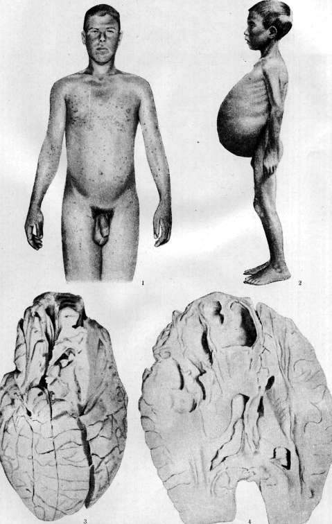
Figure 1. Hemorrhagic rash in a malaria patient. Figure 2. Spleen in chronic malaria (boy 5 years). Figure 3. Destruction of the left frontal lobe by abscesses. Figure 4. Section through the posterior part F;!. Lower part
abscesses. Some after or during attacks of M. The observations of Marcou-Mutzner on the antagonism between tbc and M. and the assumption to use the latter in the treatment of tbc are not justified. Similarly, the view of the incompatibility of cancer and M. is not justified. Since the publication of Wagner von Jauregg's observations on the beneficial effect of malaria in the treatment of progressive paralysis, inoculation of M. for therapeutic purposes has been tried in a significant percentage of cases with very satisfactory results on thousands of progressive paralytics. The use of M. in the treatment of gonorrhea is in the study stage. In leukemic hyperleukocytoses, Lucherini saw a rapid but soon passing decrease in the number of leukocytes under the influence of a malarial attack. Attempts to use M. as a therapeutic agent in some other diseases (dermatitis, interstitial keratitis, etc.) must be recognized rather unsuccessful.--For therapeutic infection, three-day malaria is usually used, more rarely four-day malaria. Infection is carried out by subcutaneously or (better) intravenously injecting several cm3 of blood containing parasites, taken from the cubital vein of a malarial patient. Some prefer infection by the natural route, i.e., through the bite of an infected mosquito, which is much more complicated. The incubation period of experimental (therapeutic) M. depending on the time and method of infection, the species of parasite, and individual characteristics of the infected person varies within rather wide limits. On average, with intravenous infection with the three-day form, it is 1-5 days; with infection with the four-day form, it is longer. The incubation periods with subcutaneous infection for both forms are longer and indefinite. In areas with endemic M. (observations of the Astrakhan malaria station), sometimes infection of local residents cannot be achieved at all and with repeated attempts. When infected with P. vivax after several days of initial irregular type fever (see above), and when infected with P. malariae from the very first attack, the typical course of fever attacks for one or another form is established. In this respect, m. quartana is particularly resistant and regular (fig. 22 on p. 637-638). Physically healthy and strong infected persons usually tolerate attacks well and do not give reason to prematurely interrupt them. If necessary (threatening condition of the patient, end of treatment), therapeutic malaria can be easily controlled and cured by ordinary methods. The diagnosis of acute M. in typical cases does not present great difficulties, especially if it is possible to observe the patient for a long time. The typical features of a malarial attack described above, its occurrence mainly in the pre-lunch hours, the short duration, and the sequential change of chills, heat, and sweat periods allow one to suspect M. immediately. The occurrence of the next attack after 48 or 72 hours confirms the diagnosis and decides the question of the form of M. It is more difficult to recognize double forms of three-day and four-day malaria and irregular forms of tropical M., when attacks occur daily, and sometimes a new attack superimposes on the previous one that has not yet ended. In such cases, it is necessary to differentiate from septic diseases, deep abscesses, with hectic fever in tbc, with late stages of typhoid fever, etc., especially since almost all the mentioned diseases can occur with chills, sweats, and enlarged spleen. A careful study of temperature, especially if it is not disturbed by the use of antipyretics, helps to clarify the diagnosis, revealing the typical curve of development of the main generation of parasites. The various manifestations of malignant, mainly tropical M., in particular its comatose form, can present great difficulties for clinical diagnosis, since in them the main features of the attack often disappear and the periodicity of symptoms is smoothed out. Such malignant forms often occur in people who have not had M. before, so in them the common features of long-term M. may also be absent - characteristic skin color, enlarged spleen. In such cases, the diagnosis of M. can sometimes be reached only by exclusion, by the absence of signs of abdominal, typhus, and relapsing fever, miliary tbc, central pneumonia, or uremia in case of comatose state. Without the help of a laboratory, the diagnosis in such cases is often extremely difficult. The diagnosis of long interparoxysmal periods of chronic M. is even more difficult. The reason for the patient to consult a doctor in such cases are indefinite states of malaise, headaches, aching in the lower back, limbs. It is not difficult to suspect M. in these complaints when the subjective sensations have an intermittent character; in other cases, suspicion of M. arises from the patient's statements about previously suffered typical attacks of M., about his stay in a malarial area. Although fever is absent, however, careful measurement of temperature in fresher cases can sometimes reveal small periodically regular fluctuations of it. Of other objective symptoms, the characteristic color of the skin and mucous membranes, enlargement of the spleen make the diagnosis of M. more or less certain. In ordinary diagnostic practice, one is satisfied with palpation of the spleen, which gives an idea of its enlargement and consistency; sometimes percussion of it is resorted to. Ossetian and Neyadomsky propose X-ray examination for detecting finer changes in the size and consistency of the spleen, but this method is difficult for most practical doctors to carry out and is not justified by necessity. When assessing the diagnostic value of an enlarged spleen, one should remember, first, that the most severe tropical forms of M. often occur without enlargement of the spleen, and second, that enlargement of the spleen is characteristic of many other pathological processes. Zito and Georgievsky attach great diagnostic importance to the presence in malarial patients of a painful point in the ninth intercostal space on the left between the middle and posterior axillary lines. Tabolov describes a painful point at the level of L1, 3 cm to the left of the spine. The absence of an absolute clinical symptom confirming the diagnosis of chronic M. forces differentiation with a number of pathological forms. Kushev lists 19 diseases with which M. can be confused. This list can undoubtedly be significantly expanded. Most often, one should keep in mind the early stages of tbc of the lungs, and in children, tbc of lymph nodes (bronchial) glands; only long-term observation of patients with the use of all available diagnostic methods for both diseases can make it possible to make the correct diagnosis. Another common cause of disorders similar to chronic M. can be various helminthic invasions leading to anemia and nutritional disorders. Such misleading conditions are especially often observed in those infected with hookworm; in more northern latitudes, carriers of the broad tapeworm, dwarf tapeworm, roundworm, or whipworm can also show symptoms similar to malaria. Krylov quite rightly warns against confusing chronic septic diseases with malaria, and Rubel with endocrine disorders in women, especially since both diseases can be accompanied by recurrent subfebrile states with subjective symptoms very similar to the complaints of a chronic malarial patient. In women in the climacteric period, subjective sensations of short chills and heat appearing at different times of the day and often several times a day are often mistaken for malarial manifestations. Further, one should keep in mind diseases of the white and red blood, deeply located malignant tumors of internal organs, syphilis of internal organs, familial hemolytic jaundice, etc. In the southern regions of the USSR or in persons who have been there, Mediterranean fever (see), characterized by a wavy course of fever attacks, or the rarer kala-azar (see Leishmaniasis), clinically similar to extreme degrees of malarial cachexia, are possible. To be sure of the correct diagnosis of chronic M., the physician must be well acquainted with the manifestations of the above-mentioned diseases, as well as with M. itself.--The prescription of quinine ex juvantibus by a practicing physician can be used as an auxiliary diagnostic method in difficult cases. However, when using this method, great caution is required. First, by using quinine for a long time without result, one can lose time valuable for correct causal treatment; second, quinine can mislead, having a temporary alleviating effect even in non-malarial conditions. Therefore, the diagnostic use of quinine is permissible only in cases where the physician does not have auxiliary diagnostic methods at his disposal and should be only short-term and sufficiently energetic, taking into account all possible reasons for its beneficial effect. The possibility of using laboratory research methods immeasurably improves, clarifies, and accelerates the diagnosis of both acute and chronic M. The most valuable laboratory method is the examination of blood for the presence of plasmodia in it.
In acute cases, the presence of vegetative forms of plasmodia in general establishes the diagnosis of M., their absence (with properly performed repeated research, of course) rejects such a diagnosis. In untreated quinine cases, plasmodia may be found in the apyrexia, but the use of quinine very quickly reduces their number to a minimum, no longer detectable by the methods available to us. The puncture of the spleen and even more so the puncture of the bone marrow, proposed for finding plasmodia in latent M., cannot be approved for practical application, representing interventions that are too indifferent to the patient. Moreover, their use is not justified by the results obtained (Telyatnikov, Puchek, Nikolaev). For blood research for plasmodia, the thick drop method is currently used: a dry, wiped, degreased slide is brought into contact with a drop of blood obtained by pricking the pulp of a finger or the earlobe; with a circular motion, the size of the blood spot on the slide is brought to approximately 1 cm in diameter, then the drop is dried in the air (protect from flies!) or in a special drying cabinet, and after drying it is treated with a solution of Giemsa stain (1-2 drops per 1 cm³ of distilled water): during this treatment, the leaching of Hb from erythrocytes by distilled water and the staining of their stroma, leukocytes, blood plates, and blood parasites by the diluted dye occur simultaneously. After drying, the drop is examined with an immersion microscope system. Rukhadze proposed drying the drop in an inclined position, which concentrates the blood parasites in the lower segment of such a crescent-shaped dried drop. - A blood smear, stained by Giemsa, is used for a more detailed acquaintance with the blood parasite and for determining its species, since in a thick drop this is not always easy to do, especially for an inexperienced researcher. The longer the period of apyrexia, the more difficult it is to detect plasmodia in the peripheral blood; in cases of chronic M., plasmodia can rarely be found on the first examination. An exception is the half-moons (gametes of tropical M.), which sometimes remain in the peripheral blood for a very long time after the acute period has passed. - For the purpose of increasing the number of plasmodia or bringing them into the peripheral blood, methods of provocation of M. have been proposed; for this purpose, massage, douches, faradization of the spleen area, small doses of quinine, injection of foreign protein, adrenaline, etc. have been used. However, a reliable method of provocation is still unknown to this day. In the absence of plasmodia in repeated examinations, for the diagnosis of M. in the interparoxysmal period, one has to resort to auxiliary laboratory methods, the aggregate of positive results of which, in connection with anamnestic and objective data, can give a certain confidence in the presence of M. even before the discovery of plasmodia in the blood. Among these auxiliary methods, the study of the leukocyte formula according to Schilling has become widespread. (The state of the white blood in acute and chronic M. is described above.) Simultaneously with the counting of leukocytes, the content of pigment in some of them can be detected, but such findings are successful only in fresh cases and more often in tropical M. A certain sign, but again more of fresh cases, can be thrombocytopenia. Of the changes in the red blood, Sarkisyan attaches auxiliary importance for the diagnosis of M. to the increased number of reticulocytes against the norm, detectable with the vital staining of blood (see). The erythrocyte sedimentation reaction in M. is accelerated, as in many other diseases: when the latter are excluded, it can be a valuable diagnostic sign of M. However, in the most difficult diagnostic chronic cases, the significance of the sedimentation rate is smoothed out. The comparative complexity of determining the resistance of erythrocytes to hypotonic NaCl solutions is not justified by the results given by these determinations. - Attempts to use immunity reactions for diagnosis cannot yet be considered successful. Indeed, RW is positive in a large percentage of cases of acute M., but with the disappearance of the paroxysms, it quickly becomes negative. Savchenko and Baronov, Mirochkin, Kingsbury proposed using the complement deviation reaction with specific antigens prepared from the organs of malaria patients or from plasmodia. The good results obtained by the authors themselves with these antigens have not yet been practically used. The precipitin reaction of Taliaferro and the intradermal reaction of German and Lifshits also require further testing and study. Finally, for the diagnosis of M., the fact of an increase in the amount of bilirubin in the blood and the appearance of urobilin in the urine can be used. However, it is precisely in the most difficult diagnostic interparoxysmal periods, when the erythrocyte-destroying activity of the plasmodium falls to a minimum, that bilirubinemia decreases to normal figures, and urobilinuria disappears. Only prolonged research and plotting the curve of bilirubin content can allow one to detect the cyclicity of this curve, coinciding with the cyclicity of relapses, especially in three-day M. (Brzhozovsky, Kataeva and Klyueva and others). The prognosis in M. disease can usually be set as quite favorable. The outcome of an acute primary disease is largely determined by the speed of establishing the correct diagnosis and the duration of the treatment applied. If after the cure of fresh M. the patient is kept under the doctor's observation for a long time and undergoes repeated anti-relapse treatment, then the transition of M. to a chronic form usually does not occur. It is self-evident that the outcome of the disease is also largely determined by the conditions of the external environment (work, nutrition, etc.). Among the individual forms of M., the three-day and four-day M. have the mildest course, but they often give persistent relapses, and patients with these forms require particularly long-term monitoring. The tropical form is more severe in clinical manifestations but is less prone to relapses with proper long-term treatment. The prognosis becomes very serious in malignant forms and especially in coma M. In the latter, even the most energetic treatment in most cases remains unsuccessful. Poorly and untimely treated or not treated at all M., passing into a chronic form and (in endemic malarial areas) maintained by repeated reinfections, is rightly compared in its effect on the body with tuberculosis and syphilis. The prognosis for such a chronic form is correspondingly much less favorable. However, in cases that have not reached severe cachexia and degeneration of organs, a quite favorable prognosis is also possible, provided proper treatment and (if necessary) elimination of external factors hindering recovery. F. Toporkov. IX. Malaria Psychoses. Malaria psychoses, i.e., mental disorders associated with malaria infection, belong to the group of rather rarely encountered infectious psychoses. Most often they arise on the basis of tropical M.; therefore, they are more likely to be found in subtropical and tropical countries (e.g., in Azerbaijan, Italy, Algeria, etc.). However, the possibility (though rare) of the occurrence of malaria psychoses on the basis of m. tertianae and quartanae is not excluded. Mild forms of M. less often give complications from the psyche. Malaria psychoses can occur both in acute and in chronic (overt or latent) M. They are most often contracted by young subjects aged 25 to 35 years. Poor general conditions-hunger, poverty, and fatigue-play an important role in predisposing to malaria psychoses, but personal predisposition and individual resistance of the psyche to the toxic effect of malaria infection in its malignant form also play a significant role. - Patho-anatomical changes in malaria psychoses are essentially reduced to the patho-anatomical changes in the brain in severe malaria infection. The lesion in this case is characterized by a severe toxic process with a relatively moderate protective reaction from the tissues. The process itself is essentially a meningo-encephalitis with regressive and at the same time progressive phenomena, having a more or less generalized character. Intoxication apparently plays a dominant role in the pathogenesis of malaria psychoses, either by specific toxins of malaria parasites (if these toxins exist), or by products of improper chemical metabolism that appeared in the body under the influence of the infection, or finally by products of the breakdown of the parasites themselves, pigment, or elements of various tissues. From the clinical and etiological point of view, malaria psychoses can be divided into 3 groups: the first includes cases where the mental disorder, of a toxic-infectious nature, arises primarily, i.e., in acute or chronic M. at the height of the infection, during the period of existence of feverish paroxysms and in the presence of a large number of malaria parasites in the blood.
The second group includes cases where the mental disorder, also of a toxic-infectious nature, arises secondarily, i.e., following the disappearance of fever attacks, during the period of waning of the malarial infection or its transition to a latent, chronic state. In such cases, malarial plasmodia may no longer be present in the blood, but a number of other somatic signs of chronic malarial infection are found, up to malarial cachexia. The third group should include cases of non-infectious psychoses (outbreaks of manic-depressive, schizophrenic, and other psychoses) that have occurred under the influence of the stimulus provided by the malarial infection. There are apparently no special clinical forms that are exclusively characteristic of malarial infection. As clinical forms of malarial psychoses, the following symptom complexes are observed: 1) dream-like hallucinatory delirium; 2) syndrome of acute confusion or amnesia; 3) manic and depressive states with hallucinatory-delusional phenomena; 4) epileptiform states of excitement; 5) stuporous states; 6) state of paranoid delirium; 7) amnestic (Korsakoff's) syndrome; 8) pseudoparalytic syndrome. These clinical forms belong to the so-called exogenous types of reaction, identified by Bonhoeffer and encountered in psychoses with various exogenous toxic-infectious etiologies, including mental disorders due to malaria. In recent times, there are data suggesting that the appearance of one or another of the above-mentioned syndromes may be associated with the constitutional predisposition of the patient. It should be noted that different stages of malarial infection are more characteristic of certain clinical pictures. Thus, dream-like hallucinatory delirium is more often observed in malarial psychoses belonging to the first group (see above), while the amnestic (Korsakoff's) syndrome, which appears as a result of prolonged infection, is more characteristic of psychoses of the second group. In their course, mental disorders in M. present characteristic features. Psychoses of both the first and second groups can proceed in the following forms: 1) episodic acute mental illness, lasting from several hours to one month, either accompanied by elevated temperature (febris perniciosa delirans) or without it (psychosis delirans). 2) Periodic attacks of mental disorder ('psychosis typica'), usually lasting no more than several hours and following the pattern of fever attacks, accompanying the latter with regular intermissions in afebrile periods. In some cases, attacks of psychosis typica can replace fever attacks and become mental equivalents of the latter. 3) Prolonged delirium (psychosis delirans protracta) - an episodic mental disorder, like febris perniciosa delirans or psychosis delirans, but extending for a longer period (more than a month, with or without elevated temperature) and which in some cases can pass into a chronic form, sometimes lasting for years (e.g., in malarial cachexia).-The prognosis in malarial psychoses should be considered relatively favorable. The most common outcome is complete recovery. Recoveries with defect are more rare; even rarer are cases that are incurable.-Treatment of malarial psychoses consists of symptomatic measures (the usual measures used in psychiatric practice) and causal measures (quinine, arsenical preparations).-Prevention of malarial psychoses can be reduced to the general preventive measures applied to malaria.
A. Perel'min. X. Treatment. M. is one of the few diseases in which the application of purely specific therapy is possible; other therapeutic interventions are usually not required. However, in chronic, persistent forms, improvement of the general condition of patients is of no less importance, especially of the nervous system (Ostroumov). With very high temperature, with cardiac failure, symptoms from the digestive tract, etc., symptomatic measures are also carried out according to the usual rules of therapy. Observance of any diet is not indicated, although there is evidence that a sugar-rich diet predisposes to relapses. The main, still unsurpassed means in M. is quinine. Of the many other drugs proposed for the treatment of M., only methylene blue, organic arsenic preparations, and especially plasmochin, introduced into practice in recent years, deserve attention. The basic principles in the therapy of M. boil down to the fact that neither quinine, nor any other means, nor a combination of these means gives therapia sterilisans magna in M., regardless of the dose of the drug and the duration of treatment. It is not always possible to prevent a relapse in each individual case. When carrying out rational therapy, the following rules must be observed: 1) strict individualization of each case (peculiarities of the patient, type of parasite); 2) treatment should be started as early as possible; each attack leads to the appearance of new sexual forms and a prolonged course of the disease; 3) correctness in the use of the drug, both in terms of the choice of the drug, its dosage, and the method of its administration and administration in the proper form; 4) treatment of the attack and prevention of recurrence; 5) prevention of habituation to the drug; 6) treatment of the consequences of infection. The first task is the fight against exhausting attacks of the disease, which usually cease quickly with proper treatment. The recovery that occurs after such basic treatment is, as a rule, only apparent. After a short time, a relapse occurs. In order to give the patient a chance to recover and to delay the onset of relapse for as long as possible, it is necessary to carry out long, systematic follow-up treatment. Complications are treated according to general rules. Treatment of relapse is carried out on the same principle as the first attack. The use of the provocation method in latent M. with a sluggish course of infection cannot be recommended because we are unable to foresee the severity of the artificially induced attack. Quinine. The mode of action of quinine in M. has not yet been elucidated. It has not been finally clarified whether the therapeutic effect of quinine in M. should be attributed primarily to its direct parasiticidal property or to its indirect influence by activating the body's protective systems. Individual species of malarial parasites respond differently to the action of quinine. The three-day form of M. is the easiest to treat, the four-day form is more difficult, and the tropical form is the most persistent. The asexual forms of the parasite are much more susceptible to the action of quinine, especially the young ones. The sexual forms are very persistent. In tropical M., it is sometimes necessary to treat for many months to achieve the disappearance of half-moons from the peripheral blood. When using even therapeutic doses of quinine, side effects of the drug are sometimes observed - headache, ringing in the ears, dizziness, trembling of the hands. Nausea, vomiting, diarrhea, sweating, confusion of ideas, clouding of consciousness occur much less frequently. Various rashes, erythemas, eczemas, urticaria, general infiltration of the skin, edema, hemorrhagic diathesis can also be noted. The administration of quinine can trigger an attack of hemoglobinuric fever. Disorders of vision and hearing are very rare. In most cases, these complications can be avoided by prescribing small doses and giving bromide at the same time (Solutio Natr. bromati 10%, 1 tablespoonful 3 times a day), calcium preparations (Sol. Calcii chlorati 10%, 1 tablespoonful 10 minutes before taking quinine). In patients who do not tolerate quinine (idiosyncrasy), in inflammations of the middle ear, and especially in developed attacks of hemoglobinuric fever, it is necessary to abandon quinine. Of the quinine preparations, Chininum muriaticum is the most widely used; Chininum sulfuricum is almost equivalent to it. Chininum tannicum is less effective. Euchinin has a good effect (almost insoluble in water, devoid of bitter taste, has no side effects of quinine); the doses are approximately the same as for quinine. It is usually used in pediatric practice. The solubility of the drug in water is not important - all are well absorbed in the intestine. The average dose is 1.0 pro die. High doses have no particular advantages. Children tolerate quinine well. The average dose is as many decigrams per day as the years of age, and as many centigrams as the months of age. During pregnancy, quinine is not contraindicated, but it should be prescribed in fractional doses with large intervals. Quinine in M. is administered orally, subcutaneously, intramuscularly, intravenously, per rectum, and externally. The simplest and most rational method of administration is per os. It is best to prescribe after meals. With proper prescription, it is almost always possible to do with the oral administration of quinine. The disadvantages are the bitter taste (prescribe in lozenges) and irritating effect on the digestive tract. Quinine tablets should first be tested for their solubility in water. It is useful to control the absorption of quinine by analyzing urine for quinine. Parenteral methods of administration of quinine - subcutaneous, intravenous, and intramuscular - differ from each other both in the strength of action and in various complications. Subcutaneously, easily soluble Chininum bimuriaticum or Chininum muriaticum with the addition of antipyrine for solubility is administered. For example, Chinini bimuriatici 50%, 1 cm3 per injection; Chinini muriat. 3.0, Antipyr. 2.0, Aq. destil. q. s. ad 6.0, 1 cm3 per injection. The absorption of quinine is not complete and is somewhat delayed. Abscesses, necrosis of tissues are observed. With intramuscular administration (the prescription is the same), necrosis of tissue occurs more often. These methods have no special advantages and are indicated in cases of insufficient absorption due to lesions of the digestive tract. Intravenous administration of quinine has the advantage that the drug is introduced directly into the blood at the right moment and in the right concentration (for example, Chinini muriatici 1.0, Sol. Natr. chlorati physiologic. ad 100.0, 50.0-100.0 for infusion). However, the possibility of serious complications, even fatal outcome, from intravenous administration of quinine limits its indications to cases where a rapid effect is required, for example in the comatose form of malaria. Contraindications - blood in the urine, vascular diseases, clearly expressed jaundice, diseases of the endocrine glands. Administration of quinine per rectum cannot be widely applied due to the inconsistency of the results obtained due to the slight absorption of quinine through the thick intestine. Rubbing into the skin is completely unjustified. Administration by iontophoresis does not give satisfactory results. Despite the large number of different treatment methods, they can be divided into two main groups: 1) more or less short-term treatment aimed exclusively at fighting attacks; 2) long-term treatment, in which follow-up treatment is carried out with the aim of delaying the onset of relapse. In treating only the attack, the administration of quinine according to the method proposed by R. Koch is often used several hours before the expected onset of the attack. Koch pointed out the rationality of administering quinine in such a way that it circulates in the blood in the maximum amount at the time of appearance of large schizonts in it, i.e., the forms of the parasite least resistant to quinine. As for the time of appearance in the blood of one or another stage of development of the plasmodium, he advised judging by means of a microscope. In practice, this is difficult to implement, and the matter comes down to administering the daily dose of quinine 4-6 hours before the expected onset of the attack. But clinically, the onset of the attack is often not possible to establish. The works of Celli, Grassi, Giemsa, and Schaumann have shown that with small doses of quinine given to the patient at short intervals, the same, if not greater, therapeutic effect can be achieved as with the treatment of a single large dose of quinine. With fractional doses, a cumulative effect of quinine is obtained, which makes it unnecessary to consider the stage of development of the parasite. These conclusions were used by Nocht and formed the basis of the intermittent treatment with fractional doses of quinine proposed by him. The therapeutic dose of quinine is distributed into several small doses and given during the day. With fractional doses over several days, the body is constantly under the action of quinine in a concentration sufficient to prevent the occurrence of new attacks. The advantage of this method is the possibility of not considering the stage of development of the parasite, the prolonged action of the drug, the reduction of its side effects, and the elimination of the possibility of habituation to it.
Most of the treatment methods proposed by various authors are based on the views of Koch or Nocht presented above. Treatment methods. Strumpell gave quinine 5-6 hours before the expected attack at a dose of 1.5-2.0 per administration. In case of a new attack, the treatment is repeated. Zakharyin proposed the following method: quinine is given daily for a month at 0.3 every hour (1.5 pro die). In case of ringing in the ears - after 2 hours. Nocht's method became widely used. Initially, Nocht, after establishing the diagnosis, gave quinine at 0.2 every 2 hours (5 times a day) for 7 days. Then followed the alternation of days with quinine prescription and pauses in the following order: 3 days pause, 3 days quinine; 4 days pause, 3 days quinine and then: 5 and 3, 6 and 3, 7 and 3, 7 and 1, 7 and 1, 7 and 1, 7 and 1. Total 72 days. The modified method is as follows: 10 days 6 times a day every 2 hours at 0.25 quinine; then pauses of 2, 3, 4, 5 and 6 days, alternating with two days in which quinine is given; 4 pauses of 7 days, alternating with one day with quinine. Total 72 days. The last scheme: quinine is given at 0.3 three times a day during the attacks and the following 5 days. Then 4 days pause, 3 days quinine and so on for 6-8 weeks. Ochsner conducts the following method: 1) in the evening before giving quinine, the patient receives 0.1 g of quinine; 2) during treatment, nutritious soups are given (rice, barley, vegetable, etc.); 3) for 48 hours continuously every 2 hours, 0.15 quinine is given; washed down with hot water; 4) quinine is not given for six days; 5) on the eighth day, a laxative; 6) from the 9th day the procedure is repeated; 7) at the end of treatment, strengthening agents are given for several weeks. Moshkovsky believes that the action of quinine manifests in the circulating blood, especially at the moment of quinine entering the blood, and therefore he seeks to ensure adequate concentration of quinine in the blood at all times during the asexual development of the parasite. He gives around the clock with four-hour intervals at 0.2-0.3 quinine, with tropical and three-day malaria - a total of 15-16 doses, and with four-day malaria - 20-24 doses; after 5-6 days of rest, treatment is repeated with a new pause (subsequent days of quinine prescription correspond to the most common times of disease relapse). Depending on the severity of the case, this plan allows for individual deviations. Methylene blue acts on the adult and sexual forms of plasmodium. Side effects - irritation of the kidneys and bladder, pain during urination (strangury), pain under the xiphoid process, nausea, vomiting, diarrhea, headache, paresthesia, convulsions in children. Urine is colored blue or green. The best results are obtained with m. quartana. To prevent stranguria, nutmeg is given simultaneously. The average dose is 0.1-0.2 pro dosi, up to 1.0 pro die in lozenges. For example, Methylenblau 0.1-0.2, Pulv. Nuc. moschat. 0.1, M. D. in capsul. gelatin, no 1 capsule 3 times a day. Arsenic. Inorganic preparations are successfully used as an additional measure in the treatment of M. Administered orally. (Ac. arsenicosum, Liquor arsenic. Fowleri) or subcutaneously (Sol. Natr. kakodylici 10%, Sol. Natr. arsenicici 1%). Good results are obtained by combining arsenic with quinine, for example, Chinini ferrocitrici 6.0, Ac. arsenicosi 0.1, Strychnini nitrici 0.06, Mas. pil. q. s. ut f. pil. № 60; no 1 pill 3 times a day. Organic arsenic preparations (Neosalvarsan and others), as having parasitotropic action, have been proposed for conducting specific therapy. These preparations have a good effect on m. tertiana; on m. quartana they are little effective, on m. tropica activation of the process is often observed. Indications: 1) severe exhaustion and depletion of the patient (in order to quickly obtain immediate results); 2) manifestations from the gastrointestinal tract; 3) quinine resistance and idiosyncrasy to quinine. Dosage of Neosalvarsan: men 0.45-0.6, women 0.3-0.45, children 0.15-0.3. Repeated infusions are necessary after 4-6-8-10 days. Tsiman injects neosalvarsan three times in a row with intervals of 6 days in doses of 0.45-0.6-0.6. Immediately after the infusion, quinine treatment should follow. On the day of infusion, quinine is not given. The organic arsenic preparation, osarsol, is also successfully used against M. orally in the usual dosage (0.25 three to four tablets a day, a total of 18-20 tablets per course). Plasmochin - a preparation prepared synthetically; its active principle is a quinoline base (diethylamino-isopentyl-8-amino-6-methoxyquinoline). Introduced into practice in recent years, it gives good results in all forms of M. Its destructive action on the sexual forms of tropical M. is particularly pronounced, while on the asexual forms of tropical M. it acts weaker. The treatment methods are the same as for quinine. Dosage: adults 3 times a day at 0.02, children from 5 to 10 years 3 times a day at 0.01, from 1 to 5 years 1-2 times at 0.01, infants 0.01 pro die. This dosage must not be exceeded due to the possibility of side effects, mainly the appearance of cyanosis as a result of methemoglobin formation. Individual fatal cases after plasmochin in severe anemias with jaundice have been described. Cardiac decompensation is a contraindication for plasmochin; in hemoglobinuric fever, plasmochin is not contraindicated. Combined treatment. In some cases of persistent M., better results are obtained with combined treatment than with the use of each agent separately. Most often, combined therapy with quinine and methylene blue is carried out. Kushev's method gives good results: for 12-15 days, 1 cm3 of a solution containing 0.5 g of bimuriatic acid and 0.03 methylene blue is injected under the skin daily. Rubashkin recommends prescribing iodine-quinine orally (T-rae Jodi 5%, Sol. Chinin. mur. spir. 10% aa 10.0; 25 drops 2 times a day) or subcutaneously (for 2 months, 5-10 cm3 of a solution containing 0.01-0.02 iodine and 0.5-1.0 quinine). Three days of injections, 3 days of pause. Good results are noted with combined treatment of quinine and organic arsenic compounds, as well as methylene blue and neosalvarsan (Lyakhovetsky). In cases of tropical malaria, combined treatment with quinine, which acts on schizonts, and plasmochin, which destroys sexual forms, is recommended. In Germany, the preparation Plasmochinura compositum has been released. A tablet contains 0.01 Plasmochin and 0.125 Chinini sulfurici. Dosage - 2 tablets 3 times a day. Treatment method - as with quinine according to Nocht. In the Trop. Institute of NKZdr. (Moscow), the following scheme of combined treatment of M. has been adopted: Plasmochin. purum 0.01 and Chininum mur. 0.2-0.3 per dose, 5 doses a day, for 3 consecutive days, with a four-day pause; such treatment lasts 4-6 weeks. In persistent cases, especially with significant splenomegalies, intramuscular injections of iodosalol according to David are prescribed; Jodi puri 0.2, Saloli 2.0, Olivarum 10.0, 5-8-10 cm3 once a week. Folk remedies. Among the population, favorite preparations are various tinctures and infusions from sunflower, eucalyptus, wormwood, lilac leaves, aspen, oak, willow bark, etc. Their spread should be explained by their inherent bitter taste, reminiscent of quinine. The use of these means as a substitute for quinine has no basis and cannot but affect the patient's condition for the worse. By taking an easily available but indifferent in terms of specificity surrogate, the patient gains confidence that he is being treated, and only delays his own disease. XI. Anti-relapse and preventive quininization in malaria. Directly related to the treatment of M. is the so-called anti-relapse therapy, the conduct of winter-spring preventive treatment of malaria patients. Its task is to complete the treatment of the malaria patient in winter and prevent the occurrence of spring relapses, at least in all those who were ill during the previous malaria season. To some extent, this measure is also carried out when conducting long-term systematic quinine treatment. This idea is far from new. At one time it was widely propagated in Italy. In a number of cases, good results were also obtained in the USSR. In implementing this measure, there are a number of obstacles, which are not always easy to overcome. However, the results obtained justify the energy expended. And here it is confirmed that the treatment of malaria patients has significance not only as a measure of an individual nature - curing the patient - but also as a measure of broad preventive significance. At present, it should be considered fully proven that organized, systematically conducted widespread treatment of malaria patients and carriers of plasmodia during the inter-epidemic period is expedient and sufficient in terms of results achieved, even in cases where it is carried out as the only anti-malarial measure. Preventive quininization. All the difficulties mentioned above must also be dealt with when conducting preventive quininization.
In the true sense of the word, this measure cannot be called purely preventive, since in its implementation it is impossible to prevent infection, to prevent the penetration of the parasite into the human body and to paralyze its life activity. Even in the case of systematic quininization, the matter comes down to not allowing an attack of M. to occur. The methods of preventive quininization are very diverse and come down to the systematic administration of quinine daily or with larger or smaller intervals. The following schemes have become most widespread: a) Daily quininization, especially popular among Italian, French and English authors. The basic method of Chelli (Italian method): 0.4 g of quinine daily, b) Intermittent quininization; popularized mainly by German authors. It has two variants. 1. Intermittent quininization with small intervals: Plean's ordinary method - 0.5 of quinine for two consecutive days, break 3 days and so on. Plean's intensified method: - 1st day 1.0, 2nd-0.5, break 3 days and so on. Method of Zimana: 1.0 every 3rd-4th day. According to Laveran, quinine is prescribed at 0.5 every other day, according to Moshkovsky-0.3-0.4 for two days with intervals of 3 days. 2. Intermittent quininization with long intervals. Koch's method: 1.0 per day every 10th or 10th and 11th day. Modified Koch's method: 1.0 for two consecutive days, break 5 days and so on. (Nocht).- None of these methods gives an absolute guarantee against contracting M. Preventive quininization should begin with the onset of warm weather and should not end before the onset of cold weather. The terms are set depending on the climatic conditions of the area. The advisability of this measure is not recognized by all. Opponents point out that for many undergoing prophylaxis, the disease is not prevented, but only the moment of its acute manifestation is delayed. As a result, a large number of latent malaria cases are obtained, which, in the opinion of some, makes this measure even dangerous from an epidemiological point of view. The disease is not prevented, not cured, but only hidden foci of malaria are created. On the other hand, defenders of preventive quininization point to a significant decrease in morbidity among those undergoing quininization, as well as to the fact that the disease observed in some undergoing preventive quininization is postponed to the winter period, which is not dangerous in terms of further transmission of the disease, which undoubtedly has a certain epidemiological significance. The possibility of protecting large masses of people at the most important moments of their activity--construction season, field work, peat extraction, bridge building, etc.--has no less important economic significance. Finally, the strategic importance of preventive quininization among military units during the camp period, during maneuvers, during a military situation is also noted. In the USSR in recent years, preventive quininization has been carried out on a large scale in some places, but the results obtained are still contradictory. The regularity of quininization is of great importance. In Rostov, during the implementation of preventive quininization among workers of a number of enterprises, 4,007 who regularly underwent quininization gave only 0.3% of cases, compared to 4.3% among 928 who underwent quininization irregularly, while 1,401 who did not undergo quininization gave 28% of cases.- When carrying out preventive quininization, it is necessary to take into account the following basic provisions. Preventive quininization should be carried out only among organized, fully disciplined groups of the population 7 who are placed in conditions of light susceptibility to M.: peat workers, workers on reclamation works, Red Army men, etc. An indispensable prerequisite is the significant development of M. in a given area. The organization of the work must guarantee the regularity of quininization and accurate accounting of results. It is mandatory to simultaneously carry out systematic long-term treatment of all malaria patients. Favorable results can be obtained only on the condition that the work is entrusted to special 'quininators' under the mandatory control of a physician.
P.
Muffle. XII. Malaria in children. The susceptibility of the child population to M. varies in different places depending on the general conditions that determine the spread of M.; during the last pandemic (1921-25) among some groups of the child population, the percentage of the diseased reached 50 and higher. No age is immune to M., but infants, who are usually more carefully protected from mosquito bites, have comparatively fewer cases; the older the group of children being examined, the higher the percentage of infected individuals. Children suffering from congenital M. in some cases are born prematurely, anemic, weak, with an enlarged liver and spleen. In other, more frequent cases, children of malarial mothers are born on time and quite normal, but after 10-20 days they begin to show symptoms of the disease, and then blood examination reveals M. parasites in them. In such cases, transmission of infection from the mother during childbirth is assumed, at the moment of placental detachment and rupture of the villi, when mixing of the mother's blood with the child's blood can occur. The clinical picture of M. in children differs more from that in adults, the younger the age of the sick child. The first apparent case of M. in a child is often detected in early spring, when infection through mosquitoes could not yet have occurred. Such cases may represent recurrences of diseases that began in the previous year, the first manifestations of which remained unnoticed or unrecognized. More often, they are the result of infection from the previous year with a very long incubation period. A child infected with M. becomes listless or irritable; his skin becomes increasingly pale, then subicteric; the mucous membranes also pale; he eats poorly, does not gain weight or even loses weight. After some time from the onset of the disease, the spleen enlarges in almost all cases, which in the beginning may be painful and very hard. Enlargement of the liver occurs less frequently and is more pronounced the younger the age of the patient. In infants, a typical attack of M. is observed very rarely; chills are almost never present in them; at the onset of an attack, they become pale and cyanotic; often there is vomiting, sometimes convulsions. The rise in temperature in many cases is slight or absent altogether. There is also no profuse sweating after an attack, usually only the head and neck become slightly moist. After an attack, the temperature sometimes decreases very little, so that the temperature curve has the character of continua. At this age, M. often manifests itself only with severe anemia, enlargement of the liver and spleen, and progressively increasing cachexia. In older children, M. generally runs as in adults, but the child's organism reacts more quickly and strongly to malarial infection with pathological phenomena than the adult organism. Severe forms of anemia develop here more frequently and quickly than in adults; diarrhea, sometimes taking the character of colitis, nephritis and nephroses, meningeal symptoms, etc., are also not uncommon in children with malaria. In advanced cases, cachexia develops more quickly in children than in adults. With prolonged M., the general development of children is affected, so that in areas with endemic M., the growth, weight of children, and other anthropometric indices lag behind the norm. By reducing resistance to various harmful influences, M. has a very unfavorable effect on the course of other diseases in children; on the other hand, various childhood diseases, such as measles, less often scarlet fever, can provoke latent M. in a child. Due to the atypical course, absence of pronounced attacks, and numerous manifestations from various organs in M. in children, symptom-complexes often arise that clinically resemble either congenital syphilis or tuberculosis, typhoid fever, meningitis, or other diseases. The outcome of childhood M. is favorable in most cases, provided the disease is recognized sufficiently early and treatment is conducted for a sufficiently long time and vigorously. For the prevention of congenital M., persistent and prolonged treatment of M. in pregnant women is required. In malarial areas, children should not be allowed to play outdoors after sunset, and in the house, they should be protected from mosquito bites by using nets on windows and doors, canopies over the bed, etc. When choosing locations for children's playgrounds, summer colonies, pioneer camps, and other children's institutions, avoid proximity to bodies of water, swamps, etc. Carry out sanitary-educational work with children in schools. Give children quinine prophylactically in the spring and after debilitating acute illnesses. Treatment. Rest, proper nutrition, and other general measures that increase resistance are of great importance. Quinine should be used for a long time and in relatively large doses; children tolerate it better than adults; the scheme of distribution of its intake is of less importance. The daily dose for older children is 0.1 g per year; in infancy, 0.02 g per 1 kg; this dose is divided into 2-3 administrations. Undoubtedly, this dose can also be increased. Easily soluble preparations are preferable due to better absorption, but if sometimes due to the bitter taste they have to be replaced with insoluble and therefore tasteless ones, such as Aristochin, Euchinin, In-sipin, etc., the dosage should be 30-50% higher. With persistent vomiting from oral administration—intramuscular injection or quinine per rectum in aqueous solution. Intravenous infusions (10% solution of quinine hydrochloride) should be used only in cases requiring emergency help, e.g., in the comatose form, etc. Until the normal temperature is established and for another 2 weeks after this, the patient receives the full dose of quinine daily, after which he should for at least two months take either half-doses daily or full doses with breaks according to one of the accepted schemes. For the chronic form, in addition to quinine, methylene blue in the amount of 0.01 g per year or plasmoquine 0.01-0.02 g 2-3 times a day (there may be poisoning phenomena) is recommended. For subsequent treatment—arsenic and iron. None of the known methods of treatment guarantees against recurrences; only very prolonged treatment with large doses of quinine, according to some authors, prevents relapses (so-called massive quinization). e. lepsky. XIII. The fight against malaria. 1. Methods of fighting M. The fight against M. is based on the modern doctrine of the essence of malarial infection, taking into account the available impact on the complex of factors that form the basis of its epidemiology. Theoretically, the principles of fighting M. have been exhaustively developed. However, in practice, the measures are often and even in most cases difficult to implement. As a result, the problem of fighting M., despite its detailed development in methodological terms, actually remains one of the most complex problems. The paths to the radical resolution of this problem usually go far beyond the capabilities of health authorities. For areas with widespread M., the fight against it is a problem of national scale. It requires systematic work of all links of the state apparatus with the mandatory cooperation of the population itself. The correct implementation of the fight against M. is possible only with the existence of special anti-malarial legislation. In accordance with modern data on the epidemiology of M., measures to combat it are divided into two main groups: 1) measures to combat the causative agent of M. (Koch's method) and 2) measures to combat the mosquito (Ross's method). In addition, 3) measures of personal and public prevention in the proper sense and 4) measures of a social order can be singled out into a separate group. A. Measures to combat the causative agent of M. consist in the systematic conduct of mass treatment of malaria patients and parasite carriers. The goal of the method is the sterilization of the virus reservoir and the exclusion of the possibility of infecting the vector. The main means of the method is quinine. In countries with seasonal M., it is particularly important in a preventive sense to carry out the method in the pre-epidemic season (March-May or April-June, depending on the start of the malaria season). The correct implementation of the method assumes systematic examination of the population and good accounting of malaria patients. The organizational form of implementation is a dispensary. The method of treating malaria patients is the main preventive means in the fight against M., because the fight against vectors, difficult in itself, is often not available in the necessary volume. The difficulty in implementing the method is associated with the need to organize a wide network of dispensaries and the massive expenditure of quinine. B. Measures to combat the mosquito aim to destroy the vector and its breeding places; this also includes methods of protection from mosquito attacks (see below). The fight against the vector is based on a direct account of its biological features. The methods of fighting are extremely diverse, in a known part very complex and expensive (fighting large-scale swampiness). The choice of methods always requires caution and careful coordination with local conditions. Ignoring the latter point easily leads to an unproductive expenditure of funds.
At the same time, properly organized measures can sometimes yield high efficiency even with a small expenditure of funds. The measures as a whole can be summarized according to the following scheme. 1. Measures for the destruction of the vector. a) The fight against winged mosquitoes has limited value due to technical inaccessibility. The most effective is the destruction of hibernating females (Anopheles maculipennis, Anopheles superpictus). This is carried out mainly by gas methods: fumigation of premises with sulfur (5-10 g of sulfur per 1 m3), disinfestation with cyanide gas at a concentration of 0.02-0.03% (dangerous!); stunning with tobacco smoke (burning tobacco dust mixed with 30% saltpeter) or simple smoke (burning pine needles, etc.) followed by collection and destruction (burning) of the stunned mosquitoes*. Spraying with appropriate liquids is also used (e.g., Malinin's liquid, 1% soap-cresol solution, 3% lysol solution). In some cases, burning mosquitoes with flame (stone cellars) or simply catching them can be used. The rational cultivation of bats, which destroy large numbers of mosquitoes, is recommended. Special towers are used for cultivating bats. b) The destruction of mosquitoes in the larval stage is much more effective and is one of the radical measures. The measures included here must be carried out over an area with a radius of up to 2-3 km from residential buildings (the average range of spread of Anopheles from their breeding places). However, the treatment of water bodies located in close proximity is particularly important. Among the methods of greatest practical significance are the use of so-called 'larvicidal' agents (chemical method) and the colonization of water bodies with pests that destroy larvae (biological method). The use of larvicidal agents is based on treating water bodies with various chemical substances that kill larvae. Various chemical compounds and preparations have been proposed as larvicidal agents: petroleum and kerosene, saprol (a mixture of mineral oil with 35-40% cresol), Panamanian liquid (a special mixture of crude carbolic acid, resin, caustic soda, and water), 'larvicide' (a mixture of aniline dyes), Paris green, calcium chloride, etc. The greatest practical significance is attached to the pouring of water bodies with kerosene or petroleum (petrolization and petroleumation) and the dusting of water bodies with Paris green. Both methods are used for the periodic treatment of water bodies every 10-15 days throughout the mosquito season, i.e., from early spring to late autumn (e.g., in the south - from April to October inclusive, every 10 days). Petrolization and petroleumation consist of pouring water bodies with kerosene or petroleum with the expectation of forming a film on the treated water surface. The action of the method is toxic and suffocating; the effectiveness is high and universal (all aquatic stages of mosquitoes of the genera Anopheles, Culex, and Aedes die within approximately 3 to 6 hours). The rate of kerosene and petroleum is from 20 to 40 cm3 per 1 m2 of water surface. It is most rational to carry out the pouring using a hydro-pulpy (ordinary or special); in the absence of the latter, garden watering cans can be used. It is practical to scatter sawdust soaked in petroleum or kerosene, as well as to pass a rag soaked in petroleum or kerosene and attached to a pole over the water surface. Depending on the width of the water body, the pouring is done from the shore (a good hydro-pulpy can spray a stream up to 5-7 m away) or from a boat. If there are significant overgrowths in the water body, preliminary cleaning is necessary. For treating channels with flowing water, the drip method can be used: boards placed across the ditch have a barrel with a hole installed on them, into which a wick is inserted; the liquid drips down the wick into the water. Petroleumation does not have a harmful effect on fish. Dusting with Paris green, introduced by Barber and Hayne (1921), is one of the most modern methods for destroying Anopheles larvae. The radical nature of its action, combined with practicality and cheapness, has made the method widely popular in America and Europe. The essence of the method is that Paris green [CuHAsO3 + Cu(C2H3O2)2] is mixed with dust and distributed over the water body in the form of a film. Anopheles larvae, which usually feed on the surface, ingest the floating particles of the arsenical preparation and quickly die. The method acts only on Anopheles larvae, but not on its pupae, or on the larvae of Culex and Aedes. Paris green must contain not less than 50% arsenious anhydride (quality test: 1 volume of green should completely dissolve in 3-4 volumes of ammonia alcohol with the formation of a blue color). The dusting technique is as follows: Paris green is thoroughly mixed with dry, sifted* road dust in the ratio of 1:100 (mixing is best done in a closed rotating box). The mixture is sprayed on the surface of the water body at the rate of 10 cm3 of mixture (1 cm3 of Paris green) per 1 m2 of water surface. Spraying is done using special dusters (garden dusters work well) or by hand. Airplanes are also successfully used for dusting large areas. The removal of vegetation from water bodies is not necessary. When using Paris green in the concentrations indicated above, the method is harmless to both fish and animals (Martini; 1927).-Among the biological methods for destroying Anopheles larvae, the colonization of water bodies with special breeds of fish deserves special attention, among which the American gambusia has gained well-deserved popularity (see). The experiment of colonizing water bodies in the south of the USSR (Transcaucasia) with gambusia was quite successful; the fish are successfully cultivated to this day (Georgia, Azerbaijan). Gambusia destroys Anopheles larvae well, but only if the water bodies are densely populated with it. As an anti-larval measure, the distribution of gambusia deserves great attention. It should be noted, however, that other fish (e.g., carp) also actively destroy Anopheles larvae and can therefore be recommended for cultivation in appropriate water bodies. On the other hand, some domestic birds (ducks) also actively devour larvae and therefore deserve attention in this regard. The same should be noted regarding the aquatic plant 'bladderwort' (Utricularia), the bladders of which trap penetrating larvae. The abundant growth of duckweed (Lemna) makes water bodies less suitable for colonization by Anopheles larvae. 2. Hydraulic measures for the destruction of stagnant water accumulations that serve as breeding places for Anopheles, when indicated and properly carried out, have very great preventive significance. They can be extremely diverse in nature, scale, cost, and accessibility. In practical terms, one can speak of small hydraulic measures that do not require complex devices and are accessible for manual execution (small bonification); these can be contrasted with large hydraulic measures carried out by engineering means (large bonification). The general scheme of measures for1 fighting stagnant water can be summarized as follows (see also Melioration): 1) evaporation, being one of the factors of natural water circulation in nature, can have great importance for drying up water accumulations under appropriate climatic conditions (example - the Mugan Steppe). Evaporation of water through plants in turn is a very strong desiccating agent (grasses extract up to 900 cubic meters of water from the soil per hectare during the growing season, sugar beets - up to 2,600 cubic meters). Hence the rationality of planting appropriate plants for drainage purposes. In particular, the planting of eucalyptus (daily evaporation of up to 35 cubic meters of water per hectare), sunflowers, willows, etc., is recommended. 2) The construction of absorption wells (vertical drainage) is used for drying up water bodies that form when water infiltration is delayed by impermeable rocks. 3) Ensuring surface runoff of water is the most common method of drainage and preventing swamping. It is used wherever it is possible to discharge water accumulations into rivers, the sea, etc. When water accumulations are located above the level of the discharge reservoir, water is diverted by a continuous system of drainage canals. In the opposite case, preliminary lifting of water by mechanical means (installation of pumps of various systems) is ensured. 4) Filling in water bodies [artificial colmatage (from the Italian colmare - to fill)] is practiced wherever possible, especially recommended for destroying small water bodies. 5) Natural colmatage or silting of flooded lowlands by admitting water rich in suspended particles is used in cases where other methods (water discharge, etc.) are inaccessible. The purpose of the method is to raise the level of the lowland by settling silt.
The method requires long-term application and is indicated only in special cases. 6) Deepening of bodies of water with corresponding treatment of the banks is practiced in cases where elimination of the water accumulation is inaccessible or impossible. The aim is to transform the body of water into one uninhabitable for Anopheles. 7) Installation of closed or open drains (see) for removing groundwater water is practiced as one of the regular measures for draining swampy areas. Open drains require constant supervision, as they can easily turn into breeding places for Anopheles. 8) Construction of protective dams, revetment of banks and their strengthening, regulation of river channels are the main means for preventing flood inundations. 9) Elimination of water stagnation in irrigation and drainage canals by cleaning them (overgrown vegetation), construction of vertical banks and their strengthening, regulation of water flow, etc., are an essential condition for the sanitary improvement of irrigation and drainage systems. 10) Application of intermittent, or alternative, irrigation and 6-refilling, recommended by Vasiliev (Tashkent; 1911) and Sergent (Ser-gent) for neutralizing stagnant waterlogging. Principle: water is admitted for a week; in the following week the water inflow is stopped, the waterlogged area dries out, and the larvae perish; water is again admitted for a week, etc. The method is practical but can only be applied when sufficient evaporation is present. As observations have shown, particularly in Azerbaijan and Armenia, the principle of intermittent irrigation can be successfully used for rice cultivation. The plantation is flooded for 10 days followed by a break in water inflow for 4 days. The four-day drying ensures the death of the larvae and is well tolerated by the rice crop (Voskresensky and Brenn; 1927). 11) Destruction of vegetation in water bodies is a very important auxiliary measure for eliminating breeding places. 12) Mechanical protection of wells, tanks, cisterns and other reservoirs with covers or nets is a mandatory measure to prevent their colonization by mosquitoes. Elimination of accumulations of broken pottery, jars, etc., prevents the formation of small water collections that can turn into breeding places for mosquitoes. By analogy with anti-larval measures, measures to combat waterlogging should be carried out within a radius of 2-3 km from residential buildings. It is important to pay special attention to the elimination of small water collections, which often have extremely great epidemiological significance. Generally speaking, the most accessible measures (small improvement) are very often not carried out; meanwhile, with their help, very significant results can be achieved. At the same time, the effectiveness of large hydraulic works can be paralyzed by ignoring small waterlogging. V. Preventive measures in the proper sense pursue the task of preventing malaria diseases in endemic areas among individuals (personal prevention) or collectives (public prevention). The methods belonging here are directed either against the causative agent of M. (preventive quinization) or against the carrier (mechanical prevention, etc.). 1. Preventive quinization consists of systematic administration of quinine for the purpose of preventing M. disease. In essence, this is preventive treatment of M., calculated on the possibility of eliminating infection when it arises or in the latent period. Preventive quinization should be systematically carried out throughout the entire malaria season or period of stay in a malarious area and additionally for 6-8 weeks after its completion or upon leaving the malarious area. (Methods of quinization see above'- anti-relapse and preventive quinization in M.) 2. Mechanical prevention aims to protect against mosquito bites. Its varieties are as follows: a) Screening of dwellings. The measure, well tested by Chelli in Italy, has received universal recognition and deserves the widest application. Screening should be applied to windows, doors, chimneys and other openings; special attention is paid to protecting sleeping quarters. Mesh cells about 1.5-2 mm in diameter; stainless steel meshes are preferable. Gauze can also be used. Screened rooms require systematic supervision (condition of screens, formation of gaps, etc.). b) Use of mosquito nets made of gauze, muslin or gauze is the most practical and widely accessible measure for individual protection against mosquitoes during sleep. It is also rational to use head nets and gloves to prevent bites in the open air. c) Use of special elevated beds for sleeping and locating living quarters in upper floors is extremely rational in connection with the very limited ability of Anopheles to fly vertically (usually not higher than a few meters, rarely up to 20 m). 3. Other preventive measures. When living in malarious areas, it is important to observe a certain regime: limited stay outdoors after sunset and during the night (period of carrier activity) is recommended; it is advisable to avoid any injuries that undermine the body's resistance (overwork, inadequate nutrition, excessive alcohol consumption, etc.). Dwellings should be built at a sufficient distance (2-3 km) from swampy areas. Immigrants should avoid settlements in the immediate vicinity of local infected residents. It is rational to keep domestic animals in the yard, which actively attract Anopheles to themselves. Special attention should be paid to measures to combat M. among incoming masses (military units, workers, settlers) entering highly malarious areas. They require special attention, as inevitable diseases among them can easily take on an epidemic spread, sometimes threatening catastrophic consequences. In relation to military units in a field situation, the necessary measures mainly consist of carrying out preventive quinization. In camp conditions, along with quinization, it is necessary to widely implement available measures to protect against mosquito bites and to destroy the carrier. In particular, it is recommended to choose locations for camp sites as far as possible from swampy areas and M.-affected villages; possible placement of units in upper floors of buildings; screening of living quarters; use of mosquito nets during sleep, where available; limitation of stay outdoors after sunset and at night; carrying out small hydraulic works to destroy stagnant water collections in the area of camp sites; destruction of Anopheles larvae (oiling, dusting); destruction of mosquitoes in living quarters (fumigation, use of appropriate liquids). Along with this, preliminary (before entering malarious areas) and sequential (during stay in malarious areas) medical examinations of units are necessary for timely identification of malaria patients and 'carriers' for their treatment. In relation to worker masses arriving in malarious areas, in general the same measures are carried out (quinization, mechanical prevention, combat against small waterlogging, combat against the carrier, organization of medical-sanitary service, etc.). It is necessary to especially note the danger of earthworks accompanied by the formation of waterlogging. Here all measures to prevent the latter or to neutralize it (oiling, dusting) are necessary. As for the protection of settlers, along with preventive measures of a general nature, the question of preventing waterlogging, which often arises in connection with construction and irrational development of land plots (water use, irrigation), deserves special attention. Among personal preventive measures, in addition to quinization, the use of mosquito nets is especially indicated; it is also rational to install elevated beds for sleeping. Settler masses should undergo preliminary medical examination to identify malaria patients and 'carriers,' who are subject to mandatory treatment. In the future, settlers should be provided with proper medical-sanitary organization, under the direct supervision of which all measures to protect them from malaria are carried out. G. Social measures. The fight against M. as a whole and the problem of its radical solution are inseparable from the general problem of social progress. The growth of culture and economic well-being are those prerequisites without which one cannot expect radical achievements in the fight against this infection, which in itself is a deeply social problem. The very nature and scale of anti-malarial measures are such that they are unthinkable for systematic implementation without ensuring them with the entire state organization. On the other hand, questions of the living conditions and economy of the rural population have a direct and often decisive significance for the successful resolution of the malaria problem. These include in particular questions of housing, water supply and water use; questions of general culture, public activity and hygienic education of the population; questions of intensification of agriculture and rationalization of irrigation; questions of rational development of 'wild' lands and their reclamation, etc.
Particular importance is attached to raising the cultural level of the population and the problem of its sanitary education (mandatory teaching of hygiene in schools, etc.). In essence, the most accessible means in the fight against M. becomes unrealistic in the absence of sanitary consciousness among the population. Finally, the question of anti-malarial legislation is of exceptional importance, without which there can be no basic approaches to the proper organization of the fight against M. For areas with endemic malaria, legislation must regulate mandatory measures for anti-malarial protection of labor with all the consequences arising therefrom. Legislative regulation of the rules for water use and irrigation, etc., is also necessary. Given the variety of methods and approaches that modern malariology offers for the fight against M., the preferential choice of certain measures presents a rather complex task. It must be solved in accordance with the specific conditions of a given locality on the basis of preliminary epidemiological study. The implementation of uniform measures (fighting according to Koch's principle or according to Ross's principle) is not rational. It is necessary to implement the entire complex of measures and means that are available and indicated under these conditions (combined method). Decisive importance is attached to the systematicity and persistence in carrying out measures to combat malaria with a steady increase in their capacity.
P. Zdrodoveky. 2. The fight against M. in foreign countries. The beginning of the systematic fight against M. in foreign countries can be considered 1901 - the time of the appearance of Italian anti-malarial legislation (the collection of the first comprehensive data for the entire country dates back to 1885). In most other countries, the systematic fight against M. begins after the imperialist war, which caused an increase in malarial morbidity in almost all countries affected by M. Thus, the USA begins the fight in 1926, Spain in 1924, Yugoslavia in 1918-1920, Palestine in 1920, Bulgaria in 1910 and 1919, Turkey in 1926 (the indicated years are the years of the enactment of the first laws relating to the fight against M.). As a rule, these legislations, with the exception of Palestine, where the British issued decrees based on experience gained in India, are fundamentally based on Italian legislation. Anti-malarial organizations. There are organizations to fight against M. of an international character: the malaria commission of the hygiene section of the League of Nations, the Rockefeller Foundation, and the Pan-American Union. The first one meets annually to discuss measures against malaria, sends special expeditions to survey the most affected countries (such an expedition was in the USSR in 1924), and organizes annual courses in Paris, Hamburg, and Rome for the training of anti-malarial workers, followed by internships in the most endemic countries. The Rockefeller Foundation maintains a number of experimental stations (Italy) for studying measures against M., subsidizes scientific institutes in a number of countries (France, Spain, Palestine, Yugoslavia, etc.) in the study of the fight against M., and provides scholarships for some of the students attending the League of Nations courses. The Pan-American Union was founded at the 2nd Pan-American Congress in 1916, at which all American republics agreed to develop a plan for the fight against malaria. Within individual states, the anti-malarial organization varies. The entire fight is directed by the department of public health under the ministry of the interior, under which there are 3 commissions: for the investigation of methods for the better treatment of M., for the radiotherapy and prevention of M., and for the development of methods and plans for bonification (small health improvement works, mainly of a hydraulic nature). The entire practical fight against malaria is conducted by the general medical network. Only in the most affected places are there Rockefeller malaria stations and stations of the Red Cross organizations. Supervision of the implementation of health improvement measures is entrusted to municipal sanitary physicians. There is a special children's sanatorium for malaria patients. In Spain, there is a central anti-malarial commission under the health department of the ministry of foreign affairs. It is subordinate to the protozoological department of the Sanitary-Bacteriological Institute of Alfonso XIII in Madrid and all malaria stations (about 25). The latter are engaged in the treatment of patients and preventive measures (see below). The organization in Yugoslavia is built on the same type, directed by the ministry of health, which coordinates all major hydraulic works with the ministry of labor. In Greece, there is an interdepartmental state anti-malarial commission. The most energetic work in the fight against M. is done by the 'Voluntary Malaria Association' and the administration of the ministry of communications. The direction of the practical fight has recently been concentrated in the hands of the ministry of hygiene, which has three special inspectors for this purpose. Similarly, only in recent years have about 100 teams for the fight against M. been created, led by medical students who have taken a short course on malaria. The teams work under the direction of a physician from the department. In Bulgaria, since 1919, a special anti-malarial inspectorate has been created in areas recognized by the Supreme Sanitary Council as malarial. The inspectorate consists of a malaria specialist physician, a hydraulic engineer, and a zoologist in each district and independently directs anti-malarial measures in its area of operation. The distribution of quinine is carried out by paramedics, clergy, and officials of rural self-government. In each village declared affected by M., a special person is designated whose duty is to distribute quinine, take blood from patients, and send it to the laboratory for examination. In Romania, there is no special anti-malarial organization. In Australia, in the federal ministry of health, there is a department of tropical hygiene, to which anti-malarial work also belongs. In the same ministry, there is an administration for hydraulic works. In Turkey, in areas declared affected by M., there are special commissions subordinate to which are courses for special personnel and other anti-malarial organizations. General supervision lies with the ministry of hygiene. In the USA, at the head of the supervision is a National Committee consisting of representatives of health and agricultural authorities, scientific organizations and universities, insurance companies and other public organizations. Its tasks are: a) to stimulate scientific and public interest in the malaria problem; b) to serve as an intermediary between societies and individuals interested in the study and prevention of M.; c) to coordinate their efforts with state and local authorities. The National Committee is divided into six sections: administrative, entomological, medical research, education and propaganda, sanitary engineering, and statistical, which carry out work according to their competence. Federal authorities subsidize the fight against malaria on condition of double investment from local authorities to the allocated amount. In Peru, which began the fight against M. earlier than other South American republics, the entire matter is organized under the department of public health, which annually allocates certain sums in the budget for the fight against M. Quinine imported for the fight against M. is exempt from duty. Railroads, individuals, and all organizations employing hired labor are obliged to organize medical assistance for their workers either themselves or through dispensaries under the supervision of the inspection. Orders have been issued for screening houses and for planting floodplain crops. A period of 1 to 4 years was given to bring land plots into sanitary condition. Those who performed this better were rewarded. System of measures. I. Compulsory registration of M. as an infectious disease exists in most countries, even in such as Norway, Latvia, and Australia, where cases of M. are very few. Very few countries do not have compulsory registration (Bulgaria). II. In the presence of significant morbidity, legislation in Italy, Spain, Bulgaria, Turkey, etc. or the ministry of health or malaria commissions is given the right to declare an area unfavorable or affected by M., which entails a whole system of compulsory measures such as screening premises, creating anti-malarial teams, free chemical treatment, etc. III. In Italy and Bulgaria, there is a monopoly on quinine. In Italy, there is a state factory preparing quinine salts from cinchona bark for the entire country. The income from sales goes to strengthen measures against M. In Yugoslavia, Greece, Turkey, and Spain, the state purchases part of the quinine for the needs of anti-malarial organizations and for free distribution to patients. IV. Special training of anti-malarial personnel is conducted in almost all countries severely affected by M. In Italy, there is a school in Nettuno, in Spain, training is conducted partly in the Alfonso XIII Institute and mainly in the anti-malarial institute in Navalmoral. In other countries, special courses are arranged for graduating students, paramedics (Bulgaria), etc. In Italy, engineers-hydraulic engineers and teachers are also trained in malaria matters. V. According to the decision of the anti-malarial commission of the League of Nations, the most essential measure in the fight against malaria is considered to be the persistent and systematic treatment of all malaria patients and the detection and treatment of gametocyte carriers. To carry out this task, special outpatient clinics have been organized at malaria stations (Spain, Yugoslavia, Italy), quinine distribution points, special quinine distributors who travel daily through their area (Spain), and finally, local medical personnel are widely involved, both for special payment (Spain) and on a compulsory basis, in case the area is declared unfavorable due to malaria. Quinine is sold not only in pharmacies but also by mail (Palestine), in tobacco shops, and in special points (Italy). Quinine is used almost exclusively orally; only in exceptional cases is it administered subcutaneously. In most organizations, quinine is given for a single dose. In case of long distances, quinine is given to the patient for several days' use; to combat the sale of quinine received by patients, free quinine is specially tabletized and colored. The sale of quinine issued free of charge is punishable by a fine of 20 times its value (Turkey).
As for preventive quinine treatment, it is applied only to workers employed in places particularly dangerous for M., in schools (Italy) and among troops (Bulgaria). Precise control over preventive quinine treatment has been established almost nowhere. -VI. Microscopic examination of blood, both for diagnostic purposes at the beginning of treatment and during mass surveys, and to control the success of treatment, is carried out both in the laboratories of malaria stations and in general laboratories, to which the attending physician can send blood for examination. In Spain, rural doctors from villages surrounding a malaria station send thick blood drops by postal vehicle and begin treatment only in case of a positive diagnosis. The response is given on the same day. Dispensarization of the population, i.e., the mass examination and treatment of all suspicious cases, is carried out almost nowhere. Only in some countries, for example in Palestine, is compulsory examination of all schoolchildren carried out three times during their entire period of education and treatment of all those with M. Thus, about 20,000 children are under observation. Selective population surveys, especially of schoolchildren, are conducted in Italy and Spain to determine the effectiveness of anti-malarial measures. These surveys are conducted in Italy during the winter months to, as far as possible, exclude acute cases. -VII. Mechanical prevention by screening living quarters has significant spread along railways in Italy, in particularly affected provinces of Spain, Yugoslavia, Greece and Bulgaria. In the latter, the anti-malarial inspectorate in heavily affected areas provides screens free of charge. Supplying mosquito nets is mandatory for hotels in Venice and for troops in Yugoslavia. -VIII. From the field of anti-malarial propaganda, it is necessary to mention the work among schoolchildren in Italy, where systematic familiarization of students by teachers is conducted, and attempts in this direction in Bulgaria and Spain, where special brochures have been published for this purpose. -IX. The fight against mosquitoes is conducted both with the larval stage and with the winged form. The legislation of a number of countries (Italy, Turkey, Palestine, etc.) provides for the maintenance of reservoirs in a certain order and cleanliness and the destruction of excess ones, not necessary for life, by filling them in, draining water, etc., with the law establishing certain punishments, mainly fines, for violation of these regulations. In those basins that cannot be destroyed, periodic cleaning from vegetation and breeding of gambusia fish are prescribed. In Spain, in many villages there is a specially designated, most cleanly maintained basin for breeding and keeping large quantities of gambusia, from which they are spread every spring into all remaining floodwaters or left for irrigation of vegetable gardens. In Palestine, the law requires the screening of wells necessary for drinking in order to prevent mosquitoes from laying eggs. In Port Said and Ismailia, a special patrol of all yards by a special anti-malarial unit has been established, which floods water bodies to fight mosquito larvae. Here the fight against flies is also combined, which is carried out by the same unit, which pours liquid into cesspits and latrines. In places where drainage is impossible and the water is not used for consumption, the destruction of larvae by mandatory oiling (Bulgaria), kerosene and oil (Palestine) is applied. The use of Paris green has received particularly widespread application. The fight against larvae is generally conducted in a three-kilometer zone around the village. A three-kilometer zone from the village is also established by legislation in a number of countries for rice fields. In places located near the sea (the Black Sea coast in Bulgaria and Ferrara in Italy), salination of fresh lakes and lagoons is carried out by introducing sea water in order to prevent the development of malaria mosquito larvae. Attempts have been made to use water-soluble chemical substances: thus, copper sulfate in the ratio of 1:50,000 was used in the valley of the Drina River in Yugoslavia during spring floods. In Spain, in one district, pyrite was used to poison larvae. During the imperialist war in Macedonia, the method of fighting the development of mosquito larvae in shallow streams by damming them and periodically draining water was widely used, thereby creating at times very rapid flow, which led to the death of larvae. Cementing the edges and bottom of reservoirs, as well as converting open wells to closed ones and using pumps, is also widely used and gives good results. Such measures have been decreed in Turkey, Yugoslavia and other Balkan states. Measures to combat winged mosquitoes, aimed at both reducing the total number of transmitters and (which is especially important) destroying infected but not yet infecting specimens, are particularly recommended by the League of Nations malaria commission and are classified as priority measures. To destroy mosquitoes in houses, smoking (Yugoslavia), spraying a patented special liquid that kills mosquitoes (Italy), and finally simply mechanical catching, regularly carried out in houses and livestock premises (in some Spanish villages) are used. The fight against mosquito hibernation is carried out far from everywhere. In Italy and Bulgaria, it is not carried out at all, based on the fact that all hibernating females cannot be destroyed, and the remaining ones can lay enough eggs to obtain the first generation, and moreover, according to observations in Italy, it has been established that in a certain size of water bodies, only a certain number of mosquitoes can be raised due to feeding conditions, since all other larvae, no matter how many eggs are laid, die from lack of food. In Palestine, mandatory smoking of sulfur for water storage tanks, where winged mosquitoes often accumulate, has been decreed. -As a measure to divert mosquitoes from human dwellings, it is recommended to arrange livestock yards nearby, where all mosquitoes flying to the dwelling rush. This measure has its supporters, especially in France and southern Italy. Adult mosquitoes are also caught to assess the effectiveness of anti-larval measures. For this purpose, 10-20 points (houses, barns, etc.) are selected in the area of operation of the station where larval control is carried out, and there mosquitoes are caught and counted daily for 1/4-7 hours. Thus, a mosquito curve is established, and it becomes possible to determine the direction from which the main mass of mosquitoes flies to the village served by the station. In cases where it is difficult to catch mosquitoes with a simple pipette, all openings and windows of the building are covered, with one window or door left open and covered only with gauze, after which smoking is carried out. The dazed flies fly to the light rectangle of the window, where they are caught on the gauze. This method is practiced when smoking large barns in Italy. -X. Measures for the destruction of swamps and places of stagnant water accumulation are provided for in almost all legislation of M.-affected countries. In the Turkish legislation adopted in 1926, the creation of water bodies that could serve as a place for mosquito breeding is prohibited. The destruction of unnecessary water bodies is carried out by the labor duty of citizens from 15 to 65 years annually for 5 days. This same law imposes on each city administration the obligation to build a water supply within two years. In Palestine, there is a special subsection in the Health Administration for sanitary facilities, through which all drainage projects pass. In Spain, the involvement of landowners in the costs of swamp drainage is provided for by the central malaria commission. In Bulgaria, the main measures against M. are considered bonification (health-improving hydraulic engineering) works, with all others, such as quinine treatment, larval control being secondary. Italy is the classic country for hydraulic engineering works. Some drainage measures, for example near Grosseto and in the Pontine Marshes, have a history of more than a hundred years. The main types of bonification works are colmatage, drainage with free outflow and with water pumping by pumping stations. Natural colmatage is carried out in Yugoslavia and around Grosseto in Italy. In Grosseto, silting of the swampy valley has been carried out for several decades by periodic flooding with the waters of the Embrone River, which carry a lot of suspended silt. At present, the first phase of work has been completed, which made it possible to improve the surroundings of Grosseto, and the second phase of work is underway, which covers tens of thousands of hectares of land. The most widespread type of bonification is drainage. It is widely practiced in foreign countries.
It is necessary to mention new methods for digging underground drains between the main water collection channels using a metal 'mole,' which creates an underground pipe from channel to channel. In places where geographical conditions do not allow for direct runoff into the sea or a large water artery, as for example in enclosed valleys or in coastal valleys separated from the sea by a dune, water is collected by open drainage channels into a main collector channel and then pumped by pumping stations into a high-lying artificial channel for discharge into the sea. Such facilities exist in Italy in a number of places and are usually organized by joint-stock companies of landowners with state subsidies, p. Sergiev. 3. Fighting M. in the USSR. Before the revolution, systematic fighting against M. on a state scale was not conducted in Russia. This struggle began only under Soviet power. Systematic fighting against M. is possible only with proper accounting of it and identification of foci. For this purpose, the People's Commissariat of Health of the RSFSR issued an order in 1921 on the mandatory registration of M. patients. Subsequently, a special card was developed for registering malaria cases. In this card, in addition to the general questions for registering contagious patients (surname, age, place of residence, profession), the following are also included: 1) diagnosis: clinical, laboratory, type of parasite, form of M.—primary, relapse, reinfection, carrier of M., chronic M.; 2) spleen enlarged, to what extent; 3) liver enlarged, to what extent; 4) complications; 5) combined disease with other illnesses, which ones; 6) treatment. For individual forms of M., the following definitions are given: 1) primary M.—illness of persons in whose history, until the present time, M. had not been noted; 2) relapse—return of acute manifestations of malarial infection in persons who had already had M. in the current or previous season; 3) reinfection: a) illness with M. under conditions not excluding the possibility of new infection in persons who had previously had M., but in neither the current nor the previous seasons had any clinical manifestations of it; b) illness with a new form of parasite, confirmed by laboratory examination of blood; 4) carriers of M. (hidden infection)—persons with the presence of the parasite in the blood, but in the absence of any clinical manifestations: elevated temperature, enlargement of the spleen, etc.; 5) chronic M.—patient with clinical signs of malarial infection, but without acute manifestations. Proper registration made it possible to identify foci of M. and the nature of these foci. At the same time, the question was raised about the systematic study of M. and methods of fighting it. For this purpose, the State Tropical Institute was created in Moscow in 1920 (see Institutes); similar institutes were subsequently opened in Kharkov, Erivan, Baku, Tiflis, Sukhumi, Bukhara, Makhach-Kala (Dagestan) and Stalinabad. At major sanitary-bacteriological institutes, protozoological departments were established (Rostov, Saratov, Tashkent, etc.). One of the first tasks assigned to the tropical institutes was the preparation of personnel for fighting M. through annual courses on M., work with interns and graduate students, etc. The creation of a contingent of specially trained physicians made it possible to gradually expand the network of anti-malarial institutions, which, along with scientific work, are primarily engaged in practical work against M; these are malaria stations (in Transcaucasia, 'tropical stations'). They were opened gradually: in 1921, 7 were opened, in 1922 there were already 24, in 1923—71, at present—more than 200 (see art. 613-614). Stations are the basic cells for fighting M. Each of them includes an outpatient clinic, a laboratory, and a museum-exhibition for sanitary-educational work. Some of them have inpatient facilities for clinical observation of patients; other stations use general hospitals for this purpose. Stations collect epidemiological material on M., compile maps of the distribution of M. and Anopheles, and direct preventive work against M. in their area of operation. The work of the stations is coordinated by the tropical institutes. Supplementing the stations are malaria posts and detachments working under the direction of the stations. They can be permanent or seasonal (for the spring-summer period), and recently have been widely deployed in malarial areas, particularly in state farms and collective farms. An idea of the nature of the activities of malaria stations can be gained from an excerpt from the 'Regulations on Tropical Stations' of Armenia. 1) In the field of scientific research, these stations study: a) etiology, epidemiology, and clinic of tropical diseases; b) the spread of tropical diseases in the given area through mass examinations of the population with the establishment of corresponding indices; c) the flora and fauna, hydrology, and meteorology of the given area; d) the peculiarities of labor and daily life of the population and their role in the spread of tropical diseases, etc. 2) In the preventive field: a) they conduct campaigns to destroy overwintering mosquitoes; b) they carry out petroleumization and dusting of swamps and other bodies of water dangerous with respect to M., as well as other methods for destroying mosquito larvae (breeding of gambusia, etc.); c) they stimulate the independent activity of the population for carrying out hydraulic engineering measures (draining swamps, repairing canals, etc.); d) they supervise proper water use with the aim of preventing damage to the irrigation network; e) they carry out preventive quininization of individual populated areas and individual social groups of the population. 3) In the organizational field: a) they direct the work of sanitary sections, health posts, etc.; b) they conduct and organize planned sanitary-educational work among the broad masses of the population; c) they prepare on the spot cadres of workers for fighting M. 4) In the treatment field: they provide free medical assistance—outpatient and inpatient—to all who come to the station, as well as actively attract for treatment patients with chronic M., carriers, etc. A 'tropical station' includes in its composition an outpatient clinic, a laboratory, a pharmacy, a museum-exhibition, and an inpatient facility. For the summer months, these stations organize a network of malaria posts, located at a distance of 3-10 km from the station in the centers of individual groups of malarial villages. Similar 'Regulations on Malaria Stations' exist in other union republics. The People's Commissariat of Health of the RSFSR approved the corresponding 'Regulations' on 14/V 1923 and 4/III 1929 (published in the official section of 'Questions of Health,' 1929, No. 7). Malaria stations serve the population on a dispensary basis: in the area of the station's activity, through repeated mass examination of the entire population or of individual most vulnerable groups of it (children, workers), all M. patients are identified, who are taken on record and undergo systematic treatment. Figure 30 gives an idea of the work carried out by the malaria station of the settlement of Krasykovskaya in the North Caucasus in 1924. The entire population of this settlement as well as a number of adjacent villages was examined for M. As a result, plans of all the populated areas examined were compiled with indication of M. patients in each hut. Persistent implementation of this system gave very favorable results in terms of a significant reduction in M. (The figure shows a section of the Krasykovskaya settlement with the number of patients indicated in individual)
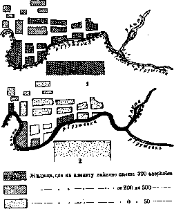
oooooo Places of breeding anopheles ^^^§^ Dried-up water bodies t*^^^*; Systematic accounting Figure 31. Condition of water bodies and quantity of Anopheles in the village of Krasykovskaya (North Caucasus): 1-from May 15 to July 15, 1924; 2-from July 16 to September 1924 houses.)-Malaria stations conduct systematic accounting of the condition of water bodies, both permanent and temporary (drying up), and mark them on corresponding maps indicating their distance from the nearest housing, marking places where mosquito larvae were found and their quantity, places where oiling was carried out, etc. (figure 31). The flora of the water body is also indicated. The Tropical Institute (Moscow) has proposed a special form for recording the species of mosquitoes found during the inspection of water bodies and the nature of the vegetation (fig. 32). Small hydraulic works are carried out directly by malaria stations or under their supervision. Large reclamation and hydraulic works of an anti-malarial nature have been legally assigned to the organs of the NKZem, which coordinate them with the health authorities, in particular with malaria stations. To unite the work of various departments in the fight against M. during the pandemic of M. in Moscow, a central malaria commission was organized (in Transcaucasia-a malaria committee), and in the periphery-regional and provincial mal. commissions (their 'Regulations' were approved by the NKZdr. RSFSR on 14/V 1923). They still function in malaria areas. To account for scientific achievements and practical results in the fight against M., congresses on the fight against M. are periodically convened: all-union (in 1923, 1924 and 1925) and regional [in the Volga region (in 1924, 1925, 1926 and 1928), in the North Caucasus, in Transcaucasia, in Central Asia], as well as on individual issues, e.g. on the fight against M. in peat extraction (in 1926 and 1928); materials of these congresses have been published in the corresponding 'Proceedings' of the congresses. From government resolutions on the fight against M., the following resolutions of the SNK RSFSR should be mentioned here: 1) of 13/IX 1923, according to which all plans for hydraulic works developed by various departments are subject to coordination with health authorities; the NKZdrav is granted the right to supervise the implementation of these works from the point of view of compliance with sanitary requirements; 2) of 12/V 1924 'On measures to combat malaria', according to which executive committees and individual departments (NKPS, VSNKh and others) are assigned the duty of carrying out sanitary and sanitary-technical measures to prevent and combat M., and to the People's Commissariat of Education-the duty to organize in schools and through schools the widespread dissemination among the population of information on issues of combating M.; 3) of 6/XII 1923 on the duty-free import of quinine (quinine in the USSR is supplied exclusively from abroad). The NKZdrav of the RSFSR has issued a number of instructions on the fight against M. (the most important of them are collected in the 'Collection of decrees, instructions and orders on issues of combating M.', Moscow, 1925). By the resolution of the SNK RSFSR of 19/VIII 1930 ('On the fight against epidemics') it is proposed: 1) to the NKZdrav and Goplan of the RSFSR when reviewing the five-year health plan to provide measures ensuring the maximum reduction in M. morbidity in the shortest possible time; 2) to the North Caucasus, Lower Volga and Middle Volga regional executive committees and the SNK of the Dagestan ASSR to develop a plan for hydraulic works related to the elimination of malaria foci; 3) to the NKZdrav and executive committees to involve the initiative of the population in the implementation of a sanitary minimum to carry out a number of works to combat malaria, such as: oiling, dusting of water bodies, filling in swamps, etc.; 4) to the NKZdrav to increase the network of malaria institutions (malaria stations, points and detachments) in unfavorable M. areas. Legislation and the fight against M. in peat extraction occupy a special place. The high incidence of M. in peat extraction (see above) raised the question of implementing a special system of combating M. in them; in general outline it comes down to the following: in peat extraction areas a network of malaria stations has been organized, the tasks of which include examining all workers arriving at the extraction sites, isolating, registering and treating all patients and parasite carriers and supervising the implementation of preventive measures (screening windows in housing, oiling and other methods of disinfecting water bodies, etc.). In places of permanent residence of seasonal workers arriving at peat extraction, a network of anti-malaria institutions (stations and points) has also been organized, which are assigned the duty of systematic treatment of the local population from M. The work of both types of stations is coordinated. This system of work has resulted in a significant reduction in M. in peat extraction. The entire anti-malarial organization in peat extraction is maintained at the expense of special credits provided for in the budgets of economic organizations according to the Resolution of the CEC and SNK of the USSR of 26/III 1926 {0.06 kopecks per each pood of peat extraction Basic-Reed-II Reed-U Rush-vv Colorful plants (Water plants. water lily Elodhi dr,<) - ^£» DismidificcflS -)) Khary-O Leafy pondweed, *ъ (Lhpn- f\ Ctrfltophyllum.Mirio-/ } icmrifl trisulcfl-j& Lemrvfl mfljor,mirwr polinphisfl-00 °° Luzyrchaytkya- ^ Spirogyrfl - Z^p С1яс1орКогя. Hydrodiction - {£) Surface PLANKTON FROM MICROORGANISMS- ф flnopKcks ♦ ♦ fled c з - a ▲ ТКсоЬя1с1|"я-иш ( JoMUHKut luju one {x>^a л^гии откачать bamwntu и,Ьгпгш.и.) In relation to individual water bodies, the following 'data are required. Shape, dimensions, depth measurements (zones with depth up to */i meters, up to 1 m, up to 2 m and above 2 m).

Character of the bottom, zone usually covered by plants, rotundity, water regime, water consumption, evaporation.
Figure 32. Conventional signs for indicating the nature of vegetation and species of mosquitoes in water bodies. фа). - Sanitary education plays a major role in the fight against M. For this purpose, special film strips, slides, posters and brochures on M. have been issued, corresponding exhibitions have been organized (including exhibition cars). Special attention is paid to involving schoolchildren and students in the fight against M. In malaria areas, M. is a subject of teaching in schools; schoolchildren study the biology of the mosquito and methods of combating it (oiling of water bodies, fighting with wintering grounds, etc.). Special legislation on the fight against M. has been issued in those union republics where malaria has particularly strong distribution (Transcaucasian and Central Asian republics). The decree of the SNK of the SSR of Armenia of 21/V 1923 concerns rice plantations and artificial irrigation. Rice can be sown 1) at a distance of at least 5 km from the inhabited outskirts of the city of Yerevan; 2) in other areas of Armenia-at a distance of at least 3 km from cities, villages, railway stations and other populated areas; 3) not closer than 2 km from main roads and 4) not closer than 1 km from country roads and railway tracks. As for artificial irrigation, the law provides for the proper maintenance of existing canals and ditches and the proper construction of new ones, and the slope of all canals should be calculated so that the flow speed per second is at least 25 cm. The resolution of the SNK of the SSR of Armenia of 9/IX 1925 provides in particular 1) the carrying out by executive committees of necessary measures for soil improvement and drainage of swamps, lakes and other standing waters; 2) submission for the conclusion of the NKZdrav in order to prevent waterlogging of projects for newly constructed and subject to major repair railways, highways and other roads, irrigation ditches and canals; 3) dissemination among the broad masses of the population and in schools of information on the fight against M.-A similar law exists in Azerbaijan (of 8/VII 1925). In particular, this law provides 1) maintaining in places of artificial irrigation when growing cotton the flow of water in order to prevent waterlogging of the soil; 2) carrying out by the NKPS hydraulic works for soil drainage in places where the railway embankment obstructs the natural flow of water; 3) carrying out by economic organs in enterprise areas works for drainage of waterlogged areas, maintaining malaria stations in these areas; 4) carrying out screening of residential houses; 5) carrying out systematic oiling of waterlogged areas in areas of settlements; 6) carrying out preventive quininization among workers and employees in malaria areas; 7) coordination with the NKZdrav of plans and projects for new settlements and new buildings for workers' housing; 8) inclusion in the retraining program for all workers of social services in the study of M.; 9) introduction in schools of I and II levels of the study of M. according to a special program. The resolution of the Council of People's Commissars of Uzbekistan of May 26, 1928 provides for the prohibition of rice cultivation within the residential part of cities and settlements of urban type, as well as a three-kilometer zone from it and more complete provision of medical assistance in those rural areas where rice cultivation is carried out. A similar law has been issued in the Tajik republic (of 26/II 1927).
I. Dobritsky. XIV. Malaria in birds and animals. Malaria in birds and animals presents exceptional interest thanks to the works of Ross (1898), who clarified the complete cycle of development of the parasite in the mosquito and proved that infection of birds occurs from the penetration of sporozoites from the salivary glands of the mosquito into the blood during a bite, which subsequently served as the key to deciphering the epidemiology of human malaria. Pigment parasites of bird erythrocytes were first discovered by V. Ya. Danilevsky (Kharkov; 1888), who established in 1890 that they cause acute disease in birds. Grassi and Feletti (1890) first identified the pigment parasites of birds as true malaria parasites and named them Haemamoeba praecox, classifying them in the genus Haemamoeba, to which they also classified the parasite of human malaria. Labbe (1894) classified the parasite of bird malaria in the genus Proteosoma, while Vennion (1926) believes that the pigment parasite of bird malaria should be classified in the same genus as the parasites of human malaria, i.e., in the genus Plasmodium, and should be called Plasmodium praecox. P. praecox is often found in birds in tropical and subtropical countries; it has also been found in more temperate countries: England, France, Germany, Austria, Italy, Switzerland, USSR, North America, Africa, Japan, India, and Australia. It is more often found in small birds: sparrows, canaries, larks, siskins [see separate table (art. 583-584), fig. 4], finches, etc.; but it is also found in larger birds—pigeons, crows, owls, partridges, ducks, etc., for whose malaria parasites separate names have been proposed. The malaria parasites in individual birds are currently still insufficiently studied. P. praecox (Grassi and Feletti) is morphologically very similar to the parasite of human malaria, from which it differs not only in that it is non-pathogenic to humans, but also in its larger size, just as the nucleus-containing erythrocyte of birds is larger in size than the human erythrocyte. In the erythrocytes of birds, the asexual development (schizogony) of this parasite occurs, while the sexual cycle occurs in mosquitoes of the genus Culex, from whose bites the natural infection of birds occurs, usually proceeding mildly, whereas laboratory infection by inoculation of blood containing parasites proceeds more severely and is often fatal, especially for canaries, siskins, etc. On autopsy in such cases, hypertrophy of the liver and spleen is noted, which become darker, and the pigment is distributed in the same way as in human malaria. The cycle of development of the bird malaria parasite in the Culex mosquito depends on the external temperature, just as in the Anopheles mosquito in human malaria. Et. and Ed. Sergent (1910-1921) studied bird malaria from the point of view of immunity. They found that the injection of sporozoites that had been in the salivary glands of Culex pipiens for several months, or sporozoites that had been preserved for a long time in vitro, reduces mortality among birds. In recent times, bird malaria has again attracted attention due to work in the search for synthetic drugs capable of replacing quinine. Among such works, the works of W. Roehl with plasmochin should be noted, which after testing on canaries was introduced for the treatment of human malaria. As for malaria in small mammals, mention should be made of the plasmodia of bats, which were first discovered by Dionisi (1899) in Vespertilio murinus. In the USSR they are also found in Vespertilio Daubentoni [see separate table (art. 583-584), fig. -6] (Shingareva A. I.; 1926) and in others. Malaria parasites in monkeys were first discovered by R. Koch in 1898. At present, parasites of malaria are known in a number of lower monkeys—macaques, as well as in anthropoid apes [see separate table (art. 583-584), fig. 5]—orangutan (Laveran; 1905), chimpanzee (Reichenow; 1917), and Plasmodium pitheci is inoculable to orangutans but not to other lower monkeys; P. inui is inoculable to macaques but not to orangutans. The parasite of human malaria, P. vivax, does not succeed in infecting monkeys; it also did not succeed in infecting humans with the malaria parasites of monkeys—P. Kochi and P. Reichenowi. Malaria in monkeys proceeds in acute and chronic forms, i.e., just as in humans. P. Popov.
Related articles
Mentioned in
Cite this page
“Malaria.” Soviet Medical Encyclopedia. English translation of Bolshaya Meditsinskaya Entsiklopediya, 1st ed. (Moscow, 1928–1936), ed. N. A. Semashko. https://sovietmedicalencyclopedia.pages.dev/article/malaria/