Protozoa (in)
Historical document, translated for reference. It reflects medical knowledge of the 1920s–30s and is not medical advice.
Summary
This article from the 1928-1936 Soviet Medical Encyclopedia provides a comprehensive overview of protozoa, including their classification, characteristics, pathogenicity for humans, and associated diseases. It details various species, their localization in the human body, cultivation methods, and the diseases they cause.
Encyclopedia article (1928–1936)
PROTOZOA in humans. Rhizopoda Main pigment Localization Pigment- | form- | Culture | Pathogenicity | for | humans | Wet fixation | Thick intestines, Heidenhain's, then ! liver, brain, lung stain with iron- | ammonium alum and hematoxylin | by Heiden- | hain
| idem idem idem Thick intestines Desna Thick intestines idem Thick intestines idem Thick intestines Rhizopoda i idem j idem idem idem idem idem p h o г a (F 1 a g e 1 1 a t a) idem
! Thick intestines idem idem idem idem idem idem idem Small intestines (duodenum), gall bladder On Boeck and Drbolilaw medium; Cleveland and Sanders liver agar idem for animals 0monkey+ Dog- Cat+ Disease caused in humans Amebiasis (dysentery), liver abscess, etc. idem idem idem idem I Walker-medium, !modification ! Musgrave i and Clegg idem idem idem idem idem Egg medium Hegner and Becker, mod. by Reichenow; coal medium Shurenkova idem idem idem Enteritis (?) idem idem idem Giardiasis (cholecystitis), periduodenitis Genus and species name Year of ! descrip- ! tion Bodo caudatus...............| 1878 Bodo edax................. 1892 Cercomonas parva (Cercomonas longica-uda Dujardin 1841)............ Copromonas subtilis Isospora belli Author Stein Klebs Hartmann and Chagas Dobell Wen yon Size of vegetative forms in /j, 6-14 Isospora tiominis.............j 1901 Railliet and Lucet Eimeria clupearum (=E. Wenyoni) ... 1892 1
Laveran 1892
Grass in Feletti 1914
Stephens i 1916 !
Marcinovsky Balantidium coli Balantidium minutum Nyctotherus faba.............i 1899 Malmsten Schaudinn Schaudinn 6-20 7-20 Motility of vegetative forms in the body + i + Coprozoan Size of cysts in ц Class Sporozoa. Oocysts 125-33x12,5-16: 16X10 30-200 x 20-70 70-100x50-70 20-30x14-20 26-28x16-18 Class 50-60 Encephalitozoon rabiei..........j 1924 Levaditi, Nicolau and Schoen Sarcosporidia Lindemani 1893 I Baraban and Remy 2,5x1,0-0,5 Г p y и п a 4,25X1,75 Mastigophora Main pigment See Tricnomonas intestinalis : Pig- | ment- | Pathogenicity | Localization | form- | Culture | ' for humans animal See Trichomonas intestinalis "~ Disease caused in humans idem idem idem idem idem idem Subclass Coccidiomorpha idem idem idem idem Unknown, found in faeces idem idem idem Henzinsky, Roma- Blood, spleen, novsky, Giemsa, liver, bone marrow Leishman idem idem idem idem idem idem idem + no Bass'y and Sinton'y idem idem Malaria tertiana Malaria quartana Malaria tropica Malaria Malaria tropica Malaria tertiana C i l i a t a Iron hematoxylin Thick intestines Teidenhain's idem idem idem idem Cnidosporidia idem Sarcosporidia idem Central nervous system Striated muscle Shurenkova's coal medium Monkey, pig Balantidiasis (enteritis, colitis) Rabies Tumors of esophagus, etc. Year of description Size of vegetative forms B r, Group Genus and species name Author Motility of vegetative forms in the body Size of cysts B fl Rhinosporidium seeberi......... Wernicke Steinberg 10-15X4---8 7-10 Trichomonas elongata (buccalis)..... Donne 10-30 x 10-15 2-4 Cunningham, Borovsky, Wright, Marcinovsky Laveran and Mesnil 2-4 Trypanosoma gambiense......... Dutton 15-30 Stephens and Fant-ham 12-35 20 Schizotrypanum cruzi (Trypanosoma cruzi) 3. Viruses causing characteristic cellular inclusions in the body; morphology unknown. - A. Septic diseases: 1) Agalaxie (agalactia of sheep and goats); 2) horse plague (virus Pferdepest, sud-afrikanische Pferdesterbe); 3) guinea pig plague (virus Meerschweinchenpest); 4) bird plague (kyanolophia, pestis gallinarum); 5) foot and mouth disease (febris aphthosa); 6) cattle plague (virus Rinderpest); 7) swine plague (cholera suum).-B. Diseases affecting the skin without severe changes: measles (morbil-H), scarlet fever (scarlatina).- C. Diseases affecting skin and mucous membranes with severe changes: warts (verrucosis), blenorrhea (non-gonococcal) (blenorrhoea) with inclusions in epithelial cells; varicella (chickenpox); variola (smallpox); verruga peruviana, rabbit mucous disease (Myxomkrankheit); trachoma.-D. Viruses affecting the brain and spinal cord: Born disease (meningoencephalitis epizootica); encephalitis lethargica; herpes febrilis, lyssa (rabies); dog plague (Hundestaupe).-E. Viruses affecting blood-infectious anemia of horses.
E. Bunina, V. Lyubarsky. Cultivation of microbes, aerobes and anaerobes--the cultivation, growing of microbes in artificial laboratory conditions, used for studying the properties of a microbe (isolated from the body or other material) and determining its species. Cultivation of microbes is performed on artificial nutrient media, both solid and liquid, the composition of which, as well as the reaction, must be adapted to the properties of the given microbe. Most pathogenic microbes grow best at body temperature (typhoid bacilli, paratyphoid, dysentery, cholera, pyogenic cocci, etc.), i.e., at 37°. Saprophytic microbes also grow well at lower temperatures. Cultivation of microbes is carried out in special cabinets, thermostats (heated by electricity, gas or kerosene), with constant temperature, regulated by regulators, as temperature fluctuations adversely affect the growth of microorganisms. Growing at lower temperatures can be done outside thermostats at room temperature. To study a microbe and its bacteriological characteristics, it must be obtained in pure form, not mixed with other bacteria, i.e., it is necessary to obtain a pure culture on an artificial nutrient medium. Thanks to the cultivation method in laboratory conditions, it is possible to constantly have a pure culture of a particular microbe, which is necessary for both practical and research purposes. For cultivating microbes (aerobes and anaerobes), methods are used, some common to both groups, some specific. Nutrient media used for growing microbes are poured into glass bacteriological dishes (test tubes, flasks with cotton plugs, and Petri dishes). All are thoroughly sterilized. Methods for cultivating aerobes. Aerobes are grown with good air access. The following nutrient media are most commonly used: meat-peptone broth, meat-peptone agar, gelatin, peptone water, sugar media, potato and other media. Seeding or Haplosp oridia Main pigment Pig- | | ment-Localization | form- | Culture | forma- | Pathogenicity for humans animals Disease caused in humans Iron hematoxylin Heidenhain's idem idem Polyps in nasal cavity + - ! Polyps in nose i Ready cavity On Boeck's media and others Vagina - 1 , (Leukorrhea) Romanovsky, Giem-! Granulations of ulcers sa, Leishman '
Skin (same) same same Spleen, liver, bone marrow same Cutaneous leishmaniasis Visceral leishmaniasis Blood (same?) Blood (same?) same Blood, muscles (same?) Trypanosomosis (sleeping sickness in Africa) Trypanosomosis (Chagas' disease in Brazil) Transferring cultures to liquid or solid media is performed (since Koch's time) with a platinum loop, spatula, or straight wire (stab seeding), inserted into a glass rod or special needle holders (fig. 1). Tubes with the culture to be transferred and an unseeded tube are taken in the left hand between the thumb and index finger, one above the other, the first below, the second above them. They are held almost horizontally to prevent microbes from the air from entering the tubes during further manipulations. With the right hand, take the handle of the platinum loop, remove the cotton plugs from the tubes, capturing one plug with the little finger, pressing it against the palm, and the other between the little finger and fourth finger, flame the edges of the tubes, sterilize the loop, insert it into the seeded tube, cool it against the wall, take a small amount of material with the loop, transfer it to the unseeded tube and mix it in the liquid medium by rubbing the loop against the wall of the tube. After this, the loop is removed from the tube, it is flamed, the edges of the tubes and plugs are flamed, and without extinguishing the plugs, they are inserted back into the tubes. The seeded media are placed in an incubator. When transferring to solid media (sloped agar, coagulated serum, etc.) the same techniques are used, and the seeding itself is done by streaking (streak cultures) on the surface of the medium; one must be careful not to damage the integrity of the medium. Transfers from liquid medium to liquid medium can also be done with a sterile Pasteur pipette. If the material to be transferred is very dense (tubercle bacilli, ray fungi), it is more convenient to work with a spatula (fig. 1 and 2).

The so-called 'direct' media (agar, gelatin) are seeded by stab (stab seeding): material is picked up on the tip of a platinum needle (fig. 1), which is then inserted into the medium (holding the tube upside down), advancing deeply along the axis of the tube. To obtain pure cultures from contaminated material, the method of plate cultures on agar or gelatin in Petri dishes (or vessels constructed on the same principle - a large smooth surface for seeding; figs. 3 and 4) is used. Seeding on dishes is done in two ways: a) by Koch's pouring method and b) seeding on dishes with solidified agar. According to Koch's method - agar in tubes is melted in a water bath or Koch's apparatus (see Koch's apparatus), cooled to 42-43°, and then into a series of tubes is introduced by gradual dilution ever smaller amounts of the original material (e.g. its various dilutions in broth or physiological solution) containing microbes. The medium thus seeded in tubes is poured into Petri dishes, with the edges of the tubes being carefully flamed before pouring. During this procedure, the dishes stand on a flat surface; with the left hand, lift the lid of the dish from one side so that the opening of the tube can be brought under and the agar poured into the dish. By slightly tilting the dish in different directions, the agar poured into it is distributed evenly over its entire bottom. When the agar has solidified, the dishes are placed in an incubator upside down. Gradual dilution of the material to be seeded can also be done in the melted agar itself, by seeding the first tube with the material and mixing it in the agar, which is then transferred in amounts of 1-2 loops into the next tube. Similarly, seedings are made on gelatin, cooling it to 30-37°. When the medium in the dishes contains a few bacteria, they are fixed far apart from each other, and each gives rise to a separate colony [see separate table (pp. 83-84), figs. 6, 7 and 8]. Seeding on dishes can also be done on agar solidified in them by smearing the material over its surface. For this purpose, so-called Conradi-Drigalski spatulas are used, which, after previously sterilizing them over a flame, pick up the seeding material, then remove the lid from the first dish and thoroughly smear the material over the surface of the medium, rotating the dish around on the plane. With the same spatula, without picking up new material, the 2nd, 3rd, 4th and 5th dishes are smeared. Thus the number of microbes on the spatula decreases with each successive dish, very few of them get into the last dishes, and in them separate colonies grow, whereas in the first ones a continuous growth is obtained [see separate table (pp. 83-84), figs. 6, 7 and 8].
Instead of a spatula, seeding on dishes can be done with a platinum loop, making successive streaks with the same loop on one dish (or successively on a series of dishes), with separate colonies growing from the last streaks [see separate table (pp. 83-84), fig. 9]. By transferring colonies to media in tubes, a pure culture of the microbe is obtained, with which further manipulations are performed. The appearance of colonies is often characteristic of a given microbe; therefore colonies must be carefully studied. For these purposes, they are examined with a magnifying glass, with the low power of a microscope, or with a special plate microscope (Plattenkulturmikroskop of Zeiss or Leitz; fig. 5), which allows observation in reflected light. With the help of this same instrument, the transfer of colonies is done by what is called harpooning them with a loop. For these same purposes, special instruments, harpoons, are used, which are screwed
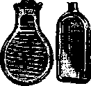
Fig. 4. Flat flasks for plate cultures.
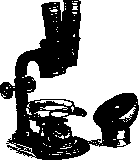
Fig. 5. Plate microscope.
onto the tube of an ordinary microscope instead of a centered objective (fig. 6). When the tube is lowered, the needle (harpoon) immerses in the colony and picks up microbes, which are then transferred from it to a drop of liquid on the loop. One can make a harpoon oneself, by applying plasticine to a plug or even better to an old objective and fixing in it strictly vertically an ordinary needle. In many cases, when colonies are large, transfer can be done with a loop under the control of the naked eye. The isolation of pure cultures of pathogenic microbes from dishes is difficult, since colonies of some pathogenic and non-pathogenic microbes (for example, paratyphoid bacilli and colon bacilli) can be extremely similar. In such cases, differential colored media (see Nutrient media) are of great help, which change their color in the place of growth of colonies of certain bacteria in one way or another. With ordinary plate cultures, a colony may arise from several stuck together bacteria of the same species. Therefore, in precise work, the method of obtaining cultures from a single cell (Einzellenkultur) is used. For this purpose, the old method of Burri with ink is applicable. Among new methods, it is necessary to mention a special micromanipulator (see) with which a single bacterium can be captured with a thin pipette (fig. 7) under microscopic control and transferred to a nutrient medium. If the original material is very

contaminated with foreign microbes, it is pre-cleaned before seeding by treatment with chemical agents (acids, antiformin), washing in physiological solution (clumps of sputum), heating (spore-bearing species). If there are few bacteria in the material, methods of artificial enrichment (seeding typhoid bacilli from blood into bile, cholera vibrio from feces into peptone water) or their elective cultivation (seeding diphtheria bacilli into coagulated serum, tubercle bacilli into Petrov's violet medium, etc.) are used. The method of enrichment should also include preliminary passage through a susceptible animal, in which only pathogenic microbes will develop (for example, tubercle bacilli, pneumococci). For the cultivation of anaerobes, many different methods have been proposed, based on creating conditions for growing the microbe without access to air. These conditions are achieved by the following methods or

Fig. 6. Objective-harpoon.
a combination of them: a) removal of atmospheric oxygen (O2), dissolved in the nutrient medium, by boiling it followed by restricting the entry of air from the outside with the help of appropriate sealing of the culture; b) displacement of the air dissolved in the medium by a gas indifferent to anaerobes; c) binding by chemical substances or physical adsorption of the air of the medium; d) removal of air with forceps.
from a hermetically sealed apparatus with cultures and from their nutrient medium with the help of an air pump. Boiling media before seeding ("refreshing" them) is part of the standard technique for cultivating anaerobes. Media that can withstand heating in Koch's apparatus are boiled; duration of boiling for test tubes-10 min., for flasks of 100-200 cm3-20 minutes, for liter flasks-60 minutes. Test tubes can be boiled in a water bath for 15 min. Then the media should be quickly cooled in snow or cold water. To reduce access to air, sealing of test tubes after seeding is used (Weinberg, Vignal-Veillon), as well as isolating liquid media from air with a layer of liquid paraffin or vaseline oil (sterile). For some anaerobes, it is sufficient to make the sowing in so-called "deep" media (agar in a column, Veillon), into the deeper layers of which access to atmospheric oxygen is limited. - For growing anaerobes in an oxygen-free atmosphere, most authors use hydrogen, formed in Kipp's apparatus by the action of sulfuric acid on metallic zinc. Before entering the vessel with cultures, the hydrogen passes through washing bottles with a 10% solution of lead nitrate and with an alkaline solution of pyrogallic acid (apparatus Novy) (fig. 8). Instead of Kipp's apparatus, bombs with condensed hydrogen or nitrogen can be used. A safety valve must be fitted to the outlet tap of the bombs to reduce the high pressure under which the gas exits from the bomb. For absorption of O2 in the nutrient medium, reducing substances are added to it, e.g. grape sugar (1-2%), sodium formate, etc. Adsorption of O2 is also achieved by adding pieces of living organs to the medium (the medium is poured in a deep layer), which are also rich in catalase, which destroys the peroxides formed during the growth of anaerobes (liver, kidneys, brain, blood, pieces of chicken egg white). The most commonly used media for anaerobes: liver broth (Kitt-Tarozzi), milk with pieces of liver, brain pulp, blood broth, sugar agar with blood, broth with pieces of chicken egg white.--The method of cultivating anaerobes in ordinary closed with cotton plugs test tubes based on the adsorption of O2 by animal and plant tissues is one of the newest methods-the Wroblewski method (fig. 9). As an adsorbent in this method, a piece of cotton (0.1 g) is placed at the bottom of a test tube with liquid medium (5-8 cm3). Sterilization at 110° for 1/3 hour.-Absorption of O2 by living cells occurs in the Fortner method-growing anaerobes on the same plate with aerobes: a plate with blood agar is divided into two halves by cutting out and removing a strip of agar; on one side a sowing of Bac. prodigiosus is made, on the other-an anaerobe; the edges of the plate are sealed with plasticine.-One of the most common methods is also the use of a fresh alkaline solution of pyrogallic acid as an O2 absorber (usually 1 cm5 of 10% solution of KOH is taken per 1 g of pyrogallic acid). A freshly prepared solution (pouring caustic potash onto pyrogallic acid in the apparatus itself) is poured into the bottom of the apparatus, into which test tubes (Buchner test tube, Omelyansky) or plates with anaerobic cultures are placed [cultivation in a desiccator, into the bottom of which a solution of pyrogallic acid is poured (Arens)]. Pyrogallic acid is used in combination with evacuation of air from the apparatus. The latter is done with a water or oil pump. A water pump is connected to the desiccator with cultures (pyrogallic acid is poured into the bottom), equipped with a good outlet tap. - Removal of air from each seeded test tube is used in the technique according to Weinberg. Tall test tubes with medium after seeding are drawn out in the upper third (above the level of the medium) on a burner flame in such a way that they can be easily sealed in this place later, then through a glass tube inserted into the plug, the test tubes are connected to a water jet pump (or oil pump), and evacuation of air occurs for about five minutes; when the medium in the test tube "boils", the lower part of the test tube is slightly heated (gas passes through a narrow tube, metal or glass, held in the hand and connected to a gas tap with a rubber tube) and tapping on it helps the air to escape from the medium, and the test tube is sealed in the area of the constriction. For growing anaerobes in plates (plate cultures) Zeissler has constructed an apparatus, manufactured by the firm Begerow (Altona) (fig. 10). Air rarefaction is achieved with the help of a Pfeiffer oil pump to 1 mm (in an empty apparatus) according to a mercury manometer, connected
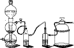
Fig. 10. Begerow's apparatus for growing anaerobes according to Zeissler.
from a hermetically sealed apparatus with cultures and from their nutrient medium with the help of an air pump. Boiling media before seeding ("refreshing" them) is part of the standard technique for cultivating anaerobes. Media that can withstand heating in Koch's apparatus are boiled; duration of boiling for test tubes-10 min., for flasks of 100-200 cm3-20 minutes, for liter flasks-60 minutes. Test tubes can be boiled in a water bath for 15 min. Then the media should be quickly cooled in snow or cold water. To reduce access to air, sealing of test tubes after seeding is used (Weinberg, Vignal-Veillon), as well as isolating liquid media from air with a layer of liquid paraffin or vaseline oil (sterile). For some anaerobes, it is sufficient to make the sowing in so-called "deep" media (agar in a column, Veillon), into the deeper layers of which access to atmospheric oxygen is limited. - For growing anaerobes in an oxygen-free atmosphere, most authors use hydrogen, formed in Kipp's apparatus by the action of sulfuric acid on metallic zinc. Before entering the vessel with cultures, the hydrogen passes through washing bottles with a 10% solution of lead nitrate and with an alkaline solution of pyrogallic acid (apparatus Novy) (fig. 8). Instead of Kipp's apparatus, bombs with condensed hydrogen or nitrogen can be used. A safety valve must be fitted to the outlet tap of the bombs to reduce the high pressure under which the gas exits from the bomb. For absorption of O2 in the nutrient medium, reducing substances are added to it, e.g. grape sugar (1-2%), sodium formate, etc. Adsorption of O2 is also achieved by adding pieces of living organs to the medium (the medium is poured in a deep layer), which are also rich in catalase, which destroys the peroxides formed during the growth of anaerobes (liver, kidneys, brain, blood, pieces of chicken egg white). The most commonly used media for anaerobes: liver broth (Kitt-Tarozzi), milk with pieces of liver, brain pulp, blood broth, sugar agar with blood, broth with pieces of chicken egg white.--The method of cultivating anaerobes in ordinary closed with cotton plugs test tubes based on the adsorption of O2 by animal and plant tissues is one of the newest methods-the Wroblewski method (fig. 9). As an adsorbent in this method, a piece of cotton (0.1 g) is placed at the bottom of a test tube with liquid medium (5-8 cm3). Sterilization at 110° for 1/3 hour.-Absorption of O2 by living cells occurs in the Fortner method-growing anaerobes on the same plate with aerobes: a plate with blood agar is divided into two halves by cutting out and removing a strip of agar; on one side a sowing of Bac. prodigiosus is made, on the other-an anaerobe; the edges of the plate are sealed with plasticine.-One of the most common methods is also the use of a fresh alkaline solution of pyrogallic acid as an O2 absorber (usually 1 cm5 of 10% solution of KOH is taken per 1 g of pyrogallic acid). A freshly prepared solution (pouring caustic potash onto pyrogallic acid in the apparatus itself) is poured into the bottom of the apparatus, into which test tubes (Buchner test tube, Omelyansky) or plates with anaerobic cultures are placed [cultivation in a desiccator, into the bottom of which a solution of pyrogallic acid is poured (Arens)]. Pyrogallic acid is used in combination with evacuation of air from the apparatus. The latter is done with a water or oil pump. A water pump is connected to the desiccator with cultures (pyrogallic acid is poured into the bottom), equipped with a good outlet tap. - Removal of air from each seeded test tube is used in the technique according to Weinberg. Tall test tubes with medium after seeding are drawn out in the upper third (above the level of the medium) on a burner flame in such a way that they can be easily sealed in this place later, then through a glass tube inserted into the plug, the test tubes are connected to a water jet pump (or oil pump), and evacuation of air occurs for about five minutes; when the medium in the test tube "boils", the lower part of the test tube is slightly heated (gas passes through a narrow tube, metal or glass, held in the hand and connected to a gas tap with a rubber tube) and tapping on it helps the air to escape from the medium, and the test tube is sealed in the area of the constriction. For growing anaerobes in plates (plate cultures) Zeissler has constructed an apparatus, manufactured by the firm Begerow (Altona) (fig. 10). Air rarefaction is achieved with the help of a Pfeiffer oil pump to 1 mm (in an empty apparatus) according to a mercury manometer, connected
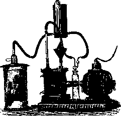
Fig. 10. Begerow's apparatus for growing anaerobes according to Zeissler.
valuable to the apparatus; when filling with cultures, the vacuum in the apparatus reaches 3 mm, often only 5 mm. By placing a rubber ring between the lid and the apparatus, a sufficiently tight seal of the apparatus is achieved, that pouring pyrogallol or other reducing substances into it is unnecessary. After the air is pumped out, the stopcock of the apparatus is closed, and the latter is placed in an incubator.- For subculturing anaerobic cultures, as well as for isolating them in pure culture, a special technique is used. Seeding into liquid media (poured in a high layer) is done with a long capillary pipette (bore 5-6 mm). The material to be seeded is introduced in large quantities into the lowest layers of the medium. Seeding into solid media (agar and gelatin) is done either into pre-melted and cooled (but not to the point of their solidification) media with the same capillary pipette, or by puncturing with a long platinum needle to the bottom of the tube. Colonies in solid media develop in the depth of the medium and are visible through the glass. To subculture them, a nick is made in the tube (previously wiped with alcohol) above the colony, the tube is broken, after which the colony is extracted with a loop or a slightly heated pipette and preferably transferred to liver broth. For seeding onto plates, the material is spread on their surface with a platinum loop or a Drigalski spatula. Direct transfer of colonies onto a plate (especially small ones) does not always succeed; therefore, it is better first to transfer the colony together with the medium into liver broth, and after obtaining growth in it, to make a seeding onto plates.-When isolating anaerobes from a mixture of microbes, heating of the material is used, successively at 80° for 20 min.; at 100°-5 min., 20 min., 30 min., 40 min., 60 min., 180 min., since various microbes and their spores die at different times, which allows them to be separated from each other. Further differentiation of anaerobes is carried out by studying the nature of their growth on media, the appearance of colonies, their biochemical properties, morphology, motility AND EXPERIMENTS ON ANIMALS. A. Togunova. Cultivation of protozoa. Practically, the cultivation of protozoa compared to other M. plays only a minor role due to the imperfection of the technique. Significant successes, relating mainly to intestinal protozoa, have been achieved only in recent years. When the original material contains other M., cultivation, especially obtaining pure cultures, is complicated by the relatively slow reproduction rate of protozoa, which are suppressed by microorganisms with more vigorous growth. The difficulty or ease of obtaining cultures of certain protozoa is related to their method of nutrition. Protozoa that feed osmotically are the easiest to cultivate, and when feeding on formed food-feeding on bacteria or erythrocytes. Pure cultures can be easily obtained in cases where the material for seeding is taken from a sterile medium; an example are blood parasites, the nutrient medium for which can also be prepared sterilely. Protozoa that need inorganic or the simplest organic compounds for their growth can also be obtained in pure cultures. Where, in the original material, protozoa are found in a mixture with other microorganisms (intestinal, free-living protozoa), obtaining pure cultures often presents insurmountable difficulties. It should be borne in mind that for some protozoa bacteria are a necessary nutritional material. Purification of cultures is carried out by the usual methods in bacteriology. In cases of a sharp quantitative predominance of bacteria over protozoa in the original material, use is made of the tendency of some protozoa to accumulate on the surface of a tube with nutrient medium, as well as the resistance of cysts to drying: drying at 37° often frees the material from bacteria. To obtain pure lines, seeding is done from an isolated individual. Large protozoa are easily isolated under a magnifying glass with a pipette. To isolate smaller ones, the liquid under examination is diluted until there is no more than one individual in a drop. From a series of cover slips with drops under the microscope, slips with one individual in a drop are selected and dropped into the nutrient medium (Lindner's method) or the liquid under examination is drawn into a very thin capillary; the part of it selected under the microscope with one protozoon, farthest from neighbors, is separated, and its contents are used for seeding (Oehler's method). (The use of a micromanipulator is possible.) In the cultivation of protozoa, a moist chamber or hanging drop (Schultz's chamber) is sometimes used. - Depending on the genus of protozoa, different nutrient media are used: agar as such, agar with broth, with hay infusion, Nelder's agar, blood, serum, protein-egg medium, etc.-As for the concentration of hydrogen ions in nutrient media, some free-living protozoa tolerate fairly wide fluctuations (Bo-do edax - pH 4.8--9.6), in others the limits are narrower, and they are especially narrow for parasitic protozoa. The strong development of bacteria in cultures quickly changes the pH of the medium.- Free-living protozoa in the vast majority are easily cultivated. Many free-living amoebae multiply well on simple agar in mixed culture with bacteria. The method of Frosch and Mouton is recommended: a plate with agar is seeded with a suitable species of bacteria in radial streaks, starting from the center. The central part of the plate is inoculated with material containing amoebae, which multiply along the bacterial culture. Free-living flagellates are partly grown on Knopp's medium and other mineral media; free-living infusoria are cultivated in mixture with bacteria. The cultivation of parasitic protozoa is of particular importance. On the one hand, it is the only diagnostic means when there is a small amount of pathogens that is insufficient for microscopic examination, on the other hand, it provides a quantitatively sufficient material for the comprehensive study of protozoa. The cultivation of parasitic protozoa is carried out either in liquid media or on solid media, which must be sufficiently moist. The presence of animal or personal protein in the medium is mandatory.-When cultivating intestinal protozoa, it must always be borne in mind that saprophytic protozoa, whose cysts passively pass through the intestine with food (Darmpassanten; amoebae of the Limax type, flagellates of the genus Prowazekia, etc.), must be considered. In freshly passed feces, they emerge from the cysts; when seeded for the purpose of isolating parasitic protozoa, they may grow and give rise to diagnostic errors. In intestinal catarrhs, emergence from cysts may already occur in the intestine (semi-parasites). The cultivation of parasitic amoebae has become common in laboratory practice after the introduction by Beck and Drbohlav (Beck, Drbohlaw; 1925) of egg-white media; the coagulated mixture of egg white and Locke's fluid is covered in tubes with the same mixture, but liquid. According to American terminology, it is designated E. L. A. (Egg-Locke-Albumen). The solid part of the Beck-Drbohlav medium can be replaced with agar containing Ringer's solution (preferably with the addition of 1% starch) or NNN-agar without detriment to growth. According to Craig and John (Craig and John; 1927), even better growth of parasitic amoebae is obtained on a simple medium: human serum mixed with Ringer's solution or physiological saline. Seeding to obtain cultures is done with a loop of mucus from freshly passed feces into pre-warmed tubes. Growth in 24-48 hours. Material for examination is taken with a capillary pipette from the surface of the solid medium. Every 24 hours it is recommended to carefully change the liquid part of the medium, in which bacteria and blastocytes multiply abundantly and give the medium an acidic reaction with their growth. In culture, amoebae feed on bacteria; when erythrocytes are added, they also absorb them; sometimes cyst formation is observed. Cultures are pathogenic for young cats (sometimes they even cause the formation of liver abscesses in them). Subcultures succeed easily in a large number of generations. On the Beck-Drbohlav medium, cultures of the following species of amoebae have been obtained: Ent. histolytica, Ent. coli (more difficult to cultivate than Ent. histolytica), Entamoeba nana, Jodamoeba Biitschlii, Dientamoeba fragilis and Ent. gingivalis, as well as Trichomonas vagi-nalis among flagellates. Of parasitic flagellates, the cultivation of Trichomonas intesti-nalis has practical diagnostic significance when microscopy is negative. Good results are obtained by Reichenow's method on a medium of horse (or another) serum and physiological saline. Several grams of blood-mucus part of feces are seeded into tubes. At 37° growth on the 3rd day. Subculturing is necessary after 3-5 days, otherwise the culture dies. At 25-30° the culture lives for up to 7 days. Material for examination is taken from the bottom of the tube. With less reliable results, Chilomastix Mesnili is grown by Reichenow's method.
Cultivation of intestinal infusorians, Balantidium coli, is carried out either on human serum mixed with physiological solution (Barret, Yarbrough; 1921) (verification by Doflein and Reichenau was unsuccessful) or on meat broth with human serum, previously seeded with Bacillus faecalis alcaligines (Reis; 1923). Addition of starch or other carbohydrates to the medium is recommended. Cultivation of flagellate protozoan parasites of blood and tissues—leishmanias and trypanosomes—is carried out on blood media, mainly on NNN-agar (in tubes or Petri dishes) or in broth with blood. When cultivated on blood media, the growth of trypanosomes occurs in the condensation water of NNN-agar, on the moist surface of Petri dishes with NNN-agar, or in blood broth in the upper layer of erythrocytes. Blood is undoubtedly an essential part of the medium. Agar apparently has protective functions, binding blood antibodies and perhaps also the metabolic products of protozoa. Agar should have sufficient density but contain much water to ensure the formation of condensation fluid. This is best achieved by mixing 1% agar with defibrinated blood in equal parts. However, for growing pathogenic species, the amount of blood can be increased to 70%. Expensive rabbit blood in the cultivation of some trypanosome species can be replaced by horse blood. The condensation water of NNN-agar and blood broth, due to the presence of acid phosphates, have a pH of 5.6. Addition of a small amount of grape sugar to blood media is useful, and besides its nutritional function, it is also attributed the function of binding certain enzymes. Seeding is done with several drops of sterile blood containing trypanosomes. When a large amount of blood is introduced, the action of antibodies present in it, as well as the formation of a blood clot, hinder growth. Noller proposed cultivating trypanosomes on Petri dishes (agar with horse blood). Some trypanosome species give characteristic growth patterns that allow individual species to be differentiated macroscopically. Relationships similar to those between leishmanias and their cultural forms—leptomonads (see below)—are also observed between trypanosomes and crithidia. In the host's blood—the trypanosome form; in cultures—the crithidial form. The optimum growth temperature for most trypanosomes is 28-30°C. At 37°C, degenerative vacuolated round leishmania-like forms are often obtained, while some trypanosomes retain their trypanosome form at this temperature (Trypanosoma theileri, Trypanosoma loxiae). Cultures of trypanosomes of cold-blooded animals, birds, rats, and cattle (Tryp. theileri) are easily obtained. All pathogenic species for animals and especially for humans are difficult to cultivate. Cultures of the causative agents of sleeping sickness, Tryp. gambiense and Trypanosoma rhodesiense, if successful, are only in the first generation. Schizotrypanum cruzi is somewhat easier to cultivate. Cultivation of leishmanias is accomplished without particular difficulty. The material for seeding is sterile blood and punctate from the spleen or—in oriental sore—from an unopened node. Seeding on NNN-agar is done in the condensation water. In culture after two days, flagellate forms of Leptomonas appear, which multiply rapidly by longitudinal division. Formation of rosettes from individuals intertwined with flagella is often observed. In old cultures, aflagellate forms reappear, which remain viable for a long time during subcultures. Cultures are pathogenic for animals. The optimum growth temperature is 22°C. This optimum temperature mainly distinguishes cultural leptomonads of leishmanias from morphologically similar Leptomonas from the intestine of completely harmless insects. The latter grow more slowly at an optimum growth temperature of about 30°C. The technique for seeding Leptomonas from insects is as follows. The intestine of the insect, removed as sterilely as possible, is washed many times in sterile physiological solution and crushed between sterile slides; the resulting liquid is drawn up with a sterile syringe and seeded into tubes or onto Petri dishes with ZN-agar. This same technique is used when seeding from the intestines of insects that are vectors of trypanosomiasis and others. - On the cultivation of malarial plasmodium—see Malaria, parasitology. Cultivation of spirochetes. The widely used in recent years laboratory cultivation of spirochetes is a very recent achievement, and here, especially with respect to some species, the transition from extremely complex methods to the simplest ones should be noted. The essential condition for cultivating spirochetes is the presence of protein (serum, ascitic fluid, etc.) in the medium. Liquid media are more often used, in which, all other conditions being equal, growth is more abundant and motility is more pronounced than on solid media. Not being absolute anaerobes, spirochetes during cultivation nevertheless require limited access to O2, which is achieved mainly by applying a layer of paraffin or vaseline oil to the surface of the seeded tube. Cultivation of Leptospira of relapsing fever is carried out by Ungerman's method in half-coiled rabbit serum diluted with Ringer's solution (preferably from young rabbits) or by Aristovsky's method, as well as Hata's method on horse serum diluted with physiological solution. Cultivation is carried out at 37°C under a layer of paraffin oil. Numerous generations are obtained. Cultures are pathogenic for humans, and infection can apparently occur through intact mucous membranes (proven by laboratory infections). After passages through mice or rats, pathogenicity for humans disappears. - Cultivation of the causative agent of infectious jaundice—Leptospira icterogenes, as well as aquatic leptospires—is carried out by Uhlenhuth's method on tap water with the addition of 30% rabbit serum under a layer of paraffin oil. When cultivating Leptospira icterogenes from blood, Manteifel successfully applied the Gildemeister method for diagnosing typhoid fever—seeding 2 cm3 of the patient's blood into 8-10 cm3 of sterile tap water. The protein necessary for the nutrition of leptospires they obtain from the seeded blood. Optimum growth temperature is 25-30°C. The virulence of cultures decreases on subculturing but can be restored by several passages through guinea pigs. - Cultivation of the pale spirochete, the causative agent of syphilis, is carried out under conditions of strict anaerobiosis on various media: half-coiled horse serum of Shereshewsky, serum agar and broth of Mülenz, medium of Schmincke, medium of Noguchi, ascitic agar with a piece of sterile rabbit kidney, etc. (optimum pH 7.2 - 7.8). Cultures are usually obtained in a mixture with non-pathogenic spirochetes. The described pure cultures of Treponema pallidum are not always conclusive. In semi-solid media, the pale spirochete grows in the form of clusters resembling a cloud; along the stab, the clusters are arranged like beads. Morphologically, pale spirochetes from cultures appear coarser than in the dark field. By Giemsa's method they are stained not pink but bluish in color.
D. Muratova. Lit.—See literature for the articles Bacteriology, Microbiology and Protozoology.
Related articles
Mentioned in
Cite this page
“Protozoa (in).” Soviet Medical Encyclopedia. English translation of Bolshaya Meditsinskaya Entsiklopediya, 1st ed. (Moscow, 1928–1936), ed. N. A. Semashko. https://sovietmedicalencyclopedia.pages.dev/article/protozoa-2/