DYSENTERY
Historical document, translated for reference. It reflects medical knowledge of the 1920s–30s and is not medical advice.
Summary
Dysentery is an acute infectious disease characterized by inflammation of the large intestine with bloody stools and painful tenesmus. The article distinguishes between amebic and bacillary forms, detailing their etiology, epidemiology, and classification of causative bacteria.
Encyclopedia article (1928–1936)
DYSENTERY, dysenteria (from the Greek dys- particle, meaning bad quality, and enteron-intestine), an acute infectious disease manifesting as inflammation of the large intestine with bloody stools and painful tenesmus; on this basis, D. is also called bloody or straining diarrhea. This disease has been known since ancient times; even contemporaries of Hippocrates were able to distinguish simple intestinal disorders (diarrhea) from specific bloody diarrhea, which they called dysenteria. Etiology. D. is widespread throughout the world, but not everywhere caused by the same cause. At present, etiologically, D. is divided into two forms: amebic and bacillary. Both forms are initially very similar in clinical signs; their differences consist mainly in the different causative pathogens. Amebic D. is bloody diarrhea caused by pathogenic amebas (see). This disease is characteristic of tropical and subtropical climates, where it usually has endemic distribution. In the temperate climate zone, amebic D. is observed only as rare individual cases, often of imported origin (see below-Amebic D.). The bloody diarrhea characteristic of temperate climates is predominantly bacillary D., which annually causes more or less extensive outbreaks of disease in summer and autumn seasons. The etiology of this form of D. remained unclear for a long time, although its bacillary nature already seemed very probable to many. In 1898 in Japan, the bacilli of D. were discovered by a student of Kitasato-Shiga (Shiga), who isolated the dysentery bacillus from the stools of dysentery patients and proved its etiological connection with the disease. Shortly thereafter, similar bacilli were obtained by Flexner and Strong (Flexner, Strong) in the Philippine Islands and America. But particularly important significance in establishing the doctrine of the etiology of D. were the works of Kruse (Kruse) in Germany (1900). In studying the epidemic of D. in Saar and Barmen, Kruse isolated the same bacillus as Shiga in Japan and thoroughly investigated its morphological and biological properties, correcting the inaccuracies made by Shiga. In addition, Kruse subjected all previously known cultures of dysentery bacilli (strains of Shiga, his own, Flexner's, Strong's, etc.) to comparative study and established that all these strains are not identical and that there is a significant difference between the Shiga-Kruse strains on the one hand and the Flexner-Strong strains on the other. Kruse attributed the latter strains to atypical dysentery bacilli and proposed to call them "pseudo-dysentery" bacilli. From this time, the bacilli of D. became the subject of the most detailed and comprehensive study. A large literature quickly accumulated devoted to the bacteriology of D. Among the relevant works, the research of Lentz, Martini, Liidke (Lentz, Martini, Liidke), Gabrichevsky, Rozental, Neporozhny, Kazarin, etc., should be mentioned. Thanks to all these studies, it was firmly established that bacillary D. is caused not by any one species of bacillus, but that this disease can be caused by various microbes, although similar in their properties, but still allowing distinction from each other in morphological and biological relations. A feature of bacillary dysentery is its tendency to assume epidemic distribution. Hippocrates points out that the army of Xerxes suffered greatly from an epidemic of D. during his campaign in Greece. Since then, there have been numerous observations indicating the existence of a constant close connection between military campaigns and epidemics of D.; this connection can be traced throughout human history. Moreover, D. has the habit of causing diseases in masses of people gathering for other reasons, for example, in prisons, in parties of workers living in temporary camps during the construction of railways and other large structures, etc. At the same time, D. is diffusely distributed in the population, and the low cultural level of the population, the lack of healthy hygienic concepts, and the insufficiency of sanitary improvements in cities and other populated places contribute greatly to the development of this disease. Depending on all these causes, a more or less large number of annual cases of D. occurs in less cultured countries; on the contrary, in states well provided for in a sanitary sense, epidemics of D. usually do not reach significant development, and accordingly D. is relatively little spread in Western Europe (see statistics). According to modern concepts, all pathogens of bacillary D. should be attributed to one group of very related bacilli, with their division into two main subgroups: 1) toxic Shiga-Kruse and 2) weakly toxic, which includes the other species of dysentery bacilli. The basis for this division is the research of L. S. Rozental (Moscow; 1902), who showed that the Shiga-Kruse bacilli differ from other similar pathogens of D. in that they produce a very active toxin. Weakly toxic bacilli, in turn, are divided into separate types based on their relationship to various sugars and serological reactions. Lentz, Hiss, and Russel (Hiss and Russel) showed that toxic strains of dysentery bacilli do not ferment mannitol, which is a second reliable distinguishing feature from weakly toxic species; finally, a third important distinguishing feature is agglutination, in which not only the ability of a given culture to agglutinate with the corresponding specific serum, i.e., its agglutinability, but also its ability to produce antibodies when introduced into an animal organism, i.e., its agglutinogenic properties, must be taken into account. Thus, the following classification of dysentery bacilli can be accepted. 1. The first subgroup: toxic, or toxigenic, Shiga-Kruse bacilli. They do not ferment mannitol, do not produce indole, and agglutinate only to the titer of the corresponding specific serum and very rarely, and only to a very slight degree, with the serum of weakly toxic species. On the contrary, their serum has a significant group effect on weakly toxic species. This group is very homogeneous and is not divided into separate types. Some authors attribute to this group the bacilli described by Schmitz and independently of him by M. I. Shutzer, which act on sugars in the same way as Shiga-Kruse bacilli and are equally toxic, but produce indole and do not agglutinate with Shiga-Kruse serum. 2. The second subgroup: weakly toxic (or oligotoxigenic) bacilli. They have a more pronounced fermentative effect on sugars, produce indole, and give sera that do not agglutinate or only very weakly agglutinate Shiga-Kruse bacilli, but they themselves are easily agglutinated by Shiga-Kruse serum, sometimes even more strongly than by their own. This subgroup is divided into the following types: a) Flexner bacilli ferment mannitol and maltose, produce indole. This includes the bacilli of d'Hérelle, which ferment the same sugars but do not produce indole and do not agglutinate with Flexner serum. b) Bacilli γ (gamma-Hiss) ferment mannitol and do not ferment maltose. Bacilli γ agglutinate with Flexner serum, and conversely, Flexner bacilli agglutinate with gamma-Hiss serum to almost the same degree. Freshly isolated cultures of gamma are very weakly agglutinated by Shiga-Kruse serum, while old laboratory races, on the contrary, agglutinate easily. c) Strong bacilli ferment mannitol and sucrose but do not ferment maltose, produce indole, do not agglutinate with Flexner serum, and weakly or not at all agglutinate with Shiga-Kruse serum. The classification presented is the most accepted, although it cannot be considered fully satisfactory. Indeed, the fermentation of various sugars, which forms the basis of this classification, is by no means a constant feature. Sucrose (Kruse, Hansen, Kempf and Metz) is the least reliable due to the very different attitude of individual strains of the same type of dysentery bacilli and even of the same strain to it under apparently equal conditions. Almost the same can be said about maltose and all sugars except mannitol, the relationship of which to different types of dysentery bacilli is the most stable. It should be noted that freshly isolated cultures usually give typical reactions with sugars, but with prolonged cultivation on nutrient media, this characteristic is lost. Therefore, according to some authors, a more reliable basis for dividing dysentery bacilli into types is agglutination with specific sera. However, agglutination also makes it possible to sharply distinguish only Shiga-Kruse dysentery bacilli from other species (Flexner, Hiss, Strong), i.e., subgroup 1 from subgroup 2 (within the second subgroup, it gives confusing results). Moreover, the grouping of dysentery bacilli based on agglutination results by no means coincides with the grouping according to biochemical properties, in particular according to their effect on various sugars (Sonne). Thus, sometimes significant difficulties arise when attributing dysentery bacilli isolated from the body to one type or another.
Particularly great difficulty for bacteriological diagnosis is presented by bacilli isolated from feces that do not agglutinate with any of the typical dysentery sera, but have a great similarity to dysentery bacilli in morphological characteristics, immobility, and their relationship to sugars. Determining their role in the etiology of this disease is extremely difficult. Therefore, Salus requires that isolated bacillary cultures be recognized as dysentery only if their pathogenicity for humans is reliably proven, which is difficult to achieve in practice. Thus, it must be admitted that not all possible variations of dysentery bacilli are known and that the etiological significance of many bacilli isolated in dysentery needs clarification. An interesting attempt to clarify the nature of atypical dysentery bacilli is made by Kalalb. Based on his numerous studies, he concludes that atypical dysentery bacilli can be considered as true dysentery bacilli that have lost their agglutinability but retained their agglutinogen-producing ability. The author substantiates his view, among other things, by the fact that atypical dysentery bacilli are never found in the feces in the first days of the disease: they appear only after the disappearance of true dysentery bacilli from the feces. It is possible that atypical dysentery bacilli arise as a result of dissociation of typical bacilli under the influence of a bacteriophage (see Bacteriophagy). According to antigenic properties, some atypical bacilli are similar to Shiga-Kruse bacilli, while others are similar to Flexner-Hiss bacilli. This leads to the division of all dysentery bacilli into the following three groups (Kalalb). 1. Shiga-Kruse type bacilli - predominantly toxic: their fermentative reactions are weakened; they do not form indole, agglutinate only with their own serum and very rarely and much more weakly with the sera of the next group; they themselves produce serum that also agglutinates the bacilli of the next, 2nd group, sometimes almost to the titer. 2. Flexner, Hiss, and Strong type bacilli have relatively weak toxicity, but their fermentative function is more pronounced; they produce indole, are agglutinated by Shiga-Kruse serum to a high degree, but they themselves produce serum that does not act on Shiga-Kruse type bacilli. 3. Atypical bacilli, which have even weaker toxicity and less constant fermentative functions with respect to sugars, are not agglutinated by sera of the 1st and 2nd groups, but they themselves produce sera that agglutinate bacilli of one or several types belonging to the first two groups. Dysentery bacilli of all types have the same rod shape with rounded ends, similar to intestinal bacilli, with sizes from 1 μ to 3 μ [see "separate table (pp. 423-424), fig. 6"]. On liquid media, in addition to short rods, elongated forms are encountered. In old cultures, atypical involutionary forms appear: coccal, filamentous, curved like a comma or like the letter S. They do not have independent mobility but possess vigorous Brownian molecular movement, which misled Shiga, who attributed active motility to his bacilli. They do not have flagella and do not form spores. They are well stained by basic aniline dyes and are decolorized by Gram's method. Dysentery bacilli grow on nutrient media at temperatures of 10-40°. The optimum growth temperature is 37°. They belong to facultative anaerobes. The growth characteristics of all types of dysentery bacilli are very similar; Flexner bacilli have somewhat more luxuriant growth. In broth, growth is noticeable at 37° after only 6 hours and reaches its maximum after 24 hours. The broth becomes uniformly cloudy, then begins to clear in the upper layers, while a viscous sediment forms at the bottom. On gelatin, isolated colonies are small, delicate, transparent, and in shape resemble grape leaves, similar to typhoid colonies. The growth pattern and appearance of colonies on agar are the same as for typhoid bacilli. On potato - a delicate, barely noticeable film, which in old cultures takes on a grayish or yellowish tint. Milk is not coagulated, growth is abundant. Dysentery bacilli break down some sugars with acid formation but without gas production. Specifically: Shiga-Kruse type breaks down glucose and levulose; it does not act on mannitol, maltose, sucrose, lactose, and dulcite. Flexner type breaks down mannitol, maltose, glucose, and levulose; it does not act on sucrose, lactose, and dulcite. Hiss type breaks down mannitol, glucose, and levulose; it does not act on maltose, sucrose, lactose, and dulcite. Strong type breaks down mannitol and sucrose; it does not act on maltose. Toxic strains of Shiga-Kruse type do not produce indole, while other types do produce indole, with Flexner type producing it somewhat less energetically than Hiss and Strong types. Neutral red is not reduced by any type of dysentery bacilli. Dysentery bacilli do not find conditions in the external environment for long-term existence, therefore the main role in the spread of dysentery belongs to humans, just as is the case with typhoid fever. In this respect, abortive and atypical forms of dysentery are of outstanding importance, especially frequently encountered in children and during winter. Due to anatomical changes in the large intestine, dysentery bacilli can be retained there for a long time. In some cases, though relatively rare, dysentery bacilli can localize in the gallbladder (Kulesha, Bruckner, etc.), which creates conditions for particularly long-term bacillary division. Bacillary carriage among people who have not had dysentery has also been proven. The infection spreads through household items, through linen, through food products, and water. Dysentery bacilli remain viable in water for one to several weeks, and on fruits for several days. Nevertheless, the significance of water sources in the spread of dysentery is very insignificant; so-called "water epidemics" of dysentery, at least in a temperate climate, are unknown, and dysentery epidemics always have a clearly expressed contact nature. The role of bacillary excretion and bacillary carriage in the spread of dysentery is undoubtedly recognized, although it is evaluated differently by various authors. Some assign it a secondary role compared to the role of bacillary carriage in typhoid fever, because they believe that the period of bacillary excretion in dysentery is short and the number of bacillary carriers is small. Others, however, hold a different position. Particular importance is attached to the circumstance that dysentery bacilli are excreted with feces only intermittently and in very small amounts as long as the intestine is healthy, but with any disorder of the intestine, as a result of dietary disturbance or other causes, they suddenly begin to be excreted in enormous quantities. In the latter case, previously safe bacillary carriers often become sources of more or less widespread epidemics. This explains the repeated occurrence of epidemics in the same place. A significant role in the spread of dysentery belongs to flies, which carry the infection to food products. The spread of epidemics predominantly in summer months is connected, as English researchers have shown with full obviousness, with the increased multiplication of flies, as well as with the greater vulnerability of the intestine in summer due to the consumption of coarse vegetables and large quantities of fruits.
Kulesha, S. Korsun. Statistics and Geographical Distribution. The history of medicine describes several pandemics of D., which affected a number of countries (1719, 1789, 1834-36). Epidemics of D. are more frequently observed, which are either focal in nature or spread over large areas. In the latter case, they are usually associated with mass national calamities—war, famine (see below). 1. Mortality. In European countries, mortality from D. is usually registered at no more than 1.0 per 100,000 population per year, dropping for individual countries (Denmark, Holland, Sweden, Norway) to 0.1-0.2 per 100,000 inhabitants. During the world war, D. in a number of countries gave a significant increase (Table 1). Very high mortality figures from D. are noted in Asian countries; in particular in Japan in non-epidemic years it is 7-10 per 100,000 inhabitants. High mortality rates from D. are also noted in some American countries: thus, in Mexico died from D. in 1924 1,917 persons, in 1925-9,854, in 1926-6,557 persons (on 13,887,000 population—according to the 1921 census). Table 1 shows mortality rates from D. (on average per year per 100,000 pop.) for the last 17 years in some countries. In Fig. 1 is shown the mortality curve from D. in Germany by years from 1892 to 1926. It, in particular, demonstrates the course of the D. epidemic during the imperialist war. The maximum occurred in 1917 (17,582 deaths). From 1914 to 1921 in Germany were registered 41,530 deaths from D. A significant part of the dead—about 20%—falls on military personnel and prisoners of war, who from 1914 to 1919 died from D. 8,040 persons. In Austria, the epidemic of D. remained at maximum figures for three years—from 1917 to 1919. Mortality figures from dysentery in Germany Figure 1. period were registered 200,879 deaths from D. The maximum occurred in 1893 (died 41,282, or 98.1 per 100,000 pop.). Mortality from D. in some cities of the USSR is given in Table 3. Table 3. Mortality from D. in Moscow, Leningrad and Odessa (on average per year per 100,000 pop.). 2. Mortality by sex and age can be illustrated by the following figures for Moscow for 1926 (Table 4: mortality per 100,000 pop. of the respective groups). The highest mortality is given by early childhood age. The lowest mortality is noted in the age group from 15 to 50 years. In old age it increases again (Fig. 4). In early childhood, mortality from D. is higher among boys than among girls. 3. Case fatality from D. varies in different epidemics; in epidemic years it is higher than outside of epidemics. For the city of Moscow, hospital mortality for the period from 1919 to 1927 was expressed in such figures (Table 5). In all cities given in Table 3, a sharp increase in mortality from D. is noted during the world and especially civil war. The highest mortality from D. was registered during this period in Leningrad, the lowest—in Odessa. D. had an epidemic character until 1922 inclusive, after which mortality from it sharply decreases, and in recent years it remains below pre-war levels (Fig. 3). In the years of greatest development of the epidemic, case fatality from D. rose to 25.8%; in years free from D., it fell to 4.3%. During the world war among military personnel of different armies, case fatality from D. was expressed as 2-3% (Vaughan, Boehncke). It is significantly higher among the civilian population when children and the elderly are included in the count, who, according to Kruse, give mortality from D. up to 50% relative to the sick. In Vilnius from July to October 1917, an epidemic of D., observed mainly among children and the elderly, gave, according to Gross (Gross), 50% case fatality. In Königsberg, the epidemic of D. in the spring of 1915 gave 24% case fatality, in Memel-38.2% (Schütz), in Lithuania from April 1 to November 1, 1917-17.27%, and in August of the same year-27.5% (Kalle). According to data from the city of Leningrad, case fatality from D. in general hospitals and the Filatov Children's Hospital for the period from 1919 to 1923 was expressed in such indicators (Table 6). Table 6. Hospital mortality from D. in Leningrad (%). Years General hospital In Filatov Children's Hospital 28.6 19.3 26.0 29.6 15.5 38 33 37 31 27 Case fatality in the children's hospital for all years was significantly higher than in general hospitals. For individual age groups, case fatality in the Filatov Children's Hospital for the indicated years was: for children under 1 year-50%, 1-2 years-47%, 2-3 years-44%, 3-4 years-50% (Danilevich). 4. Morbidity. The number of registered cases in individual countries in recent years is as follows (Table 7). from 1901 to 1927 (average morbidity per year per 10,000 population). Table 8. Morbidity from D. in the USSR, Moscow, Leningrad and Ukraine (per 10,000 pop.). Years Moscow Leningrad Ukraine 1891-1895...... 24.3 26.1 1.0 1896-1900...... 21.4 19.4 1.0 - 1901-1905...... 20.8 17.7 0.4 28.8 1906-1910...... 22.5 - 0.7 31.1 28.5 1911-1913...... 20.1 3.3 34.4 1914-1918...... 21.7 15.1 9.0 21.9 33.5 18.2 62.3 5.8 65.7 24.2 115.2 57.0 24.1 31.3 27.7 29.7 10.7 25.1 20.9 7.6 9.1 10.2 10.9 7.0 16.1 10.2 10.1 8.8 16.9 19.6 12.6 9.0 7.4 3.6 9.2 9.1 In the USSR (Russia) before the world war, about 20 cases were registered annually per 10,000 population. The highest increases were noted in 1892 (31 per 10,000 pop.) and in 1913 (31.4). In the years of the civil war, D. rises to unprecedented heights: in 1920-65.7 and in 1921-57.0 per 10,000 population. From 1925, the morbidity curve from D. falls below pre-war levels (Fig. 5). In Ukraine, D. before the war was registered higher than the average in the USSR. Significant increases were noted in 1903. Table 7. Number of registered cases of D. in individual countries from 1919 to 1926 (approximate figures). ! I Countries 1919 1920 |1921 | 1922 1923 1924 1925 1926 Bulgaria .... Hungary ..... Germany .... Holland .... Italy..... Lithuania...... Poland..... Romania .... Finland . . . France .... Czechoslovakia . Sweden..... Japan ..... 7,734 271 573 800 7,120 53 246,529 346 345 251 2,090 150 109 3,286 979 175 148 705 106 2,030 14,720 1,990 For most European countries, the first years of the period given significantly higher figures than the last years. This is a continuation of the epidemic that began during the world war. Only some countries—Holland, Denmark, Sweden—remained free from D. epidemics all these years and gave only individual cases annually. Table 8 shows morbidity rates from D. (number of registered cases) in the USSR (Russia), Moscow and Leningrad from 1891 to 1927 and in Ukraine—(35.8 per 10,000 pop.), in 1909-11 (38.3- 34.0-37.4) and in 1913 (41.5). In 1917, D. gives the highest figures (47.7 per 10,000 pop.), further the curve decreases, and from 1923 it remains below the pre-war level. In Moscow, morbidity rates from D. from 1890 to 1903 are close to the average figures for the whole country. The highest figures were noted in 1892 and 1893 (32.4 and 31.3). From 1904, dysentery in Moscow is registered lower than the average in Russia. A noticeable increase, compared to previous years, was given by 1920 and 1921 (24.2 and 24.1). In Leningrad until 1911, dysentery was registered in very small figures, with morbidity from it in most years being less than 1.0 per --U4 --52 --J.0 5(Ь-~ 41)---------4a =F32 -2S 33=F=F= Mortality from D. in Japan (per 100,000 population) oa 9Г £ C" Тг / 1«1 f \ !ftl i \ ?!l" |H \ ' / 1 I/ 1/ / t in \ r' " a fi- \ / jq: л з 1 A (* ;4 i s: ' Го- ды 1880 1890 1900 1910 1S20 Figure 2. 10,000 population (significantly lower than in Moscow). From 1911, a gradual increase begins; in 1916, D. reaches unprecedented for Leningrad figures—3,905 cases, or 12.1 per 10,000 population. The character of a sharply expressed epidemic of dysentery
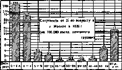
Рис. i.
reaches its peak in 1919 (5,606 cases) and in 1920 (7,525). The epidemic remains high through 1922 inclusive. The incidence of dysentery in Leningrad for 1919-22 is significantly higher than in Moscow and on average in the USSR. Subsequently, dysentery in Leningrad gradually decreases, but through 1926 it continues to remain above the pre-war level ('epidemic tail'). The incidence of dysentery in individual republics of the USSR (per 10,000 population) is given in Table 9. Dysentery is most frequently recorded in the Transcaucasian republics. The incidence of dysentery in individual regions of the RSFSR (per 10,000 population) is given in Table 10. 5. War and dysentery. Dysentery is the most dangerous disease of wartime. Unfavorable living conditions in the field, in trenches and camps lead to soil contamination, an increase in contact infections, and the development of dysentery outbreaks and epidemics (Kriegsruhr). The same is observed among refugee populations. Military units and refugees introduce the disease into the civilian population, among whom epidemics of dysentery also develop during wartime (see above). Military units suffered particularly severely from dysentery in previous wars. During the Franco-Prussian War of 1870-71, 38,652 people in the German army contracted dysentery, or 49 per 1,000 personnel; of these, 2,380 died, or 6.1% of the number of cases. German troops suffered enormous losses from dysentery during expeditions to China and Africa (K. Boehncke). During the Civil War of 1861-1865, among the troops of North America, 287,522 people contracted dysentery, of whom 9,431 died; 1,451,623 people had diarrhea, of whom 35,125 died; a total of 1,739,145 cases of dysentery and diarrhea were recorded, and 44,556 people died from these diseases (Vaughan). During World War I, dysentery was also one of the most formidable epidemics. In the German army, over 4 years of war, among field units 12.0 people, and among rear units 7.38 people per 1,000 personnel contracted dysentery. In terms of the number of cases, dysentery was second only to malaria. Thus, for the period from 1914 to 1918 in the German army per 1,000 personnel, the following contracted:
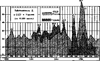
Table 11. Forms of disease Field units Rear units Typhus . Cholera . Typhoid fever . Dysentery . Malaria . . 0.64 0.62 7.9 12.0 15.96 0.20 0.17 1.62 7.38 5.61 Over 4 years in the German army (with a strength of 11,000,000 people), approximately 120,000 people contracted dysentery. The incidence was not the same on different fronts, as can be seen from the following table. Table 12. Incidence of dysentery in the German army on different fronts in 1914-1918 (per 1,000 personnel). Table 14. Western Eastern Balkan Italian Turkish front front front front front 1st .... 2nd ... . 4.6 2.0 - 103.1 3rd .... 1.0 4.2 - 98.4 4th .... 1.2 10.0 15.3 1.9 24.9 The highest incidence was on the Turkish front, the eastern front was second in terms of incidence. The incidence and mortality from dysentery in the German army on the eastern front were expressed in such absolute figures (Table 13). Table 13. Years Cases Deaths Mortality 1918 (Oct 1-31) 5,389 14,480 20,751 145 352 651 125 2.1 2.1 3.2 2.7 Table 14 shows the number of cases of dysentery and dysentery-like diseases recorded (according to Vaughan) in the troops of the U.S. Army in 1918 with a strength of 2,500,000 people. Table 15 shows the number of cases and deaths from dysentery in the Russian army during the war of 1914-17 (according to Avramov). Form of illness Cases Deaths Mortality Bacillary dysentery . Balantidiasis . Amoebic dysentery . . . Other protozoan diseases . Without form designation .... 325 6 428 30 3 31 2.5 2.6 10.0 1D Total . . . 3,573 | 53 1.5 Table 15 Years Cases Deaths total % total % 1914 (Aug-Dec) . . 1915 ........ 1917 (Jan-Sep) . 7,531 14,251 26,722 15,760 Total . . 16.5 6.7 According to materials of the Military Sanitary Administration of the People's Commissariat of Health, the incidence of dysentery in the pre-revolutionary Russian and Red armies per 1,000 personnel is expressed by the following indicators (Table 16). Table 16. Years % % 0.6 1919 ...... 1 17.5 0.8 15.3 0.7 6.9 1.1 5.7 0.6 0.53 0.87 4.08 0.58 3.94 The incidence sharply increased from the first year of World War I, but rose particularly high during the Civil War.
6. Dysentery as a social disease. The importance of one of the greatest social calamities—war—for the development of dysentery epidemics was noted above. Mass malnutrition and famine are also factors contributing to the development of dysentery. On the one hand, famine worsens the sanitary living conditions of the population (refugee status, peddling) and thus contributes to the development of dysentery; on the other hand, an exhausted organism becomes more susceptible to contracting dysentery (Danilevich, Dobreytser). This in particular should explain the unprecedented outbreak of dysentery epidemics in the USSR during the Civil War and famine. The situation in which various population groups find themselves during an epidemic sharply affects the incidence in these groups. Thus, during World War I, the officer corps in German prisoner-of-war camps almost never contracted dysentery: of 7,776 British prisoner officers, 3 died from dysentery; of 10,865 French officers, 1; and of 17,050 Russian officers, none (A. Gartner). 7. Seasonality of spread. Dysentery has a clearly expressed seasonal character with the greatest peak in summer and early autumn months. In the RSFSR, the highest figures for dysentery are recorded in June-September; in Ukraine and Transcaucasia, from June to October; in Hungary, from August to October; in Japan, from June to October. The corresponding curves are shown in Fig. 6. Epidemics', 'Figure 5.', 'Dysentery is also most frequently observed in summer months. Characteristic in this regard is the movement of dysentery by months in 1915-18 among the units of the eastern German army (according to Boehncke); over these 4 years, dysentery had an epidemic character: in 1915—in July-August, in 1916 and 1918—from June to October. Figure 7 shows the course of the dysentery epidemic in Petrograd from 1919 to 1923. Dysentery epidemics in these years in Petrograd occurred mainly from July to September, during which months the number of recorded cases reached 2,900, while in other months the number dropped to several dozen cases. Winter epidemics are observed much less frequently, usually they are limited to small outbreaks in individual foci.'
I. Dobreytser. Pathological Anatomy. The pathological-anatomical changes characteristic of dysentery, as stated, are localized in the intestine, specifically in the section of the large intestine. Only in severe cases is sometimes a segment of the ileum adjacent to the large intestine also affected, whereby colitis dysenterica thus turns into entero-colitis dysenterica. The intestinal lesions in bacillary dysentery manifest as acute inflammation of the intestine, the degree of which varies in different cases. A characteristic feature of these lesions is their gradual intensification as they move downward along the intestine, so that the lower part of the intestine and in particular the rectum are always most severely affected. In mild cases, as well as at the very beginning of more severe forms, the inflammation is of a catarrhal nature. The mucous membrane is more or less strongly hyperemic, swollen, pink-red, with small hemorrhages, covered with bloody mucus. The lesion may be focal, located mainly on the tops of the intestinal folds, or sometimes takes on a continuous spread. Despite the superficiality of the intestinal lesions, death may occur depending on the toxic effect of the infection. In more severe cases, along with increased hyperemia, swelling, and intensified hemorrhages, the inflammation takes on a catarrhal-purulent, and then diphtheritic character, which leads to the appearance of necroses of the mucous membrane of varying depths and the formation of fibrinous membranes. Necrosis and membranes first appear on the tops of the intestinal folds in the form of gray stripes and spots, with a sharply hyperemic periphery. In the further development of the inflammation, with the simultaneous spread of necrosis deep into the intestinal wall, there occurs impregnation of the mucous membrane with fibrin, as a result of which dense, granular, dry membranes are formed, firmly attached to the inner surface of the intestine. These membranes, depending on their impregnation with bile pigments, have a brown or yellowish-brown color, receive either a focal or predominantly continuous location, are especially strongly developed on the tops of the intestinal folds and in certain parts of the large intestine, particularly in the rectum and sigmoid flexure and its bends (flexura colica dextra et sinistra, S-Romanum). With these described lesions, the intestinal wall is found to be greatly thickened (edematous) and indurated, which is also facilitated by the severe spasm of the intestinal musculature, causing significant narrowing of its lumen; the tissue of the wall on sections is edematous and impregnated with blood, and from the side of the serous membrane there are ecchymoses. Necrosis, and this rather early, can be so deep that it involves the entire wall of the intestine down to the serosa (gangrenous form of dysentery), on the basis of which acute peritonitis may develop (see below). If the patient survives the first stage of the disease, the latter enters the second period: the inflammatory changes undergo reverse development. The formed strap-like membranes begin to gradually detach with the help of demarcative suppuration, at which time a purulent admixture is noticed in the feces. In place of the detached membranes, ulcers form (the second ulcerous period of dysentery), either shallow with flat edges or, with significant necroses, deeply penetrating with raised, swollen edges and a gray base. In especially severe cases, the ulcers have extensive spread, appearing continuous or interrupted only by strips and islands of surviving mucous membrane unaffected by the lesion. The healing of dysenteric ulcers (the third period of the disease) proceeds through granulation and the formation of a scar. With extensive and deep ulceration, healing usually requires a considerable time for its completion, sometimes, especially in severely emaciated or starving individuals, it may be prolonged and take a chronic course (outcome in the so-called chronic dysentery with widespread necrosis of the mucous membrane and severe edema of the submucosal layer gives the entire picture a characteristic appearance). There are indications of severe changes in the cells of the sympathetic ganglia of Meissner's and Auerbach's plexuses.-It is customary among anatomical forms of intestinal lesions in dysentery to distinguish the so-called "follicular dysentery" (colitis dysenterica follicularis, s. nodularis, s. cystica); here the matter concerns the formation in the large intestine, in the locations of the follicular apparatus, of small follicular sinusoidal ulcers, as well as unique sago-like inclusions (cysts) in the mucosa and submucosa, consisting of mucopurulent masses. The ulcers themselves have a sinusoidal character, have a small opening into the lumen of the intestine and expand significantly in depth. Sometimes this expansion takes the form of a submucous phlegmon of the intestine, which is accompanied by secondary ruptures to the surface of the mucous membrane; the latter in prolonged cases therefore takes on a unique appearance of a system of bridges, loops or gratings. The very origin of ulcers is conceived in two ways. On the one hand, it is pointed out that the covering epithelium of the intestine first grows into the depth of the follicles with subsequent separation and formation of a cyst, resembling grains of sago from the surface; later these grains become suppurated and open into the lumen—follicular ulcers form. Others believe that here there is primary suppuration and ulceration of the lymph follicles with subsequent ingrowth of the covering epithelium into the defect and rapid epithelial and connective tissue coverage of the ulcer. Since this coverage of the ulcer occurs before the elimination of the process in the depth, the cavity stretching under the epithelium with mucus and pus creates the impression of a sago grain or nodule. This form, like the purely catarrhal form of dysentery, is relatively rare and is observed in distant cases in these cases the exudate in the peritoneum is usually sterile (toxic peritonitis). Abscesses of the liver, in contrast to amebic dysentery, are very rare; pyelitis and pyelonephritis, according to the data from the autopsy of the Model Children's Hospital in Moscow (565 autopsies), are noted only in 1.4%. Nephritis, myocarditis belong to rare complications. The spleen in dysentery is usually not enlarged or this increase is insignificant; sometimes necroses in its pulp. In cases where dysentery ends in chronic ulcerative colitis [see separate table (pp. 55-56), Fig. 1], with the development of severe exhaustion and marasmus, hydroremic, so-called protein-free edemas, as well as amyloid degeneration of organs are observed.
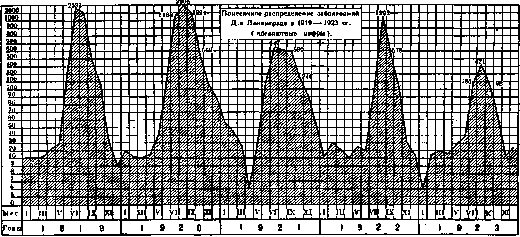
Fig. 7.
in the advanced periods of the disease. Among other peculiarities of the course of D. should be mentioned the diphtheritic process affecting the ileum; in approximately */z of all cases of D. such an affection is observed for several cm from the Bauhinian valve; extremely rarely - affection of only the small intestines (enteritis dysenterica).-With respect to other organs, attention is drawn to the diphtheritic inflammation of the lower third of the esophagus (in 3-4% of all cases at autopsy), less frequently - of the stomach, pharynx; moreover, in the upper quarter of the esophagus at the level of the larynx, necrotic strips of decubital character are sometimes found, most likely associated with the special looseness of the mucous membrane of this area and in particular with the presence here of the so-called glands of Schaffer. The retroperitoneal peri-aortic lymph glands (less than the mesenteric) are enlarged, hyperemic, sometimes with hemorrhages, necroses, and phenomena of myeloid hyperplasia. Peritonitis in D. is generally a rare phenomenon (not exceeding 1% of all fatal cases); even in the gangrenous form, peritonitis usually does not have time to develop, and the patient dies earlier from general intoxication. More frequently observed is microperforative peritonitis (Durchwanderungsperitonitis of German authors), when the purulent process unnoticedly penetrates to the serous coat through the lymphatic fissures. The observed Pathogenesis. In humans, dysentery infection penetrates the body by a single route - through the mouth along with food and drink, by means of contaminated hands and other objects introduced into the mouth. Following infection, the disease does not develop immediately, but is usually preceded by a short latent period (incubation) lasting up to 3 days. The dysentery bacilli that have entered the body multiply in the intestine, causing the changes described above through their toxins. The presence of bacilli can be proven in the excretions of the sick, in the contents of the intestines at autopsy, in the intestinal mucus, at the edges of ulcers (which in appropriate cases may lead to carriage), and in the mesenteric glands. From the intestine, the bacilli apparently do not penetrate into the blood; at least their detection in the blood of patients has not yet been achieved by anyone. Similarly unsuccessful have been the searches for bacilli in urine, into which they could only have entered through the blood. The negative results of searches for bacilli in the internal organs of patients and those who died from D. in most cases are presumably also connected with the non-finding of bacilli in the blood. The data presented on the distribution of dysentery bacilli in the bodies of patients and corpses leave no doubt that D. represents a pure form of intestinal toxicosis, when the patient's body is poisoned by poisonous products - toxins - produced by dysentery bacilli in the intestine. The very mechanism of the lesion of the intestine (and in rare cases - the esophagus) should however be conceived not only as the direct action of toxins on the mucous membrane, but mainly as a process associated with the excretion by the intestine of already absorbed toxins (by analogy with uremic, sublimate and other toxic forms of colitis). Almost all authors unanimously note the different character of diseases D. depending on which of the dysentery bacilli causes these diseases. The most dangerous in terms of severity, toxicity, and number of fatal outcomes are diseases caused by the Shiga-Kruse bacillus: they give the highest mortality (10-20%, and sometimes up to 50%); on the contrary, bacilli of the Flexner, Strong, and Hiss types do not possess great toxicity, the diseases they cause run more mildly and less frequently lead to death (0-5%, in rare cases 8-13%). Experimental study of D. Under natural conditions, the causative agents of bacillary D. are pathogenic exclusively for humans. Among other animals (wild, domestic, laboratory), spontaneously occurring dysenteric diseases are still unknown. All laboratory animals, when dysentery bacillus is introduced into them through the mouth, as is done naturally in nature, turn out to be little sensitive to this poison, and it is not possible to cause disease under such conditions. However, according to experiments of Bezredka, the disease occurs if young animals (rabbits) are taken and the virus is introduced to them on an empty stomach. Much more sensitive to dysenteric microbes are animals when the poison is introduced under their skin and especially into their vein. Even small doses cause disease and rapid death. The disease manifests itself by an increase in temperature, diarrhea, sometimes mucous, sometimes bloody, strong emaciation, paralysis of the extremities. Death occurs after several days following infection with symptoms of collapse with a drop in temperature below normal. At autopsy, along with hyperemia of internal organs and the entire intestinal tract, inflammatory changes are found in the mucous membrane of the large intestine with hemorrhages, necrosis, and the formation of scab-like films. All this shows that the lesion of the intestine is the result of the action of toxic substances. The same disease phenomena and death are also observed when killed cultures of dysenteric microbes are injected, from which it becomes obvious that the animal gets sick and dies not because the infectious agent introduced into the body appears in it and multiplies, but mainly due to the toxic substances contained in the microbes. In all these experiments, the most toxic of the dysentery bacilli proved to be the Shiga-Kruse bacilli. If autopsies are performed on animals that died from infection with D. bacilli, these microbes, especially the Shiga-Kruse bacilli, are rarely found by bacteriological cultures in the blood and internal organs. When death occurs shortly after infection, it is for example very common to find bacilli in the intestinal contents and in the bile, regardless of how the infection was introduced - into the blood or under the skin. On the basis of similar experiments, it must be concluded that dysentery bacilli have a special affinity for the intestinal wall (Vezredka).
G. Kulesha. Symptomatology. Although dysentery is an infectious disease caused by specific pathogens, having a definite pathogenesis and course, nevertheless in clinical practice difficulties often arise in diagnosis, since the diagnosis of dysentery cannot always be confirmed by the isolation of specific pathogens. The multiplicity of forms of these pathogens also extremely diversifies the course of the disease itself. Undoubtedly mild forms of the disease also occur, which no longer give the impression of true clinical dysentery, whereas bacteriological examination of the feces proves the presence of dysentery bacilli. Difficulty may also arise due to the similarity of the clinical picture of dysentery and colitis, since neither the character of the feces nor the severity of the disease can provide distinguishing guiding principles. Such an extremely varied course of dysentery, depending not only on the above-mentioned causes but also on the individual characteristics of the subject (age, sex), the nature of the epidemic, etc., forces one, for the purpose of describing the clinical picture, to take only the expressed clinical form as a guiding basis, omitting the various diverse forms of the disease which may not have the basic clinical symptoms of dysentery, yet are caused by specific dysentery bacilli. The incubation period lasts 2-6 days. The onset of bacillary dysentery is often abrupt. Sometimes there are preceding symptoms of malaise, loss of appetite, aching in the limbs, but more often the disease manifests itself immediately with abdominal pains and diarrhea. The pains are located sometimes on the right, sometimes on the left, often in the region of the transverse colon. There may be vomiting of food or bile. A slight fever appears. The feces are initially liquid and have the usual fecal character; however, after their evacuation the patient does not get relief - after a few minutes the pains and urges to defecate reappear, leading to a new evacuation of liquid stool. After several hours the character of the feces changes, mucus joins the watery feces, the evacuation becomes more scanty, and gradually the dysenteric syndrome develops, consisting of the following main symptoms. Sharply expressed colicky pains in the abdomen, sometimes of constant character, sometimes periodic, are concentrated either in the right or left iliac region, or near the navel corresponding to the position of the transverse colon. These pains sometimes give the sensation of a constant painful band in the abdomen or occur in the form of colic or agonizing tenesmus before defecation. This tenesmus continues during defecation and for some time after its completion. The pains depend on the peristaltic contraction of the affected large intestine. Tenesmus, a constant urge to defecate due to the sensation of the need to evacuate a seemingly solid body. There is straining, followed only by the evacuation of a small amount of mucus. These tenesms force the patient to almost constantly be on the toilet or in the lavatory, but each evacuation, even after the most severe straining, does not bring relief. Tenesms depend on the inflammation and swelling of the rectal mucosa (dysenteric proctitis) and on reflex spasmodic contractions of the sphincter of the rectum. Often simultaneously a painful spasm of the neck of the bladder appears, hindering urination. Figure 8. A case of moderately severe dysentery.
nation Strong straining can give prolapsus ani. Tenesmus can sometimes be so agonizing as to cause fainting. Character of the stools. In contrast to enteritis, in dysenteric colitis the stools are frequent and each time in small amounts. The number of stools is 10-20-50 times a day, but can be more. The character of the stools has a special imprint. Initially, as already mentioned above, the stools are of fecal character, then they gradually decrease in quantity and acquire the appearance of mucous masses. Individual flakes of mucus merge with each other, forming large jelly-like clots containing red streaks. Soon the stools take on a clearer bloody character and consist of bloody mucus alone. Purely bloody stools are also often observed. With progression of the process, pus appears in the secretions. The objective findings, first described by V. P. Obraztsov, give a definite picture. In dysentery, the process is most often concentrated in the lower segment of the colon, for which reason the S-Romanum appears as a dense, infiltrated, non-rumbling cord the thickness of a thumb, sometimes more. If the process also involves the transverse colon, it can be easily determined as a shortened painful transverse intestine lying at the lower border of the stomach. Along with this, the general condition of the patient suffers, general weakness develops as a result of severe intoxication, coated tongue, loss of appetite, intense thirst; fever is not always observed, but in more severe cases in the first days of the disease an increase in temperature is still noted (fig. 8 and 9). In severe forms of bacillary dysentery, in parallel with the intensification of the main symptoms, the phenomena of intoxication are also sharply expressed, the pulse is small and frequent, general decline of strength; in the end death occurs - more often during the second week of the disease. With favorable course, the main agonizing symptoms usually weaken soon, the stools on the 6-7th day take on a more fecal character, and recovery occurs. Rarely, bacillary dysentery, in contrast to amebic, passes into a chronic form with temporary exacerbations and improvements, emaciation and cachexia. Usually dysentery ends in 2-4 weeks, but sometimes in the period of recovery a new flare-up of the process and a new exacerbation of all clinical phenomena is noted. A severe form of dysentery with involvement of the rectum, S-Romani, transverse and ascending colon is characterized by extreme frequency of stools, discharged every 10-15 minutes; their character is sero-hemorrhagic with periodic discharge of fragments of necrotized mucosa. Severe pains are constant, and patients experience terrible suffering causing fainting. Along with this, the general condition suffers greatly, cardiac weakness with small irregular pulse appears, and patients gradually pass into the algid stage with cyanosis, cooling of the extremities, cold sweat, convulsions, aphonia. In the final period the pains subside, the abdomen becomes non-painful, the stools are discharged involuntarily, and with signs of rapid decline of strength death occurs in a soporous state. Less resistant individuals, children, the elderly, persons with poor nutrition more easily become victims of the disease than people of robust health. In addition, it has been noted that the course of individual dysentery epidemics depends on the type and virulence of the causative agents, as indicated above. Other symptoms of the disease. The tongue is dry and often heavily coated, appetite is absent, but thirst is sharply expressed. Often nausea, sometimes vomiting. The HCl content in the stomach is sharply reduced, as in other infectious processes. The spleen is not enlarged or slightly enlarged. Urine is scanty and usually contains a small amount of protein, hyaline cylinders; however, the development of nephritis is rare. The indican content is elevated. Temperature is not characteristic. More often - moderate fever in the first 2-3 days, then temperature approaches normal (fig. 8), sometimes with individual new elevations (fig. 9); in severe cases with expressed toxemia temperature at the height of the disease falls below normal (a grave symptom). Individual new elevations of temperature after the end of the disease are attributed to secondary infection (Brauer, Lyon). The condition of the cardiovascular system depends on the degree of toxemia, and with its expressed form the pulse is small and very frequent. In the blood at the height of the disease moderate leukocytosis; significant leukocytosis speaks for suppuration. Of the complications of bacillary dysentery, peritonitis should be noted first, arising due to the spread of the pathological process into the thickness of the intestine and transition of inflammation to the serous membrane. It can remain local, giving in the clinical picture vomiting and exacerbation of pains, but still does not yet have a grave prognosis and can end in complete recovery, whereas general peritonitis with perforation of the intestine gives the worst prognosis. It is observed only in severe forms of dysentery and clinically may give no symptoms (neither pains nor changes in temperature and pulse) and is often only an unexpected finding at autopsy. This is explained by the fact that peritonitis arises already at that period when there is no subjective reaction from the organism, in consequence of which all new phenomena remain unnoticed. Besides these complications, which are the result of the spread of the local pathological process in the intestine, complications caused by the dysentery toxin or secondary infection are sometimes also observed. These include neuritis and paralysis of the upper or lower extremities, depending on poliomyelitis, and arthropathies, occurring both during the dysentery itself and in the period of recovery from it (dysenteric rheumatism). More often the joints of the lower extremities are affected (joints of the foot, knee). The affected joint swells, is painful on pressure and movement. The swelling of the joint depends either on the periarticular process or on effusion into the joint itself. The effusion is usually serous and sterile. However, prolonged disturbance of joint function is rare. As for liver abscess, in bacillary dysentery, in contrast to amebic, it is almost never observed.
k.
Rutkevich. Diagnosis. The diagnosis of bacillary dysentery does not present particular difficulties when an established epidemic already exists, but the diagnosis of the first cases is not always correctly established, since dysentery very often develops during those months when ordinary colitis also exists. The clinical picture of hemorrhagic colitis and dysentery is very similar, and only bacteriological research can indicate the specific nature of the disease. Therefore, in every suspicious case, it is necessary to examine the feces, especially the bloody, glassy mucus, from which the specific causative agent must be isolated. The material for bacteriological research is the stool of patients, collected in sterile dishes. Dysentery bacilli are most easily isolated from the stool of fresh cases, which mostly has a mucous-bloody character. In such mucous masses, dysentery bacilli are often contained in large quantities, which can be easily verified upon examination of smears from the stool mucus, stained with diluted carbolic fuchsin or methylene blue with eosin. From the moment the stool becomes purulent, the examination is complicated by the admixture to the feces of various microbes from the intestinal flora, the number of which in the intestine increases significantly during this period of the disease. Examination of feces. For examination for dysentery, it is necessary to use fresh feces, if possible, just obtained from the patient. Otherwise, the percentage of positive results is greatly reduced. Dysentery bacilli disappear from the feces at room temperature within a few hours, and on ice - within 11/2-2 days. But even after a shorter period, the detection of dysentery bacilli is complicated by the growth of intestinal bacilli and saprophytes. In case of a negative result, the examination must be repeated several times. The method of examination is as follows: characteristic mucous-bloody feces are distributed in a thin layer in a Petri dish or in a saucer placed on a dark background; a platinum loop is used to pick up a mucous or mucous-bloody clump, which is repeatedly rinsed in a sterilized salt solution and sown on special media (see below). Bacteriological research must be preceded by microscopic examination of mucus clumps, which gives valuable data for diagnosis. For this, a mucus clump is crushed between a slide and a coverslip without rinsing. The preparations are examined in the unstained state, as well as stained with Löffler's blue or thionin. A large number of poly- and mononuclear cells indicates dysentery. The presence of blood, epithelial cells, and mucus is not very characteristic. The preparations are carefully examined for the presence of dysentery amoebas and cysts. Fecal masses having a normal appearance (from convalescents or bacillus carriers) are triturated with the addition of physiological salt solution into a homogeneous, porridge-like consistency. In the first days of the illness, there are few foreign bacteria in the feces, and colonies of dysentery bacilli are easily obtained even on ordinary meat-peptone agar. However, it is preferable to use colored agar media (Conradi-Drigalski, Endo, or Padlewski), on which dysentery bacilli form colonies that do not change the color of the medium. When using the Conradi-Drigalski medium, it is necessary to exclude crystal violet, which inhibits the growth of dysentery bacilli. It is better to use freshly prepared and not too alkaline Endo medium. An excess of fuchsin, especially unreduced, inhibits the development of dysentery bacilli. Some authors consider litmus unsuitable as an indicator and recommend using alizarin (Guth). The advantage of the latter over litmus is that due to reduction it undergoes only insignificant changes, while litmus is easily decolorized, turning into leuco-compounds. Alizarin is also more convenient for work under artificial lighting, as it gives sharper color contrasts. As a nutrient medium, it is best to use agar with lactose, which gives the most reliable results; an indicator (litmus tincture or, better, alizarin) is added to the medium. The sowing is done with a Drigalski glass spatula on two dishes with lactose and one with mannitol, which are placed in an incubator for 18-24 hours. Colonies of the toxic Shiga-Kruse group do not change the color of the mannitol medium, which remains red (with alizarin) or blue (with litmus). Colonies of the weakly toxic group (Flexner, Hiss, Strong bacilli) on the mannitol medium acquire a yellow color (with alizarin indicator) or red (with litmus) due to acid development and do not change the color of the lactose medium. Colonies selected for further research are transferred 1) to ordinary broth to study the motility of the bacilli (after 7-8 hours) and for the indole reaction (after 3 days); 2) into a tube with agar to which glucose and alizarin are added; 3) into a tube with agar with lactose and alizarin. If material remains, it is also transferred to simple slant agar. Sowing into tubes (items 2 and 3) is done as follows (Piibram, Halle): agar (3-4 cmг) in tubes is melted and cooled to 45°; 1-2 drops of an alkaline solution of alizarin (0.4% solution of alizarin with 0.3% caustic soda) are added to it. The alizarin solution is boiled for several minutes before use, during which the precipitate formed dissolves again upon standing. After adding alizarin, the agar takes the color of red wine. The culture introduced into the melted agar is evenly distributed in the tubes by careful shaking, which are then placed in an incubator. The results are noted already after 5-6 hours. Indeed, if we are dealing with dysentery bacilli, then in the glucose tube an acidic reaction develops, due to which the medium turns yellow, gases are not formed, in the lactose tube the color does not change. Sonne (Sonne) uses milk serum with litmus for transferring suspicious colonies. Identification of individual species (types) of dysentery bacilli. First, the presence of signs common to the entire dysentery group is established, then the features that allow distinguishing individual types are determined. The following properties are common to all species of dysentery bacilli: a rod, not staining by Gram, very similar to typhoid, but not possessing active motility, not forming spores, not decomposing lactose, not releasing gases during sugar fermentation, not reducing neutral red. The signs by which individual species of dysentery bacilli are distinguished are given in the table. Agglutination with specific immune sera is the final link in the bacteriological diagnosis of dysentery, but the agglutination reaction gives clear results only for delimiting the toxic (Shiga-Kruse) subgroup of dysentery bacilli. Agglut. Types of dysentery bacilli with sera | Toxicity. B. Shiga + + Very toxic B. Stutzer-Schmitz + Toxic B. Flexner + + + or + + + Weakly toxic B. Hiss (y) + + + or + + + B. Strong + + + The use of Flexner and Hiss sera to distinguish the corresponding species of bacilli leads to erroneous results, as Kruse already pointed out. Therefore, Przibram and Halle believe that for diagnostic purposes it is necessary to have a Shiga-Kruse serum and another polyvalent one, prepared with the help of all types of bacilli of the weakly toxic group. In any case, however, besides the Shiga-Kruse serum, it is also necessary to have Flexner serum (see the table above). Serodiagnosis at the patient's bedside. The Widal reaction can give valuable results. Schmidt (Schmidt) considers the diagnosis positive if the patient's serum agglutinates Shiga-Kruse bacilli in a dilution of 1:50 after two hours and in a dilution of 1:100 after 20 hours, and types of dysentery bacilli that decompose mannitol, i.e., weakly toxic ones, in a dilution of 1:200 after 2 hours. Serodiagnosis is especially important in chronic forms and in late stages of the disease, when the serum titer reaches 1:640 and 1:1280, all the more so since at this time it is almost impossible to detect dysentery bacilli in the feces (Frankel, Strauss and others). In diseases caused by Shiga-Kruse type bacilli, the patient's serum agglutinates not only these bacilli but also weakly toxic ones, the latter sometimes even to a greater degree. Conversely, in diseases caused by weakly toxic bacilli, the serum agglutinates only the corresponding bacilli and has no effect on Shiga-Kruse bacilli. A positive Widal reaction in healthy people indicates the possibility of bacillus carriage in them or that they have recently had an abortive form of dysentery. Thus, the Widal reaction is sometimes of great importance for clarifying the sources of the epidemic. Prognosis. The prognosis depends on the type of pathogenic agent, on the patient's resistance, and on the living conditions in which he is located.
Weak individuals, children, and the elderly give a higher percentage of mortality than healthy people of middle age. The nature of the epidemic is also of great importance. There are epidemics with insignificant mortality (1-2%), while other epidemics give mortality of 20-40% and more. Sporadic cases in general run a milder course. A high percentage of mortality is given by epidemics in mental asylums, prisons, barracks, and trenches. When making a prognosis, the frequency of bowel movements is not as important as the general condition of the patient. The frequency of bowel movements depends on the localization of the lesion. When the lower part of the rectum is affected, tenesmus is more pronounced, and urges are more frequent. Special attention should be paid to the general condition, on account of which the algid stage, rapid weak pulse, sunken eyes, subnormal temperature, etc., indicate a poor prognosis. Treatment. Often dysentery ends in recovery without any treatment. Everything depends on the strength of the body's resistance and the nature of the epidemic. There are epidemics of extremely mild course, and the means used during this epidemic are already recommended as the most reliable and effective, whereas de facto during subsequent epidemics they are found to be unsuitable; many such medicinal substances, which previously had the reputation of being specific, have already been consigned to the archives. An extremely important factor in the treatment of dysentery is the care of the patient and nutrition. The patient must be kept in bed in a warm room. After each bowel movement, the area of the anus must be thoroughly cleaned with cotton wool. For painful tenesmus - suppositories with morphine or cocaine; smooth pieces of ice can also be introduced into the rectum in the form of suppositories (Obraztsov). As for the diet of a dysentery patient, no definite list of dishes and sequence of their application can be given. Here there must be strict individualization depending on the severity of the case. The patient should be given easily digestible food that gives little residue - rice water, oatmeal, barley soup, sago, broth, soft-boiled eggs, crackers. All milk products should be avoided. Beverages - cold tea or cold boiled water, if it does not increase pain and diarrhea. Some authors recommend in fresh cases on the first and second day a starvation diet - tea, boiled water, red wine and intravenous infusions of 6% grape sugar (Noorden, Salomon), Normosal (Straub). In addition, they advise (Salomon, Wallace) to add to the starvation diet sugar solutions in water (200-300 g per day) to increase the calorie content. If diarrhea and tenesmus continue for 5-6 days, then a more concentrated diet is prescribed to avoid the patient's weakening - rice porridge, porridge made through a sieve; jelly, to soups add egg white or nutrient preparations, such as: somatose, plasmon, etc. After the disappearance of tenesmus and frequent stools, chicken meat, minced meat, pigeons, potato puree, rice porridge are added, and gradually a return to normal food is made. However, for a long time the patient should avoid sour, fatty and spicy dishes, sausages, canned fish, raw fruits, vegetables, pastries, cakes, beer, kvass. - Medicinal treatment of bacillary dysentery should begin with the use of a laxative with the aim of cleansing the large intestine of contents that irritate the mucous membrane. Some authors recommend for this purpose calomel in a dose of 0.3-0.5, others - castor oil. others - saline laxatives. The disadvantage of calomel is the possibility of the appearance of stomatitis, for which many avoid it and prefer the prescription of castor oil, sodium sulfate, magnesium. Sodium sulfate is given either as a single dose - 15.0, or as repeated 4-6-8 times a day at 4.0, or as a 2% solution (Ivashentsev). At the same time, for existing abdominal pains, fomentations, hot water bottles are prescribed, which give significant relief. For severe colic, one has to resort to pantopon, morphine or a mixture of morphine with codeine (Morphii mur. 0.1, Codeini phosphor. 0.1, Aq. destillat. 10.0 three times a day at 10 drops - Cohnheim); atropine under the skin or in suppositories, microclysters of suprarenin. Often after 2-3 days it is again necessary to prescribe saline laxatives or Oleum Ricini in order to remove fecal masses that maintain irritation of the mucous membrane, or to systematically prescribe a 2% solution of sodium sulfate. The use of disinfectants, including new preparations, gives no results. After cleansing the intestine, bismuth preparations are used at 1.0 three times a day or as a single dose of 5.0-10.0 on an empty stomach in the morning and astringent means - tanalbin, tanigen, tanismut. If one wants to give the patient rest at night, a small (72 glass) starch enema is prescribed with the addition of 10 drops of tincture of opium. Many also recommend for the bacillary form ipecacuanha, which is especially suitable for amoebic dysentery, in the form of an infusion 4.0:160.0 (Ruge), but in such a dose it often causes vomiting and increases pain, for which it is better to give Pulv. Doveri or emetine under the skin. Enemas with Kali hypermanganici (0.1-0.3 per 1.000) with tannin (V2%), Argent, nitrici (0.1-0.2 per 1.000) sometimes bring benefit, but in many cases cause pain and colic; therefore one has to abandon them. Enemas with animal charcoal (Schittenhelm) or with Bismutum sub-gallicum with Mucilago gummi arabici (Schmidt) are also recommended. Schiff recommends a 1% solution of formalin, Geissler - 15 drops of 10% Tinctura Jodi per 1 liter of chamomile infusion, Lutsch - a 2% solution of sodium salicylate, Leo - a 1% solution of calcium chloride. For cardiac weakness - caffeine, camphor under the skin, digalen, digipuratum. For persistent vomiting and hiccups - morphine under the skin. The introduction subcutaneously or intravenously of a physiol. solution to replenish the body's loss of water is also useful. One must also care for the patient's sleep. Already the reduction of tenesmus and urges gives the patient rest during the night; if necessary - the use of morphine or eukodal helps to relieve the patient from suffering, to induce sleep and strengthen. Specific treatment of bacillary dysentery is serotherapy. Experience in its application has already given practical results in terms of rapid improvement of all pathological phenomena and reduction of mortality. The experience of war, however, showed that only dysentery caused by the Shiga bacillus is successfully treated with anti-Shiga serum, whereas in Flexner and G dysentery the specific serum does not give such brilliant results. In sporadic and in the first cases of dysentery at the beginning of an epidemic, a polyvalent serum is used, and when the type of causative agent is established - the corresponding monovalent one. In Shiga dysentery, already a few hours after the introduction of serum, pains and tenesmus decrease, the stools lose the character of bloody mucus and after 1-2 days take on the usual fecal appearance, decrease in frequency, the general condition improves, the phenomena of toxemia pass. At the same time, the earlier the serum is applied, the faster the improvement occurs, while in late application in severe cases, when there is a sharp infection and intoxication, brilliant results cannot be expected from the use of serum. The serum is injected under the skin or into a vein, the dose depends on the severity of the disease and the age of the subject. In dysentery of moderate degree at the beginning of the disease, 20 cm3 of serum is sufficient to obtain an effect. If the first injection does not give a noticeable improvement of all pathological phenomena, then a new injection is indicated the next day, and sometimes a third injection on the 3rd day. In severe cases, 50-100 cm3 of serum is injected immediately and even 2-3 times a day. In very severe cases, the serum is injected into a vein. Some authors have used vaccine therapy or autovaccine therapy (the use of an emulsion of microbes killed by heat or sterilized with iodine), and in some cases encouraging results have been obtained. Observations, however, are still too few to allow definite conclusions. Prevention. In terms of prevention, dysentery does not differ essentially from other acute intestinal infections - cholera, typhoid fever, etc. To prevent dysentery diseases, the most important things are timely isolation of patients, rational organization of hospital care, disinfection of excretions and disinfection at the patient's home. In addition, enormous importance is attached to general sanitary-hygienic measures aimed at the correct and rational organization of water supply, sewerage, sanitary supervision over the production, storage and sale of food products, over the impeccable sanitary arrangement and clean maintenance of markets, trade premises, public toilets, to the fight against flies, etc.
All these measures, when rationally applied, limit the spread of dysentery diseases and prevent them from growing to the size of large or moderately significant epidemics. *In addition to them, it is necessary to organize sanitary education for the masses, thereby promoting among the population correct concepts about the properties of dysentery infection, its sources, routes of spread, measures of personal prevention, methods of disinfecting the sick and their excretions, etc. In the fight against the spread of dysentery, artificial immunization of the population against dysentery infection has recently acquired great value. Attempts to create artificial immunity to dysentery toxin existed long ago, since the discovery of dysentery bacilli (Shiga), but the application of this measure on a large scale encountered a major obstacle in the extreme toxicity of dysentery bacilli, which caused such a strong reaction when using ordinary immunization methods (subcutaneous injection of killed cultures) that it was necessary to stop any further actions in this direction. For this reason, subcutaneous immunization for dysentery was practically not carried out. The situation changed after research in this field by Bezredka, who proposed to carry out immunization not subcutaneously, but orally (per os), using for this purpose killed cultures of dysentery bacilli, prepared in the form of tablets. Testing of this method, which ensures not only local immunity to dysentery toxin from the intestine, but also phenomena of general immunity (Zabolotny), gave very encouraging results; therefore, at present, oral immunization is assessed as one of the effective measures in the fight against dysentery. Below are several observations, the most demonstrative and impeccable in their accuracy, establishing the positive value of enterovaccination by Bezredka's method (see Vaccination) in the fight against a dysentery epidemic. The first observation refers to 1923, when in the Versailles garrison out of 1,132 soldiers, 546 people were vaccinated by taking dry dysentery tablets. Of the vaccinated, 42 fell ill, i.e., 7.6%, and of the unvaccinated, 235 fell ill, i.e., 40%. In the same year in Leningrad, where a significant dysentery epidemic was raging, in one almshouse out of 2,768 inhabitants, 1,000 people were vaccinated per os. Of the vaccinated, 92 fell ill, including 10 within the first ten days after the end of vaccination; after deducting these ten, the morbidity rate among the vaccinated is determined to be 0.3%, while the morbidity rate among 1,768 unvaccinated during the same period was 3.11%, i.e., ten times higher. In May of the same 1923, an epidemic of dysentery broke out on the island of Hydra in Greece: out of 700 refugees, 22 people fell ill in a short time. In June, all these refugees were vaccinated, and none of them fell ill anymore, although they were in the focus of severe dysentery and used the same contaminated water as the sick population. Further, on August 4, 1923, 2,800 refugees were admitted to a hospital, during which in the preceding seven days of the sea voyage, 36 of them died from dysentery, and in the next two days another 44; 200 sick patients were placed in the hospital. Immediately, the entire group of these refugees was vaccinated with tablets, and dysentery ceased. With the same success, up to 30,000 refugees were vaccinated during the summer and autumn of 1923, with remarkable consistency, the morbidity rate among the vaccinated completely ceased or decreased significantly, while among the unvaccinated population groups, the epidemic continued to rage for several months. Very favorable results were obtained in the Moscow province in 1927-1928 and in the Red Army.
S. Korshun, G. Kulesha, K. Rutkevich. Dysentery in children. If completely unusual conditions (war, famine) are excluded, then the morbidity from D. and mortality from D. in children are so predominant that it can be classified as a predominantly children's infectious disease. Dysentery in children differs somewhat in its course and treatment compared to adults. The greatest differences are found in early childhood and especially in infants. In the main, these features are as follows. In younger children, anatomical changes are expressed less strongly. Often there is only catarrhal inflammation of the large intestine, and in necrotic-ulcerative lesions, the process as a rule does not have a continuous character, but is focal, with predominant localization in the lower sections. Furthermore, with the relatively more frequent finding in excreta not of Shiga bacilli, but of paradyssentery bacilli, the agglutination reaction, especially in children under 6 months of age, occurs less frequently. The stool, even in cases with significant anatomical changes, rarely completely loses its fecal character. Usually the stool is liquid, fecal, with an admixture of mucus and blood, green in color, with white curds, and alternates with a stool consisting only of mucus and blood. The odor of the excreta has a characteristic nature, reminiscent of the smell of semen. The frequency of stool and the intensity of tenesmus often depend on the reactive excitability of the child. Among the features of the course must be included the often observed discrepancy between anatomical findings and the severity of intoxication. Thus, on a large amount of material, a number of cases have been established where, with bacteriologically established clinical picture of D. and with severe intoxication, changes in the large intestine at autopsy had the character of simple catarrh. Dysenteric intoxication in children is characterized by coldness of the extremities, spastic vomiting, a sharp drop in weight, apathy passing into coma, convulsions, a decrease in cardiac activity, and in some cases - hydrocephalus with meningeal symptoms on the basis of rapid increase in cerebrospinal fluid without the presence of inflammatory phenomena. Skin hemorrhages are also noted during intoxication, mainly on the abdomen. Along with the specific form of toxemia, there may also be intoxication developing on the basis of dehydration of the body due to frequent vomiting and diarrhea. This form of intoxication (Exsikationstoxikose) resembles the specific one, but always proceeds with severe shortness of breath and glycosuria. Mainly due to insufficient and improper nutrition, one often has to deal with relapses, edema, and secondary nutritional disorders. For a long time after D., general weakness may remain, atony of the intestinal musculature with a characteristic tendency to persistent constipation, or conversely, an extremely high sensitivity of the large intestine, which is the cause of repeated colitis in connection with the most insignificant causes; in rare cases, a picture of Barlow's disease may develop. Among the complications during the disease, gaping of the anus is noted on the basis of paralysis of the sphincters, prolapse of the rectum, excoriations, skin abscesses, furunculosis, erysipelas lesions, stomatitis, thrush. Complications from the respiratory and excretory systems (pneumonia, cystitis, pyelitis, nephroses) are also not uncommon. Pneumonia as a rule ends fatally. In addition to pneumonia in infants at the beginning of the disease and during exacerbations, acute pulmonary distension, volumen pulmonum augmentum, is noted, which leads to covering the area of cardiac dullness. In addition to the severity of the dysenteric process itself, the presence of symptoms of intoxication and complications, in prognosis one must take into account the child's constitution, method of feeding, and nutritional status. Infants, artificially fed, with weakened nutrition (hypo- and atrophies) and who have recently had some infection, tolerate D. particularly poorly. The state of nutrition in D. plays an extremely important role not only in early childhood but also in older children. In no infectious disease is this significance so pronounced as precisely in dysentery (Danilevich). Prevention of D. in children's institutions consists in the possible rapid and complete isolation of the sick child or, in extreme cases, placing him in a box (see.) with attention to the staff on the possibility of transmitting the disease through hands, linen, and care items. Thorough disinfection of the patient's excreta and linen is necessary. It is very important to accustom children to observe the rules of personal hygiene (washing hands before eating, proper use of the toilet, etc.).-Treatment of D. in children presents great difficulties. The younger the child, the greater these difficulties. In children, as in adults, specific sera are finding increasingly wide application each year. But unfortunately, despite the continuing qualitative improvement of sera, their standardization, as well as despite the clarification and acceleration of the bacteriological diagnosis, this method of treatment so far cannot be considered exhaustive. Even less place should be given to drug therapy. It has only symptomatic significance. At the beginning of the disease and during exacerbations, a laxative (Ol. Ricini, Calomel, medium salts) is given. For at least temporary relief of the child from painful attacks associated with tenesmus and false urges, narcotics, atropine, belladonna in suppositories are used to a limited extent. The patient is given the opportunity to sleep and thereby somewhat restore his strength (Luminal-natrium). Narcotics are administered subcutaneously and in toxic vomiting. Treatment with enemas is suitable only during the period of subsidence of the process. Stimulants (Camphora, Coffein, Adrenalin, hot baths) must also be used, and in cases of intoxication and dehydration, the introduction of fluids should be widely used not only per os, but when possible also in enemas, but also by subcutaneous administration of physiological salt solution or Ringer's solution. Furthermore, care plays a major role. The latter is based on bed rest, cleanliness, warming by all means, careful care of the mouth, the anal area, and the skin; the act of defecation must be facilitated by performing it directly on a diaper or under a bedpan. The main, basic moment in the treatment of D. in children at present is nutrition. Until recently, the widespread and unfortunately not eradicated even to this day use of hungry and semi-hungry diets was in common use. This light, sparing diet (schonende Diat) consisted of mucilaginous decoctions, rice broth, rice porridge for older children, astringent jellies from blueberries. Without speaking of qualitative insufficiency, the caloric value of such food is so small that it is not only anti-diarrheal but also masked starvation. This diet leads to atrophy of the intestine, decrease in fermentative energy, cessation of fermentation processes, disappearance of free acids that stimulate peristalsis. Its prolonged use leads to exhaustion of the body, decrease in tolerance, relapses or prolonged forms, secondary nutritional disorders. The sad result of such disorders was often the death of the child. The conflict between the increased need for nutrition in D. in children and the impossibility of satisfying it due to anatomical lesions of the digestive tract is fortunately at present largely resolved. Now the diet is based on the use of full-fledged preparations with sufficient caloric value and moreover easily tolerated and assimilated even in early childhood. Such preparations are concentrated rice broth, women's milk, protein milk, concentrated protein milk, cottage cheese, meat puree. Mixtures are more often used with the addition of nutritive sugar Soxlet, and if the latter is not available - also ordinary sugar within the range of 3% to 12%. Concentrated rice solution, proposed for the treatment of D. in 1925 by Bessau, contains in 1 liter 400 calories with 0.11% protein; by adding sugar its caloric value can be brought to 800 calories and higher. This preparation is quickly digested, is a poor substrate for the causative agents of fermentation, and is a good irritant to the secretion of alkaline intestinal juice, which leads to the neutralization of free acids and the calming of intestinal peristalsis. Protein milk contains 437.5 calories, concentrated protein milk - 875, with the appropriate addition of sugar the caloric value can be increased by 1½ and even 2 times. Protein milk, cottage cheese, and to some extent meat puree also represent an environment unfavorable for the development of acid fermentation processes, and in addition have a high secretory effect on the intestine. Of the nitrogenous products, casein is only to a small degree subject to the action of proteolytic enzymes, and therefore also to putrefaction. The plan of treatment is approximately as follows. After giving castor oil at the beginning of treatment, immediately or, in the presence of vomiting and toxic phenomena, after a 4-6-hour tea diet, concentrated rice broth with saccharin in unlimited quantity is prescribed, according to the child's desire.
After 1-1½ days, 3% sugar is added to the broth and 200-400 g of protein milk with saccharin or the same with 3% sugar is given. In the following days, the amount of sugar is gradually increased to 8-12% with a simultaneous reduction of the rice broth to 200 g. To increase the caloric content, concentrated protein milk can be used instead of ordinary protein milk. The preparations are better given in alternation with each other, but if the child refuses to take one rice broth, they can also be given as a mixture. In children over 3 years old, protein milk can be replaced with cottage cheese, ground in rice broth with sugar, and later with meat puree in an amount of 50-100 g pro die. Further supplementary feeding, in connection with the disappearance of pathological symptoms and the appearance of digested stool, is carried out by adding mucilaginous soups on broth, cereals, jelly, and finally milk, butter, fats. In the treatment of children in the first half of the year and those with atrophy, the preferential use of mother's milk is indicated. The pace of prescriptions is determined by the condition of the patient, his appetite. During exacerbations, treatment has to be started by following the scheme from the beginning. The treatment itself must be carried out with mandatory consideration of the quality of food and the amount of calories introduced. To enrich the food with vitamin C, fresh juices can be added to the mixtures early on, and during recovery, the amount of juices should be increased. Acid buttermilk, acid mixtures, and fat mixtures are used in the treatment of D. in children. When using acid mixtures, there is a replacement of one acid with another, which leads to the displacement of pathogenic intestinal microbes by the beneficial lactic fermentation bacillus. Acid mixtures, as well as kefir, soured milk, and yogurt, are substitutes for protein milk preparations that are readily accepted. The matter is different with fat (cream) mixtures (Balaban, Uffenheimer, Lamprecht). Sufficient confirmation of their usefulness has not yet been obtained. It can be noted that in recent years, in connection with a change in views on diet, the problem of treating D. in children has been greatly advanced. m. danipievich. Amebic dysentery. The isolation of this disease as a special form became possible due to establishing the etiological role of Ent. histolytica in amebiasis and in particular in the so-called tropical, or amebic, dysentery. Thus, the latter is distinguished from D. of the temperate zone, caused by various bacteria, as well as similar intestinal diseases caused or associated with the presence of other protozoa (infusoria, flagellates, spirochetes) and worms. (Details concerning etiology, pathology, and epidemiology-see Amoebas.)-The clinic of amebic D. is extremely diverse. In some cases, the disease has an acute fulminating course, but more often it takes a chronic recurrent subacute form, sometimes stretching out for many years, while it can occur with insignificant clinical manifestations. The very onset of the disease is not always the same, and in connection with this, it cannot always be established with sufficient accuracy. In more or less typical cases, after the incubation period, which lasts from several days to several (6-12) weeks and is sometimes accompanied by nausea and indefinite abdominal pains, amebic D. begins suddenly in the form of an acute bloody-mucous diarrhea (up to 15-20 and even up to 80 times a day), accompanied by colic and tenesmus. Patients are restless, depressed, greatly weakened, lose their ability to work and appetite; the skin is pale, dry; the tongue is coated and moist; complaints of bloating and pain in the abdomen. However, diffuse abdominal pains, characteristic of bacillary D., may be absent, and instead, pains along the course of the sigmoid colon and in the region of the cecum are felt; sometimes special tenderness is noted at the points of flexure of the large intestine. Temperature is not always elevated; in some cases, nausea and vomiting appear, as well as nasal bleeding. The indican content in the urine is elevated. The appearance and consistency of the stool are very characteristic. Unlike in bacillary D., the excreted mucus is diffusely colored red and has the appearance of raspberry jelly; the stool is mostly neutral or alkaline in reaction and has a peculiar sweetish odor. On microscopic examination of the feces, glassy mucus, erythrocytes, a small number of neutrophils, eosinophils-mostly in large quantities, large mononuclear cells, often with phagocytosed erythrocytes, and cellular detritus at various stages of destruction are found; in many cases-Charcot-Leyden crystals, sometimes very numerous. In addition to typical cases with acute onset and course, cases are observed that begin gradually and proceed uncharacteristically, with the general condition being slightly disturbed and the intestinal manifestations having rather the character of a mild disorder (slight increase in frequency, mushy stool with mucous-blood layers); however, these cases can also exacerbate and give a picture typical both clinically and in terms of the nature of the stool. It is noted that in some cases the character of amebic D. may differ depending on the geographical region. Thus, the acute form described, for example, is found in North China, Panama, Tiflis. On the other hand, for Hong Kong, for example, up to 60% of cases with acute onset and fulminating, sometimes cholera-like course leading to death on the 4-5th day in a state of collapse are described. In these cases, there is a breakdown of the mucous membrane and the excretion of up to 40 times a day of foul-smelling fecal masses of chocolate or gray-green color with pieces of necrotic gangrenous tissue. In other cases (Philippines), the manifestations from the intestine recede into the background-they are either completely absent or have the character of mild diarrhea without blood excretion and without tenesmus, but with a dragging pain in the region of the large intestine; in some cases, constipation is even observed; the patient complains of indefinite abdominal pain at night, bloating, nausea, weakness. This condition is usually accompanied by anemia, weight loss; strong sweating of the palms and soles is noted. Pathological-anatomical manifestations can be very pronounced in such cases, but are discovered only on X-ray examination or at autopsy. Course. In typical cases of moderate severity, the acute course changes after 4-6 weeks-even without specific treatment-to a latent state, with the stool becoming thicker and even formed, losing the admixture of mucus and blood, the patient gains weight, and apparent recovery occurs. Such a remission can last from several days to many months, only to be interrupted, often without a definite cause, but more frequently from physical and even mental strain, dietary indiscretions, colds, intercurrent diseases (bacillary D., pneumonia, typhoid fever, malaria, beriberi), and again give a picture of an acute attack. Such alternation of remissions and attacks is very common in amebic D. and can last for many years. During remissions, the character of the stool can gradually return to normal, but in many cases of chronic amebic D., it has the characteristic appearance of a frothy-mushy mass of grayish or reddish-brown color with an admixture of glassy mucus and traces of blood; mucus clots usually contain amoebas; the reaction is alkaline. In chronic course, the thickened ascending colon and appendices epiploicae are often palpable, there is tenderness of the large intestine on pressure, a burning sensation in it, loss of appetite, belching after eating, heaviness in the stomach, nausea, tendency to vomit, neurasthenic phenomena (insomnia, palpitations), vascular disorders (cold sweats), decreased reflexes; subfebrile temperatures are often noted. The onset of unquenchable thirst and vomiting is usually an alarming symptom. Chronic cases can end in death with manifestations of bleeding (destruction of the vessel wall), but they can also drag on indefinitely long with manifestations of spastic constipation, being discovered only at autopsy in the form of solitary ulcers in the region of the cecum. Complications. 1. Deep destruction of the intestinal wall is a frequent cause of its perforation, mainly in the region of the cecum and sigmoid colon. In these cases, a fatal outcome is possible if a delimited adhesive peritonitis does not form. 2.
Chronic lesions of the intestine and corresponding inflammations of the peritoneum often lead to a picture of pericolitis accompanied by enlargement of the mesenteric glands; depending on the localization, peritonitis can give symptoms simulating diseases of the liver and gallbladder (in lesions of the flexura sigmoidea); lesions in the region of the transverse colon may give rise to suspicion of duodenal or gastric ulcer (radiation to the back); lesions in the region of the flexura lienalis can cause pains in the region of the spleen, which, in connection with attacks of elevated temperature and corresponding clinical phenomena, may suggest malaria; lesions of the cecum may give rise to suspicion of its tuberculosis as well as appendicitis; lesions of the sigmoid colon can cause in men a picture of prostatitis, in women—left-sided salpingitis or disease of the uterus (retroflexio); cases of narrowing of the intestinal lumen due to compression have been described; periproctitis is rarely observed; in lesions of the lower part of the large intestine, symptoms resembling sciatica may arise. 3. Abscesses of the liver are observed independently of the severity and even of the presence of intestinal phenomena; they develop from several days to several (2-20!) years after the onset of the disease, and cases have been described in which an abscess of the liver already developed after the cure of intestinal phenomena. Symptoms of liver abscess in amebic dysentery: pains, often unbearable, in the region of the liver and stomach; radiation to the right shoulder and the corresponding half of the chest (the characteristic inclination of patients to the right side), chills, diarrhea, vomiting, temperature 39°-40°, spasmodic cough; pressure determines the painful focus corresponding to the forming focus (trial puncture of the liver); jaundice often develops, bile pigments are usually present in the urine; leukocytosis is not always present; the abscess is usually single, superficial, in the upper-posterior part of the right lobe; more rarely there is a second abscess or multiple ones (up to 36); size—from a hazelnut to a child's head. Changes in the liver consist of coagulation necrosis of the liver tissue with subsequent softening in the center of the necrotic foci and formation of abscesses. The latter are filled with contents of chocolate to red-brown color, gelatinous consistency and contain a large amount of cellular detritus, drops of fat, crystals of hematoidin, cholesterol, Charcot-Leyden. Pus cells are comparatively few. Amoebae are usually not found in such an abscess; they can be found mainly in the walls of the abscesses. The frequency of liver abscesses after amebic dysentery reaches 30-40%. On the other hand, statistical data indicate that out of 100 cases of liver abscess, 95 occur after amebic dysentery (Rogers, Stroganov, Vasiliev). In addition to liver abscesses, hepatitis is also observed, often in acute form, however, not leading to suppuration. In this case, the liver is enlarged in volume and painful, jaundice phenomena are present. Ruptures of abscesses into surrounding organs (peritoneum, lungs, pleura, renal pelvis, intestine, pericardium, vena cava, etc.) have been described. In connection with liver abscesses, perihapatitis often forms. Liver abscesses are often accompanied by intestinal hemorrhages. 4. Amebic bronchitis may arise in connection with amebic dysentery or independently of it; origin: metastatic or rupture; some allow the development of bronchitis as a result of inhaling dust containing cysts of amoebae. Symptoms: high temperature, severe cough, mucopurulent, viscous sputum with layers of blood, containing amoebae—vegetative forms and cysts; X-ray shows shadows in the region of the root of the lung (to differentiate from tuberculosis and bronchopneumonia). 5. Amebic cystitis in men is probably of metastatic origin; in women, infection of the bladder with amoebae excreted from the intestine is possible. 6. Amebic epididymitis. 7. Subacute nephritis with the presence of amoebae in the urine. 8. Infarcts of the kidneys, spleen, liver. 9. Parotitis. 10. Phlebitis. 11. Abscesses of the brain. 12. Iritis. 13. Lesions of the skin. 14. Some authors connect amebic dysentery with subsequent polyneuritis and arthritis. Symptom complexes of a rheumatoid character have also been described (Ravaut), when intensification of pains in the bones and joints accompanies the exacerbation of intestinal phenomena. The above-mentioned conditions yield to specific antiamebic treatment. Phenomena from the adrenal glands have also been described (lowering of arterial pressure; Ravaut). 15. There are authors who express the opinion that sprue occurs in connection with previously suffered amebic dysentery. 16. An unfavorable effect on the course of amebic dysentery is exerted by the addition of bacillary dysentery, typhoid, paratyphoid, pneumonia, malaria, beriberi, nephritis, as well as invasion with ascarids, ankylostomes and schistosomes (bilharzias). Cases of simultaneous infection with balantidia have also been described. Pathological anatomy. In amebic dysentery, the large intestine, mainly its lower part, and the cecum with the vermiform appendix are mainly affected; lesions of the Bauhinian valve on the side facing the lumen of the large intestine have also been described. Lesions of the small intestine occur much less frequently (about 3%), a case of finding an amebic ulcer in the stomach is known. (The method of penetration and the paths of spread of amoebae into the tissues of the intestine—see Vol. I, p. 518.) Penetration of amoebae into the submucous layer causes the formation of abscesses in it, the first macroscopic traces of which from the side of the mucous membrane are sharply limited small red spots with a yellow center and a red periphery. Ulcers, up to the size of a silver ruble, have a round or oval shape and hard undermined or raised edges. Their interior is filled with yellowish necrotic masses. Unlike in bacillary dysentery, their development proceeds in depth towards the serous membrane and may end in rupture into the peritoneum. In lesions of the follicles, flask-shaped abscesses develop, which may merge, forming under the mucous membrane long passages. Secondary infection of amebic ulcers by bacteria can lead to their gangrenous disintegration and the formation of diffuse lesions. If a local lesion of the serous membrane forms, adhesions with neighboring organs as well as with the peritoneum arise. In the late period, sclerosis of the mesocolon may be observed. The lumen of the intestine can be narrowed by 4-5 cm due to thickening of the intestinal wall as a result of proliferation of connective tissue. In other cases, the formation of polypoid growths of the mucous membrane is observed. Spreading to the mesentery and mesenteric glands, proliferation can lead to the formation of dense strands that disrupt the mobility of the stomach and intestine and can cause a number of stagnation phenomena. From the microscopic side, the characteristic feature is the formation of coagulation necroses around those places where amoebae have penetrated. The affected follicles are filled with amoebae, the surrounding layers of the submucosa are hyperemic, swollen, impregnated with deposits of fibrin; hyaline degenerations and necroses are observed in them. The affected follicles are surrounded by a zone of cellular infiltrate. Through the vessels and capillaries, amoebae penetrate to the muscular and serous membranes (Fig. 10), however, neither inflammatory infiltrates nor diphtheritic deposits are formed. In case of healing of the ulcer, the glandular tissue is mostly replaced by connective tissue. Macroscopically, scars with raised edges remain, sometimes pigmented, often resembling scars remaining after bacillary dysentery. Prognosis. In the absence of treatment and in cases not yielding to it, amebic dysentery, with its tendency to chronic course and metastatic complications, gives in most cases an uncertain prognosis. Of the complications in this respect, the most serious are abscesses of the liver and especially of the lungs and brain, as well as intestinal hemorrhages, peritonitis and thrombophlebitis. Leukocytosis over 25,000, left nuclear shift of neutrophils and decrease in the number of eosinophils usually serve as unfavorable prognostic signs. The earlier the case is taken, the more favorable the prognosis. Parasitological diagnosis (see Vol. I, p. 520). The finding in the contents of ulcers, respectively—in the intestinal secretions, of Charcot-Leyden crystals is quite characteristic but has no absolute value, since on average it is observed only in 10% of cases, and on the other hand they can also be found in other infections. X-ray examination. X-rays 28-30 hours after taking barium reveal shadows with blurred contours in the affected areas. Rectoscopic examination gives a characteristic picture mainly in subacute cases (in the presence of round ulcers surrounded by a roller and serving as an entrance to deeply located flask-shaped lesions under the submucous layer). Examination in acute cases of amebic dysentery (on the second day after the onset of the disease) gave a picture of diffuse mucopurulent proctocolitis, with no changes observed above 20 cm. The surface of the mucous membrane was of raspberry color, had a fleshy rough appearance and resembled granulation tissue. No ulcers and abscesses
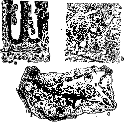
Figure 10. Ent. histolytica: a—in the Lieberkühn glands; b—in the submucous tissue; c—in the venous capillary.
at this time there was not yet. In subacute and chronic cases, general infiltration, hardenings, swellings and hyperemia of the mucous membrane are most often encountered, in places covered with ecchymoses. Phenomena of proliferation with the formation of polypoid growths, desquamation, formation of superficial ulcers and false membranes are often observed. In general, however, the proctoscopic picture cannot provide reliable distinctions from certain changes that also occur in bacillary D. In the differential diagnosis (see table on the next page), it is necessary to consider the possibility of infection of the intestine with other protozoa, as well as invasion by some helminths, which in some cases can create a clinical picture similar in certain respects to amebic D. In this regard, infection with infusoria (Balantidium coli) is of particular interest. Some authors consider the possibility of dysentery phenomena also in flagellate infections. Among the sporozoans, it is necessary to consider malarial plasmodia; infection with which in some cases can give intestinal manifestations, as well as coccidia. Some also admit the possibility of a dysenteric symptom complex associated with the presence of spirochetes. Among parasitic worms, it is necessary to consider Schistosoma haematobium, hookworms, roundworms, Strongyloides. Therapy. In the acute period, bed rest, quiet and warmth are mandatory; hot water bottles on the abdomen. Diet - initially complete abstinence from food (1-2 days), then Differential diagnosis of amebic and bacillary D. Geographical and seasonal distribution. Incubation, onset and course of the disease. Fever. Leukocytosis. Metastases. Relapses. Pathological anatomy. Pathogen. Experiments on animals. Serodiagnosis. Chemotherapy. Serotherapy. Causes of death. Amebic D. Endemic mainly in the tropical and subtropical zones, from where it is also carried to temperate countries. The predominant season is autumn, but it occurs throughout the year. Incubation from 3 to 90 days (approximately); onset mostly gradual, less often acute; course mostly chronic, less often fulminating. Spontaneous recovery is doubtful. In uncomplicated cases, fever is mostly absent and in any case not high. Observed in complications. Very frequent, especially in the liver. Very frequent. In the large intestine, limited undermined and deep ulcers; the submucosal tissue is predominantly affected. Perforations of the serous membrane, adhesive peritonitis, thickening of the intestinal walls and mesentery, drying up of the intestinal lumen are not uncommon. Gangrenous superficial ulcers are rare. Entamoeba histolytica. Infection of cats with vegetative amebae per anum and cysts per os is possible. Practically not applied. Emethine, yatrine, stovarsol, salvarsan, rivanol are more or less specifically effective. Unknown. Exhaustion, perforation of the intestines, hemorrhages, abscesses, etc. Bacillary D. Epidemic in hot and temperate zones. In the latter, mainly in summer. Incubation about seven days, acute onset and course with not infrequent outcome in complete recovery. Usually high, but only at the beginning of the disease. Frequent phenomenon. Not observed. Very rare. Superficial, flat ulcers with diphtheritic coating. Various species of Bac. dysenteriae. Infection of cats with dysentery bacilli per anum is impossible, per os was successful. Serum from patients agglutinates corresponding dysentery bacilli. Unknown. Corresponding anti-dysentery sera are specifically effective. Exhaustion, toxemia. mucilaginous decoctions (rice, oat, and barley), tea, protein milk; milk diet is recommended because lactic acid fermentation prevents the development of amebae. As the symptoms subside - broth with egg, softened crackers, potato puree, rice with milk, fish. When the stool becomes formed - careful addition of white meat. Beef, black bread, legumes are undesirable. During remissions, avoid colds, physical exertion, dietary indiscretions. General medicinal treatment: at the beginning of the disease, cleansing of the intestine (1-2 tablespoons of Oleum Ricini; Magn. sulph. or Natrii sulphi. after 3 hours, one tablespoon of concentrated solution; Calomel 0.5). Some recommend combined treatment with Bism. subnitr. or Karlsbad salt (Ziemann) according to the following scheme: in the evening, cleansing of the intestine with one tablespoon of Oleum Ricini, after a few hours one teaspoon of Karlsbad salt in 300 cm³ of warm water, the next morning and evening one teaspoon of Karlsbad salt and during the day Bism. subnitr. 6-10 times at 0.5. Usually mucus and blood disappear in 2-3 days. Previously, before the introduction of chemotherapeutic agents, Radix Ipecac was widely used in the form of Infusum ex 4.0:160.0 at 80 cm³ three times a day with the addition of peppermint oil to prevent vomiting movements, as well as Cortex Simarubae 1.0-1.5 pro dosi (5.0-15.0 pro die). Chemotherapy. At present, the basis of treatment for amebic D. is the use of various chemotherapeutic agents. The first chemotherapeutic agent for amebic D. was emethine (Rogers; 1913). Indeed, subcutaneous injections of Emet. hydrochlor. at 0.1 per day give in most cases very noticeable clinical improvement in a short time (approximate treatment scheme with emethine: 0.1 x 6 days, 2 days break; 0.1 x X 6 days, 2-3 weeks break; 0.1 x 6 days, 2 days break; 0.1 x 6 days). However, hydrochloric acid emethine could not finally resolve the question of treating amebic D. because, while providing clinical relief, in most cases (not less than 60%) it does not destroy the amebae but only promotes their encystation. Thus, without preventing the possibility of relapse, it also turns patients into chronic cyst carriers. In addition to subcutaneous and intramuscular application (the latter often less painful), hydrochloric acid emethine is also injected into the vein (0.02-0.05 in physiological salt solution). This method is apparently in many stubborn cases the most effective (Ravaut, 1918; Pexetakis, 1924). However, there are known cases when hydrochloric acid emethine has no effect at all and even does not cause encystation of the amebae. All these circumstances, as well as its known degree of toxicity and its cumulative action, have forced the search for other means to treat amebic D. One such agent proved to be Bismut-Jodide-Emethin (B. J. E.) - the double iodide salt of emethine and bismuth (Dale, Dobell; 1916). Containing 20% emethine, B. J. E., with certain precautions, is easily tolerated when taken internally and often provides radical help even in cases that stubbornly resist the action of hydrochloric acid emethine. According to English data, B. J. E. cures up to 90% of cases of amebic D. and its complications. The course of treatment consists of 12 doses of B. J. E. at 3.0 per day. A further improvement of this preparation is the less toxic and better tolerated (in terms of less frequent nausea and vomiting) compound emethine-per-iodide (E. P. J.). The mechanism of action of emethine is not exactly known. Assumptions about the possibility of its direct action on amebae have not been justified. It seems more probable that its action is indirect. In particular, the mechanism of the often observed emethine-resistance in amebic D. is also not clarified. The following figures can give an idea of how the introduction of emethine affected the mortality from amebiasis. Mortality from amebic D. Place of observation Before the introduction of emethine After the introduction of emethine Egypt .... India (Calcutta) . . . Panama . . . 52% (1911-12) 34.6% (before 1913) 18% (1908) 12% (1913) 0% (after 1913) 1.7% (1923) In the chemotherapy of amebiasis, arsenic preparations are also successfully used. First proposed by the French author Ravaut (Ravaut; 1918), these methods have since been developed mainly by the French school. Of the arsenic preparations, novarsenobenzol is used, which has a very noticeable and often very rapid effect on both the clinical course of the disease and the amebae themselves. Novarsenobenzol is used both intravenously and per os (0.05 x 2 for 10 days), in the latter case in the form of special tablets (Narsenol Billon). The internal method of using novarsenobenzol is often more effective. Recently, stovarsol (acetylaminooxyphenylarsinic acid) has also been used at 0.25 x 2 for 10 days (Marchoux; 1928), as well as treparsol (a derivative of meta-aminoparaxyphenylarsinic acid) at 0.25 3-4 times a day (Flandin). But of all preparations used internally, according to French authors, sanluol (arsenobenzol, old salvarsan, '606') is apparently most suitable. In doses of 0.1 1-10 per day, it can be systematically used for a long time without any side effects, which are common with the use of other arsenic preparations (Ravaut). Arsenic is also used in the form of enemas (0.15-0.30 novarsenobenzol in 60 cm³ physiological solution) in combination with intravenous infusions and per os administration. Treatment of amebic D. often presents great difficulties, and therefore many recommend the combined use of both emethine and arsenic. An example of such combined therapy can be the following scheme. Novarsenobenzol (N.
A.) 0.3 in vein- Emetin hydrochlor. (E. H.) 0.4 under the skin- E. H. 0.6-E. H. 0.8-N. A. 0.3-E. H. 0.4-E. H. 0.6-E. H. 0.8-N. A. 0.3-E. H. 0.4-E. H. 0.6-E. H. 0.8-N. A. 0.3-0-0-0-N. A. 0.3-0-0-0-N. A. 0.3-0-0-0-N. A. 0.3-E. H. 0.4-E. H. 0.6-E. H. 0.8-N. A. 0.3-E. H. 0.4-E. H. 0.6-E. H. 0.8-N. A. 0.3-E. H. 0.4-E. H. 0.6-E. H. 0.8-N. A. 0.3. In many cases, chemotherapeutic agents are successfully combined with internal administration of ipecacuanha. A very significant step forward in the chemotherapy of amebiasis is the introduction of yatren (Yatren "105", iodo-oxychinolinsulfonic acid, containing about 28% iodine). Proposed by Mühlens (1921), yatren is administered both orally (1.0x3 for 8-14 days) and in enemas (1.0-5.0 in 200.0-500.0 water). Yatren easily penetrates tissues, but in therapeutic doses is completely non-toxic, and German authors consider it a more perfect remedy for treating all forms of amebiasis than emetin. The exact mechanism of action of yatren is not known. Yatren belongs to the group of agents with so-called paradoxical action. 1. In the organism its action is beyond doubt. This is particularly evident from the fact that it is an excellent therapeutic agent not only for amebic dysentery but also for amebic liver abscesses not complicated by bacterial processes. 2. Meanwhile in vitro it has no effect on amebas even in a 1% solution. The latter is particularly remarkable in view of the fact that with respect to staphylococci, dysentery, diphtheria, and typhoid bacilli, yatren is a very powerful bactericidal agent. However, the contradiction between the given data can be easily reconciled if we take into account that yatren prevents the encystment of amebas. The latter circumstance may indicate that it inhibits the formation of precystic stages of amebas, in other words, that it inhibits their reproduction. From this point of view, it seems quite possible that the bactericidal action of yatren is expressed primarily in its inhibitory effect on the reproduction of bacteria. It is very likely that the parasitotropism of yatren in the organism is combined with its purely mechanical action. Easily penetrating deep into tissues and being a fairly strong laxative, yatren undoubtedly promotes the mechanical washing out of amebas from deep tissues and their removal from the intestine. Due to this, yatren has proven to be an extremely active agent also in the rapid sterilization of the intestine in chronic and stubborn cyst carriers in cases when emetin had no effect. However, it must be noted that cases have been described where yatren also proved powerless. In recent years, rivanol has also been used in the chemotherapy of amebiasis. Testing of rivanol showed that it possesses, compared with other chemotherapeutic substances, a higher parasitocidal and consequently more favorable therapeutic effect. While at a dilution of 1:1,000 yatren, emetin, and trypanflavine exert only a weak effect in vitro, rivanol at the same dilution kills Ent. histolytica in 24 hours. When the indicated solutions were administered to infected cats in the form of enemas, the strongest effect was shown by rivanol (1:1,000-1:1,250), which proved twice as active as trypanflavine and ten times as active as yatren (Wagner; 1928). In clinical trials, Rivanol at 0.03-0.05 three times a day for 6 days (max. 0.2 per dose; pediatric dose 0.004-0.008 three times a day) proved to be a very effective agent against both vegetative forms and cysts. Enemas of 1:5,000-1:3,000 were also used with good success (Urchs; 1926). On average, complete sterilization of the body occurred in 4.1 days with a total amount of rivanol of 0.843 g. Due to its antispasmodic properties, rivanol is also an analgesic (Schaumann; 1928). Rivanol is also successfully used in amebic infection of the bladder. In addition to the usual treatment (0.25 three to six times a day, 10 days), the bladder was also irrigated with a 1:3,000 solution (Peter; 1928). g. Epstein.
Related articles
Mentioned in
Cite this page
“DYSENTERY.” Soviet Medical Encyclopedia. English translation of Bolshaya Meditsinskaya Entsiklopediya, 1st ed. (Moscow, 1928–1936), ed. N. A. Semashko. https://sovietmedicalencyclopedia.pages.dev/article/dysentery/